Firmicutes under Conditions Limiting for Growth and Survival
A dissertation submitted to the University of Neuchatel for the degree of
Docteure ès Sciences
bySevasti Filippidou, MSc Molecular Genetics
Accepted by the Jury:
Prof. Pilar Junier, thesis director, University of Neuchatel Prof. Maarten Voordow, University of Neuchatel
Prof. Melanie Blokesch, EPFL, Lausanne Dr. David Russel Johnson, Eawag, Dübendorf Dr. Paul Herron, University of Strathclyde, Glasgow, UK
Defended the 11th April 2016
Rue Emile-Argand 11 2000 Neuchâtel - Suisse Tél: + 41 (0)32 718 2100 E-mail: secretariat.sciences@unine.ch
IMPRIMATUR POUR THESE DE DOCTORAT
La Faculté des sciences de l'Université de Neuchâtel
autorise l'impression de la présente thèse soutenue par
Madame Sevasti Filippidou
Titre:
“Sporulation capability and metabolic
mechanisms of endospore-forming Firmicutes
under conditions limiting for growth and
survival”
sur le rapport des membres du jury composé comme suit:
- Prof. Pilar Junier, directrice de thèse, Université de Neuchâtel, Suisse - Prof. ass. Maarten Voordouw, Université de Neuchâtel, Suisse
- Prof. Melanie Blokesch, EPF Lausanne, Suisse - Dr David R. Johnson, EAWAG, Dübendorf, Suisse - Dr Paul Herron, University of Strathclyde, Glasgow, UK
Neuchâtel, le 28 avril 2016 Le Doyen, Prof. B. Colbois
This work is dedicated to
The memory of Marilena Vourkou, for her inspiration to life,
Entropia, that has shaped me to what I am, Dimitris Serafis, without whom I wouldn’t have reached that far.
Firstly, I would like to thank my professor Pilar Junier for being a very supportive tutor and a real mentor throughout all these years. Pilar has taught me, guided me, encouraged me, and gave me opportunities I never imagined I could have. She is constantly showing how academic science should be and inspires her team, being a reassurance for brighter days.
I would also like to thank Mrs. Nicole Jeanneret for having introduced me to the lab and who has been a good-hearted advisor on techniques, laboratory practices and French since day one.Many thanks also go to all my lab collaborators, and more specifically the ‘spores’ team, Dr. Matthieu Bueche, Dr. Ludovic Roussel-Delif, Dr. Sathiya Ganesan, Mr. Loic Sauvain, Mrs. Marion Jaussi and Mr. Christophe Paul for their help, contribution and pleasant atmosphere in the group. In particular, Dr. Tina Wunderlin has generously coached, advised, supported, and shared with me in any possible way. I thank her for being there to encourage me since I could never imagine for a better colleague, office mate and friend. I am most grateful to Dr. Thomas Junier for almost all bioinformatic analysis and for patiently spending his time teaching me phylogenetic tools and rules of scientific writing. Moreover, I would like to thank Dr. Radu Slobodeanu for his statistics advice for the article presented in chapter three. Finally, I am greatful to all our collaborators, and especially the team of Dr. Patrick S. Chain, without whose valuable contributions, this work would never be completed.
Thanks also go to all the students who have worked with me, during their learning stage: Mrs. Amandine Pillonel, Mr. Andrej Al-Dourobi, Mrs. Ilona Palmieri, technicians; Mr. Patricio Munoz-Ruffat, undergraduate student and Mr. Fabio Palmieri, master student. I hope their learning procedure was as constructive as mine during our collaboration. Special thanks to Mrs. Anna Voulgari for being the perfect ecological eye during sampling campaigns, and who, along with Mrs. Vassiliki Mpelogianni, Dr. Tina Wunderlin and Dr. Nikos Mitakidis have critically read my general introduction. Mrs. Monica Albini has patiently helped me during the last hours before submission and I thank her for that. Dr. Vincent Hervé has translated my summary into “good” French, for which I am most grateful.
I thank my thesis jury for accepting to evaluate this work and provide feedback.
Special thanks go to what I call “an army of people” that helped and supported me throughout these years and which include my family and dearest friends. Although some of them are already acknowledged, all of them have always been available to support and encourage me in any possible manner.
Cover photo: Endospore-forming Firmicutes forming a sphere with copper carbonate crystals. Scanning Electron Microscopy. Photo taken by S. Filippidou. LAMUN 2012
Life, as we know it, has physical and chemical limits. More precisely, physical and chemical environmental parameters set the thresholds for reproduction, metabolism and survival for the organisms known to date. Environmental conditions unsuitable for survival and development are the rule rather than the exception in most habitats. Microorganisms have developed various strategies to withstand environmental conditions that limit active growth. A microbial group that displays a large array of strategies to resist adversity is endospore‐forming Firmicutes. These strategies range from the formation of resting states (endospores), to biofilms and metabolic adaptation. These strategies are a costly biological investment and therefore might affect their success.
Endospore‐forming Firmicutes have been isolated from various environments, including extreme habitats. Paradoxically, in diversity studies they are either absent from these environments or they represent a small fraction of the microbial community. Using a profile analysis of the spo0A gene, unique to endospore‐forming Firmicutes, we have shown that they cannot be found in publically available metagenomic datasets, despite the fact that many of these datasets correspond to well‐known habitats for endospore‐ forming Firmicutes. We have shown that this bias is likely due to the fact that commonly used methodological approaches are inefficient. Moreover, we have shown experimentally that an improved DNA extraction can improve detection in amplicon sequencing; however, this was not the case for shotgun classification. Although this group is known to colonize every habitat on Earth, endospore‐firming Firmicutes are not
prevalent in all habitats. Their energy‐demanding survival strategies become an actual
benefit only when multiple physical and chemical limits of life are present. Extremity favors the presence of the survival strategies deployed by endospore‐forming Firmicutes. More specifically, we have shown that extreme environmental conditions are an important factor for the survival strategy of sporulation to evolve. Two novel discoveries support this suggestion. Firstly, we have shown that extremity is a driving force for sporulation in a species that was not known to sporulate, Serratia ureilytica. Secondly, we demonstrated that the ability to sporulate is not lost when there is environmental pressure. That was the case of Kurthia spp. a previously known asporogenic genus. The common ancestor of Kurthia, and the endospore‐forming Firmicutes was able to produce spores, however sporulation is considered to have been lost within the lineage of Kurthia. The genomic analysis and the microscopic observation of a Kurthia sp. strain isolated from a geothermal reservoir reveal that the sporulation pathway has not been lost, and that Kurthia is not an asporogenic but rather a cryptosporulating genus.
The survival strategy of sporulation makes endospore‐forming Firmicutes capable of tolerating adverse conditions and thriving in extreme environments. A novel species that has a particular ecological niche in geothermal reservoirs was discovered; Anoxybacillus
geothermalis was revived under laboratory conditions and is hypothesized to have
remained inactive in the reservoir since the Permian age. More isolates belonging to the same species were also discovered in different geothermal reservoirs.
sporulation strategy but also their high metabolic diversity. Manganese oxidation and copper reduction of endospore‐forming Firmicutes in natural, uncontaminated environments, were studied leading to the conclusion that metal tolerance is a widespread phenomenon in unrelated aerobic endospore‐forming Firmicutes from natural uncontaminated environments. Finally, in saline habitats, both metabolic strategies are deployed, resulting in an impressive diversity of endospore‐forming Firmicutes.
Biochemical, genomic, ecological and environmental data are pieces to fill in the puzzle of adaptations of endospore‐forming Firmicutes in extreme habitats.
Keywords: Microbial Ecology, Firmicutes, endospore‐forming Firmicutes, sporulation,
Kurthia, Serratia, Anoxybacillus geothermalis, Manganese Oxidation, Copper Tolerance,
La vie, telle que nous la connaissons, possède des limites physiques et chimiques. Plus précisément, des paramètres environnementaux physiques et chimiques limitent la reproduction, le métabolisme et la survie des organismes vivants décrits à ce jour. Des conditions environnementales défavorables à la survie et au développement biologique sont la règle plutôt que l'exception dans la plupart des habitats. Les microorganismes ont cependant développé différentes stratégies pour résister aux conditions environnementales qui limitent leur croissance. Parmi ces microorganismes, le groupe des Firmicutes endosporulantes présente de nombreuses stratégies pour résister à ces contraintes environnementales, allant de l'état de dormance (endospore), à la formation de biomembranes en passant par l'adaptation métabolique. Ces stratégies sont un investissement biologique coûteux pour les organismes et par conséquent, peuvent influencer leur succès.
Les Firmicutes endosporulantes ont été isolées dans des environnements variés, y compris dans des habitats extrêmes. Paradoxalement, dans les études de diversité elles sont, soit absentes de ces environnements, soit elles représentent une faible fraction de la communauté microbienne. A l'aide d'une analyse de profil du gène spo0A, un gène spécifique des Firmicutes endosporulantes, nous avons montré que ces dernières ne pouvaient pas être détectées dans les métagénomes publiques disponibles, et ce malgré le fait que plusieurs de ces métagénomes étaient issus d'habitats connus pour abriter des Firmicutes endosporulantes. Nous avons mis en évidence que ce biais était dû au fait que les approches méthodologiques couramment utilisées étaient inefficaces. De plus, nous avons démontré expérimentalement qu'une extraction d'ADN optimisée permettait d'améliorer la détection de Firmicutes endosporulantes par séquençage d'amplicon. Cependant ce n'était pas le cas pour le séquençage shotgun. Bien que ce groupe bactérien soit connu pour sa capacité à coloniser tous les habitats sur Terre, les Firmicutes endosporulantes ne sont pas fréquemment retrouvées dans tous les habitats. Leurs stratégies de survie étant énergétiquement coûteuses, elles ne deviennent un avantage uniquement lorsque plusieurs contraintes physiques et chimiques sont présentes. Les conditions extrêmes favorisent la présence des stratégies de survie déployées par les Firmicutes endosporulantes.
Plus spécifiquement, nous avons démontré que des conditions environnementales extrêmes étaient un facteur important pour l'apparition de la sporulation comme stratégie de survie. Deux nouvelles découvertes supportent cette hypothèse. Premièrement, nous avons montré que les conditions extrêmes étaient une force motrice de la sporulation pour une espèce qui n'était pas connue pour sporuler, Serratia ureilytica. Deuxièmement, nous avons démontré que la capacité de sporuler n'était pas perdue lorsqu'il y avait une pression environnementale. C'était le cas de Kurthia spp., un genre décrit jusqu'à présent comme asporogénique. L'ancêtre commun de Kurthia, une Firmicutes endosporulante, était capable de produire des spores, cependant cette capacité était considérée comme perdue dans la lignée de Kurthia. L'analyse génomique ainsi que des observations microscopiques d'une souche de Kurthia sp. isolée d'un
que Kurthia n'est pas un genre asporogénique mais un genre cryptosporulant.
La stratégie de survie de sporulation rend les Firmicutes endosporulantes capables de tolérer des conditions défavorables et de prospérer dans des environnements extrêmes. Une nouvelle espèce, Anoxybacillus geothermalis, a été découverte dans une niche écologique particulière, les réservoirs géothermiques. Cette souche a été remise en culture au laboratoire et nous avons émis l'hypothèse que cette souche était inactive dans le réservoir depuis le Permien. D'autres isolats appartenant à la même espèce ont également été découverts dans différents réservoirs géothermiques.
Les environnements extrêmes permettent aux Firmicutes endosporulantes d'utiliser leur stratégie de sporulation mais également de tirer profit de leur grande diversité métabolique. L'analyse des processus d'oxydation du manganèse et de réduction du cuivre par les Firmicutes endosporulantes dans des environnements naturels et non contaminés a révélé que la tolérance aux métaux est un phénomène largement répandu dans les environnements non contaminés, y compris parmi des Firmicutes endosporulantes aérobies ne présentant pas de lien de parenté. Enfin, dans les milieux salins, ces deux stratégies de survie sont utilisées et génèrent une impressionnante diversité de Firmicutes endosporulantes.
En conclusion, l'utilisation de données biochimiques, génomiques, écologiques et environnementales a permis de mieux comprendre l'adaptation des Firmicutes endosporulantes aux environnements extrêmes.
Mots-clés: Ecologie Microbienne, Firmicutes, Firmicutes formatrices des endospores,
sporulation, Kurthia, Serratia, Anoxybacillus geothermalis, Oxidation du Manganese, Resistance au Cuivre, Halophiles.
Summary ... 1 Résumé ... 3 Table of Contents ... 5 1. General Introduction ... 11 1.1. Thesis outline ... 11 1.2. Background ... 12 1.2.1. What is Ecology? ... 12
1.2.2. Microbial Ecology: from microscopes to metagenomes ...14
1.2.3 Extreme habitats ... 16
1.2.4. Survival strategies in the microbial world ... 18
1.3 Sporulation as a survival strategy in adverse conditions ... 19
1.3.1. Sporulation ... 19
1.3.2. Sporulation in endospore‐forming Firmicutes ... 20
1.3.3. Sporulation in Cyanobacteria ... 20
1.3.4. Sporulation in Actinomycetes ... 21
1.3.5. Sporulation in Myxococcus ... 21
1.3.6. Costs and benefits of sporulation ... 22
1.4. Survival by adaptation ... 22
1.4.1 Adaptation under extreme conditions at a molecular level ... 24
1.5 Research Objectives... 26
1.6 References ... 27
Chapter 2 Under‐detection of endospore‐forming Firmicutes in metagenomic data ... 33
Abstract ... 34
2. 1. Introduction ... 35
2.2. Material and methods ... 36
2.2.1. Genome sequence retrieval ... 36
2.2.2. Detection of orthologous sporulation genes common to all endospore formers ... 36
2.2.3. Profile construction and validation ... 36
2.2.4. Metagenomic datasets retrieval ... 37
2.2.5. Environmental sampling, DNA extraction and quantitative PCR ... 37
2.2.6. Amplicon sequencing of the 16S rRNA and spo0A genes ... 37
2.2.7. Metagenomic sequencing ... 38
2.2.8. Metagenome data annotation ... 38
2.3.2. Profile analysis of sporulation genes in metagenomes ... 40
2.3.3. Amplicon sequencing of an environmental sample with high prevalence of endospore‐forming Firmicutes ... 42 2.3.3. Metagenomic sequencing ... 43 2.4. Conclusions ... 46 2.5. Acknowledgments ... 46 2.6. References ... 47 2.7. Supplementary material ... 52
2. 8. References on Supplementary Material ... 54
Chapter 3 Extreme conditions favor the prevalence of endospore‐forming Firmicutes in natural springs ... 57
Abstract ... 58
3.1. Introduction ... 59
3.2. Materials and methods ... 60
3.2.1. Sampling and environmental factors ... 60
3.2.2. DNA extraction ... 60
3.2.3. Quantitative PCR assays ... 61
3.2.4. Amplicon pyrosequencing and analysis ... 62
3.2.5. Statistical analysis ... 63
3.3. Results ... 63
3.3.1. Characterization of the natural springs ... 63
3.3.2. Single environmental factors do not influence EFF relative abundance ... 64
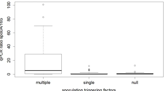
3.3.3. EFF are prevalent in multiple‐limiting environmental factors ... 64
3.3.4. Diversity of EFF in environmental samples ... 64
3.4. Discussion ... 65
3.5. Acknowledgements ... 68
3.6. References ... 70
3.7. Supplementary material ... 75
Chapter 4 Another path to survival: discovery of sporulation in Serratia ureilytica... 93
Abstract ... 94
4.1. Introduction ... 95
4.2. Materials and methods ... 95
4.2.1. Sampling and isolation ... 95
4.2.2. Physiological characterization of Lr5/4 ... 96
4.2.3. Molecular characterization ... 96
4.2.6. DPA measurement ... 98
4.2.7. Genome sequencing and analysis... 98
4.2.8. Horizontal gene transfer analysis... 99
4.3. Results ... 99
4.3.1. An unusual spore‐former isolated in a thermal spring ... 99
4.3.2. Sporulation in S. ureilytica Lr5/4 ... 100
4.3.3. A framework for the emergence of sporulation in Serratia ... 102
4.3.4. Genomic imprints of sporulation ...103
4.5. Acknowledgments ... 106
4.6. References ... 107
4.7 Supplementary Material ... 112
Chapter 5 Cryptosporulation in Kurthia spp. forces a rethinking of asporogenesis in Firmicutes ... 123
Abstract ... 124
5.1. Introduction ... 125
5.2. Materials and methods ... 126
5.2.1. Sample collection and isolation ... 126
5.2.2. Strain identification ... 126
5.2.3. Phylogenetic analysis ... 127
5.2.4. gDNA extraction and sequencing ... 127
5.2.5. Retrieval of sporulation genes sequences ... 127
5.2.6. Sequence data analysis ... 127
5.2.7. DPA measurement ... 128
5.3. Results and discussion ... 128
5.3.1. Sampling and characterization of the isolate ... 128
5.3.2. Genomic imprints of sporulation in Kurthia sp. strain 11kri321 ...130
5.3.4. Presence of dipicolinic acid inside the spores of strain 11kri321 ... 132
5.3.5. Comparative analysis of all Kurthia genomes available ... 133
5.3.5. Cryptosporulation and asporogenesis ... 135
5.4. Acknowledgements ... 135
5.5. References ... 137
Chapter 6 Anoxybacillus geothermalis sp. nov., a facultative anaerobic endospore‐forming bacterium isolated from mineral deposits in a geothermal station ... 143
Abstract ... 144
6. 1. Introduction and results ... 145
6.4. References ... 155
6.5. Supplementary Material ... 159
Chapter 7 Comparative genomics of endospore‐forming bacteria isolates for the discovery of potential biomarkers of extremity... 165
7.1. Published genomes ... 166
7.1.1. Genome sequence of Bacillus alveayuensis strain 24KAM51, a halotolerant thermophile isolated from a hydrothermal vent ... 166
7.1.2. Genome sequence of Aeribacillus pallidus strain GS3372, an endospore‐forming bacterium isolated in a deep geothermal reservoir ... 167
7.1.3. Genome Sequence of Anoxybacillus geothermalis strain GSsed3, a novel thermophilic endospore‐forming species ... 168
7.2. Publically available, non‐published genomes ... 169
7.4. References ... 171
Chapter 8 Manganese‐II oxidation and copper‐II resistance in endospore forming Firmicutes isolated from uncontaminated environmental sites ... 173
Abstract ... 174
8.1. Introduction ... 175
8.2. Materials and methods ... 176
8.2.1. Site description and sample collection ... 176
8.2.2. Enrichment and isolation ... 176
8.2.3. Identification of the isolated strains ... 177
8.2.4. Manganese (II)‐oxidation ... 177
8.2.5. Copper (II) resistant/tolerance ... 177
8.2.6. Statistical analysis ... 178
8.2.7. Phylogenetic analysis of 16S rRNA, Mn (II) oxidation and Cu (II) resistance .... 178
8.3. Results ... 178
8.3.1. Isolation and identification of endospore‐forming Firmicutes ... 178
8.3.2. Mn (II)‐oxidation... 179
8.3.3. Cu (II) resistance/tolerance ... 180
8.3.4. 16S rRNA phylogeny and co‐existence of Cu (II) resistance and Mn (II)‐ oxidation ... 180 8.4. Discussion ... 181 8.5. Conclusions ... 182 8.6. Acknowledgments ... 183 8.7. References ... 184 8.8. Supplementary Material ... 188
Abstract ... 196
9.1. Introduction ... 197
9.2. Materials and Methods ... 198
9.2.1. Site description and sample collection ... 198
9.2.2. Enrichment and isolation ... 198
9.2.3. Identification of the isolated strains ... 198
9.2.4. Phylogenetic analysis of 16S rRNA gene and biochemical data ... 199
9.2.5. Whole genome sequencing ... 199
9.2.6. Biofilm DNA extraction ... 199
9.2.7. Amplicon Pyrosequencing and Analysis... 199
9.2.8. Statistical analysis ... 200
9.3. Results and Discussion ... 200
9.3.1. Characterization of Isolates ... 200 9.3.2. Genome Analysis ... 201 9.3.3. Environmental Samples ... 202 9.4. References ... 205 9.5. Supplementary Material ... 208 Chapter 10 Synthesis ... 211
10.1. Under‐detection of endospore‐forming Firmicutes in metagenomics datasets .... 211
10.2. Extreme conditions favor the prevalence of Endospore‐forming Firmicutes in natural springs ... 211
10.3. Extremity: a driving force for sporulation in species that are not known to sporulate ... 212
10.4. Anoxybacillus geothermalis: a novel species that thrives in geothermal reservoirs ... 214
10.5. Spore‐forming Isolates for the description of potential biomarkers of extremity 215 10.6. Manganese‐II oxidation and Copper‐II resistance in endospore forming Firmicutes isolated from uncontaminated environmental sites ... 216
10.7. Active versus Dormant: Adaptations of Firmicutes to Thrive in Saline Environments ... 216
10.8. References ... 218
Chapter 11 Parallel collaborations ... 221
11.1 Exploiting the fungal highway: development of a novel tool for the in situ isolation of bacteria migrating along fungal mycelium ... 221
Abstract ... 222
11.3 Gains of bacterial flagellar motility in a fungal world ... 223
Abstract ... 223
11.4 Emergent spatial patterns allow the coexistence of competing bacterial species in a homogeneous environment ... 224
Abstract ... 224
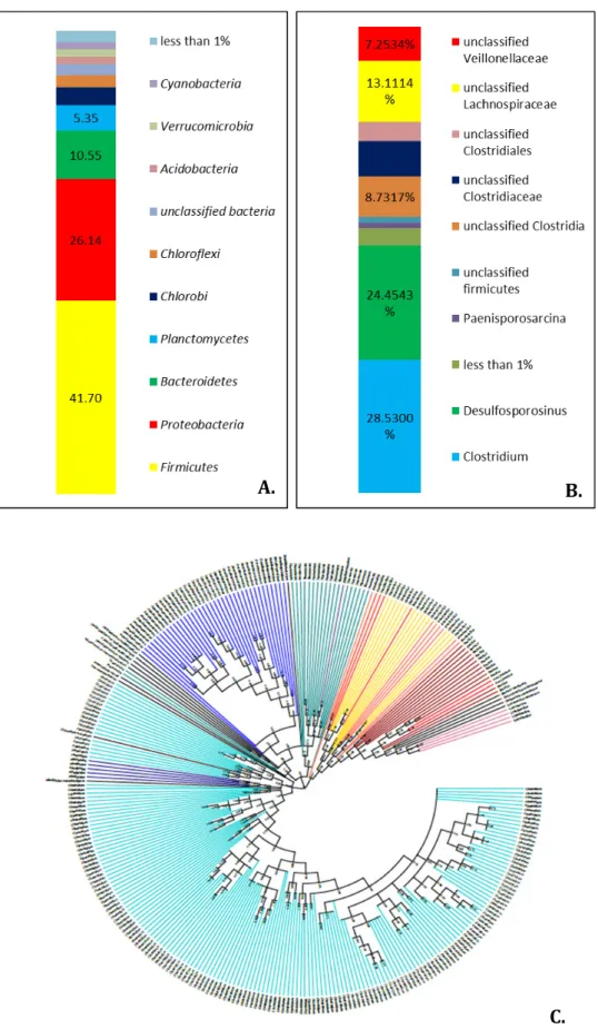
Chapter 12 Bacterial diversity in the sulfur and iron springs of Ponts‐de‐Martel, Neuchâtel ... 225
Abstract ... 226
12.1. Introduction ... 227
12.2. Materials and methods ... 228
12.2.1. Description of the site ... 228
12.2.2. Physicochemical measurements made in situ ... 229
12.2.3. Microelectrode analysis ... 229
12.2.4. Samples for molecular studies ... 229
12.2.5. DNA extraction, sequencing and analysis ... 229
12.3. Results and discussion ... 230
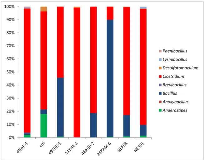
12.3.1. The sulfur spring (NeSul) ... 230
12.3.2. The iron spring (NeFer) ... 234
12.3.3. Overall conclusion ... 236
12.4. Acknowledgements ... 236
12.5. References ... 237
Annex 1 ... 239
Oral presentations at conferences ... 239
1.1. Thesis outline
This thesis studies two survival strategies that drive the ecology of endospore‐forming Firmicutes (EFF): their sporulation capability and their diverse metabolic activity. Although both abilities are encoded in the genetic material of EFF, they are mostly expressed under conditions limiting growth and survival.
In the first part of this dissertation, sporulation, observed in four bacterial taxa so far, is explored from an ecological point of view, trying to understand the costs and benefits of this survival strategy. A primary hypothesis is formulated: since sporulation is a beneficial strategy, spore‐forming bacteria should survive under unfavorable conditions and prevail everywhere. The ubiquity of EFF, however, has until now not been confirmed, based on previous literature.
Chapter two explores whether the under‐detection of EFF is due to methodological biases. This hypothesis was evaluated by testing the detection of EFF in metagenomic datasets. Two molecular markers unique to this bacterial group (spo0A and gpr) were screened for in 73 datasets, and found to be absent, with the exception of spo0A present in three mammalian gut microbiomes. An improved DNA extraction method resulting in the detection of a large diversity of endospore‐formers in amplicon sequencing of the 16S rRNA and spo0A genes, once applied to a sample from a geothermal spring, was insufficient to overcome the limitations for detecting EFF in whole‐genome metagenomic analysis. The results showed limitations in sequencing depth, coverage and annotation. The methodological biases, however, are not sufficient to explain the non‐detection of EFF in every environment. In chapter three, we argue that there is a pattern for the prevalence of EFF under limiting conditions. Indeed, we have observed that the co‐ occurrence of limiting environmental conditions increases the relative abundance of EFF to the whole bacterial community in mineral springs.
We have shown that environmental conditions, and more specifically, multiple limiting factors, play a role in the prevalence of EFF in the environment. Whether these factors can also be a driving force for the evolution of sporulation in species that are not known to sporulate, is discussed in chapters four and five. Serratia ureilytica, a hospital‐acquired pathogen, was isolated in a geothermal spring and found to produce spore‐like resistant structures. Biochemical analysis showed similarities to EFF spores and genome analysis revealed a potential lateral gene transfer of crucial sporulation genes from EFF to this strain. This discovery is described in chapter four. In chapter five, the discovery of another species that is not known to sporulate, Kurthia sp. str. 11kri321 is described. Its ecology and distribution as well as the genomic imprints of sporulation are shown.
The second part of this thesis contains numerous examples of metabolic capabilities found in EFF that make them capable of tolerating adverse conditions and thriving in extreme environments.
Chapter six includes the discovery of a novel Anoxybacillus species, Anoxybacillus
geothermalis, which was isolated from a geothermal reservoir. This strain was revived
reservoir since the Permian age. More isolates belonging to the same species were also discovered in different geothermal reservoirs.
Chapter seven focuses on other genomic imprints found in the genomes of EFF strains isolated from natural environments. These genomes have been sequenced for the first time and have been published as novel genome announcements.
Manganese oxidation and copper reduction of EFF in natural, uncontaminated environments, was studied and is presented in chapter eight. Our results lead to the conclusion that metal tolerance is a widespread phenomenon in unrelated aerobic EFF from natural uncontaminated environments.
Chapter nine discusses the limits of salt tolerance in aerobic and anaerobic EFF isolated from marine environments or salt lakes.
Finally, chapter ten is a general synthesis of this work and gives perspectives on future research. The ecological importance of sporulation under limiting conditions is also discussed. All conclusions drawn from these examples are summarized in chapter ten. Chapter eleven summarizes all the collaborations that have been made in parallel to this thesis. Chapter twelve contains an article submitted to the Bulletin de la Société des Sciences Naturelles de Neuchâtel, addressed to the Swiss broad public interested in nature, and describes the microbial community structure of the natural mineral springs of Ponts‐de‐Martel, located in the Jura of Neuchatel, from an ecological perspective.
1.2. Background 1.2.1. What is Ecology?
Pascal Acot, in the introduction of his Histoire de l’Ecologie [1] in 1988, described that since the beginning of mankind, man has observed the relationships between plants and animals, and even tried to use these relationships to favor him, like in the case of fishing using a worm as bait. However, he states that the role of an ecologist is not simply to observe but also to study the relationships among organisms and their environment and to reveal patterns that drive ecosystems and formulate laws to describe these patterns. This study, along with the proposed laws that govern habitats, is defined as Ecology. This definition is not far from those proposed already since 1866 by Ernst Haeckel (“the comprehensive science of the complex relationship of organisms to the environment [...] described by Darwin as the necessary conditions for existence”) [2], by Odum, in 1963 as the “study of the structure and function of nature” [3], and finally by Krebs, in 1972 as the “study of the interactions that determine the distribution and abundance of organisms” [4].
Mostly in the last two centuries, laws, principles and patterns about abundance, distribution and function of ecosystems have been proposed, at first for plants and then extrapolated or adapted to the animal kingdom. In 1970, Pierre Dansereau described ecological patterns and collected them in 27 laws [5], a selection of which are summarized herein. Any given environment cannot offer the optimum conditions for all of the functions for a given species (law of the inoptimum). However, a species can survive and grow in a habitat, depending on its limits of tolerance with regards to every
environmental factor (law of tolerance). Irrespective of the tolerance limits of an organism, stress will eventually occur and define the species’ distribution (law of climatic
stress). As a consequence, a species can spread in a geographical area, to a larger or
lesser extent, due to variations in environmental factors (law of valance), that in their turn create overlapping ecological niches, which allow a gradual change in community composition and structure (law of the continuum). However, these gradients, community or environmental, cannot be considered as static: they steepen or smoothen at various times and places. Species with fitness potential cease the opportunity to prevail (law of
cornering). Organisms share resources in a way that allows a greater portion to the most
efficient (law of competition and cooperation). Species tend to survive in their habitat, even when the latter is altered (law of persistence). The habitat (i.e. the nature and population structure of the communities, the geography and the environmental factors) defines the ecological success of a species (law of evolutionary opportunity). Species abundance is controlled by the scarcest resource, rather than by the most abundant (law
of factorial control or law of the minimum).
An unoccupied environment should be first inhabited by organisms with high tolerance and generally with low requirements (law of ecesis). The first settlers alter the environment, creating favorable conditions for other invaders, who, if fitter, may displace first settlers (law of succession). Succession should reach an equilibrium (law of regional
climax). During climate change, elimination of some species or abundance reduction may
occur through migration (law of association segregation). Migration is influenced not only by climate change, but also by population pressure (law of migration).
The local distribution of a plant depends on the long‐range geographical distribution, since short‐ and long‐scale environments are determined similarly (law of geoecological
distribution). Drastic events put a selective pressure on organisms: therefore, it is more
likely that differentiation occurs during these events than in other periods (law of
geological alternation) [5].
Rather than reviewing all 27 laws, in this introduction, a selected few on abundance and diversity are brought into focus.
Liebig’s law of the minimum [6] states that the abundance of species or biomass growth of an organism are controlled by the scarcest resource, rather than by the most copious. In more detail, for a specific organism in a given ecosystem, it should always be that a particular nutrient, or another environmental factor that influences growth, should determine the biomass or abundance of this organism in the ecosystem. Experimentally, the addition of this limited factor ought to increase the abundance of the species, provided that all the other requirements for growth are met. This law, however, overlooks biological alternation of environments, therefore of the provided nutrients, and is difficult to be applied in ecosystems, where more than one factor is scarce. It also focuses on the specific pair “organism‐given environmental factor”.
A fundamental ecological principle, Shelford’s law of tolerance, relates the role of environmental factors to plant and animal niche differentiation [2]. This law postulates that “[an organism] is absent or found in minimal numbers only […] should a(n)
[environmental] condition vary outside the limits tolerated by the animal” [7]. Such conditions, or factors, can be ‘operationally significant’ for the distribution of an individual, but they have also been extended to explain the distribution of a population or even a community [8–10].
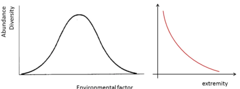
If abundance and diversity are taken into account, based on this ecological law, a general theoretical distribution model with maximum diversity and abundance towards the middle range of each environmental factor can be predicted. This model suggests that the distribution is unimodal. For some environmental factors, this is indeed the case: extremity acts the same in both directions, for example in the case of temperature or pH. For other environmental factors, however, the distribution is rather monotonic, as in the case of salinity. In both cases, therefore, a monotonic relationship can be presented as shown in Figure 1.1.
1.2.2. Microbial Ecology: from microscopes to metagenomes
The roots of all ecological laws described so far lie in the observations that ecologists made in nature based on plants and animals specimens, and then tested experimentally. The same principle of observation and experimental testing would not be possible for the microbial world, if it were not for the discovery of the light microscope. Indeed, Antonie
van Leeuwenhoek, an amateur microscope maker in the 17th century, was the first to
observe and describe in detail almost all the unicellular microorganisms that are known today: protozoa, algae, unicellular fungi, and bacteria [11]. These microscopic organisms were named animalcules [12], and soon a debate started concerning their origin. On the one hand, the theory of spontaneous generation was already well established. On the other hand, the idea that animalcules were transferred through air in boiled infusions, started being tested experimentally (Spallanzani). The debate lasted for more than a century until 1861, when Louis Pasteur performed a series of experiments to prove that firstly animalcules exist in air (by air‐sampling and microscopic observation); secondly, that such an air‐sample provokes microbial growth in sterile medium; and finally that a sterilized infusion remains sterile if placed in his renowned bent‐neck flasks, thus not in contact with air. Once this neck breaks, the sterile medium is contaminated and
Figure 1.1. Theoretical curves representing abundance and diversity of species. At extreme values of a given environmental factor, both abundance and diversity are lower. This bell‐shaped distribution curve can be summarized to a monotonal one, when conditions become in general adverse.
important animalcule growth is observed [12]. However, Pasteur did not win the battle until a decade later when Tyndall’s experiments on spontaneous generation supported Pasteur’s findings and led to the discovery of the sterilization method of discontinuous heating, known as tyndallisation. In parallel, the foundations of many biological processes of microorganisms were demonstrated, such as fermentation by yeasts and microbial anaerobic growth. In 1876, Robert Koch in his experiments on Bacillus anthracis showed that the causative agents of infectious diseases are bacteria. The same experiments were performed independently by Pasteur’s group and supported Koch’s findings.
A big step towards the establishment of Microbial Ecology was taken with the discoveries that microorganisms are geochemical agents and that they participate actively in geochemical cycles, as those of carbon, sulfur and nitrogen. This was mainly the work of M.W. Beijerinck and S. Winogradsky, who also developed the technique of enrichment cultures. This application is used until today, and for many decades almost all microbial ecology research has been based on this technique. Enrichment cultures for example depict natural selection at a microscale. Also, the successful creation of pure cultures has resulted in the isolation of many new species and their physiological description. In 1985, however, it was first demonstrated that microbes, which can be cultured, represent approximately 1% of the total diversity in soil [13]. This was called “the great plate anomaly” [14,15] and methodologies to overcome this issue started to emerge. The use of genes as indicators of biodiversity led to the direct detection of those genes in various environments. A milestone in the direct detection of genes was the description of the 16S rRNA gene as a phylogenetic marker by Carl Woese [13]. The 16S rRNA gene is a universal gene encoding the rRNA constitutive to the small subunit of the ribosome for bacteria. This gene contains both well‐conserved and variable regions. Since the discovery of the 16S rRNA gene marker and its application to environmental samples, the number of potentially novel species has steadily increased. This phylogenetic marker, along with other markers such as universal genes (rpoB, recA) or group‐specific genes (nif) has paved the way to a more detailed description of microbial communities in various environments.
For the last two decades, novel methodologies have emerged, resulting into the production of large gene sequence datasets and collection of associated environmental parameters in different habitats. This “era of metagenomics”, as it is more commonly named [16,17] has been facilitated by an exponential technological advancement, in favor of microbiology.
Although technological advances have provided scientists with the tools to describe the microbial world and answer the question “who”, still, the questions “why” and “how” need to be addressed.In 2007, an article published in Nature by Prosser et al., explained the importance of ecological theory in microbial ecology, the need to propose hypotheses, laws and patterns based on observations then reject, confirm or discuss them based on experiment [18]. This article also highlights the problem of many contemporary microbiological research articles that often resemble more to the fisherman’s than the ecologist’s conclusions in Pascal Acot’s example.
Whether plant and animal ecological laws are applicable to the microbial world is still to be determined. There is no doubt, however, that this question is an interesting challenge, since microbes set the limits beyond of what has been known for macro‐organisms: limits of tolerance and distribution, as well as survival strategies, are already discovered concepts, but need to be mapped against ecological laws. This is no easy task; it is still very difficult to conclude whether theories that are already established in the macro‐ organismal world, can be extrapolated to microbes. The difficulties lie mostly in the fact that microbes have set new grounds on the species concept and have extended the limits of life, the rates of dispersal, and the laws of reproduction.
Bacteria and archaea can be differentiated from other organisms based on several fundamental biological processes. First, prokaryotic microorganisms are morphologically simple: bacterial or archaeal cells come only in a few shapes and forms. This morphological simplicity does not allow distinction between species, as is often the case for eukaryotic organisms. Second, a new generation in the prokaryotic world can be produced as quickly as within twenty minutes. This short reproduction time, along with differences between the bacterial/archaeal and the eukaryotic replication machinery, influences dramatically the mutation rate and, as a consequence, the evolutionary context of microbes. Third, in bacteria and archaea there is only asexual reproduction. In addition, horizontal gene transfer among closely related but also between distinct species is a phenomenon mostly observed in prokaryotes.
Besides differences in fundamental biological processes, there are differences in the ecology, i.e. their interactions with the environment. Firstly, bacteria and archaea have high dispersal rates; this observation led to the suggestion that there is no geographical divergence for these microbes: they can be distributed everywhere and the environmental factors alone would drive the selection of each habitat. This concept, that “everything is everywhere, but the environment selects” was first described by M.W.
Beijerinck in the early 20th century and persists today. Second, their metabolisms are
more diversified than those found in plants and animals. Anaerobic growth, anoxygenic photosynthesis, chemolithotrophy are only few examples of metabolisms restricted to bacteria and archaea. Third, microorganisms have expanded the limits of life, as we know
them. They are able to survive temperatures as high as 120oC, pH as low as 1 and as high
as 13, high pressure and UV radiation. Survival to these conditions needs special adaptations rarely found in the macro‐organismal world.
Microbes, thus, can colonize previously well‐known habitats in potentially different patterns than those that drive the colonization by plants and animals.
1.2.3 Extreme habitats
Extreme is in the eyes of the beholder, stated Rothschild and Mancinelli about life in
extreme environments [19]. Most of the time we tend to follow anthropocentric definitions of normality and extremity; this results in a mesophilic and extremophilic categorization of the (microbial) world. A number of physicochemical parameters, such as temperature (10 to 45°C), pH (5.5 to 8), atmospheric pressure (~1 atm), salinity (up to
0.3M salt content), humidity (water activity (aw)> 0.8) and concentrations of elements or
chemical compounds (minimum inhibitory concentrations), are used to describe mesophilic conditions. In that context, any conditions departing from such given norms are by default defined as extreme. Habitats that present these conditions are also characterized as extreme (for life). In the case of multiple extreme conditions co‐ occurrence in natural ecosystems, habitats are then defined as ‘multiple extreme’.
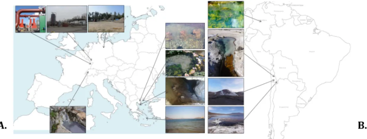
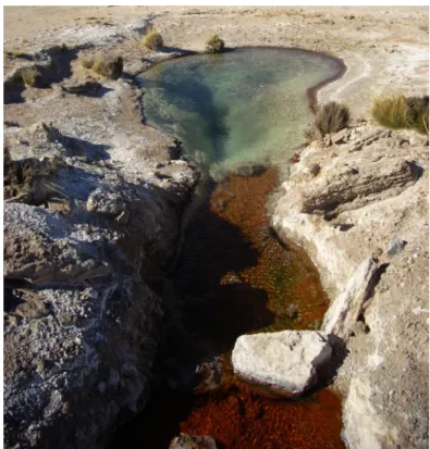
For this dissertation, the extreme habitats of geothermal reservoirs, geothermal natural springs, brines, desserts and highly contaminated environments with trace metals are considered.
Geothermal sites are terrestrial and hydrothermal (oceanic) hot springs or underground reservoirs that consist of highly pressurized water heated up from the earth mantle, in which minerals from the earth crust are dissolved. These environments house a large
variety of microorganisms. These microbes not only survive in the presence of CO2
(exclusively), inorganic constituents and with other minimal requirements, but they also exhibit high diversity [20,21]. As conditions in geothermal sites differ, so does the microbial metabolic activity and diversity.
In a community study published in 2013 at the Lower Geyser Basin, Yellowstone, Schubotz
et al. suggested that differences between communities in proximate but different sites
occur because of variances in microbial metabolism and because of the exogenous carbon input into these systems [20]. In other words, different cellular functions serving various microbial needs occur in different environments. Indeed, 16S rRNA gene sequencing and metagenomic analysis showed abundant Crenarchaeota and Aquificales, Thermales and Thermotogales in all sites, however the community composition changed significantly when sampling progressively downstream of the natural spring’s pool (decreasing temperature).
Environmental stress factors play an important role in the formation of community patterns. Distinct community types have been observed according to four environmental factors (pH, temperature, dissolved sulfide and sulfur, and physiographic site description) [22]. In addition, one single environmental factor is not enough to determine taxon segregation in a study of two alkaline hot springs that share temperature conditions but not water chemistry, as proposed by Weltzer and colleagues. They also introduced the idea that thermal gradients can create habitat gradients because of different temperature fitness of various taxa, resulting in differences in species richness and dissimilarity among sites [23]. Finally, studying the community composition and associated metabolism can also provide very informative insights. A study of Arctic and Antarctic microbial mat communities suggested that communities dominated by different phyla may vary in the response to stress at a genetic level [24]. In a later study, Takacs‐ Vesbach et al. discussed that different environments can favor different characteristics, even among members of the same family [25]. Indeed, evolutionary pressure in a given environment is what triggers these intra‐genera and intra‐family differentiations.
Saline environments are also considered extreme, since the concentrations of Na+, K+,
osmosis. Such saline environments are often natural, for example salt lakes, seashore evaporations or estuaries, but they can also be artificial brines [26]. Microorganisms capable of surviving under these conditions of total salinity 10 times higher than seawater [26] require specific cellular adaptations to withstand dehydration. These adaptations differ in regards to whether these microorganisms are salt‐requiring (halophiles) or salt‐ tolerant (halotolerant). Halophilic microorganisms withstand osmotic dehydration of their cytoplasm by accumulating potassium and chloride. High concentrations of potassium and chloride however can be toxic to proteins (denaturation) thus, extended proteomic modifications have been observed in these organisms. These proteins are now more acidic than those found in mesophilic organisms and denature in low salt concentrations [27]. For this reason, these organisms require salt for growth. Salt‐ tolerant organisms use a different strategy. When halotolerant bacteria are found in an environment where the external NaCl concentration is higher than the cytoplasm, water osmoses out of the cell, resulting in cell shrinkage. This decrease in cellular volume triggers the synthesis of compatible solutes and as a result, the cellular volume returns to the acceptable size for growth [26,27].
Environmental contamination by toxic metals, such as mercury, lead, copper, cadmium, zinc, manganese, nickel, cobalt, silver, gold, uranium, and thorium, can also have a significant impact on microbial populations. Metal contamination at toxic levels has a strong influence on biological processes, since metals are involved in practically every metabolic path, as ligands to enzymes, and consequently every index of microbial metabolic activity (respiration, methanogenesis and nitrogen fixation, among others) can be adversely affected by elevated concentrations of toxic metals [26,28–30]. Therefore, microorganisms that thrive in metal‐contaminated environments have developed a variety of strategies for their survival including detoxifying mechanisms such as bioaccumulation, biotransformation, biomineralization or biosorption [31–35].
Another important environmental factor is pH. Enzymes (and proteins in general) are very sensitive to changes in pH; however cells are able to control the cytoplasmic pH in order to avoid enzyme degradation and consequent death. The range of pH that most macro‐organisms can tolerate is from pH 5.5 to pH 8. However, acidic and alkaline environments exist, for example when soluble sulfur compounds are present or under high sodium carbonate concentrations, a pH as low as one and as high as eleven can be measured, respectively [26,36]. pH is an environmental factor that is influenced by other environmental parameters, such as temperature and salt and metal concentrations. Thus, thermophilic, halophilic or metal‐contaminated environments are frequently multiply extreme, since pH is an associated factor.
1.2.4. Survival strategies in the microbial world
In order to avoid extinction during unfavorable conditions, microbial communities have developed diverse survival strategies. One of the most common is the formation of biofilms. By using minerals as a substrate, bacteria of different species can form biofilms in which they survive and multiply. They are able to exchange plasmids, and genes that
code for traits that allow them to adapt and evolve. Biofilms consist of “bacterial neighborhoods” in which the community deals with the problem of limited nutrients in the “economical” way of using common resources. This strategy is considered to be an altruistic behavior among bacterial individuals in a microbial community, contributing to the survival, fitness and evolution of species [37].
Apart from community survival strategies, bacteria often develop individual characteristics in times of adversity. They can develop morphological plasticity [38], they can switch to low catabolic activity or to a metabolic stand‐by, or exhibit post‐ transcriptional modifications to optimize their fitness [39].
Dormancy is a well‐studied survival strategy, which simultaneously fulfills both categories described above, community and individual survival. It is defined as “a state whereby metabolism and normal progression of life activities and development are dramatically reduced or brought to a halt” [40]. It is an individual characteristic, since not all bacteria possess genes that allow them to switch from a vegetative to a dormant state. However, dormancy has also been described as a community strategy: many hypotheses have been advanced, suggesting that there is an “altruistic behavior” among dormant cells before, during and after entering the state of dormancy [41–43].
1.3 Sporulation as a survival strategy in adverse conditions 1.3.1. Sporulation
Sporulation is a dormancy state. It is described as a condition of low metabolic activity and shrinking in size (in most cases) [44]. Four taxa are known to produce spore or spore‐ like forms, structures that are more resistant to environmental stress than the equivalent vegetative forms. Spores are found within Firmicutes, Actinobacteria, Cyanobacteria, and in a genus of Proteobacteria, Myxococcus spp. Among these phyla, not all members can enter sporulation. A series of genes are necessary for this process. Absence or malfunction of one or more genes results in the failure of sporulation. Indeed, there are bacteria, which are not known as endospore‐formers, but that do encode some (but not all) sporulation genes in their genome. These bacteria cannot produce spores and for that reason they are called asporogenic. Sporulating bacteria undergo an intricate sequence of cell differentiation events leading to the formation of spores. These developmental processes can be used as a model of the evolution of structural and cellular functions. The spores, or spore‐like forms, that are produced by bacteria are significantly different between taxa, in terms of structure, sporulation procedure and resistance to limiting environmental conditions. Although the sporulation procedure differs significantly among these taxa, it can be described as a general 4‐step process. First, cells detect, in most cases, unfavorable conditions in their environment. Second, commitment to sporulation follows, as an irreversible step. Third, a dormant structure, mostly called a
spore, is then produced either as a result of a special cell division or a modification of the
vegetative cell structure. The spore structure is lighter, denser and more resistant than the vegetative mother cell they derived from, and less metabolically active. In all four taxa, this structure does not allow growth and replication, it does, however, permit
survival and dispersal. Finally, a mature spore is produced, ready to germinate once conditions are favorable again.
1.3.2. Sporulation in endospore-forming Firmicutes
Sporulation in EFF can be schematically described in five steps [44]. The initiation of sporulation takes place when the bacterial cell senses adverse conditions. Before changing morphologically, the vegetative cell programs the initiation of sporulation by activating the early sporulation genes. These are the 0/I stages. At stage II, the vegetative cell enters a particular cell division state, during which it creates a septum that is located at the one side of the cell and not in the middle, as in normal cell division. This special cell division and subsequent engulfing results in the production of a forespore inside the original vegetative cell, during stage III. However, before entering stage IV, this forespore transforms into a protoplast that has a double membrane layer. Between the two membranes the spore wall and cortex is formed. The spore coat is also produced to surround the forespore. These structures, which are not present in a vegetative cell, are all formed during stage IV. During stage V, the vegetative cell is lysed and the endospore is released [44].
The spore itself can alter its size, depending on humidity, without exiting dormancy [45]. During this dynamic state of low or no metabolic activity, the spore can survive for a very long time and once conditions are favorable again, it initiates germination to return to the vegetative state. Indeed, there are many publications claiming revival of endospore‐ forming bacteria, which have been in a dormant state for millions of years. An impressive example of such revival is the isolation of a 250 million years old Bacillus from a salt crystal [46].
Not all bacteria can enter sporulation. A series of genes are necessary for this process [47]. All endospore‐forming bacteria, known so far, belong to the phylum Firmicutes (Gram‐positive, low G+C content bacteria), although not all Firmicutes form these structures [48].
1.3.3. Sporulation in Cyanobacteria
Cyanobacteria are a very diverse phylum, in terms of metabolism and morphology. Some filamentous cyanobacteria are able to produce differentiated cell types that are capable of nitrogen fixation, called heterocysts [49]. These filamentous forms of cyanobacteria also form a resting cell, called an akinete. These forms are produced after light or
phosphate deprivation, but also at low temperatures, low K+ availability, and when their
C:N ratio is fluctuating [40]. Akinetes are often larger than the vegetative form, and have a certain metabolic activity, although lower than the vegetative cell or the heterocyst [40]. Through the process of akinete maturation, akinetes are subjected to different
metabolic stages. Young akinetes are able to fix CO2, however, metabolic rates decrease
progressively, and mature akinetes do not possess any functional photosystem and lack chlorophyll. A few reaction centers do exist, however, in the mature akinete, so that photosynthesis can be activated quickly during germination. Akinetes contain double the
quantity of DNA than their mother vegetative cell, and up to a ten‐fold of the protein content [40].
Not all Cyanobacteria produce akinetes. Some genera are able to produce other spore‐ like structures, such as exospores, baeocytes, and hormocytes [50].
1.3.4. Sporulation in Actinomycetes
Sporulation in Actinomycetes is a complex procedure, tightly related to germination, cell division and colony growth. Among Actinomycetes, the genus Streptomyces is a model for the description of the cell germination and sporulation cycle. Firstly, a Streptomyces spore germinates, producing one or more hyphae, which branch to form a vegetative mycelium [51]. That mycelium becomes more complex during exponential growth; apical growth and branching occur during this step. Upon nutrient limitation, sporulation‐ programmed hyphae are formed and multiple cell divisions take place [52]. Septal peptidoglycan synthesis takes place, and peptidoglycan is also formed between the spores. The final step includes lysis of the peptidoglycan in between the spores and release of the mature spore [11].
A family of activators, the SALP proteins (SsgA‐like proteins), controls sporulation in
Streptomyces. They are exclusively found in Actinomycetes. The SsgA protein
accumulates in the cell during mycelium growth, and upon reaching a crucial concentration it up‐regulates a series of genes involved in aerial growth and spore formation resulting in the activation of sporulation‐specific cell division. SsgB is responsible for the cessation of the aerial tip growth. SsgD regulates the formation of the peptidoglycan cell wall, while SsgG ensures that all sporulation septa are formed simultaneously. Finally, SsgE and SsgF are responsible for the correct autolysis of the peptidoglycan between spores during the maturation step [53].
1.3.5. Sporulation in Myxococcus
Sporulation in the genus Myxococcus is mostly studied in Myxococcus xanthus. Two different sporulation pathways are described; the first is a response to nutrient deprivation and the second one is induced by an increase of glycerol concentration in the microenvironment of the bacterium. The starvation‐induced sporulation takes place in nature, while the chemically (glycerol and other organic chemical compounds) induced pathway has been observed in the laboratory [54].
In the first sporulation pathway, upon detection of limiting nutrients in the environment, a re‐modeling of the rod‐shaped cell, rather than a cell division, takes place resulting in a spherical structure [55]. The whole developmental program from a vegetative cell to a resting spore lasts approximately 72 hours and is a complex procedure. Unlike endospore‐formation in Firmicutes that occurs individually, sporulation in Myxococcus is a multicellular process that involves intra‐cellular communication and sacrifice of the majority of the cells for the survival of the minority [56]. Upon nutrient deprivation, an
average of 105 cells aggregate into a mound, called a fruiting body. At the completion of
(approximately 80%) lyse. The peripheral cells maintain their rod shape and do not sporulate. A series of inter‐ and intra‐cellular signals induces sporulation in the rest of the cells [57]. During this last step, the bacterial genome is duplicated. In the second case, the glycerol‐induced sporulation, fewer cells are involved and all of them differentiate into spores within eight hours. Spore coats are thinner and each spore contains multiple genome copies [58].
A series of proteins are specific to sporulation and some of them are homologous to proteins responsible for sporulation in Firmicutes, such as the CbgA and FdgA that are homologs to SpoVR, and the ActA and ActB proteins that are homologs with CsgA in Firmicutes. Finally, the nfs operon encodes a series of proteins responsible for the production of viable spores [59].
1.3.6. Costs and benefits of sporulation
Whether sporulation is a costly or beneficial survival strategy is considered based on the fitness of a sporulating species in a given environment. According to Levin’s definition of fitness, it is “the extent to which an individual contributes its genes to future generations relative to other individuals in the same population; i.e. the individual’s relative reproductive success” [60]. This is a general definition that applies to all organisms. When it comes to bacteria, however, it is not only the number of offspring (generation time and growth rate) that is taken into account but also the resistance and survival under given conditions [61].
As previously presented, sporulation in any of the above‐mentioned taxa, is a complex developmental procedure that is controlled by a series of factors unique to spore‐ formation. These extra genes that encode for these factors are an addition to the bacterial chromosome. To maintain a larger chromosome is energetically costly. Thus, under constant optimal conditions, spore‐forming bacteria tend to lose their sporulation genes, and as a consequence, their sporulation capability, in order to maximize fitness. When environmental conditions are adverse, in order to enter sporulation, the bacterial cell needs to decide whether entering sporulation would be beneficial or not. During sporulation, extra proteins are synthetized to produce a specialized structure. This procedure is very energetically demanding and commitment to sporulation is therefore a decision that the cell takes as a last resort. Under catastrophic conditions, sporulation has a clear survival advantage, as it may be the only way to avoid cellular death.
1.4. Survival by adaptation
In order to withstand adverse conditions, microbes alter their physiology to adapt accordingly, and consequently thrive in extreme habitats.
In hotspots, such as geothermal reservoirs, physiological processes are generally less efficient than in mesophilic conditions. At high temperatures, microbes face an irreversible breakdown of their biomolecules and a disruptive high fluidity of their plasma membrane [62]. Consequently, thermophilic and hyperthermophilic organisms evolved thermostable proteins and enzymes and their cell membranes have a different
composition. The stability of their proteins is guaranteed through extra chemical bonds (S‐S bridges, H‐bonds, metal bindings) [63], by hydrophobic amino acids, by producing multiple small subunits for the formation of an enzyme, and by the presence of chaperone proteins. Stability of proteins by extra chemical bonds results in rigid enzymes that seem to be less productive catalysts than mesophilic enzymes. Thermophiles seem to use thermal energy to overcome this reduction. A second adaptation is the accumulation of chemicals, such as amines and polyamines, which increase the stability of NADH, ATP and amino acids [50]. A final adaptation concerns the integrity of the plasma membrane, as at high temperatures, the fluidity of the plasma membrane increases. The stability of the membrane is guaranteed through an increase in the length of carbon chains and an extension of the branching of the phospholipids, an accumulation of saturated phospholipids, and through changes in the heads of the phospholipids [50, 64]. At low temperatures, microorganisms face the low availability of liquid water, and the damages to cellular integrity caused by the formation of ice crystals. The cellular response to cold is a change of the membrane composition; as expected, the opposite modifications of those taking place in thermophiles are made: unsaturated and shorter fatty acids are incorporated into the cellular membrane, while branching between lipids is also limited [65]. These fatty acids make the membrane more fluid because they introduce gaps that push apart the components of the membrane. At the interior of the cell, they contain antifreeze agents (mainly sugars) and small acidic proteins, which prevent ice formation [62]. Moreover, their proteins tend to have more α‐helices than β‐ folded sheets, to allow flexibility [63]. Finally, psychrophiles are found to have a unique cold‐stable translational system [50].
In highly acidic or highly alkaline habitats, microbes enable processes for gaining energy and carrying out chemical reactions inside the cell in order to regulate the intracellular pH to neutral. This is mainly accomplished through pumping protons out or into the cell,
respectively. In an acidic environment, a K+/H+ antiporter is pumping K+ in and H+ out to
make the cytoplasm alkaline. In a basic environment, a Na+/H+ antiporter is pumping H+ in
and Na+ out to produce the opposite effect [50].
In highly saline as well as dry habitats, microorganisms struggle with high osmotic pressure and low water availability. Moreover, the membrane integrity is threatened by disruption due to dryness or salinity. In both saline and dry habitats, microbes control the water loss from the cell by producing compatible solutes [66]. These molecules are polar, water‐soluble and are capable of stabilizing proteins [50]. Examples of organic compatible solutes in bacteria are glycine betaine, ectoine, and trehalose.
Osmoprotection can be accomplished by accumulation of potassium chloride. Cl‐ and K+
are transported separately into the cytoplasm. KCl is formed to counterbalance the high concentration of NaCl that is found outside the membrane [67].
Osmotic pressure is only one challenge that prokaryotes face. There is also high atmospheric pressure, under which the tight packing of molecules and the loss of fluidity of the cellular membrane result in impaired cellular functions. Microorganisms have developed mechanisms of alternative gene expression in order to produce molecules
that enhance the uptake of nutrients and a change in the membrane structure, by incorporating unsaturated fatty acids to guarantee fluidity.
Microorganisms are in contact with light in most habitats, and for phototrophs light is their energy source. However, ultraviolet (UV) light and ionizing radiation are extremely harmful because the cells only perform a limited repair of damaged DNA. Bacteria can survive only low exposure to this radiation since they can only repair limited DNA damage. Some extremophilic bacteria can survive very high levels of radiation (thousand times higher than other cells) due to advanced DNA repair mechanisms, and due to their specialized cell membrane and cell wall. Moreover, these organisms often have multiple copies of their genomes at stationary phase. However, this resistance is highly costly: DNA damage repair and the replication of a 9‐fold genome both demand high amounts of energy [50].
Finally, the chemical composition of the environment plays an important role in microbial survival. Low nutrient availability or presence of harmful compounds result in a fatal reduction of biomolecule synthesis and enzyme productivity. Slow metabolic activity and production of extracellular ‐often polysaccharides‐ mucus or an impermeable cell wall, in the case of harmful chemicals, are the main adaptations for microorganisms under such conditions [62].
Needless to say that impaired cellular functions under the above‐mentioned extreme conditions are confronted by entering dormancy, as well. At the edges of extremity, this could be the last resort solution.
1.4.1 Adaptation under extreme conditions at a molecular level
From a genetic point of view, the above‐mentioned adaptations to extreme environments are imprinted as molecular modifications; they are either genomic imprints, or post‐transcriptional and post‐translational modifications. The metabolic adaptations that are imprinted in the genomes of microbes and the modifications at an intracellular level are summarized herein.
In thermophilic and hyperthermophilic bacteria and archaea a series of modifications at the genomic level are necessary for the cells to withstand high temperatures. Firstly, changes at the amino acid level are observed; proteins of thermophiles contain mostly alanine, threonine, arginine and glutamic acid residues, while amino acids that enable flexibility of the protein are rare [68]. Moreover, the overall codon usage and nucleotide content, especially concerning rRNA and tRNA, vary significantly between mesophiles and thermophiles [69]. Secondly, multiple chaperone genes are found in the genomes of thermophiles [50, 70]. Thirdly, genes that can be used as thermophily‐specific biomarkers have been defined and are related to the supercoiling of the circular DNA [71], DNA repair and transcription regulation [70]. Genes that encode for proteins related to metal detoxification have also been identified [50, 70]. Finally, differences at the level of gene expression are also observed between mesophiles and thermophiles, especially concerning genes for amino acid synthetases and the regulation of these genes [72].