Drosophila germ-line modulation of insulin signaling
and lifespan
Thomas Flatt†, Kyung-Jin Min†‡, Cecilia D’Alterio§, Eugenia Villa-Cuesta†, John Cumbers†, Ruth Lehmann¶,
D. Leanne Jones§, and Marc Tatar†储
†Division of Biology and Medicine, Department of Ecology and Evolutionary Biology, Brown University, Box G-W, Providence, RI 02912;‡Department of Biological Sciences, University of Alaska, Anchorage, AK 99508;§The Salk Institute for Biological Studies, Laboratory of Genetics, 10010 N. Torrey Pines Road, La Jolla, CA 92037; and¶Howard Hughes Medical Institute, Developmental Genetics Program, Skirball Institute of Biomolecular Medicine and Department of Cell Biology, New York University School of Medicine, New York, NY 10016
Edited by Allan C. Spradling, Carnegie Institution of Washington, Baltimore, MD, and approved March 6, 2008 (received for review September 25, 2007)
Ablation of germ-line precursor cells in Caenorhabditis elegans extends lifespan by activating DAF-16, a forkhead transcription factor (FOXO) repressed by insulin/insulin-like growth factor (IGF) signaling (IIS). Signals from the gonad might thus regulate whole-organism aging by modulating IIS. To date, the details of this systemic regulation of aging by the reproductive system are not understood, and it is unknown whether such effects are evolu-tionarily conserved. Here we report that eliminating germ cells (GCs) in Drosophila melanogaster increases lifespan and modulates insulin signaling. Long-lived germ-line-less flies show increased production of Drosophila insulin-like peptides (dilps) and hypogly-cemia but simultaneously exhibit several characteristics of IIS impedance, as indicated by up-regulation of the Drosophila FOXO (dFOXO) target genes 4E-BP and l (2)efl and the insulin/IGF-binding protein IMP-L2. These results suggest that signals from the gonad regulate lifespan and modulate insulin sensitivity in the fly and that the gonadal regulation of aging is evolutionarily conserved. aging兩 endocrine regulation 兩 reproduction 兩 longevity 兩 metabolism
A
side from dietary restriction, inhibition of reproduction is one of the most effective ways to extend animal lifespan (1–3). Despite the generality of this effect, the mechanisms by which reproduction regulates aging remain unknown (4–6). Progress toward this goal has been made with the nematode Caenorhabditis elegans (7–9). Ablation of germ-line precursor cells is sufficient to extend lifespan, and overproliferation of germ cells (GCs) shortens lifespan. In contrast, ablation of the entire gonad has no impact on longevity (7–8). These observations suggest that there is a balance between longevity assurance signals from the somatic gonad and signals from the germ line that promote aging (7). Longevity extension by germ-line ablation depends on DAF-16, the C. elegans ortholog of the forkhead transcription factor (FOXO), which is also required for longevity extension by reduced insulin/insulin-like growth factor (IGF) signaling (IIS) (7–8). To date, however, little is known about how signals from reproductive tissues systemically affect lifespan and whether the model developed in C. elegans is relevant to animals beyond the nematode (4–6, 9). Here we investigate this problem in the fruit fly, Drosophila melanogaster.Several methods to inhibit reproduction extend Drosophila life-span: removing oviposition substrate (10), reducing egg production (10–13), and inhibiting mating (14, 15). However, reproduction can also be reduced without affecting lifespan (3, 16–19), and whether loss of GCs extends fly lifespan remains unclear (4). Irradiation or the female-sterile mutation ovoD1induce sterility and extend life-span (12, 13), but whether these manipulations do so because they damage GCs or disrupt processes upstream of germ-line activity is unknown (4, 20, 21). Interestingly, a recent study suggests that GC ablation might not extend lifespan in D. melanogaster (21). Failure to form primordial GCs in grandchildless-like mutants (tudor, germ cell-less, oskar) increases lifespan only slightly or not at all (ref. 21 and our unpublished data). However, this finding is at odds with the observation that lack of a primordial germ line in a Drosophila
subobscura grandchildless mutant extends lifespan (11, 22). Thus, in contrast to the worm, how reproductive processes modulate aging in the fly remains poorly understood (3–5, 23).
One reason for the discrepancies in fly studies might be that some grandchildless-like mutations impact the development of the so-matic gonad (21, 24), perhaps precluding the capacity of this tissue to produce longevity assurance signals (7, 21). If so, GCs might modulate aging, but only when the somatic gonad has matured in the presence of the germ line during development. Moreover, because grandchildless-like mutants act during development (25), their impact on adult demography might involve pleiotropic effects independent of aging (5, 21). We therefore sought an alternative system that eliminates GCs exclusively in late development or the adult to test whether the D. melanogaster germ line modulates aging. Here we investigate the impact of GC loss induced through misexpression of bag of marbles (bam). In females, bam is necessary and sufficient for differentiation of GCs, and overexpression of bam⫹in GCs leads to precocious differentiation and subsequent loss of GCs (26–28). In males, bam limits mitotic amplification divisions of spermatogonia, which occur before the initiation of terminal differentiation into spermatocytes. Overexpression of bam⫹ in early male GCs causes GC loss, presumably through apoptosis (29). By manipulating bam, we investigate the impact of GC ablation on aging and find that loss of GCs in female and male flies extends lifespan and modulates insulin signaling.
Results and Discussion
Ectopic misexpression of bam⫹in the female germ line, by using the binary GAL4⬎UAS system or heat shock-induction, eliminates GCs (Fig. 1) (27, 28). Previous data suggest that the lost GCs are germ-line stem cells (GSCs): heat shock-induced bam⫹expression causes GC loss, but GCs that were not GSCs at the time of heat shock develop normally (27). Although grandchildless-like mutants lack pole cells and cannot form primordial GCs (21–22, 25, 30), heat shock-induced bam⫹overexpression eliminates female GSCs in the third larval instar (L3) or later but not before the L3 stage (27). When driving constitutive overexpression of UASp-bam⫹(28) with the germ-line-specific nanos (nos)-GAL4::VP16 driver (31), we found that GC loss continues in adult females, after the ovary has completed development. Females initially have the capacity to lay a small number of eggs but become fully sterile by day 7 [Fig. 1 and supporting information (SI) Fig. S1]. Similarly, in males, bam⫹ overexpression induced GC depopulation in the L3 stage or later
Author contributions: T.F., R.L., D.L.J., and M.T. designed research; T.F., K.-J.M., C.D., E.V.-C., J.C., and D.L.J. performed research; R.L. and D.L.J. contributed new reagents/analytic tools; T.F. analyzed data; and T.F., R.L., D.L.J., and M.T. wrote the paper.
The authors declare no conflict of interest. This article is a PNAS Direct Submission.
储To whom correspondence should be addressed. E-mail: marc㛭tatar@brown.edu. This article contains supporting information online atwww.pnas.org/cgi/content/full/ 0709128105/DCSupplemental.
© 2008 by The National Academy of Sciences of the USA
(Fig. 1) (29). Moreover, bam⫹overexpression caused a dramatic expansion of somatic cells in ovaries and testes (Fig. 1), reminiscent of the enlarged somatic gonads of agametic grandchildless-like mutants (21, 24). Thus, grandchildless-like mutants and flies misex-pressing bam⫹have expanded somatic gonads but complete GC loss at different times.
GC loss induced by misexpression of bam⫹significantly increased lifespan in females and males, in several independent experiments
(Fig. 2 andTable S1). Lifespan was increased by 31.3% and 50% in females and 21% and 27.8% in males by GC ablation in a y w background by driving y w;UASp-bam⫹ with nos-GAL4::VP16; effects are relative to a coisogenic control (y w;UASp-bam⫹; control 1) and a control with a heterozygous background (y w/w1118; nos-GAL4::VP16; control 2) (Fig. 2 A and B and Table S1). Longevity was also extended when UASp-bam⫹ was driven by
nos-GAL4::VP16 in an independent background (w1118) lacking
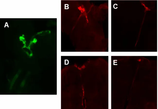
one copy of genomic bam (Fig. 2 C and D;Table S1). The capacity for GC ablation to extend lifespan was likewise effective with the germ-line driver nos-GAL4-tubulin (NGT-GAL4) (32) in the y w and w1118backgrounds (Fig. 2 E–H andTable S1). Thus, bam⫹ misexpression in the germ line is sufficient to force GC loss and to increase lifespan in multiple genetic backgrounds and with different germ-line drivers. Because the failure of grandchildless-like mutants to develop GCs has no consistent major effects on lifespan (ref. 21 and our unpublished data), we hypothesize that GC loss during late Fig. 1. GC loss and expansion of somatic cells in gonads from flies misexpressing
bam⫹. (A–C) GC loss in adult females misexpressing bam⫹in the germ line (y w/w1118;UASp-bam⫹::gfp/⫹; nos-GAL4::VP16/⫹). GCs are stained for GC specific antigen vasa (green), somatic cells (FasIII, red), and DNA (DAPI, blue). Asterisk (*) denotes somatic cap cells. (A) GCs are rarely observed in ovarioles from 2-day-old females overexpressing bam⫹[24/225 ovarioles (10.7%) contained GCs]. (B) GC loss is virtually complete by 7 days [3/325 ovarioles (0.9%) contained GCs]. See also
Fig. S1. (C) Germarium from a 2-day-old control female. All ovarioles from control females contained GCs at 2 (n⫽ 120) and at 7 days (n ⫽ 152). (A⬘–C⬘) shows FasIII⫹ somatic cells only. Note the expanded somatic gonad in GC-less females. (D and E) GC loss in third instar larval (L3) males overexpressing bam⫹. Control testes from L3 males (D) have a normal number of GSCs (vasa) in contact with hub cells (*) at the apical tip. (E) Males overexpressing bam⫹show loss of GSCs by this stage. (E⬘) GCs present near the hub have branched fusomes (arrows) as detected by staining with antibodies to␣-spectrin. Note the reduced size of the gonad. Asterisk (*) denotes the FasIII⫹apical hub. (F and G) Expansion of somatic cells in testes from adults misexpressing bam⫹. (F) Control testes show normal distribution of FasIII⫹ hub cells (red) and TJ⫹somatic cells (green). (G) An expansion of FasIII⫹and TJ⫹ somatic cells is observed in testes. (Scale bars: 50m in A–C, F, and G; 20 m in D and E.)
Fig. 2. Adult GC loss extends lifespan in D. melanogaster. (A–D) Driving UASp-bam⫹in germ line (no germ line, UASp-bam⫹/⫹; nos-GAL4::VP16/⫹) ex-tends lifespan, both in a y w background (A, females; B, males) and a w1118 background lacking one copy of bam (C, females; D, males), relative to two controls. Controls were, in the y w background, y w/y w;UASp-bam⫹::gfp/⫹ (control 1) and y w/w1118; nos-GAL4::VP16/⫹ (control 2), and in the w1118 back-ground, w1118/w1118;UASp-bam⫹::gfp/⫹; bam⌬86/⫹ (control 1) and w1118/w1118; nos-GAL4::VP16/⫹ (control 2). (E–H) Misexpressing UASp-bam⫹with an alterna-tive germ-line-specific driver, nos-GAL4-tubulin (UASp-bam⫹::gfp/NGT-GAL4), also extends lifespan, both in the y w background (E, females; F, males) and in a background lacking one copy of bam (G, females; H, males) compared with two controls (control 1: y w/y w;UASp-bam⫹::gfp/⫹ or w1118/y w;UASp-bam⫹::gfp/⫹; bam⌬86/⫹, respectively; control 2: y w/y1w*; NGT-GAL4/⫹). SeeTable S1for statistics.
Flatt et al. PNAS 兩 April 29, 2008 兩 vol. 105 兩 no. 17 兩 6369
development or in the adult might promote longevity because GCs associate and interact with somatic cells before loss.
If the germ line produces a signal that shortens lifespan or represses a somatic signal that extends lifespan, GC overprolifera-tion should decrease lifespan (7). To test this predicoverprolifera-tion, we examined a sterile heteroallelic null mutant of bam (bam⌬86/ bam⌬59) in which mitotically active, nondifferentiating GSCs over-proliferate (26). Mutant flies were short-lived relative to two fertile controls (Fig. S2andTable S1). Thus, eliminating GC proliferation slows aging, whereas GC overproliferation shortens lifespan in the fly, as in the nematode (7, 8). However, we cannot fully exclude the possibility that the longevity effects of bam are independent of its effects on GCs.
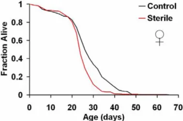
Germ-line loss might slow aging simply by abolishing the survival costs of producing gametes (1–2, 4, 21, 23). To rule out that egg production is required for GCs to shorten lifespan, we examined a female-sterile mutant of egalitarian (egl) (33). Mutants of egl prevent differentiation of cystoblasts into oocytes (34). Conse-quently, flies produce eggs with 16 rather than 15 nurse cells, and egg chambers degenerate before they acquire yolk (34). Lifespan of sterile egl mutant females (eglPR29/eglwu50) was reduced compared
with fertile controls (Fig. S3andTable S1), suggesting that oogen-esis per se might not be sufficient for reproduction to shorten
lifespan. This result adds to a growing number of cases showing that the tradeoff between reproduction and survival can be decoupled (3, 16–19, 21, 23).
In C. elegans, lifespan extension by GC loss requires the FOXO transcription factor DAF-16; FOXO activity is normally repressed by IIS (7–8). Because reduced IIS slows Drosophila aging [by mutations disrupting IIS (13, 35), constitutive activation of
Dro-sophila FOXO (dFOXO) (18), or ablation of insulin-producing cells
(36, 37)], we reasoned that GC loss might extend lifespan by down-regulating IIS. Accordingly, we measured message abun-dance for the three Drosophila insulin-like peptides (dilps) pro-duced by median neurosecretory cells (mNSCs), the major insulin-producing cells (IPCs) in the brain of the adult (Fig. S4A) (38–40). Rather than reduced message from the dilp2, dilp3, and dilp5 loci, we found that these transcripts were induced upon GC loss by 1.8-to 26-fold relative 1.8-to controls, in two independent genetic back-grounds (Fig. 3 A–C and D–F).
Previous attempts to quantify DILPs by Western blot analysis have failed because of low ligand abundance (37), and current technology does not permit detection of circulating DILPs in the hemolymph. However, several observations suggest that increased
dilp message in GC-ablated flies might be biologically meaningful.
Immunostaining of brains with DILP antibody indicated that the Fig. 3. GC loss up-regulates dilp message but activates expression of dFOXO target genes. (A–D) GC-less flies (UASp-bam⫹/⫹; nos-GAL4::VP16/⫹) exhibit increased production of dilp 2, dilp 3, and dilp 5, both in the y w (A–C) and w1118backgrounds (D–F), relative to controls. (G–H) GC loss causes up-regulation of dFOXO targets 4E-BP (G) and l (2)efl (H) in both backgrounds. For details of genotypes, see Fig. 1.
IPCs of GC-less flies produced as much and, in some cases, more DILP protein than controls, and DILP⫹staining of IPC axonal projections was strong, suggesting functional DILP transport (Fig. S4 B–E). Furthermore, neural DILPs homeostatically regulate sugar levels in the hemolymph (37, 40), and GC-less flies had reduced amounts of stored and circulating carbohydrates (Fig. S5 A and B).
The hyperinsulinism of GC-less flies is a paradox because lifespan should not be extended in the face of increased DILPs. Because high DILP levels should activate IIS in peripheral tissues and repress dFOXO, we measured transcripts of two major dFOXO targets from body tissue, the translational regulator thor (encoding 4E-BP), and the small heat shock protein l (2)efl, which are normally induced when IIS is repressed and dFOXO is activated (41–43). Message levels of both dFOXO targets were up-regulated in GC knockout flies (Fig. 3 G and H). Although we cannot rule out that these targets have transcriptional inputs other than dFOXO (44), flies with GC loss, despite elevated DILPs, express markers consistent with active dFOXO and reduced IIS.
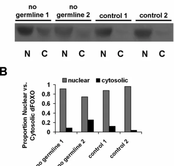
Because reduced IIS causes dephosphorylation and nuclear translocation of dFOXO, nuclear accumulation of dFOXO can be used to assess IIS pathway activity. To confirm that dFOXO is active in GC-less flies, we examined its localization with immunostaining in peripheral fat body, a major site of IIS activity, and by Western blotting analysis with cell fractionation in whole-body tissue (Figs. S6 and S7). As expected, dFOXO was predominantly nuclear in GC flies, indicating that dFOXO is active. Yet, despite differential up-regulation of dFOXO targets, GC-less and control flies did not differ in nuclear dFOXO localization (Figs. S6 and S7), which suggests that GC loss might affect dFOXO activity independent of its subcellular localiza-tion, as recently found in C. elegans (45).
There are many mechanisms by which IIS can be impeded between the site of insulin production and FOXO-dependent responses of peripheral tissues: at the level of insulin secretion or transport and at many steps within intracellular IIS of target tissues (46, 47). To initiate an understanding of IIS impedance in GC-less flies, we explored whether GC loss might change transcript abun-dance of two DILP cofactors, dALS and IMP-L2 (48–50). In mammals, circulating IGFs form a complex consisting of IGF-1, IGF-binding proteins (IGF-BPs), and the liver-secreted scaffold protein acid labile substrate (ALS); by creating a pool of circulating IGFs, this ternary complex limits ligand availability (51). The
Drosophila homolog of ALS (dALS) is expressed in
DILP-expressing IPCs and the fat body (48, 52) and is up-regulated in
dFoxo null mutants (53). Consistent with the model that dALS
functions as a DILP cofactor, dALS forms a circulating trimeric complex containing DILP2 and IMP-L2, an Ig-like homolog of IGF-BP7 (48, 54). Binding of dALS requires prior formation of a dimeric complex containing DILP2 and IMP-L2 (48). In cell culture experiments, IMP-L2 binds mammalian insulin and IGF-1/-2, and fall army worm (Spodoptera frugiperda) IMP-L2 inhibits IIS through the human insulin receptor (55). Because overexpression of dALS and IMP-L2 can systemically antagonize DILP function and IIS in
Drosophila in vivo (48, 49), we measured message abundance of dALS and IMP-L2 upon GC loss. Although dALS levels did not
change, IMP-L2 message was increased 7-fold in GC-less flies (Fig. 4 A and B). Although this observation is correlational, it might suggest a potential explanation for why IIS might be impeded in GC-less flies in the face of elevated DILP production. It will be of major interest to determine whether GC loss can modulate DILP availability and IIS by affecting IMP-L2.
Together, our results show that GCs regulate aging and modulate IIS in the fly. Although future work is required to fully characterize IIS state upon GC loss, we observed that GC-less flies exhibit characteristics of both increased and decreased IIS. Increased DILPs and hypoglycemia are suggestive of increased IIS, but GC-less flies also have markers of IIS impedance. The
induction of dFOXO targets is consistent with the finding that lifespan extension by GC loss in the nematode requires FOXO/ DAF-16 (7–8). In the worm, GC loss induces nuclear translo-cation of DAF-16 and activates DAF-16 targets, but nuclear accumulation is also observed in worms that lack the entire gonad and have normal lifespan (45). Similarly, we find that GC-less and control flies differ in dFOXO target activation, but not dFOXO localization, suggesting that IIS can affect aging by modulating FOXO/DAF-16 activity independent of subcellular localization. Indeed, dietary restriction in C. elegans extends longevity by activating AMP-activated protein kinase (AMPK), which phosphorylates and activates DAF-16 but does not pro-mote DAF-16 nuclear translocation (56).
Because extended longevity by GC loss is associated with up-regulation of DILPs, GC loss might impede IIS downstream of DILP production. In humans, compensatory hyperinsulinemia is a hallmark of severe insulin resistance (57), and mutations in the tyrosine kinase domain of the insulin receptor can cause hyperin-sulinemic hypoglycemia coupled with insulin resistance (58). Re-cent studies with fly and mouse also suggest that lifespan can be extended despite hyperinsulinemia (59, 60). In Drosophila
target-of-rapamycin (dTOR) mutants, longevity extension is associated
with elevated DILP2 and hypoglycemia (59), and brain-specific
insulin receptor substrate-2 (Irs-2) knockout mice are
hyperinsuline-mic but insulin-resistant and long-lived (60). Clearly, further ex-periments are needed to unravel the mechanisms by which insulin production can be uncoupled from IIS sensitivity and modulation of lifespan.
Our finding that GC loss affects neural DILP production also adds to growing evidence suggesting evolutionary conservation of endocrine feedback between brain and gonad (61). In Drosophila, neural DILPs bind to the insulin-like receptor (dINR) on GSCs to regulate GC proliferation (62, 63), and neuronal InR knockout (NIRKO) mice show impaired spermatogenesis and ovarian follicle maturation (64). Conversely, in rats, ovariectomy decreases IGF-1 receptor density in the brain but increases circulating IGF-1 levels (65). Together with progress made in the worm (7–9, 66) and mouse (67), the Drosophila system will allow us to dissect the mechanisms underlying the fundamental and intricate relationship among IIS, reproduction, and aging.
Materials and Methods
Fly Strains and Maintenance. For bam⫹misexpression, we used UASp-bam⫹in a y w background, obtained by backcrossing w; [w⫹;UASp-bam⫹::gfp]/CyO; Fig. 4. GC loss up-regulates message of IMP-L2, but not of dALS. (A) GC loss does not alter message abundance of the DILP cofactor dALS. (B) In contrast, GC loss strongly up-regulates expression of the insulin/IGF- binding protein IMP-L2. See Fig. 1 for genotype information.
Flatt et al. PNAS 兩 April 29, 2008 兩 vol. 105 兩 no. 17 兩 6371
bam⌬86/TM3 (28) for six generations into y w and eliminating the bam mutant allele. We also misexpressed bam⫹in a w1118background lacking one copy of bam (w; [w⫹;UASp-bam⫹::gfp]/CyO; bam⌬86/TM3). For each UAS responder, we in-duced expression with two germ-line-specific nanos (nos)-GAL4 lines (w1118;⫹/⫹; nos-GAL4::VP16 [MVD1] and y1w*; NGT-GAL4 [nos-GAL4-tubulin]) (31, 32). Thus, we examined how bam⫹misexpression affects lifespan in four sets of genotypes: (i) no germ line: y w/w1118;UASp-bam⫹::gfp/⫹; nos-GAL4::VP16/⫹; control 1: y w/y w;UASp-bam⫹::gfp/⫹; control 2: y w/w1118; nos-GAL4::VP16/⫹; (ii) no germ line: w1118/w1118;UASp-bam⫹::gfp/⫹; nos-GAL4::VP16/bam⌬86; control 1: w1118/w1118; UASp-bam⫹::gfp/⫹; bam⌬86/⫹; control 2: w1118/w1118; nos-GAL4::VP16/⫹ (control 2); (iii) no germ line: y w/y1w*;UASp-bam⫹::gfp/NGT-GAL4; control 1: y w/y w;UASp-bam⫹::gfp/⫹; control 2: y w/y1w*; NGT-GAL4 (Fig. 2 E and F); and (iv) no germ line: w1118/y1w*;UASp-bam⫹::gfp/NGT-GAL4; bam⌬86/⫹; control 1: w1118/y w;UASp-bam⫹::gfp/⫹; bam⌬86/⫹; control 2: y w/y1w*; NGT-GAL4/⫹. For the assay inFig. S2, we used a heteroallelic null mutant (ry506e1bam⌬86/red e bam⌬59) and two heterozygous controls (w1118/⫹; red e bam⌬59/⫹ and w1118/⫹; ry506e1
bam⌬86). bam⌬59is an undescribed deletion, and bam⌬86is described in ref. 26. The assay inFig. S3was performed with a heteroallelic mutant (w; cn bw eglPR29/cn bw
eglwu50) and a control overexpressing egl⫹in the mutant (w; cn bw eglPR29/cn bw
eglwu50; CA8B(egl⫹)/⫹) (33, 34). UASp-bam⫹, bam mutants, and w1118were
do-nated by D. McKearin (University of Texas Southwestern Medical Center, Dallas); y w by E. Rulifson (University of California, San Francisco); nos-GAL4::VP16 and egl by R.L.; NGT-GAL4 strain by the Bloomington Stock Center (Bloomington, IN); dilp2-GAL4 by E. Hafen (ETH Zu¨rich); and UAS-CD8::gfp by R. Stocker (Universite´ de Fribourg, Fribourg, Switzerland). Flies were reared and experiments were conducted at 25°C and 40% relative humidity on a 12-hour light– dark cycle and using a standard cornmeal/sugar/yeast/agar diet.
Gonad Immunocytochemistry. Immunofluorescence experiments on squashed
testes were performed as described in ref. 29. Ovaries were dissected into PBS, fixed in fresh 4% formaldehyde/PBS for 30 min, and blocked in PAT (PBS/0.1% Triton X-100/1% BSA) for 2 h at room temperature. Primary and secondary antibodies were diluted in PAT; incubation with primary antibodies was carried out overnight at 4°C. Ovaries were washed quickly twice, followed by four 30-min washes at room temperature in PBT (PBS with 0.1% Triton X-100). Primary antibodies used were mouse monoclonal anti-fasciclin III (FasIII) (7G10) and anti-␣-spectrin (3A9) at 1:10 (Developmental Studies Hybridoma Bank, University of Iowa, Iowa City, IA), rabbit anti-Vasa at 1:2,000 (gift from P. Lasko, McGill University, Montreal, QC), and guinea pig anti-Traffic jam (Tj) at 1:3,000 (gift from D. Godt, University of Toronto, Toronto, ON). Secondary antibodies were ob-tained from Molecular Probes. Samples were mounted in Vectashield medium with DAPI (Vector Laboratories). Images were obtained by using a Zeiss Axiovert 200 microscope and processed with Zeiss AxioVision (version 4.5) and Adobe Photoshop software.
Lifespan Assays. Adult survival was determined by using previously described
methods (23, 35, 68). Newly eclosed adult flies were collected within a 24-hour period. To minimize stress-induced mortality in very young flies, we lightly anesthetized flies with moist CO2. Each 1-liter demography cage (68) was initi-ated with⬇150 newly eclosed adults, mixed sexes (unless otherwise noted;Table S1). Dead flies were recorded and removed every 2 days, at which time fresh food was provided in a vial with 3 ml of medium. We used four to five replicate cages for each treatment/genotype; data were combined across replicates for each treatment/genotype. Survival, lx, was estimated as Nx/N0, where Nxis the number of flies alive at the beginning of each census interval and N0is the initial cohort size (69). We tested for significant differences in survival between pairs of cohorts
using log-rank tests (69). Data were analyzed with JMP (SAS Institute) (70). We also inspected and analyzed patterns of age-specific mortality (69) to verify that differences in survival were caused by continuous differences in mortality rate (data not shown).
Quantitative PCR (qPCR). mRNA transcript levels were measured with
reverse-transcription qPCR. Ten-day-old live females were snap-frozen in liquid nitro-gen and stored at⫺80°C. Heads were separated from bodies by using a funnel with a fine mesh. For neural dilps, we measured message from heads, whereas for all other transcripts we measured message from decapitated bodies. Because heads can thaw rapidly and mRNA degrades, all sample preparations were performed with iced reagents and containers before RNase inactivation. We prepared total RNA from three to four replicates per genotype, each replicate with 75 heads or bodies, using TRIzol reagent (Invitrogen). The purity and amount of RNA was determined spectrophotometrically (NanoDrop, ND-1000). DNase-treated total RNA was reverse-transcribed by using the iScript cDNA synthesis kit (Bio-Rad) according to the manufacturer’s protocol. For qPCR we used iTaq SYBR Green Supermix with ROX (Bio-Rad) and an ABI prism 7300 Sequence Detection System (Applied Biosystems). Each PCR was performed by using three to four biological replicates; each biological repli-cate was replirepli-cated three times (technical replirepli-cates). For each transcript, we normalized message levels relative to a GAPDH2 control by the method of 2⫺⌬⌬CT(71). Previous work, confirmed by independent microarray analysis, suggested that GAPDH2 is a robust control when analyzing dilp transcript levels (18); statistical analysis of CT values of GAPDH2 controls confirmed that GAPDH2 did not differ among genotypes (Fig. S8). For information on primers
seeSI Materials and Methods.
Fecundity Assay. Details of the fecundity assay are described inSI Materials and Methods.
Carbohydrate Measurements. Hemolymph and total carbohydrates were
mea-sured as described (37, 40). For details seeSI Materials and Methods.
DILP Immunocytochemistry. DILPs were detected by immunostaining of brains
with DILP antibody as described (19, 72). Details are given inSI Materials and Methods.
FOXO Immunocytochemistry and Western Blot Analysis. We examined dFOXO
subcellular localization with anti-dFOXO antibody in peripheral fat body tissue, as described in ref. 18, and in nuclear and cytosolic extracts of whole-body samples by Western blotting. Details are given inSI Materials and Methods.
ACKNOWLEDGMENTS. We thank H. Broihier, M. Brown, D. Godt, E. Hafen, P.
Lasko, D. McKearin, E. Rulifson, R. Stocker, the Bloomington Stock Center, and the Developmental Studies Hybridoma Bank at the University of Iowa for reagents and fly stocks; J. Bauer, R. Butler, S. Morris, S. Megumi-Naylor, M. Rocha, B. Sage, D. Warburton, and R. Yamamoto for technical assistance; P. Le´opold, H. Stocker, and E. Hafen for sharing results before publication; and two referees for helpful comments on the manuscript. This work was sup-ported by National Institute on Aging (National Institutes of Health) Grants AG024360 and AG021953 (to M.T.). R.L. is a Howard Hughes Medical Institute Investigator; D.L.J. is an Ellison Medical Foundation (EMF) New Scholar; and M.T. is an EMF Senior Scholar. This research was conducted while T.F. was a Roche Research Foundation Fellow and a Swiss National Science Foundation Postdoctoral Fellow.
1. Bell G, Koufopanou V (1986) in Oxford Surveys in Evolutionary Biology, eds Dawkins R, Ridley M (Oxford Univ Press, Oxford), Vol 3, pp 83–131.
2. Reznick D (1985) Costs of reproduction: An evaluation of the empirical evidence. Oikos 44:257–267.
3. Partridge L, Gems D, Withers DJ (2005) Sex and death: What is the connection? Cell 120:461– 472.
4. Leroi A (2001) Molecular signals versus the loi de balancement. Trends Ecol Evol 16:24 –29.
5. Harshman LG, Zera AJ (2007) The cost of reproduction: The devil in the details. Trends
Ecol Evol 22:80 – 86.
6. Tatar M (2002) Germ-line stem cells call the shots. Trends Ecol Evol 17:297–298. 7. Hsin H, Kenyon C (1999) Signals from the reproductive system regulate the lifespan of
C. elegans. Nature 399:362–366.
8. Arantes-Oliveira N, Apfeld J, Dillin A, Kenyon C (2002) Regulation of life-span by germ-line stem cells in Caenorhabditis elegans. Science 295:502–505.
9. Mukhopadhyay A, Tissenbaum HA (2007) Reproduction and longevity: Secrets re-vealed by C. elegans. Trends Cell Biol 17:65–71.
10. Partridge L, Green A, Fowler K (1987) Effects of egg-production and of exposure to males on female survival in Drosophila melanogaster. J Insect Physiol 33:745–749. 11. Maynard Smith J (1958) The effects of temperature and of egg-laying on the longevity
of Drosophila subobscura. J Exp Biol 35:832– 842.
12. Sgro CM, Partridge L (1999) A delayed wave of death from reproduction in Drosophila.
Science 286:2521–2524.
13. Clancy DJ, et al. (2001) Extension of life-span by loss of CHICO, a Drosophila insulin receptor substrate protein. Science 292:104 –106.
14. Fowler K, Partridge L (1989) A cost of mating in female fruitflies. Nature 338:760 –761. 15. Chapman T, Liddle LF, Kalb JM, Wolfner MF, Partridge L (1995) Cost of mating in
Drosophila melanogaster females is mediated by male accessory gland products. Nature 373:241–244.
16. Marden JH, Rogina B, Montooth KL, Helfand SL (2003) Conditional tradeoffs between aging and organismal performance of Indy long-lived mutant flies. Proc Natl Acad Sci
USA 100:3369 –3373.
17. Simon AF, Shih C, Mack A, Benzer S (2003) Steroid control of longevity in Drosophila
melanogaster. Science 299:1407–1410.
18. Hwangbo DS, Gersham B, Tu M-P, Palmer M, Tatar M (2004) Drosophila dFOXO controls lifespan and regulates insulin signalling in brain and fat body. Nature 429:562–566. 19. Tu M-P, Tatar M (2003) Juvenile diet restriction and the aging and reproduction of
adult Drosophila melanogaster. Aging Cell 2:327–333.
20. Extavour C, Garcia-Bellido A (2001) Germ cell selection in genetic mosaics in Drosophila
melanogaster. Proc Natl Acad Sci USA 98:11341–11346.
21. Barnes AI, Boone JM, Jacobson J, Partridge L, Chapman T (2006) No extension of lifespan by ablation of germ line in Drosophila. Proc R Soc London Ser B 273:939 –947.
22. Spurway H (1948) Genetics and cytology of Drosophila subobscura IV: An extreme example of delay in gene action, causing sterility. J Genet 49:126 –140.
23. Flatt T, Kawecki TJ (2007) Juvenile hormone as a regulator of the trade-off between reproduction and life span in Drosophila melanogaster. Evolution (Lawrence, Kans) 61:1980 –1991.
24. Margolis J, Spradling A (1995) Identification and behavior of epithelial stem cells in the Drosophila ovary. Development 121:3797–3807.
25. Boswell RE, Mahowald AP (1985) Tudor, a gene required for assembly of the germ plasm in Drosophila melanogaster. Cell 43:97–104.
26. McKearin DM, Ohlstein B (1995) A role for the Drosophila bag-of-marbles protein in the differentiation of cystoblasts from germline stem cells. Development 121:2937– 2947.
27. Ohlstein B, McKearin DM (1997) Ectopic expression of the Drosophila bam protein eliminates oogenic germline stem cells. Development 124:3651–3662.
28. Chen D, McKearin DM (2003) A discrete transcriptional silencer in the bam gene determines asymmetric division of the Drosophila germline stem cell. Development 130:1159 –1170.
29. Schulz C, et al. (2004) A misexpression screen reveals effects of bag-of-marbles and TGF class signaling on the Drosophila male germ-line stem cell lineage. Genetics 167:707–723.
30. Williamson A, Lehmann R (1996) Germ cell development in Drosophila. Annu Rev Cell Dev Biol 12:365–391.
31. Van Doren M, Williamson AL, Lehmann R (1998) Regulation of zygotic gene expression in Drosophila primordial germ cells. Curr Biol 8:243–246.
32. Tracey WD, Jr, Ning X, Klingler M, Kramer SG, Gergen JP (2000) Quantitative analysis of gene function in the Drosophila embryo. Genetics 154:273–284.
33. Schupbach T, Wieschaus E (1991) Female sterile mutations on the 2nd chromosome of Drosophila melanogaster. 2 Mutations blocking oogenesis or altering egg morphol-ogy. Genetics 129:1119 –1136.
34. Navarro C, Puthalakath H, Adams JM, Strasser A, Lehmann R (2004) Egalitarian binds dynein light chain to establish oocyte polarity and maintain oocyte fate. Nat Cell Biol 6:427– 435.
35. Tatar M, et al. (2001) A mutant Drosophila insulin receptor homolog that extends life-span and impairs neuroendocrine function. Science 292:107–110.
36. Wessells RJ, Fitzgerald E, Cypser JR, Tatar M, Bodmer R (2004) Insulin regulation of heart function in aging fruit flies. Nat Genet 36:1275–1281.
37. Broughton SJ, et al. (2005) Longer lifespan, altered metabolism, and stress resistance in Drosophila from ablation of cells making insulin-like ligands. Proc Natl Acad Sci USA 102:3105–3110.
38. Brogiolo W, et al. (2001) An evolutionarily conserved function of the Drosophila insulin receptor and insulin-like peptides in growth control. Curr Biol 11:213–221. 39. Ikeya T, Galic M, Belawat P, Nairz K, Hafen E (2002) Nutrient-dependent expression of
insulin-like peptides from neuroendocrine cells in the CNS contributes to growth regulation in Drosophila. Curr Biol 12:1293–1300.
40. Rulifson EJ, Kim SK, Nusse R (2002) Ablation of insulin-producing neurons in flies: Growth and diabetic phenotypes. Science 296:1118 –1120.
41. Puig O, Marr MT, Ruhf ML, Tjian R (2003) Control of cell number by Drosophila FOXO: Downstream and feedback regulation of the insulin receptor pathway. Genes Dev 17:2006 –2020.
42. Teleman AA, Hietakangas V, Sayadian AC, Cohen SM (2008) Nutritional control of protein biosynthetic capacity by insulin via Myc in Drosophila. Cell Metab 7:21–31. 43. Wang MC, Bohmann D, Jasper H (2005) JNK extends life span and limits growth by
antagonizing cellular and organism-wide responses to insulin signaling. Cell 121:115– 125.
44. Zhang Y, et al. (2008) Caenorhabditis elegans EAK-3 inhibits dauer arrest via nonau-tonomous regulation of nuclear DAF-16/FOXO activity. Dev Biol 315:290 –302. 45. Yamawaki TM, Arantes-Oliveira N, Berman JR, Zhang P, Kenyon C (2008) Distinct
activities of the germline and somatic reproductive tissues in the regulation of Cae-norhabditis elegans’ longevity. Genetics 178:513–526.
46. Saltiel AR, Kahn CR (2001) Insulin signalling and the regulation of glucose and lipid metabolism. Nature 414:799 – 806.
47. Biddinger SB, Kahn CR (2006) From mice to men: Insights into the insulin resistance syndromes. Annu Rev Physiol 68:123–158.
48. Arquier N, et al. (2008) Drosophila ALS regulates growth and metabolism through functional interaction with insulin-like peptides. Cell Metab, 7:333–338.
49. Honegger B, et al. (2008) Imp-L2, a putative homolog of vertebrate IGF-binding protein 7, counteracts insulin signaling in Drosophila and is essential for starvation resistance. J Biol 7:10.
50. Ge´minard C, et al. (2006) Control of metabolism and growth through insulin-like peptides in Drosophila. Diabetes 55 (Suppl. 2):S5–S8.
51. Boisclair YR, Rhoads RP, Ueki I, Wang J, Ooi GT (2001) The acid-labile subunit (ALS) of the 150 kDa IGF-binding protein complex: An important but forgotten component of the circulating IGF system. J Endocrinol 170:63–70.
52. Colombani J, et al. (2003) A nutrient sensor mechanism controls Drosophila growth. Cell 114:739 –749.
53. Zheng X, Yang Z, Yue Z, Alvarez JD, Sehgal A (2007) FOXO and insulin signaling regulate sensitivity of the circadian clock to oxidative stress. Proc Natl Acad Sci USA 104:15899 –15904.
54. Garbe JC, Yang E, Fristrom JW (1993) Imp-L2: An essential secreted immunoglobulin family member implicated in neural and ectodermal development in Drosophila. Development 119:1237–1250.
55. Andersen AS, Hansen PH, Schaffer L, Kristensen C (2000) A new secreted insect protein belonging to the immunoglobulin superfamily binds insulin and related peptides and inhibits their activities. J Biol Chem 275:16948 –16953.
56. Greer EL, et al. (2007) An AMPK-FOXO pathway mediates longevity induced by a novel method of dietary restriction in C. elegans. Curr Biol 17:1646 –1656.
57. Tritos NA, Mantzoros CS (1998) Syndromes of severe insulin resistance. J Clin Endocrinol Metab 83:3025–3030.
58. Hojlund K, et al. (2004) A novel syndrome of autosomal-dominant hyperinsulinemic hypoglycemia linked to a mutation in the human insulin receptor gene. Diabetes 53:1592–1598.
59. Luong N, et al. (2006) Activated FOXO-mediated insulin resistance is blocked by reduction of TOR activity. Cell Metab 4:133–142.
60. Taguchi A, Wartschow LM, White MF (2007) Brain IRS2 signaling coordinates life span and nutrient homeostasis. Science 317:369 –372.
61. Narbonne P, Roy R (2006) Regulation of germline stem cell proliferation downstream of nutrient sensing. Cell Div 1:29 –38.
62. LaFever L, Drummond-Barbosa D (2005) Direct control of germline stem cell division and cyst growth by neural insulin in Drosophila. Science 309:1071–1073.
63. Hsu HJ, LaFever L, Drummond-Barbosa D (2008) Diet controls normal and tumorous germline stem cells via insulin-dependent and -independent mechanisms in Drosoph-ila. Dev Biol 313:700 –712.
64. Bruning JC, et al. (2000) Role of brain insulin receptor in control of body weight and reproduction. Science 289:2122–2125.
65. El-Bakri NK, et al. (2004) Ovariectomy and gonadal hormone treatment: Effects on insulin-like growth factor-1 receptors in the rat brain. Growth Horm IGF Res 14:388 – 393.
66. Gerisch B, et al. (2007) A bile acid-like steroid modulates Caenorhabditis elegans lifespan through nuclear receptor signaling. Proc Natl Acad Sci USA 104:5014 –5019. 67. Cargill SL, Carey JR, Muller H-G, Anderson G (2003) Age of ovary determines remaining
life expectancy in old ovariectomized mice. Aging Cell 2:185–190.
68. Tatar M, Chien SA, Priest NK (2001) Negligible senescence during reproductive dor-mancy in Drosophila melanogaster. Am Nat 158:248 –258.
69. Parmar MKB, Machin D (1995) Survival Analysis: A Practical Approach (Wiley, Chich-ester, UK).
70. Sall J, Lehman A (1996) JMP Start Statistics: A Guide to Statistics and Data Analysis Using JMP and JMP IN Software (SAS Institute Inc, Duxbury Press, Belmont). 71. Livak KJ, Schmittgen TD (2001) Analysis of relative gene expression data using real-time
quantitative PCR and the 2⫺⌬⌬CTmethod. Methods 25:402– 408.
72. Cao C, Brown MR (2001) Localization of an insulin-like peptide in brains of two flies. Cell Tiss Res 304:317–321.
Flatt et al. PNAS 兩 April 29, 2008 兩 vol. 105 兩 no. 17 兩 6373
Supporting Information
Flatt et al. 10.1073/pnas.0709128105
SI Materials and MethodsPrimers for Quantitative PCR (qPCR).We used the following primer pairs: dilp2-F, TGA GTA TGG TGT GCG AGG; dilp2-R, CTC TCC ACG ATT CCT TGC; dilp3-F, GAA CTT TGG ACC CCG TGA A; dilp3-R, TGA GCA TCT GAA CCG AA CT; dilp5-F, CAA ACG AGG CAC CTT GGG; dilp5-R, AGC TAT CCA AAT CCG CCA; 4E-BP-F, GAA GGT TGT CAT CTC GGA TCC; 4E-BP-R, ATG AAA GCC CGC TCG TAG A; l (2)efl-F, AGG GAC GAT GTG ACC GTG TC; l (2)efl-R, CGA AGC AGA CGC GTT TAT CC; dALS-F, TAC ATT CGG CAC CAA AAA GCT; dALS-R, AGA TTG GCG AAC GGC AAG T; IMP-L2-F, CAC TGG CTC CAA GAC CAT CT; IMP-L2-R, AGG TAT CGG CGG TAT CCT TT; GAPDH2-F, GCG GTA GAA TGG GGT GAG AC; GAPDH2-R, TGA AGA GCG AAA ACA GTA GC.
Fecundity Assay. For each genotype (GC-less y w/y w; UASp-bam⫹/⫹; nos-GAL4::VP16/⫹; and fertile y w/y w; UASp-bam⫹/ ⫹), we measured fecundity using 10 single adult (1-day-old) females kept with 2 y w males in vials containing standard fly food medium with live yeast sprinkled on top. Flies were passed daily to new vials, and fecundity was measured over 10 days posteclosion.
Carbohydrate Measurements. To measure circulating carbohy-drates (glucose plus trehalose), hemolymph samples were col-lected from 10-day-old females by decapitating the heads. For each genotype, we collected a total of 0.3l of hemolymph by extracting⬇20–30 flies; hemolymph samples and standards were added to 75l of Infinity Glucose Reagent (ThermoElectron) in 96-well plates. To determine stored glucose (whole-body glucose), we homogenized flies in 1 ml of ice-cold homogeni-zation buffer [0.01M KH2PO4, 1 mM EDTA, pH 7.4, plus protease inhibitors (CompleteMini, Roche)] and used 25l of homogenate to measure glucose with the Infinity Glucose Reagent. Total glucose was measured by reading absorbance at 340 nm after 3 min of incubation at 37°C. To measure trehalose in the samples, porcine kidney trehalose (Sigma Chemical, T8778) at 5l per 5 ml was added, the solution returned to 37°C overnight, followed by a second reading of absorbance at 340 nm. The amount of trehalose was calculated from the second absor-bance reading minus that of the first reading.
DILP Immunocytochemistry. For DILP immunocytochemistry, brains of 10-day-old adult females were dissected into PBS, fixed for 1 h with fresh 4% paraformaldehyde, washed in PBS three times for 5 min, washed in PBT (PBS with 0.3% Triton X-100)
three times for 20 min, and then blocked in PBT plus 1% BSA three times for 20 min at room temperature. Brains then were incubated overnight in 1:1,000 primary DILP antibody in PBT (polyclonal anti-rabbit antibody against A chain of D. melano-gaster DILP; 440D antiserum; courtesy of M. Brown, University of Georgia) (1) at 4°C. The next day, samples were washed in PBT three times for 20 min and incubated for 2 h in 1:500 secondary antibody (Alexa Fluor 594-conjugated goat anti-rabbit IgG; Molecular Probes) in PBT at room temperature. After incubation in secondary antibody, brains were washed in PBT three times for 20 min, followed by three 5-min washes in PBS, and mounted in ProLong Gold mounting medium (Invitro-gen). Images were processed using a Zeiss AxioVision Z1 fluorescent microscope with ApoTome optics (Zeiss) and Zeiss AxioVision software suite (version 4.5). The IPCs inFig. S4A were visualized by overexpressing GFP under the control of a
dilp2-GAL4 driver (dilp2-GAL4; UAS-CD8::gfp) (2).
FOXO Immunocytochemistry.As described in ref. 3, whole-mount samples of peripheral fat body tissue from the posterior abdo-men of 10-day-old adult females were dissected into PBS, fixed in fresh 4% paraformaldehyde, incubated in anti-dFOXO (1:500, guinea pig antiserum; courtesy of H. Broihier, Case Western Reserve University), followed by incubation in Alexa Fluor 568-conjugated secondary antibody (1:2,000; Molecular Probes), and then mounted in ProLong Gold mounting medium with DAPI (Invitrogen). Samples were visualized and images processed using a Zeiss Axioplan 2 fluorescent microscope and Zeiss AxioVision software (version 4.5).
FOXO Western Blot Analysis. For each genotype, we generated lysates from 160 decapitated bodies from 10-day-old females. Cytoplasmic and nuclear extracts were prepared from lysed tissues by using the Nuclear Extract Kit (Active Motif, #40010), and protein concentration was determined with the BCA Protein Assay Kit (Pierce), following the manufacturers’ protocols. For Western blot analysis, we loaded 10g of protein per lane using a NuPAGE 4–12% Bis-Tris Gel (Invitrogen). Antibodies used were guinea pig anti-dFOXO (a gift from H. Broihier, Case Western Reser ve University), mouse anti-Lamin DMO (ADL84.12, Developmental Studies Hybridoma Bank, Univer-sity of Iowa) as nuclear control, and rabbit anti-Hsp90 (SPA-846, Stressgen) as a cytoplasmic control. The intensity of each band was analyzed by using Image J software (http://rsb.info.nih.gov/ ij/). For intensity analysis, the amount of dFOXO protein was corrected for nuclear contamination of the cytoplasmic fraction by quantifying and subtracting the amount of Lamin protein found in the cytoplasmic fraction.
1. Cao C, Brown MR (2001) Localization of an insulin-like peptide in brains of two flies.
Cell Tiss Res 304:317–321.
2. Ikeya T, Galic M, Belawat P, Nairz K, Hafen E (2002) Nutrient-dependent expression of insulin-like peptides from neuroendocrine cells in the CNS contributes to growth regulation in Drosophila Curr Biol 12:1293–1300.
3. Hwangbo DS, Gersham B, Tu M-P, Palmer M, Tatar M (2004) Drosophila dFOXO controls lifespan and regulates insulin signalling in brain and fat body. Nature 429:562–566.
4. Broughton SJ, et al (2005) Longer lifespan, altered metabolism, and stress resistance in
Drosophila from ablation of cells making insulin-like ligands. Proc Natl Acad Sci USA
102:3105–3110.
5. Brogiolo W, et al (2001) An evolutionarily conserved function of the Drosophila insulin receptor and insulin-like peptides in growth control. Curr Biol 11:213–221. 6. Rulifson EJ, Kim SK, Nusse R (2002) Ablation of insulin-producing neurons in flies:
Growth and diabetic phenotypes. Science 296:1118 –1120.
Fig. S1. GC loss causes female sterility. Early fecundity over the first 10 days posteclosion in females ectopically misexpressing bam⫹in the germ line (no germ line: y w/y w; UASp-bam⫹/⫹; nos-GAL4::VP16/⫹) relative to a fertile control (y w/y w; UASp-bam⫹/⫹). GC loss in females misexpressing bam⫹ causes sterility but occurs after the ovary has completed its normal develop-ment. Females lay some eggs before they become fully sterile at around day 7 posteclosion. Also see Fig. 1.
Fig. S2. GC overproliferation shortens lifespan. Survival is reduced in a heteroallelic bam null mutant (overproliferation: ry506 e1 bam⌬86/red e bam⌬59) compared with fertile controls (control 1: w1118/⫹; ry506e1bam⌬86; control 2: w1118/⫹; red e bam⌬59), among both females (A) and males (B). For survival statistics, seeTable S1.
Fig. S3. Loss of oocytes is not sufficient for slowing aging. Lifespan is reduced in a female-sterile, heteroallelic mutant genotype of egl (sterile: w; cn bw eglPR29/cn bw eglwu50) compared with a fertile control carrying a transgenic rescue construct [control: w; cn bw eglPR29/cn bw eglwu50; CA8B(egl⫹)/⫹]. See
Table S1for survival statistics.
Fig. S4. DILP immunostaining of IPCs and axonal projections. (A) The pars intercerebralis of the fly brain contains the major insulin-producing cells (IPCs; in green, with GFP), which are functionally equivalent to the mammalian pancreatic cells (2, 4–6). The IPCs form two bilaterally paired clusters of seven median neurosecretory cells (mNSCs), which contact the heart via axonal projections and release DILPs into the circulatory system to homeostatically regulate circulating sugar levels (4 – 6). The image shows the IPCs in the brain of a 10-day-old adult female fly overexpressing a GFP construct, driven with a dilp2-GAL4 line (dilp2-GAL4; UAS-CD8::gfp). (B and C) IPCs in GC-less (y w/w1118; UASp-bam⫹::gfp/⫹; nos-GAL4::VP16/⫹) flies. (D and E) IPCs in fertile control flies (y w/y w; UASp-bam⫹::gfp/⫹). DILP protein in IPCs and their axonal projections was visualized with antiserum against DILP in 10-day-old adult females; shown are IPCs from two different individuals for each genotype. Qualitative inspection of z stack projections (n⫽ 15 for each genotype; exposure time: 14 s) suggests that brains of GC-less flies produce as much and, in some cases, more DILP protein in the cell bodies of the IPCs than in fertile controls. Strong axonal staining is indicative of functional transport of DILP vesicles along the axon.
Fig. S5. GC loss lowers carbohydrate levels. In the fly hemolymph, circulating carbohydrates consist of trehalose (a glucose disaccharide) and glucose (free, monomeric glucose); together these sugars represent the combined total pool of hemolymph glucose (4, 6). (A and B) GC-less flies (y w/w1118; UASp-bam⫹::gfp/⫹; nos-GAL4::VP16/⫹) have lowered circulating levels of hemolymph glucose (glucose plus trehalose) (A) and exhibit a reduction in total stored glucose levels compared with fertile control flies (y w/y w; UASp-bam⫹::gfp/⫹) (B). Because IPC-produced DILPs tightly regulate sugar levels in the fly (4, 6), these data suggest that increased neural DILP production in GC-less flies causes reduction in carbohydrate levels.
Fig. S6. Effects of GC loss on dFOXO localization in fat body. Reduced IIS can cause nuclear localization of dFOXO in target tissues, and subcellular localization of dFOXO can be used as a proxy for IIS activity (3). Peripheral fat body tissue (whole-mount tissue) of 10-day-old females was stained with anti-dFOXO antibody. In both fertile control flies (left lane; y w/y w; UASp-bam⫹::gfp/⫹) and GC-less flies (right lane; y w/w1118; UASp-bam⫹::gfp/⫹; nos-GAL4::VP16/⫹), most fat body cell nuclei are dFOXO⫹, suggesting that dFOXO is mainly nuclear (blue, DAPI, nuclear counterstain; red, anti-dFOXO; purple, merge), which suggests that dFOXO might be active in both GC-less and control flies (also seeFig. S7).
Fig. S7. Subcellular localization of dFOXO in whole-body tissue. (A) Western blot analysis with anti-dFOXO antibody, performed on nuclear (N) and cytosolic (C) fractions of whole bodies (without heads) of 10-day-old females. GC-less genotypes 1 and 2 have identical genotype (y w/w1118; UASp-bam⫹::gfp/⫹; nos-GAL4::VP16/⫹); however, as an internal control, crosses were set up in two ways: (i) by crossing UAS females to GAL4 males and (ii) reciprocally, by crossing GAL4 females to UAS males. Control 1 flies are y w/y w; UASp-bam⫹::gfp/⫹; control 2 flies are y w/w1118; nos-GAL4::VP16/⫹. In both GC-less and control flies, dFOXO is predominantly nuclear. (B) Quantification of band intensity of the Western blot shown in A. dFOXO is mainly nuclear, both in GC-less and control flies, suggesting that the nuclear localization pattern of dFOXO does not differ between flies with and without GC loss. Despite the lack of a difference in the nuclear localization pattern among genotypes, GC-less flies show strong up-regulation of transcriptional targets of dFOXO (Fig. 3).
Fig. S8. CT values of GAPDH2 controls used in qPCR experiments shown in Fig. 3. (A–H) CT values for the GAPDH2 normalization control do not differ among GC-less flies and control genotypes 1 and 2 (one-way ANOVA with factor ‘‘genotype’’: F(2,9)⬎ 0.29, P ⬎ 0.05, in all experiments), which suggests that differences in mRNA transcript levels between flies with and without GC loss are not confounded by changes in the normalization control.
Table S1. Statistical analysis of lifespan
Genotype Sex Fig. Treatment N(0)
Mean lifespan SE Median lifespan 2 P Percentage change
y w/w1118;UASp-bam⫹::gfp/⫹; nos-GAL4::VP16/⫹ F 2 A No germ line 285 40.8 0.60 42 — — —
y w/y w;UASp-bam⫹::gfp/⫹ F 2 A Control 1 303 30.9 0.53 32 189.3 ⬍0.0001 31.3
y w/w1118; nos-GAL4::VP16/⫹ F 2 A Control 2 306 29.4 0.64 28 169.0 ⬍0.0001 50.0
y w/w1118;UASp-bam⫹::gfp/⫹; nos-GAL4::VP16/⫹ M 2B No germ line 278 45.8 0.61 46 — — —
y w/y w;UASp-bam⫹::gfp/⫹ M 2B Control 1 237 37.1 0.78 38 67.7 ⬍0.0001 21.0
y w/w1118; nos-GAL4::VP16/⫹ M 2B Control 2 290 36.4 0.77 36 67.1 ⬍0.0001 27.8
w1118/w1118;UASp-bam⫹::gfp/⫹;
nos-GAL4::VP16/bam⌬86 F 2C No germ line 240 40.2 0.63 42 — — —
w1118/w1118;UASp-bam⫹::gfp/⫹; bam⌬86
/⫹ F 2C Control 1 249 32.5 0.64 32 60.2 ⬍0.0001 31.3
w1118/w1118; nos-GAL4::VP16/⫹ F 2C Control 2 247 35.6 0.65 34 17.7 ⬍0.0001 23.5
w1118/w1118;UASp-bam⫹::gfp/⫹;
nos-GAL4::VP16/bam⌬86 M 2D No germ line 312 46.8 0.82 48 — — —
w1118/w1118;UASp-bam⫹::gfp/⫹; bam⌬86
/⫹ M 2D Control 1 300 34.0 0.72 30 130.2 ⬍0.0001 60.0
w1118/w1118; nos-GAL4::VP16/⫹ M 2D Control 2 323 40.4 0.72 36 29.9 ⬍0.0001 33.3
y w/y1w*;UASp-bam⫹::gfp/NGT-GAL4, F 2E No germ line 315 63.5 0.84 66 — — —
y w/y w;UASp-bam⫹::gfp/⫹ F 2E Control 1 303 30.9 0.53 32 609.7 ⬍0.0001 100.1
y w/y1w*; NGT-GAL4 F 2E Control 2 291 47.5 0.87 46 186.0 ⬍0.0001 43.5
y w/y1w*;UASp-bam⫹::gfp/NGT-GAL4, M 2F No germ line 230 64.1 0.87 68 — — —
y w/y w;UASp-bam⫹::gfp/⫹ M 2F Control 1 237 37.1 0.78 38 403.5 ⬍0.0001 78.8
y w/y1w*; NGT-GAL4 M 2F Control 2 252 53.9 0.88 56 79.3 ⬍0.0001 21.4
w1118/y1w*;UASp-bam⫹::gfp/NGT-GAL4; bam⌬86
/⫹ F 2G No germ line 315 57.1 0.81 62 — — —
w1118/y w;UASp-bam⫹::gfp/⫹; bam⌬86
/⫹ F 2G Control 1 360 47.9 0.68 48 99.4 ⬍0.0001 29.2
y w/y1w*; NGT-GAL4/⫹ F 2G Control 2 354 45.0 0.58 44 225.8 ⬍0.0001 41.0
w1118/y1w*;UASp-bam⫹::gfp/NGT-GAL4; bam⌬86
/⫹ M 2H No germ line 308 60.0 0.87 64 — — —
w1118/y w;UASp-bam⫹::gfp/⫹; bam⌬86
/⫹ M 2H Control 1 337 55.5 0.75 56 18.9 ⬍0.0001 14.3
y w/y1w*; NGT-GAL4/⫹ M 2H Control 2 348 50.7 0.61 52 157.8 ⬍0.0001 23.1
ry506e1bam⌬86 /red e bam⌬59 F S2 A Overproliferation 434 51.1 0.92 54 — — — w1118/⫹; ry506e1bam⌬86 F S2 A Control 1 447 61.1 0.60 64 40.1 ⬍0.0001 ⫺15.6 w1118/⫹; red e bam⌬59 /⫹ F S2 A Control 2 406 61.2 0.83 62 60.49 ⬍0.0001 ⫺13.0 ry506e1bam⌬86 /red e bam⌬59 M S2B Overproliferation 359 59.2 1.0 62 — — — w1118/⫹; ry506e1bam⌬86 M S2B Control 1 432 68.9 0.83 72 40.8 ⬍0.0001 ⫺13.9 w1118/⫹; red e bam⌬59 /⫹ M S2B Control 2 368 68.4 0.96 72 46.2 ⬍0.0001 ⫺13.9
w; cn bw eglPR29/cn bw eglwu50 F S3 Sterile 445 31.1 0.52 32 — — —
w; cn bw eglPR29/cn bw eglwu50; CA8B(egl⫹)/⫹ F S3 Control 446 35.7 0.62 34 56.9 ⬍0.0001 ⫺5.9 Results grouped by experiment and sex (F, female; M, male). Reported are initial cohort size [N(0)]; mean lifespan, standard error (SE), and median lifespan in days;2test statistic and P value from log-rank test; and percentage change in median lifespan of experimental treatment relative to each control. We performed pairwise comparisons between treatment (e.g.,⬙no germ line⬙) and each control with log-rank tests. Significant increases in median lifespan relative to control are indicated in bold, and significant decreases relative to control are in italics.