CHROMATIN STRUCTURE OF THE INACTIVE X CHROMOSOME
by
Sandra L. Gilbert
B.A. Biology and B.A. Economics University of Pennsylvania, 1990
Submitted to the Department of Biology in Partial Fulfillment of the
Requirements for the Degree of
Doctor of Philosophy in Biology
at the Massachusetts Institute of Technology September, 1999
© 1999 Massachusetts Institute of Technology All rights reserved
Signature of Author
...
.. ... .. . ... .. . ... .. .. . ..HUS Department of Biology
August 19,1999 MASSACHUSETTS INSTITU
C e rtifie d by
...
... ... ... Phillip A. Sharp Salvador E. Luria Professor of Biology Thesis SupervisorA cce pte d by ... ...-- . * ... . Terry Orr-Weaver Professor of Biology Chairman, Committee for Graduate Students
ABSTRACT
X-inactivation is the unusual mode of gene regulation by which most genes on one of the two X chromosomes in female mammalian cells are transcriptionally silenced. The underlying mechanism for this widespread transcriptional repression is unknown. This thesis investigates two key aspects of the X-inactivation process.
The first aspect is the correlation between chromatin structure and gene expression from the inactive X (Xi). Two features of the Xi chromatin - DNA methylation and late replication timing - have been shown to correlate with silencing of individual genes. This thesis describes a third feature that correlates with silencing of individual genes on the Xi: promoter-specific hypoacetylation of histone H4. Chromatin immunoprecipitation experiments demonstrated that transcriptionally active genes had elevated levels of H4 acetylation at their promoters on both the active and inactive X. In contrast, promoters of X-inactivated genes were markedly deacetylated, which coincided with the methylation of adjacent CG dinucleotides. This suggests that promoter hypoacetylation may be a key component of an X-inactivation machinery that operates at the level of individual genes.
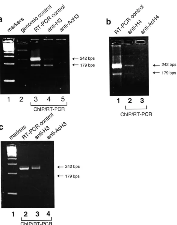
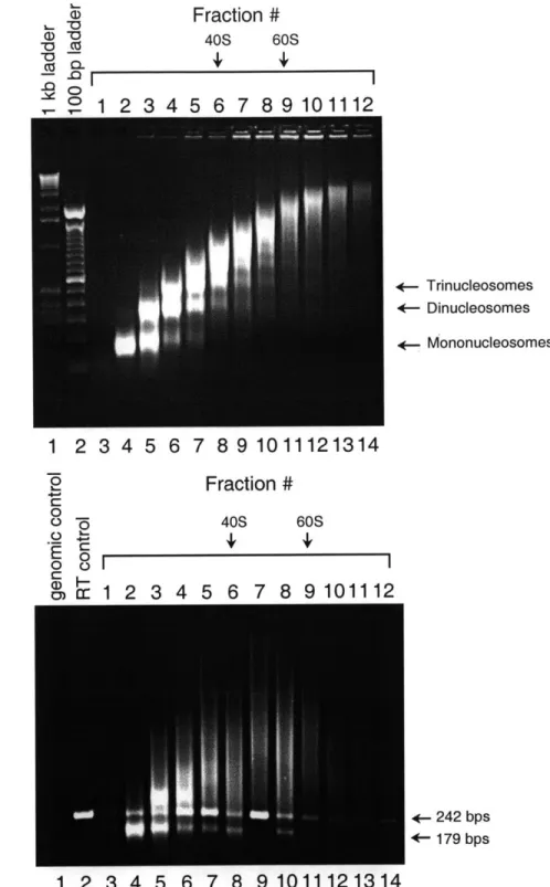
The second focus of this thesis is the nature of the association between XIST RNA and the Xi chromatin. Microscopy studies have shown that the noncoding XIST RNA colocalizes with the Xi. It is unclear, however, if this colocalization is due to physical association of XIST RNA with the Xi chromatin, or if it is a secondary consequence of XIST RNA and the Xi being sequestered to the same nuclear territory. This thesis provides evidence from chromatin immunoprecipitation experiments that XIST RNA is part of the Xi chromatin. First, XIST RNA can be co-precipitated by antisera against macroH2A, a histone H2A variant enriched in the Xi. Second, XIST RNA can be co-precipitated by antisera that recognize unacetylated, but not acetylated, isoforms of histones H3 and H4. As demonstrated in this thesis, hypoacetylated histone H4 is enriched at promoters of X-inactivated genes, whereas hyperacetylated histone H4 is found only at promoters of active genes. The preferential association of XIST RNA with unacetylated histones therefore suggests that the RNA is not uniformly associated with the Xi chromatin. Further evidence for this conclusion comes from fluorescence in situ hybridization in mouse cells, in which Xist RNA is shown to localize with the inactivated Zfx locus, but not with the Sts locus which escapes X-inactivation. These results raise the possibility that association with XIST RNA may be a fourth feature that correlates with silencing of individual genes on the Xi. This physical association between XIST RNA and the Xi chromatin may facilitate X-inactivation.
Thesis Supervisor: Dr. Phillip A. Sharp, Title: Salvador E. Luria Professor of Biology
ACKNOWLEDGMENTS
First, I would like to thank Phillip Sharp for providing guidance over my graduate career. I have had the privilege of observing his intellect, fairness and work ethic first-hand. This has set a standard for which I will continue to strive.
I would also like to thank Margarita Siafaca, who has been an enormously effective Lab Manager and a wonderful resource for everyone.
I am grateful to members of my thesis committee - David Page, Rudolf Jaenisch, Rick Young, and Steve Buratowski - for their generous advice and interest in my work. In addition, I am indebted to Dave Allis, who encouraged my work on the chromatin project.
I wish to thank members of the Sharp lab, past and present, for their interest and
assistance throughout my career. I would like to thank Barbara Panning for getting me interested in X-inactivation; Julian Borrow, Derek Dykxhoorn, Hristo Houbaviy, Jae B. Kim and Bruce Lahn for critically reviewing my work; and Hong Tang, K.B. Lee, Tom Tuschl, Grace Jones, Ben
Blencowe, Dan Chasman, Charles Query, Vicki Wang, Carl Novina and Dean Tantin for many thought-provoking discussions. I also appreciate the insights of my collaborator, John Pehrson, with whom I worked on the macroH2A project.
Many people throughout the Biology Department have enriched my time at MIT. My classmates Bruce Lahn, Alok Srivastava, Gillian Stanfield, and Elizabeth Hong have added another dimension to my experience. Shuguang Zhang, Amanda Shearman, Julian Borrow, Lisa Spirio and Charles Tilford were also generous and knowledgeable companions.
Finally, my family is an energetic bunch whose curiosity about my work has been stimulating. I feel very lucky to have had their support throughout the years.
TABLE OF CONTENTS
TITLE PAGE ... ... .. ... 1
ABSTRACT ... ... ... ---... 3
ACKNOW LEDGMENTS ... 4
TABLE OF CONTENTS ... 5
LIST OF FIGURES AND TABLES ... 7
CHAPTER ONE: ... 9
1. OVERVIEW OF DISSERTATION ... 10
II. DOSAGE COMPENSATION MECHANISMS ... 11
i) Dosage Compensation in Drosophila ... 12
ii) Dosage Compensation in Nematodes ... 13
iii) Dosage Compensation in Mammals ... 14
I1. MECHANISMS OF X-INACTIVATION ... 15
i) X-Inactivation is Controlled by the X-Inactivation Center (XIC) 16 ii) Counting and Choice ... 18
iii) Initiation and Spread of X-inactivation ... 19
iv) Maintenance of the Inactive State ... 20
IV. ESCAPE FROM X-INACTIVATION ... 21
V. FEATURES OF THE INACTIVE X CHROMATIN ... 23
i) Colocalization of XIST RNA and the Xi ... 23
ii) Heterochromatinization ... 24
iii) DNA Methylation ... 25
iv) Replication Timing ... 26
v) Histone Acetylation ... 27
VII. CHROMATIN STRUCTURE IN TRANSCRIPTIONAL REGULATION 30
i) N ucleosom e Structure ... 30
ii) C ore H istone Variants ... 31
iii) Nucleosomes and Gene Repression ... 32
iv) H istone Acetylation ... 32
v) Histone Acetyltransferases and Deacetylases ... 33
vi) Site Usage of H4 Acetylation ... 35
vii) Histone Hyperacetylation Correlates with Gene Expression 36 viii) Histone Hyperacetylation Modulates Higher-Order Chromatin S tructure ... 37
ix) DNA Methylation Alters Chromatin Structure and Function ... 37
VIll. SUM M ARY ... 38
REFERENCES ... ... ... 39
CHAPTER TWO: PROMOTER-SPECIFIC HYPOACETYLATION OF X-INACTIVATED GENES... 48
REFERENCES ... 67
CHAPTER THREE: XIST RNA IS A COMPONENT OF THE INACTIVE X CHROMATIN... 70
REFERENCES ... ... 83
CHAPTER FOUR: D IS C U S S IO N ... ... 85
APPENDIX:
LOCALIZATION OF XIST RNA RELATIVE TO X-LINKED GENES ... 98 REFERENCES...S ... 105
LIST OF FIGURES AND TABLES
CHAPTER TWO
Figure 1. Histone H4 Acetylation at Promoter Regions of X-Linked Genes .... 52 Table I. H4 Acetylation Status of 5' Region of X-Linked Genes ... 53 Figure 2. Site-specific Acetylation of Histone H4 at Promoter Regions .... 55 Figure 3. Quantitative PCR Analysis of Histone H4 Acetylation along
X -linked G enes ... 57-58 Figure 4. Methylation Analysis of Promoter Regions of X-Linked Genes .... 60 Table II. Primers for PCR Analysis ... 66
CHAPTER THREE
Figure 1. Immunofluorescence and Immunoprecipitation Analysis of
H istone m H 2A ... 73 Figure 2. Immunoprecipitation profile of XIST RNA with various anti-histone
antisera. ... ... 75-76 Figure 3. Sedimentation of non-crosslinked, MNase-digested chromatin in
10-30% glycerol gradients ... 78
APPENDIX
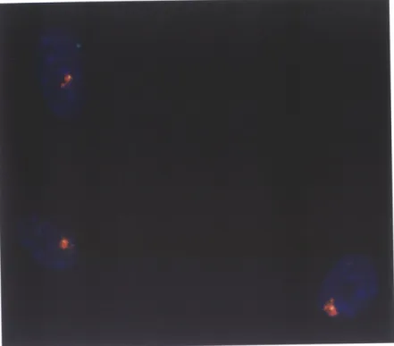
Figure 1. Xist RNA Overlaps with the Inactivated Zfx Locus ... 102 Figure 2. Xist RNA Rarely Overlaps with the Sts Gene,
w hich Escapes X-inactivation ... 103 Table 1. Overlap Between XIST RNA and X-Linked Genes ... 104
SECTION I: OVERVIEW OF DISSERTATION
This dissertation describes an in-depth investigation of the chromatin structure of the mammalian inactive X chromosome (Xi). The primary goal of this investigation was to identify distinct features associated with the Xi chromatin, and to understand how these features may contribute to gene silencing. The dissertation is organized in the following manner:
Chapter One outlines a broad survey of past research in X-inactivation and other relevant fields. Topics include: (1) dosage compensation mechanisms in model organisms; (2) molecular events associated with the X-inactivation process; and (3) the role of chromatin structure in transcriptional regulation.
Chapter Two describes a series of experiments designed to examine the histone acetylation status of individual genes along the Xi. A major emphasis of these experiments was to compare the acetylation status of genes that are subject to X-inactivation with those that escape this process. This study revealed a perfect correlation between the promoter-specific acetylation status of histone H4 and the expression of individual genes on the Xi: genes which escape silencing on the Xi show elevated levels of acetylation at their promoters, while
X-inactivated genes are markedly hypoacetylated at their promoters.
Chapter Three describes studies aimed at understanding the molecular nature of the colocalization between XIST RNA and the Xi. These studies demonstrated that XIST RNA may be a physical component of the Xi chromatin. In addition, results suggested that the distribution of XIST RNA along the Xi is perhaps not uniform, but is instead concentrated at hypoacetylated regions of the Xi chromatin.
Chapter Four discusses the significance of the findings reported in this thesis. A model for how XIST RNA controls X-inactivation is proposed, in which the RNA serves as a bridging factor that recruits transcriptional silencing machinery to particular genes of the Xi.
One prediction of this model is that genes not subject to X-inactivation should fail to associate with XIST RNA. This prediction is partly supported by experiments described in the
Appendix. These experiments demonstrated that Xist RNA colocalizes with Zfx, an X-inactivated locus in the mouse, but not with Sts, a locus that escapes X-inactivation.
SECTION II: DOSAGE COMPENSATION MECHANISMS
One of the fundamental genetic distinctions between males and females of XY-bearing species is the number of X chromosomes that they possess. Dosage compensation is the process by which the sex difference in X chromosome dosage is attenuated through transcriptional regulation of X-linked genes.
The evolution of dosage compensation is thought to be an integral part of sex chromosome evolution. In XY-bearing species, the genetically distinct X and Y chromosomes were once a pair of identical autosomes, but differentiated as most genes on the Y chromosome degenerated. One consequence of this Y chromosome degeneration is the dosage imbalance of X-linked genes between males and females. This imbalance, which is detrimental to proper development, drives the evolution of dosage compensation mechanisms as a countermeasure.
Evolutionary studies have firmly established that sex chromosomes in distantly related lineages evolved on multiple, independent occasions. For each lineage, therefore, the dosage compensation mechanism has also arisen separately. This notion is confirmed by the observation that dosage compensation mechanisms in flies, nematodes and mammals - three lineages with the best characterized sex chromosomes - are disparate in their molecular characteristics. Dosage compensation in Drosophila is accomplished by upregulating transcript levels from the single male X to achieve a level comparable to that obtained from the two female X chromosomes. C. elegans employs the reverse strategy, in which transcript levels are
normalized between males and females by downregulating expression from both X(s) in the hermaphrodite. A third strategy evolved in mammals, in which one of the two female X chromosomes is transcriptionally silenced, almost in its entirety.
Although these three dosage compensation strategies - hypertranscription, hypotranscription, and transcriptional silencing - are evolutionarily unrelated, and appear
mechanistically disparate, each involves alterations in the chromatin structure of the regulated X chromosome. Furthermore, two of the three strategies, hypertranscription in Drosophila and transcriptional silencing in mammals, involve noncoding RNA molecules that localize to the regulated X chromosome from which the RNA is transcribed. These commonalities suggest that noncoding RNAs may have the ability to recruit various activities to a chromosome in cis.
i) Dosage Compensation in Drosophila
Dosage compensation in Drosophila is achieved through an approximately two-fold overexpression of transcripts from the single male X. This process requires a number of protein factors, many of which have been identified in genetic screens where their loss-of-function resulted in male-specific lethality. These factors include the male-specific lethal proteins 1, 2, and
3 (MSL-1, -2, and -3) and the maleless protein (MLE) (Belote and Lucchesi, 1980; Fukunaga et al., 1975).
Biochemical and cell biology studies have substantiated the involvement of these genetically identified factors in chromosome-specific regulation of the male X. In these studies, the MSL-1, -2 and -3 proteins were shown to form a heteromeric complex (Copps et al., 1998), which localizes to hundreds of sites along the polytene male X. Although the biochemical mechanism of dosage compensation has not been fully elucidated, potential activities of the MSL and MLE proteins have been characterized to some extent. The MLE protein has RNA/DNA helicase, adenosine triphosphastase (ATPase), and single-stranded (ss)RNA/ssDNA binding activities (Lee et al., 1997). Mutant MLE proteins with defective helicase/ATPase activites still localized to the male X chromosome, but failed to rescue an m/e phenotype (Lee et al., 1997). This suggests that the helicase/ATPase activities of MLE are needed for transcriptional regulation. Sequence analysis of the MSL proteins revealed several motifs common to transcriptional regulators, suggesting that the dosage compensation machinery indeed regulates transcription (Bashaw and Baker, 1995; Kelley et al., 1995; Palmer et al., 1993).
Binding of the MSL proteins to the upregulated male X is also coincident with the specific acetylation of lysine 16 on histone H4 (Bone et al., 1994; Turner et al., 1992). Males that fail to dosage compensate because they carry mutations in male-specific lethal genes also ak the lysine 16 acetylation on histone H4 (Bone et al., 1994). The reason for this appears to be that the MSL proteins recruit the MOF (males-absent-on the first) protein to the male X (Gu et
al., 1998), which encodes a putative histone acetyltransferase (Hilfiker et al., 1997). Thus, one way the MSL complex may effect dosage compensation is by alterating chromatin structure of the male X in a manner that enhances transcription of its resident genes.
In addition to protein factors, two noncoding RNA molecules - roX1 and roX2 - which originate from the upregulated male X have been implicated in dosage compensation. The roX1 and roX2 genes are expressed in many cell types during early development, but become restricted primarily to cells of the central nervous system in adults (Amrein and Axel, 1997; Meller et al., 1997). Both roX1 and roX2 produce noncoding RNA molecules which can associate with X-derived chromatin either in cis or in trans (Amrein and Axel, 1997; Meller et al., 1997). These transcripts bind to the male X in a pattern that coincides with MSL binding (Meller et al., 1997). However, roX1 is clearly not required for dosage compensation, as its disruption produces no obvious phenotype in males (Meller et al., 1997). It is possible that there are additional, unidentified RNAs that associate with the hypertranscribed male X. In fact, association of the putative RNA helicase, MLE, with the male X is RNase-sensitive (Richter et al., 1996), although it is not affected by disruption of roX1 (Meller et al., 1997).
ii) Dosage Compensation in Nematodes.
A different dosage compensation strategy has evolved in the nematode C. elegans. Here, the active modulation of the dosage imbalance occurs in hermaphrodites (which are XX), rather than in
males (which are XO). In hermaphrodite worms, transcription from both X chromosomes is downregulated, such that the steady-state transcript levels of most X-linked genes are
comparable between hermaphrodite and male cells (Donahue et al., 1987; Meyer and Casson,
1986).
Similar to studies of Drosophila dosage compensation, genetic screens for sex-specific lethal phenotypes have identified a number of factors in C. elegans that contribute to transcriptional downregulation of the hermaphrodite X chromosomes. Functional analysis has led to the identification of a protein complex that includes many of these factors. This protein complex localizes to both of the dosage-regulated X chromosomes of hermaphrodites (Chuang et al., 1994; Chuang et al., 1996). Thus far, the complex has been shown to contain the following proteins: SDC-2, SDC-3, DPY-26, DPY-27, and MIX-1. Some of these proteins, such as DPY-27, appear not to have any function beyond their role in dosage compensation. However, several proteins have other essential cellular functions. For example, the DPY-26 protein initially associates with all mitotic chromosomes in embryos of both sexes. Yet, the initiation of dosage compensation changes this pattern, such that DPY-26 is selectively localized to the hermaphrodite X's in somatic cells and with all meiotic chromosomes in germ cells (Lieb et al., 1996). Similarly, the MIX-1 protein is required not only for downregulation of transcript levels in hermaphrodites, but also for chromosome segregation during mitosis in both sexes (Lieb et al., 1998). SDC-2 and SDC-3 also have several functions, one of which is to target the dosage compensation machinery to X chromosomes (Dawes et al., 1999). The substantial overlap of components between the dosage compensation machinery and the meiotic and mitotic machinery is strong evidence that dosage compensation in nematodes - and conceivably in other species as well
-evolved by adapting existing chromosome regulatory mechanisms for a de novo task.
iii) Dosage Compensation in Mammals
Mammals employ yet a third strategy for dosage compensation. During the early stages of embryogenesis, one of the two X chromosomes in each female somatic cell is randomly chosen to undergo a widespread transcriptional silencing. Once the silenced state of the chosen X is established, it is clonally inherited through cell divisions and differentiation (Monk and Harper,
1979). A deviation from the random nature of X chromosome choice is observed in marsupials
and in the extraembryonic membranes of placental mammals, in which the paternal X is invariably chosen for silencing. The mechanisms of X-inactivation will be described in greater detail in following sections of this chapter.
SECTION III: MECHANISMS OF X-INACTIVATION
The approaches that have been employed to study the mechanisms of X-inactivation are numerous and varied. Most of these approaches can be divided into three broad categories: The traditional approach is a genetic one, whereby regions of the X chromosome that control the X-inactivation process are mapped using mutant chromosomes that are derived from X-autosome translocations. Such an approach led to the original identification of the X-inactivation center (XIC) (Brown et al., 1991), and the isolation of the X/ST gene within the XIC (Brown et al., 1991). The genetic approach has been expanded enormously in recent years to include targeted deletions and transgenic insertions which have enabled fine dissections of the chromosomal regions that control various aspects of X-inactivation. A second major approach in the study of X-inactivation is to follow developmental events that occur during the X-inactivation process. In this case, the objective is to chronicle the sequence of events that occur when female cells undergo X-inactivation, either in vivo during embryogenesis, or in tissue culture during embryonic stem (ES) cell differentiation. A third approach to study X-inactivation, which has gained greater popularity in recent years, is to identify molecular features that distinguish the inactive X chromosome from its active counterpart. Comparative analysis has identified the following features unique to the XI chromosome: association with XIST RNA, late timing of DNA replication, general histone hypoacetylation and methylation of CG islands associated with the 5' ends of X-inactivated genes. Intense investigations with combinations of the various approaches have provided detailed descriptions of the X-inactivation process. Although these descriptions are often phenomenological rather than mechanistic, they are beginning to yield clues as to how this intriguing biological process may be regulated at a molecular level.
i) X-Inactivation is Controlled by the X-Inactivation Center (XIC)
X-inactivation is a multi-step process which involves: (1) counting the number of X chromosomes and choice of the one that will remain active, (2) initiation of silencing on all remaining X's, and (3) clonal maintenance of the inactive state once it is established. Most of these functions - other than maintenance - are specified by a region on the X termed the X-inactivation center (XIC). The
XIC is located in interval Xq13 in humans, and was originally defined as a region common to all
rearranged X's that were capable of being inactivated (Brown et al., 1991). More recently, the transgene-based approach has been used to define the minimum functional Xic in the mouse by its ability to induce inactivation at ectopic sites in ES cells (Lee et al., 1996; Herzing et al., 1997; Lee et al., 1999). Such studies have suggested that an 80 kb region surrounding the mouse Xist gene may be sufficient to induce many features associated with X-inactivation at ectopic sites, such as hypoacetylation of histone H4, late replication timing, and downregulation of X-linked gene expression (Lee et al., 1999). However, a caveat of the transgene-based approach is that most transgenes are present in multiple copies. One study demonstrated that single copy transgenes were insufficient to induce inactivation, suggesting that some of the elements needed for initiation or spread of X-inactivation are lacking in these transgenes and are artificially supplied
by multimerization (Heard et al., 1999).
Another strategy to dissect the function of the XIC is to identify and characterize genes located within this critical interval. Several genes have been mapped to the XIC, but only one -the XIST gene - has been conclusively implicated in X-inactivation. In addition to its physical location within the XIC, XIST is the only known X-linked gene that is expressed exclusively from the inactive X in differentiated somatic cells (Brown et al., 1991). Furthermore, XIST RNA is apparently not transported out of the nucleus as most mRNAs are; rather, it colocalizes with the inactive X chromosome from which it is transcribed (Brown et al., 1992). Human XIST encodes a
17 kb spliced and polyadenylated transcript. Initially, evidence for the role of XIST in X-inactivation had been circumstantial. More recently, targeted deletion studies provided conclusive
evidence that the XIST gene is required for initiation of X-inactivation (Marahrens et al., 1997; Penny et al., 1996).
To date, the mode by which the X/ST gene functions in X-inactivation is unknown. Several models have been proposed (Brockdorff et al., 1992; Brown et al., 1992), including: (1) the XIST gene encodes a critical protein factor that participates in X-inactivation; (2) the XIST genomic locus acts as a chromatin organizing center from which X-inactivation is propagated along the chromosome; or (3) X/ST encodes a structural RNA which is a crucial component of the X-inactivation machinery. The first model is most likely incorrect, as there is no evidence that XIST encodes a protein. First, the nuclear localization of XIST RNA suggests that it is not accessible to the cytoplasmic translation machinery. Second, XIST lacks any significant open reading frames (Brockdorff et al., 1992; Brown et al., 1992). Finally, the few short open reading frames that do exist are not conserved amongst mammals. The other two models for X/STfunction - either that
it is a chromatin organizing center or that it encodes a functional RNA - are thus far both consistent with the data. The observation that XIST RNA colocalizes with the Xi tends to favor the functional RNA model, although it does not exclude the possibility that the X/ST locus functions as a chromatin organizing center.
Genetic studies have also identified a sequence element within the XIC which controls the likelihood that the chromosome bearing this element will be chosen for X-inactivation. This X
a,
b
c
d
controlling element (Xce) has at least four alleles in mice - Xcea
(weakest),
Xceb, Xce, Xce _such that X-inactivation is biased towards the chromosome carrying the weaker allele (Migeon, 1994). Targeted deletion of a large region encompassing this element resulted in constitutive Xist expression from the disrupted X (Clerc and Avner, 1998). The only gene identified in this interval to date is Tsix. Tsix is a 40 kb nuclear transcript that is antisense to Xist RNA (hence, its name is
"Xist' spelled backwards)(Lee et al., 1999). In addition to its antisense orientation relative to Xist,
the expression pattern of Tsix makes it a candidate regulator of Xist Prior to X-inactivation, Tsix is expressed biallelically. At the onset of X-inactivation, however, its expression becomes restricted to the future Xa and persists until Xist expression is repressed. While the evidence
that Tsix is an antisense regulator of Xist is tentative at best, it is worth noting that antisense transcripts have also been detected in the vicinity of imprinted genes, which are also subject to allele-specific regulation (Moore et al., 1997; Rougeulle et al., 1998).
In addition to Xist and Tsix, several other genes have been mapped to the XIC in both human and mouse. These include the brain-specific gene Brx (Rougeulle and Avner, 1996), the testes-restricted gene Tsx (Simmler et al., 1996), the ribosomal protein pseudogene pS19X (Rougeulle and Avner, 1996), and a homologue of the caudal gene, Cdx4 (Horn and Ashworth,
1995). The expression patterns and the putative functions of these genes are not consistent with
their having a role in X-inactivation, although this has not been conclusively demonstrated by genetic analysis.
ii) Counting and Choice
In placental mammals, the selection of X chromosomes for inactivation proceeds according to the "n-1 rule", which silences all but one X per diploid genome. The term "counting" reflects the fact that one X is kept active per diploid genome. Increasing amounts of ploidy are able to support
additional active X's in a cell, as triploid individuals with a 69, XXY karyotype are able to maintain two active X's (Weaver and Gartler, 1975). However, the term "counting" can be somewhat misleading, since supernumerary X's in a diploid genome do not necessarily need to be counted to become X-inactivated. For instance, it may be that all X chromosomes are normally silenced by default, but that there exists a mechanism that actively prevents just one X from undergoing the default process. It has not been possible to experimentally distinguish whether the "n-1 rule" is implemented through counting the total number of X's, or merely by protecting one X from the
inactivation process. Regardless of whether the cell actually counts the number of X
chromosomes in the nucleus, it has been hypothesized that "blocking factors" produced in diploid cells prevent one XIC from initiating X-inactivation such that it remains active, while all remaining unblocked Xics inactivate their host chromosomes (Lyon, 1996).
The element(s) that are required for counting and/or choice have not been fully delineated. The random nature of X-inactivation in the embryonic cells of eutherian mammals can be biased by alleles at the X-controlling element (Xce), as described above. The genetic relationship between Xist and the Xce is unclear. Targeted deletion of a region encompassing the mouse Xce and Tsix caused constitutive expression of the linked Xist gene (Clerc and Avner,
1998). Another element that contributes to choice is present in the Xist gene itself. Targeted
deletion of sequences corresponding to exon 1 through intron 5 of Xist disrupted the ability to choose the mutant X, such that only the intact chromosome became inactivated (Marahrens et al.,
1998). Further genetic studies will be needed to better define the sequences and the effector molecules that are critical for choice.
iii) Initiation and Spread of X-inactivation
Time course analysis of X-inactivation has relied primarily on mouse ES cells, which are experimentally more tractable than developing embryos. Female ES cells possess two active X chromosomes, one of which becomes inactivated as cells differentiate in culture. This process of
in vitro differentiation recapitulates many of the stages of X-inactivation that occur during embryonic development.
Temporal observation of X-inactivation during ES cell differentiation, and in some cases in the developing mouse embryo, has revealed a series of events that occur in a defined sequence over a period of several days (Keohane et al., 1996). The first detectable event in the process of X-inactivation is the increase in levels of Xist RNA from the incipient Xi (Kay et al., 1993; Lee et al., 1996; Panning et al., 1997). Prior to this event, Xist is transcribed at a low-level from both X chromosomes of undifferentiated ES cells. Yet, this high-level Xist expression initiates X-inactivation only if it occurs within a critical developmental period: Reactivation of mouse Xist in differentiated cells did not induce silencing of genes in cis, despite the fact that the ectopic Xist
RNA localized to its chromosome-of-origin (Clemson et al., 1998). That Xist RNA can initiate X-inactivation during embryogenesis, but not at later stages of development, suggests that a
change occurs either in the composition of available auxiliary factors, or in the RNA itself. It is possible that one contributing factor to this difference is the change in levels and location of de novo and maintenance methyltransferases in early embryonic cells relative to terminally differentiated cells (Razin and Shemer, 1995).
Xist upregulation coincides almost immediately with a shift to late S phase in the replication timing of the Xi (Keohane et al., 1996). Within 2 days of Xist upregulation, transcriptional downregulation of several genes on the Xi is completed. Although it is commonly held that X-inactivation spreads out from the Xic in both directions, RT-PCR analysis of transcript levels has not detected a gradient in the timing of inactivation for X-linked genes (Keohane et al.,
1996). Nevertheless, the notion that X-inactivation spreads laterally from the XIC is not an invalid
one, since studies of X:autosome translocations have shown that X-inactivation can traverse the translocation boundary to affect the proximal portion of the autosomal region. However, the incomplete spreading of X-inactivation on these translocation chromosomes has led to the proposal that "way station" elements evolved on the X chromosome to facilitate the spread of X-inactivation (Gartler and Riggs, 1983). Mary Lyon has proposed that LINE elements, which are disproportionately enriched on the X and which correlate with the X-inactivation potential of autosomes, may facilitate XIST RNA association with the Xi and contribute to its X-inactivation (Lyon, 1998).
iv) Maintenance of the Inactive State
Once an X chromosome has been chosen for inactivation, its inactive state is stably and clonally transmitted during subsequent cell divisions and differentiation. The mechanism responsible for the maintenance of the inactive state is apparently separable from that responsible for its initiation. This assessment is based on the observation that maintenance of the inactive state, unlike initiation of X-inactivation, does not require the XIC. In cultured human/rodent somatic cell hybrids in which the human X is stably inactivated, induced deletion of the XIC - including the
XiST gene - from the Xi does not alter its transcriptionally repressed state (Brown and Willard,
1994; Rack et al., 1994).
Because CG islands associated with the 5' ends of silenced genes on the Xi are hypermethylated, in contrast to their counterparts on the Xa or on most autosomes, methylation is thought to provide "memory" of the inactive state. The greater stability of the Xi in eutherian cells
is also attributed to the methylation of CG islands, which is not detectable in marsupials (Kaslow and Migeon, 1987; Loebel and Johnston, 1996) and is believed to have evolved in eutherians as
a means to "lock-in" the repressed state (Wakefield et al., 1997). Another molecular feature which distinguishes the Xi from the Xa and from all other chromosomes is a chromosome-wide histone hypoacetylation evident in metaphase microscopy (Belyaev et al., 1996; Boggs et al., 1996; Jeppesen and Turner, 1993). Time course analysis of differentiating ES cells suggested that both methylation and the microscopically observable deacetylation of the Xi may follow, rather than precede, downregulation of gene expression, suggesting that they are important for maintenance of the inactive state (Keohane et al., 1996).
SECTION IV: ESCAPE FROM X-INACTIVATION
Important clues about the mechanism of X-inactivation have come from analyzing exceptions to the rule - namely, genes which are present on the Xi but which are not inactivated. Despite the apparently sweeping nature of X-inactivation, transcription from the inactive X is not uniformly repressed. The first indication that gene expression from the Xi is needed for important cellular functions came from studies of Turner syndrome (TS). Patients with TS have partial or complete sex chromosome monosomy (an XO karyotype), and exhibit a range of defects including short stature, gonadal dysgenesis and multiple anatomical abnormalities. TS individuals can be thought of as either missing an X (when compared to normal XX females), or a Y (when compared to normal XY males). Based on this reasoning, Ferguson-Smith postulated that TS is due to the haploinsufficiency of "TS genes" which should be present on both X and Y, and should escape X-inactivation on the X (Ferguson-Smith, 1965). The immediate implication of
this hypothesis is that X-inactivation is incomplete; that is, there are genes on the X that escape silencing to fulfill the critical requirement of their double dosage during development.
The hypothesis that X-inactivation is incomplete has been validated by experimentation in the ensuing years. Since the 1980's, genes that escape X-inactivation have been continually discovered. Currently, the most powerful method to identify such genes is to assay X-linked gene expression in human/rodent somatic cell hybrids that retain either the active or the inactive human X (Mohandas et al., 1980). Human genes that undergo X-inactivation are expressed only in the Xa-containing hybrid, whereas genes that escape X-inactivation are expressed in both types of hybrids. This and other approaches have led to the identification of nearly two dozen genes that escape X-inactivation in humans (Brown et al., 1997). These genes fall into two categories: those in the pseudoautosomal regions, and those in the X-specific region.
Pseudoautosomal regions (PARs) are located at the distal tips of the sex chromosomes. Within PARs, the X and Y undergo meiotic recombination just as two homologous autosomes do. As a result, PARs on the X are identical to corresponding PARs on the Y. In males, PAR genes are expressed biallelically, from both the X and the Y. For these genes to be expressed at a comparable level in females, they must escape X-inactivation. Indeed, all pseudoautosomal genes examined, other than SYBL1, are expressed from both Xi and Xa in females. They constitute approximately one-third of the human genes known to escape X-inactivation. The remaining genes that escape inactivation reside in the X-specific portion of the X - the portion that does not recombine with the Y in meiosis. Consistent with Ferguson-Smith's hypothesis, many of these genes have functional homologs on the Y chromosome.
Interestingly, X-inactivation appears to be much more complete in the mouse, as only four genes have been shown to escape X-inactivation (reviewed in (Disteche, 1995)). The fact that fewer mouse genes should escape X-inactivation is consistent with the observation that the XO karyotype in the mouse produces a much milder phenotype in comparison to TS in humans: XO female mice are fertile, and are often indistinguishable from normal XX females (Banzai et al., 1995).
The mechanism by which some genes escape X-inactivation is largely unappreciated. The fact that genes which escape X-inactivation are dispersed throughout the X suggests that the mechanism by which genes escape X-inactivation operates on a rather localized basis. Occasionally, however, several genes which escape X-inactivation are found clustered together, suggesting the possibility that local control may extend beyond the level of a single gene (Miller and Willard, 1998).
Another way to understand the mechanism by which some genes escape X-inactivation is to perform time course analyses of X-linked gene expression in the course of development. These studies have suggested that the X-inactivation status of a gene can be dynamically altered during development. Several studies have reported that the mouse Smcx gene is originally silenced during the initiation of X-inactivation in either differentiating ES cells (Penny et al., 1996) or in early embryos (Lingenfelter et al., 1998). However, Smcx expression from the Xi is reestablished later in development such that expression from the Xi and Xa are nearly comparable (Lingenfelter et al., 1998; Sheardown et al., 1996). These results reveal the highly complex nature of both initiation and maintenance of X-inactivation.
SECTION V: FEATURES OF THE INACTIVE X CHROMATIN
Several features distinguish the chromatin of the Xi from that of the Xa. Some of these features, such as hypoacetylation and association with XIST RNA, have been described at the level of the entire chromosome. Other features - such as DNA methylation and late replication timing - distinguish genes that are silenced on the Xi from those on the same chromosome that escape inactivation. Studies of these features have provided important clues to the mechanism of gene silencing on the Xi.
i) Colocalization of XIST RNA and the Xi
The observation that the RNA product of XIST localizes to the inactive X Barr body (Brown et al., 1992; Clemson et al., 1996) is the primary reason to favor the model that XIST functions as a
noncoding RNA during the inactivation process. There are at least two possibilities that may account for the observed colocalization: either XIST RNA may be sequestered to the same nuclear compartment as the Xi, perhaps by mechanisms that confine the Xi to a transcriptionally repressive environment, or XIST RNA may be physically attached to the Xi chromatin.
Several attempts have been made to better characterize the colocalization between XIST RNA and the Xi. In one experiment, procedures that extracted 90-95% of DNA and most of the histones from the nucleus hardly diminished the in situ XIST RNA signal. This led to the speculation that XIST RNA is attached to the nuclear matrix and that it is not part of the chromatin (Clemson et al., 1996). However, the results of this experiment are subject to multiple interpretations, as there exist alternative technical or biological reasons for the inability to extract XIST RNA from the nucleus along with chromatin. In addition, it was reported that XIST RNA does not hybridize directly to the Xi DNA, as it is not degraded by RNase H which digests
RNA/DNA hybrids (Clemson et al., 1996).
ii) Heterochromatinization
A number of observations suggest that the Xi is distinguished from other chromosomes, including the Xa, by higher-order changes in chromatin structure. First, the fact that the majority of genes on the Xi are silenced, more or less simultaneously, suggests that their repression is coordinated at the level of the chromosome. In addition, the Xi appears to be visibly condensed into a heterochromatic Barr body that is usually located at the nuclear periphery (Barr ML, 1961). It is thought that higher-order packaging of chromatin is responsible for an altered conformation of the Xi, including a bend in its Xq arm during metaphase (Flejter et al., 1984), and interactions between the telomeres that produce a looping of the Xi (Walker et al., 1991).
Molecular analyses of individual genes has also revealed differences between the higher-order chromatin structure of the Xi and Xa. Three DNase I-sensitive regions were detected for the X-linked Pgk gene that are unique to the Xa allele (Riley et al., 1986). These differences in nuclease accessibility reflect differences in chromatin structure that correlate with gene expression.
Similarly, the promoter region of human HPRTcontains a DNase I-sensitive region on the Xa, but not on the Xi (Lin and Chinault, 1988). In addition, chromatin fractionation by differential centrifugation showed that several genes on the Xi were enriched in the heterochromatin fraction, whereas their counterparts on the Xa were enriched in the euchromatic fraction (Endo et al., 1998; Endo et al., 1999).
iii) DNA Methylation
Mammalian DNA is methylated at cytosine residues within CG dinucleotides and maintained
through cell division by the action of maintenance methylases on hemimethylated templates. C G islands that are associated with 5' ends of X-inactivated genes are heavily methylated on the X but completely unmethylated on the homologous Xa alleles (Norris et al., 1991; Tribioli et al., 1992). Furthermore, CG islands associated with genes on the Xi that escape silencing are not methylated, demonstrating a correlation at the level of individual genes.
Although there appears to be a perfect correlation between methylation and X-inactivation status (Jegalian and Page, 1998; Tribioli et al., 1992), the precise role of methylation in the process has not been fully elucidated. Several lines of evidence suggest that methylation contributes to maintenance of the inactive state, once it has been established by other methods. First, treatment of cells with 5-azacytidine results in DNA hypomethylation and consequent reactivation of genes on the Xi (Mohandas et al., 1981). Second, marsupial Xi genes, which are known to lack CG methylation (Loebel and Johnston, 1996), are more easily reactivated in cell culture (Kaslow and Migeon, 1987). The fact that marsupials can achieve X-inactivation without methylation suggests that methylation was not essential for the process in mammalian ancestors, but that it was a later evolutionary invention to help "lock-in" the repressed state (Wakefield et al., 1997) Although methylation is implicated in maintenance of the inactive state in eutherian mammals, it may be dispensible for initiation of X-inactivation. The strongest evidence for this claim is the observation that Xist-mediated X-inactivation can be initiated in cells which are deficient for DNA methyltransferase activity (Beard et al., 1995; Panning and Jaenisch, 1996).
iv) Replication Timing
Many heterochromatic regions of the genome replicate late in S phase, after the majority of the genome has already undergone DNA synthesis. Consistent with its heterochromatic state, the A also replicates late in S phase with respect to its active counterpart in female cells (Gartler and Riggs, 1983). This feature is one of the earliest detectable changes to the Xi chromatin, immediately following the upregulation of Xist RNA during embryonic development (Keohane et al., 1996; Takagi et al., 1982). Replication timing has also been assessed at the level of several individual X-linked genes in human cells. Genes that are subject to X-inactivation replicate earlier on the Xa than do their counterparts on the Xi. In contrast, genes that escape X-inactivation were found to replicate synchronously on Xi and Xa, as measured either by RNA FISH analysis or by chromatin fractionation following BrdU incorporation (Boggs and Chinault, 1994; Hansen et al.,
1996; Schmidt and Migeon, 1990). This correlation was further substantiated by the finding that reactivation of several silenced genes on the Xi with the hypomethylating agent 5-azacytidine coincided with a shift to an earlier timing of replication, although there were some exceptions to this rule (Hansen et al., 1996).
Although late replication is a prominent feature of the Xi chromatin which coincides temporally with X-inactivation, some studies suggest that it is neither necessary nor sufficient for the process: Several cell lines have been isolated that contain an early-replicating Xi which is
nevertheless transcriptionally repressed (Yoshida et al., 1993). Thus, X-inactivation can b e
maintained, at least in cell culture, without late replication of the Xi. Furthermore, a shift to a late replication timing, induced by the deacetylase inhibitor Trichostatin A, was not sufficient to induce silencing of X-linked genes in the absence of other features associated with X-inactivation (O'Neill et al., 1999).
Immunofluorescence microscopy with antibodies against acetylated isoforms of histones revealed that the metaphase Xi in both human and murine cells is distinguished by a marked reduction in the acetylation of H2A, H3 and H4 (Jeppesen and Turner, 1993; Belyaev et al., 1996; Boggs et al., 1996). Antibodies which could distinguish amongst acetylation of individual lysine residues confirmed that the metaphase Xi was reduced in all acetylated isoforms of histone H4. This is similar to the pattern of reduced H4 acetylation observed on metaphase chromosomes for other
regions of markedly reduced transcriptional activity, such as centric heterochromatin and G bands (Jeppesen and Turner, 1993). The role of histone hypoacetylation in gene silencing on the Xi is further corroborated by the finding that the histone deacetylase inhibitor Trichostatin A prevented
the silencing of X-linked genes in differentiating ES cells (O'Neill et al., 1999).
Xi chromosomes from mouse metaphase spreads occasionally showed three major bands of H4 acetylation, as did human Xi chromosomes that were exposed to the deacetylase inhibitor sodium butyrate (Jeppesen and Turner, 1993). These three regions of persistent acetylation correspond to homologous regions on the mouse and human Xi, suggesting that the modification has been conserved. Despite the imprecision inherent in cytological localization, it appears that these persistent acetylation signals correspond, at least in part, to regions which contain genes that escape X-inactivation.
While most analyses of the acetylation status of the Xi have been at the level of the whole metaphase chromosome, several recent studies have attempted to examine the acetylation status of individual genes on the Xi. One report showed that there is a promoter-proximal increase in acetylation levels of the Xist gene in female cells (McCabe et al., 1999). A more comprehensive analysis of the acetylation status of X-linked genes reported in this thesis (Chapter Two) indicates that promoters of genes that undergo X-inactivation are enriched in hypoacetylated isoforms of H4, whereas promoters of genes that escape transcriptional silencing are acetylated on H4 at elevated levels.
SECTION VI: EVOLUTION OF X-INACTIVATION
Mammals are the only living descendants of synapsid reptiles, from which they first emerged 200 million years ago, as estimated by fossil records. Extant mammals belong to three subclasses - monotremes, marsupials, and eutherian (placental) mammals. All three subclasses are thought to employ X-inactivation as the means of X chromosome dosage compensation. Comparisons of the molecular features associated with X-inactivation amongst these three subclasses, and amongst different species within the same subclass, have provided clues to both the evolution and the mechanism of the X-inactivation process.
Monotremes, the only egg-laying mammals, are the most primitive of the three mammalian subclasses. They diverged from the therian lineage (marsupials and eutherians) approximately 150-170 million years ago (Air et al., 1971). Many of the features associated with eutherian X-inactivation have not been detected in monotremes to date. In fact, the only indication that X-inactivation may occur in monotremes is the asynchronous replication in some tissues of the short arm of the monotreme X, which has no counterpart on the Y chromosome (Wrigley and Graves, 1988). Using asynchronous replication of the Xp arm as a marker, monotreme X-inactivation was judged to be incomplete and tissue-specific, occurring in lymphocytes, but not in fibroblasts (Wrigley and Graves, 1988).
X-inactivation is also observed in marsupials, which diverged from eutherian mammals 120-150 million years ago (Hope et al., 1990). The occurrence of X-inactivation in marsupials has been confirmed by a variety of observations, including late replication of one of the two female X chromosomes in kangaroos, monoallelic expression of kangaroo isozymes (Richardson et al.,
1971), and transcriptional downregulation (Wakefield et al., 1997). The fact that no XIST homologs have been detected in either monotremes or marsupials to date could indicate either that XIST is not present in these primitive mammals, or that its poor conservation has made it difficult to detect.
The primary feature which distinguishes X-inactivation in marsupials from that observed in eutherians is that the paternal X is invariably inactivated (Sharman, 1971). Interestingly, the same
imprinted manner of X-inactivation is also observed in the trophectoderm of placental mammals, in which the paternal X is always silenced (Takagi, 1974). Given that imprinted X-inactivation is employed by both marsupials and the extraembryonic tissues of placental mammals, it is likely that it represents the evolutionarily more primitive state, and that the random nature of X-inactivation observed in the embryo proper of placental mammals is a later invention. In the eutherian lineage, this imprinted manner of X-inactivation persisted in extraembryonic tissues, but was replaced by random X-inactivation in the embryo proper.
The short arm of the marsupial X (Xp) has homology to and replicates synchronously with the Y chromosome. The long arm (Xq), however, is asynchronously replicating and subject to tissue-specific inactivation (Graves and Dawson, 1988). This Xq region also appears underacetylated on histone H4 by metaphase microscopy analysis (Wakefield et al., 1997). The strong evolutionary conservation of histone underacetylation implies that it was a component of X-inactivation in ancestral mammals. The fact that DNA methylation is not as conserved led to the suggestion that DNA methylation was a later evolutionary development which contributes to maintenance of the inactive state in eutherians (Wakefield et al., 1997). This is consistent with the finding that the marsupial Xi, which lacks methylation of CG islands, is more susceptible to reactivation in cell culture (Kaslow and Migeon, 1987).
The evolution of X-inactivation was an integral part of sex chromosome evolution. Mammalian sex chromosomes are believed to have arisen from a pair of autosomes approximately 300 million years ago. This initially homologous pair of chromosomes continually differentiated as the X remained conserved, while the Y lost most of its genes. It is thought that functional degeneration of the Y created the selective pressure that drove the X to become dosage compensated (Adler et al., 1997; Jegalian and Page, 1998). Evolutionary comparisons of sex chromosome genes in diverse eutherian orders have revealed that Y degeneration is an ongoing process, which has advanced to varying degrees in different eutherian orders. The
RPS4Y gene for example, is conserved only on the Y chromosome of primates, but has
eutherians except in rodents. The fact that Y degeneration evolved on a gene-by-gene basis raises the possibility that X-inactivation, which is a counterresponse to Y degeneration, also evolved on a gene-by-gene, or at least a region-by-region basis. This hypothesis is supported
by two observations. First, when a particular X gene is examined across diverse eutherian orders, its X-inactivation status is correlated with the presence or absence of a homologous gene on the Y chromosome. That is, in species where the Y homolog is present, the X gene escapes inactivation, whereas in species that lack a conserved Y homolog, the X gene is subject to X-inactivation (Jegalian and Page, 1998). Second, when multiple X genes are examined within the same species, the same general correlation holds: most X genes that lack Y homologs undergo
X-inactivation, while X genes that have conserved Y homologs escape X-inactivation.
The gene-by-gene or region-by-region evolution of X-inactivation implies that the mechanism responsible for transcriptional repression on the Xi should operate at a local level, and that this mechanism should be sufficiently malleable to allow particular genes to change their X-inactivation status as their Y homologs become degenerate.
SECTION VII: CHROMATIN STRUCTURE IN TRANSCRIPTIONAL REGULATION
i) Nucleosome Structure
In eukaryotes, DNA is packaged by histone and non-histone proteins into a highly ordered and extremely condensed nucleoprotein complex known as chromatin. At the first level of packaging is a nucleoprotein structure called the nucleosome, which contains 146 basepairs of DNA wrapped in 1.75 superhelical turns around a octamer of core histones. There are four types of core histones - H2A, H2B, H3 and H4 - which are present in two copies each in a nucleosome (Kornberg and Thomas, 1974). Repeating arrays of nucleosome cores are further organized into higher-order chromatin structure by the linker histone H1, which brings neighboring nucleosomes into closer proximity. At this level of compaction, chromatin exists as 30-nm nucleosome fibers
(Shen et al., 1995). These fibers are further organized into 50- to 100-kb loop domains by attachment to either the nuclear matrix or the nuclear membrane (Gasser and Laemmli, 1987).
ii) Core Histone Variants
Histones are amongst the most conserved eukaryotic proteins, reflecting their ancient origins and their fundamental importance in many cellular processes. Each of the four core histones is comprised of a globular domain that participates in histone-histone interactions required for nucleosome formation, as well as a highly charged, unstructured tail at its N-terminus that protrudes out of the nucleosome. Histones H3 and H4 are especially conserved, showing very few amino acid substitutions or deletions between human, Tetrahymena, and yeast. Histones
H2A and H2B are much more divergent between species, especially at their N-termini.
Several variant species of nucleosomal histones have been described. Although their exact functions are not known, they are typically implicated in more specialized chromatin functions. One variant of histone H3 is CENP-A, which is localized to centromeres (Sullivan et al., 1994). Several variants of histones H2A and H2B have been reported in metazoans. One particular variant of H2A which has been conserved from protozoa to humans is H2A.Z. The H2A.Z homolog in Tetrahymena, which is known as hvl, is enriched in nucleoli (Allis et al., 1982). Although the functional significance of this enrichment is not yet known, the Drosophila homolog, H2A.vD, is essential for early development (van Daal and Elgin, 1992). To date, no variants of H4 have been described.
A histone variant that may be particularly relevant to the understanding of X-inactivation is macroH2A (mH2A), which contains an N-terminal region that is 64% identical to conventional H2A, and a non-histone region which includes a basic region, a leucine zipper, and a region that is homologous to viral RNA-binding proteins. Based on the homology to viral RNA-binding proteins, it has been suggested that mH2A may also have RNA-binding activity (Pehrson and Fuji, 1998). Immunofluorescence analysis indicates that mH2A is enriched in the inactive X chromosome in female cells, similar to the pattern of Xist RNA localization (Costanzi and Pehrson,
1998). Although its function is not yet known, the fact that there is greater than 95% identity
between the rat and chicken proteins suggests that mH2A plays a critical role in nucleosome structure and that this role is not limited to X-inactivation (which is not present in birds).
iii) Nucleosomes and Gene Repression
The ability to compact a large amount of DNA into the nucleus has enabled eukaryotes to have genomes that are orders of magnitude larger than prokaryotic genomes. This may have facilitated the evolution of enormous genetic complexity in eukaryotes. The packaging of DNA into chromatin also provides an important means of transcriptional regulation. Many studies have shown that chromatinized DNA is significantly more refractory to transcription than is naked DNA, presumably due to reduced accessibility of DNA to the transcriptional machinery. It has been shown by in vitro studies that the inhibition of transcription by nucleosomes can occur at the level of activator binding (Owen-Hughes and Workman, 1994), transcriptional initiation, or elongation (lzban and Luse, 1991; Studitsky et al., 1994).
iv) Histone Acetylation
Core and linker histones are subjected to a variety of post-translational modifications at their N-termini, including acetylation, ubiquitination, methylation, ADP-ribosylation, glycosylation, and phosphorylation (Bradbury, 1992). These covalent modifications change the biochemical properties of histones, which then alter the structure of chromatin in ways that have important consequences for gene expression.
Most of the present studies on histone modifications and their role in transcriptional regulation have focused on histone acetylation. Although it was first proposed that acetylation of core histones may correspond to transcriptional activity 35 years ago (Allfrey et al., 1964), it was not until recently that mechanistic insights emerged as to how acetylation might affect chromatin structure and transcriptional competence. Genetic studies of yeast, for example, have shown that mutants which are unable to acetylate the N-terminus of histone H4 have disrupted patterns
of transcription (Durrin et al., 1991). In another study, it was shown that treatment of mammalian cells with histone deacetylase inhibitors, which increased the acetylation level of bulk chromatin, led to a coincident alteration in the expression of many genes (Yoshida et al., 1995). In addition to these studies which attempt to directly address the effect of acetylation in transcription, numerous lines of circumstantial evidence are consistent with the role of histone acetylation in promoting transcription. Chief among them is the recent recognition that many transcriptional regulatory complexes possess histone acetylase or deacetylase activities. This recognition has served as an intellectual bridge to unite the chromatin and transcription fields.
While the precise effects of histone acetylation on transcriptional regulation are still not fully understood, the consensus in the field is that acetylation of nucleosomes results in a more accessible chromatin structure which facilitates transcription. There are several possible mechanisms by which histone acetylation may affect the transcriptional competence of chromatin. The first is that acetylation of lysine residues at the N-termini of histones neutralizes their positive charge, thus reducing the net charge of histone tails. This reduction in charge weakens the histones' interaction with DNA, such that transcription factors are better able to access their binding sites (Cary et al., 1982; Lee et al., 1993). Second, acetylation of histones may facilitate transcription by disrupting inter-nucleosomal contacts that are also inhibitory to transcription factor binding (Luger et al., 1997). The third mechanism by which histone acetylation may enhance transcription is by direct recruitment of transcription factors. Some transcriptional regulators have been shown to have affinity not for DNA, but for histones in a hyperacetylated state. These regulators contain the so-called bromodomain, a motif that specifically binds to acetyl-lysines in the N-termini of H3 and H4 (Winston and Allis, 1999). Thus, histone acetylation may function both directly and indirectly in recruiting transcriptional regulators to chromatin.
v) Histone Acetyltransferases and Deacetylases
Histone acetyltransferases (HATs) are the enzymes that transfer the acetyl moiety from acetyl coenzyme A to the e-amino group of target lysine residues in the N-terminal tails of histones.