†Martin Scoglio, http://orcid.org/0000-0003-2566-3524
Received: May 1, 2020; Revised: June 1, 2020; Accepted: June 6, 2020
© The Author(s) 2020. Published by Oxford University Press. All rights reserved. For Permissions, please email: journals.permissions@oup.com
This is an Open Access article distributed under the terms of the Creative Commons Attribution Non-Commercial License (http://creativecommons.org/li censes/by-nc/4.0/), which permits non-commercial re-use, distribution, and reproduction in any medium, provided the original work is properly cited. For commercial re-use, please contact journals.permissions@oup.com
235
Oxford Medical Case Reports, 2020;7,235–237
doi: 10.1093/omcr/omaa053 Clinical Image
C L I N I C A L I M A G E
Visualization of the Thebesian veins during coronary
angiography in a 57-year-old woman
Martin Scoglio
∗,†
and Stéphane Cook
Cardiology, University and Hospital, Fribourg, Switzerland
∗Correspondence address. Cardiology, University & Hospital Fribourg, Chemin de Pensionnats 2-6, 1708 Fribourg, Switzerland. Tel: 0041263062443; E-mail: martin.scoglio91@gmail.com
Abstract
A 57-year-old woman was hospitalized due to a type 2 respiratory failure. Electrocardiogram at admission showed inverted T waves in leads V2–V4. Coronary angiogram was performed showing no relevant stenosis. However, this exam allowed visualization of the Thebesian veins outlining the left ventricle.
CASE REPORT AND DISCUSSION
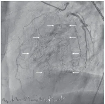
A 57-year-old woman with a history of a severe chronic obstruc-tive pulmonary disease was hospitalized due to type 2 respira-tory failure. She denied having chest pain, dyspnea, palpitations or syncope. Electrocardiogram at admission showed inverted T waves in leads V2–V4 (Fig. 1), a finding which was not present in the previous exams. Cardiac enzymes were negative. Transtho-racic echocardiography showed hyperkinetic left ventricular sys-tolic function (67%) without any wall motion abnormalities nor valvular problems. There was no coronary sinus ostial atresia nor persistent left superior vena cava. Given the multiple car-diovascular risk factors, a coronary angiogram was performed showing no relevant stenosis. However, direct drainage of blood into the left ventricle due to the presence of the Thebesian veins was noted (movie 1,Fig. 2).
The venous drainage system of the heart can be subdivided into a greater and lesser cardiac venous system. The latter runs in the myocardial layer of the heart and includes the Thebesian veins, which are present in all four heart chambers but are more abundant in the right side of the heart, especially in the right atrium, and drain directly into the heart chambers [1]. These vessels, also called ‘venae cordis minimae’ were first studied and described by German anatomist Adam Christian Thebesius in
1708 in his anatomy dissertation ‘Disputatio medica inauguralis de circulo sanguinis in corde’ [2].
The Thebesian veins are believed to have distinct functions. First, they provide a route of myocardial drainage driving carbon dioxide and other waste away from the tissue and back into the general circulation. Second, they may be an alternative route of nourishment for the myocardium, though they cannot provide adequate blood flow during ischemia due to their small num-ber and their venoluminal nature [3]. Moreover, they contribute along with the bronchial veins to the physiological shunting of blood, which explains the difference in the partial pressure of oxygen between the alveolar and the arterial blood, also called the alveolar–arterial (A–a) gradient [1]. Finally, it has been considered that the presence of Thebesian veins protects against myocardial edema, fibrosis and ventricular diastolic dysfunction [4].
We stated in the pre-procedural echocardiography finding that there was no coronary sinus ostial atresia nor persistent left superior vena cava. These two conditions are often found together leading retrograde caval flow into innominate vein and more prominent Thebesian veins [5], thus the importance of actively looking for them when confronted with such angio-graphic images.
236 Scoglio and Cook
Figure 1: ECG demonstrating T wave inversion in leads V2-V4.
Figure 2: Angiographic image of the left coronary artery showing the Thebesian
veins.
In their case series, Carlyle and Pitney [6] reviewed 5800 angiograms finding relevant Thebesian drainage in only 0.1% of cases. Subjects were often old elderly females with a history of hypertension. Tortuous coronary vessels were often visualized during angiograms, which is also the case in our patient. In conclusion, the significance and the origin of Thebesian venous drainage visualized during coronary angiograms are still unclear, further investigation is warranted.
SUPPLEMENTARY DATA
Supplementary data mentioned in the text are available to sub-scribers in OMCREP online.
ACKNOWLEDGEMENTS
None.
CONFLICT OF INTEREST STATEMENT
None.
FUNDING
None.
CONSENT
Written consent was obtained from the patient and is available upon request.
GUARANTOR
Martin Scoglio, corresponding author.
REFERENCES
1. Nordick K, Singh P. Anatomy, thorax, heart Thebesian veins (veins of Thebesius, vessels of Thebesius, vasa Thebesii). In:
StatPearls [Internet]. Treasure Island (FL): StatPearls Publishing,
Thebesian veins 237
2. Mazurak M, Kusa J. Adam Christian Thebesius’ channels into the human heart: the Thebesian veins and the Thebe-sian valve. Tex Heart Inst J 2019 Jun;46(3):175–178. doi: https://doi.org/10.14503/THIJ-18-6604.
3. Ansari A. Anatomy and clinical significance of ventricular Thebesian veins. Clin Anat N Y N 2001;14(2):102–110. doi: https://doi.org/10.1002/1098-2353(200103)14:2
<102::AID-CA1018>3.0.CO;2-4.
4. Kurbel S, Mari´c S, Gros M. Do Thebesian veins and arterioluminal vessels protect against myocardial
edema occurrence? Med Hypotheses 2009. doi:
https://doi.org/10.1016/j.mehy.2009.01.042.
5. Ogawa K, Hishitani T, Hoshino K. Absence of the coro-nary sinus with corocoro-nary venous drainage into the main pulmonary artery. Cardiol Young 2013 Oct;23(5):759–762. doi: https://doi.org/10.1017/S1047951112001709.
6. Carlyle A, Pitney M. Is Thebesian venous drainage a part of a clinical syndrome? Heart Lung Circ
2013;22:S140–1. doi: https://doi.org/10.1016/j.hlc.2013.05. 335.