HAL Id: hal-01987719
https://hal.archives-ouvertes.fr/hal-01987719
Submitted on 21 Jan 2019
HAL is a multi-disciplinary open access
archive for the deposit and dissemination of
sci-entific research documents, whether they are
pub-lished or not. The documents may come from
teaching and research institutions in France or
abroad, or from public or private research centers.
L’archive ouverte pluridisciplinaire HAL, est
destinée au dépôt et à la diffusion de documents
scientifiques de niveau recherche, publiés ou non,
émanant des établissements d’enseignement et de
recherche français ou étrangers, des laboratoires
publics ou privés.
Heterozygous HTRA1 mutations are associated with
autosomal dominant cerebral small vessel disease
Edgard Verdura, Dominique Hervé, Eva Scharrer, Maria del Mar Amador,
Lucie Guyant-Maréchal, Anne Philippi, Astrid Corlobe, Françoise Bergametti,
Steven Gazal, Carol Prieto-Morin, et al.
To cite this version:
Edgard Verdura, Dominique Hervé, Eva Scharrer, Maria del Mar Amador, Lucie Guyant-Maréchal,
et al.. Heterozygous HTRA1 mutations are associated with autosomal dominant cerebral small vessel
disease. Brain, 2015, 138 (8), pp.2347–2358. �hal-01987719�
Heterozygous HTRA1 mutations are associated
with autosomal dominant cerebral small vessel
disease
Edgard Verdura,
1,2Dominique Herve ´,
1,2,3,* Eva Scharrer,
4,* Maria del Mar Amador,
5Lucie Guyant-Mare ´chal,
6Anne Philippi,
1,2Astrid Corlobe ´,
7Franc1oise Bergamettii,
1,2Steven Gazal,
8Carol Prieto-Morin,
1,2,5Nathalie Beaufort,
4Benoit Le Bail,
9Irina Viakhireva,
10Martin Dichgans,
4,11Hugues Chabriat,
1,2,3Christof Haffnner
4and
Elisabeth Tournier-Lasserve
1,2,5*These authors contributed equally to this work.
Cerebral small vessel disease represents a heterogeneous group of disorders leading to stroke and cognitive impairment. While most small vessel diseases appear sporadic and related to age and hypertension, several early-onset monogenic forms have also been reported. However, only a minority of patients with familial small vessel disease carry mutations in one of known small vessel disease genes. We used whole exome sequencing to identify candidate genes in an autosomal dominant small vessel disease family in which known small vessel disease genes had been excluded, and subsequently screened all candidate genes in 201 unrelated probands with a familial small vessel disease of unknown aetiology, using high throughput multiplex polymerase chain reaction and next generation sequencing. A heterozygous HTRA1 variant (R166L), absent from 1000 Genomes and Exome Variant Server databases and predicted to be deleterious by in silico tools, was identified in all affnected members of the index family. Ten probands of 201 additional unrelated and affnected probands (4.97%) harboured a heterozygous HTRA1 mutation predicted to be damaging. There was a highly significant diffnerence in the number of likely deleterious variants in cases compared to controls (P = 4.2 10 6; ; ; E odds ratio = 15.4E 95% confidence interval = 4.9–45.5), strongly suggesting causality. Seven of these variants were
located within or close to the HTRA1 protease domain, three were in the N-terminal domain of unknown function and one in the C-terminal PDZ domain. In vitro activity analysis of HTRA1 mutants demonstrated a loss of function effnect. Clinical features of this autosomal dominant small vessel disease diffner from those of CARASIL and CADASIL by a later age of onset and the absence of the typical extraneurological features of CARASIL. They are similar to those of sporadic small vessel disease, except for their familial nature. Our data demonstrate that heterozygous HTRA1 mutations are an important cause of familial small vessel disease, and that screening of HTRA1 should be considered in all patients with a hereditary small vessel disease of unknown aetiology.
1 INSERM UMR 1161, Gené´ne´tique et Physiopathologie des Maladies Cerébro-vasculaires, Paris, France 2 Universite´Paris Diderot, Sorbonne Paris Cite ´, UMR-S1161, Paris, France
3 AP-HP, Groupe Hospitalier Saint-Louis Lariboisière -Fernand-Widal, Service de Neurologie, Centre de Re ´fe´rence des Maladies Vasculaires Rares du Cerveau et de l’Oeil (CERVCO), Paris, France
4 Institute for Stroke and Dementia Research, Klinikum der Universita ¨t Mu¨nchen, Ludwig Maximilians University, Munich, Germany
5 AP-HP, Groupe Hospitalier Saint-Louis Lariboisie `re-Fernand-Widal, Service de Ge´ne´tique Mole´culaire Neurovasculaire, Centre de Re´fe´rence des Maladies Vasculaires Rares du Cerveau et de l’Oeil (CERVCO), Paris, France
6 Service de Neurophysiologie, Ho ˆpital Charles Nicolle, Rouen, France 7 Service de Neurologie, Ho ˆpital Gui de Chauliac, Montpellier, France
8 Plateforme de Ge´nomique Constitutionnelle du GHU Nord, Assistance Publique des Hopitaux de Paris (APHP), Hopital Bichat, Paris, France
9 Service de Neurologie, CH Bretagne Sud, Lorient, France
10 Service de Neurologie, Hopital de la Cavale Blanche, Brest, France 11 Munich Cluster for Systems Neurology (SyNergy), Munich, Germany Correspondence to: Pr. Elisabeth Tournier-Lasserve,
INSERM UMR 1161,
Faculte´ de Me´decine Paris Diderot (site Villemin), 10 avenue de Verdun,
750010 Paris, France
E-mail: tournier-lasserve@univ-paris-diderot.fr
Keywords: small vessel diseaseE vascular dementiaE HTRA1; ; ; E CARASILE CADASIL
Abbreviations: CADASIL = cerebral autosomal dominant arteriopathy with subcortical infarcts and leukoencephalopathyE CARASIL = cerebral autosomal recessive arteriopathy with subcortical infarcts and leukoencephalopathyE SVD = small vessel disease
Introduction
Cerebral small vessel disease (SVD) is a heterogeneous group of disorders affnecting small arteries, arterioles, veins, and/or capillaries of the brain (Pantoni, 2010E Wardlaw et al., 2013). Their two main clinical conse-quences are stroke and cognitive impairment. Typical neu-roimaging features include white mattier lesions associated with small infarctions, microbleeds, and macrobleeds. In most cases, SVD is sporadic, with age and hypertension representing the prevailing risk factors. However, several early-onset monogenic forms of SVD have been reported in adult patients, the majority of them with a dominant inheritance pattiern. They include CADASIL (cerebral auto-somal dominant arteriopathy with subcortical infarcts and leukoencephalopathy), inherited cerebral amyloid angiopa-thies and angiopaangiopa-thies associated with mutations of COL4A1 and COL4A2 genes (Joutel et al., 1996E Gould et al., 2006E Revesz et al., 2009). CADASIL is caused by NOTCH3 mutations and is by far the most common her-editary SVD with more than 500 families reported and a 10–15% mutation rate in patients with SVD referred for NOTCH3 molecular screening, the second one being COL4A1/COL4A2 angiopathies (unpublished data) (Chabriat et al., 2009). By contrast, CARASIL (cerebral autosomal recessive arteriopathy with subcortical infarcts and leukoencephalopathy) is a very rare autosomal reces-sive SVD caused by biallelic mutations of the HTRA1 gene (high temperature requirement protease A1)E only 12 HTRA1 mutated CARASIL families have been reported worldwide so far (Hara et al., 2009E Nozaki et al., 2014). While the identification of mutated genes has provided invaluable tools for diagnosing monogenic SVD forms, molecular screening of those genes in routine diagnosis identifies the causative mutation in 520% of patients referred for a familial SVD, which strongly suggests that other genes may be involved.
Herein, we used genome-wide linkage analysis and whole exome sequencing to identify the gene involved in an
autosomal dominant SVD family in which SVD genes previously known to lead to an autosomal dominant SVD were excluded. We then screened a panel of 201 unrelated patients with a familial SVD of unknown aeti-ology to estimate the frequency of mutations of this gene in familial SVD.
Materials and methods
Participants and study design
Two siblings and one of their first cousins (Family F1) were referred for stroke and/or cognitive impairment associated with diffnuse white mattier hyperintensities (genealogical tree shown in Fig. 1). Detailed clinical and neuroimaging features of these patients are presented in the ‘Results’ section.
A panel of 201 unrelated SVD probands referred for NOTCH3 molecular screening in the Genetics department of Lariboisie`re hospital were also included in this study on the basis of the following criteria: (i) having at least one first or second degree relative with a clinical history of stroke and/or vascular dementiaE and (ii) being screened nega-tive for mutations in the 23 exons of NOTCH3 known to be involved in CADASIL. Mean age of this cohort at referral for molecular screening was 57 years (standard deviation = 11.6). This panel was used to search for mutations in the candidate genes identified in Family F1 (see below and Supplementary material).
All patients and participating relatives provided writtien informed consent for participation in genetic studies, in accordance with ethical recommendations in France for genetic disorders. Informed consent forms were approved by local medical ethics comittiees or CPP Ile de France (N 007110588).
One hundred and ninety-two healthy controls of French origin were used as a control group for HTRA1 candidate variants analysis. Additional control data were obtained from the public Exome Variant Server (EVS) database (n = 4300 European Americans) and 1000 Genomes database (n = 379 Europeans).
Procedures
Whole-genome linkage analysis was performed in Family F1 with data obtained with Affnymetrix GeneChipÕ Human
Mapping 250K arrays (Supplementary material). Parametric multipoint linkage analysis was performed with MERLIN assuming an autosomal dominant inheritance, a complete penetrance, a disease allele frequency of 0.00011, and no phenocopy. Intervals reaching the maximum theoretical LOD score achievable in this family were considered as possibly linked. Intervals with LOD scores lower than 2 and larger than 2 cM were considered as excluded for linkage.
Whole exome sequencing of the three patients of Family F1 was performed at IntegraGen platform. Variant annotation was done by an internal bioinformatics pipeline (IntegraGen). Only missense, nonsense, insertion/deletion variants or variants potentially affnecting splicing were kept for further analysis. Variants present in dbSNP132, as well as those reported with a minor allele frequency 40.1% in the 1000 Genomes Project, EVS or IntegraGen exome database (96 French exomes) were excluded. Candidate variants were further se-lected when (i) shared by all three affnected patients in a het-erozygous stateE and (ii) located outside regions previously excluded by genome-wide linkage analysis.
Figure 1 Genealogical trees of the 11 mutated probands. Square = maleE circle = femaleE diagonal black line = deceased individual Eblack filled symbol = clinically and MRI proven affnected individualE empty symbol = clinically healthy relative Eblack dot = affnected individual based on family history and/or clinicalchartsE question mark = unknown status E syringe symbol = blood sampled individuall Easterisk: individualshowing an HTRA1 deleterious variant.
For screening of candidate genes identified in Family F1, we used Fluidigm multiplex PCR combined with next generation sequencing (191 unrelated patientsE see Supplementary mater-ial) and whole exome sequencing data (10 unrelated patients for whom whole exome sequencing was also performed at IntegraGen platform).
Standard PCR amplification and Sanger sequencing were used to confirm all candidate variants detected by Illumina sequencing. When available, patients’ relatives were also sequenced. In addition, Sanger sequencing of 96 DNA samples of healthy French individuals was performed to screen exons of candidate genes in which candidate variants were located. HTRA1 exon 1 was sequenced in 192 DNA samples of healthy French individuals.
Candidate variants were analysed by three prediction pro-grams: PolyPhen-2, SIFT, and MutationTaster. Potential splice-affnecting variants were analysed by the Alamut Splicing Analysis package (Interactive Biosoftwware).
Frequency of likely deleterious variants was compared between cases and two control samples from online databases: European-American controls from Exome Variant Server (EVSE n = 4300) and European controls from 1000 Genomes project (n = 379). The frequency of HTRA1 variants in cases and controls was compared by a Fisher’s exact test on a 2 2 contingency table. All the tests were two-sided, with a P-value of 0.05 considered significant and odds ratios (OR) and 95% confidence intervals (CI) were calculated. Analyses were performed using the statistical analysis package R.
Expression constructs, conditions used for transfection, collection of conditioned media, protein electrophoresis, immunoblottiing and procedures used to measure the activity (Beaufort et al., 2014) of the various HTRA1 mutants are detailed in the Supplementary material.
Results
Clinical and neuroimaging features of
Family F1 patients
The genealogical tree of Family F1 is shown in Fig. 1.
Patient F1-9, the proband of this family, is a senior
execu-tive 69-year-old male with moderate hypercholesterolaemia
but no other vascular risk factor. He had a history of
sciatica related to a lumbar disk prolapse. Since age 66
he reported a progressive reduction of cognitive
perform-ances without significant impact on his daily activities and
complained of gait disturbance since age 68. At age 69 he
experienced an acute episode of leftw-sided hemiparesis that
completely resolved within 8 days. His neurological
exam-ination performed at the time of MRI examexam-ination showed
a subtle leftw-sided ataxia. His blood pressure was 134/
70 mmHg. Neuropsychological testing revealed mild alter-ations in processing speed, executive functions, and verbal episodic and visuospatial memory performances suggestive of subcortical cognitive impairment. Mini-Mental State
Examination score was 29/30. Mood was found to be
mildly depressed. The standard vascular work-up (EKG,
echocardiography, cervical and transcranial ultrasound
examination, blood cell count, sedimentation rate, creatin-ine level) and fundus examination were normal.
His 64-year-old brother (Patient F1-10) had no vascular
risk factor. He had a history of arthritis of small and large
joints. At age 55, he presented a major depressive episode. He subsequently had cognitive complaints and decided to
retire early at age 58 years. At 62 years of age, he
experi-enced recurrent attiacks of visual, sensory and aphasic
migraine aura with or without headache. His neurological
examination was normal. His blood pressure was at 136/
78 mmHg. Cognitive testing showed alterations of perform-ances in tests evaluating attiention, executive functions and verbal episodic memory, and executive functions. Mini-Mental State Examination score was 25/30. The
standard vascular work-up was normal at time of
MRI examination, as well as the fundus examination.
In addition, the following exams were in the normal
range: 24-h blood pressure monitoring, anticardiolipid anti-bodies, antinuclear antianti-bodies, anti-beta-2 GP1 antianti-bodies, and homocysteine level.
Patient F1-8, a first cousin of Patients F1-9 and F1-10, is
a 65-year-old female with a long history of well-controlled
hypertension and hypercholesterolaemia. At age 60 years,
she had some cognitive complaints and at 61 years, an
acute episode of right hemiparesis that lasted 24 h. Her
neurological examination showed mild bradykinesia,
micrographia, pyramidal signs with diffnuse increased
reflexes. Blood pressure was 140/90 mmHg. She had a cog-nitive slowing, but cogcog-nitive testing including Mini-Mental
State Examination, Mattiis Dementia Rating Scale, Grober
and Buschke Verbal Learning Test, verbal fluencies, Rey
figure copy and oral naming were normal. The standard
vascular work-up was normal at time of MRI examination. Complete ophthalmological examination, electrophysiolo-gical studies of the limbs and electronic microscopy skin biopsy were normal.
The father of Patients F1-9 and F1-10 had a history of dementia starting during the sixth decade of life. He died at
75 years while he was still able to walk with help. His
father, died in his late seventies several days aftwer the
sudden onset of a hemiplegia. Patient F1-7, sister of
Patient F1-8, presents a cognitive impairment that started
aftwer 60 years of age. Their mother died at age 70 years of
unknown cause.
MRI showed in all three patients diffnuse white mattier
hyperintensities affnecting supratentorial white mattier,
exter-nal capsules and pons but sparing the temporal lobes
(Fig. 2). Patients F1-9 and F1-8 had confluent and diffnuse
white mattier hyperintensities in the centrum semi-ovale
associated with hemispheric and pontine lacunes, while
Patient F1-10 had early confluent, less diffnuse white
mattier hyperintensities and no lacune. Patient F1-8 was
the only one to show microbleeds on gradient-echo
imagesE those numerous microbleeds were located in the
deep grey nuclei and subcortical areas. All patients had
images and leading to so-called e ´tat crible´ in Patients F1-9 and F1-10.
Sequence analysis of the 23 exons encoding the 34 epi-dermal growth factor (EGF)-like repeats in the extracellular
region of the NOTCH3 receptor was negative in all
patients. Screening of genomic DNA of Patient F1-8 for
APP mutations and duplication was also negative.
Identification of candidate variants in
Family F1
Genome-wide linkage analysis conducted in Family F1
excluded 79% of the genome (LOD score 5 2), including all known SVD loci. We identified 28 regions reaching the
theoretical maximal LOD score achievable in this family
(LS = 0.903) over a minimum distance of 2 cM
(Supplementary Fig. 1).
Subsequent whole exome sequencing of the three affnected family members was performed. Filtering strategy and data
obtained are presented in Supplementary Table 1. A total
of 11 candidate variants, including six single nucleotide
variants and five insertion/deletion (Indel) variants were identified. Six of 11 variants were located in regions excluded by linkage analysis and were therefore not considered. The five remaining variants were missense
variants located in HTRA1 (NM_0020775.4), TTBK2
(NM_1730500.3), C15orf43 (NM_1520448.2), CPEB1
(NM_0300594.4) and ZNF785 genes (NM_1520458.6)
(Supplementary Table 2). They were all absent from 1000
Genomes, EVS and IntegraGen databases, and confirmed
by Sanger sequencing to cosegregate with the affnected
Figure 2 MRI data obtained in Patients F1-9, F1-10 and F1-8. Leftw column: T2-weighted images. Centralcolumns:FLAIR images. Right
column:T1-weighted image (top), 3D time of flight image (middle) and gradient-echo image (bottiom).Extensive white mattier hyperintensities in
Patients F1-9 and F1-8 and less diffnuse white mattier hyperintensities in Patient F1-10 are detected on FLAIR images.Multiple dilated perivascular spaces corresponding to so-called e´tat crible´ were observed in Patients F1-9 and F1-10 on T2-weighted images. Lacunes are found in Patients F1-9
phenotype in Family F1. The HTRA1 variant (c.497G 4 T, p.Arg166Leu) was predicted to be pathogenic by all three in silico prediction tools (PolyPhen-2, SIFT and MutationTaster) (Table 1). The TTBK2 variant (c.11010A 4 T, p.Lys367Ile) was considered benign by PolyPhen-2, and the other three variants, located in C15orf43 (c.307A 4 C, p.Ser103Arg), CPEB1 (c.699G 4 C, p.Met233Ile), and ZNF785 (c.10111C 4 G, p.Ser337Arg) were considered benign by at least two pre-diction tools.
Heterozygous HTRA1 variants are
associated with familial cerebral SVD
To test the relevance of the five candidate genes (HTRA1, TTBK2, C15orf43, CPEB1, ZNF785) in additional patients with SVD, multiplex Fluidigm PCR amplification of exonic regions of these genes followed by massive parallel sequen-cing was conducted in 191 unrelated patients with familial SVD and in Patient F1-9, used as a positive control. In add-ition, mutations of these five genes were searched for in whole exome sequencing data of 10 additional unrelated familial SVD probands. Eighteen exonic and/or splice site rare candidate variants were identified in 17 unrelated pa-tients of the cohort. Fourteen of those heterozygous variants
were located in HTRA1. Each of the four remaining vari-ants, located in the four remaining genes, was identified in only one patient. They were all excluded based on either their prevalence in control genomic databases, or their pre-dicted benignity by at least two in silico prediction tools or their occurrence in a proband showing a deleterious HTRA1 variant (Supplementary Table 3).
Sanger sequencing was then used to confirm the presence of candidate HTRA1 variants in all mutated probands. In addition, Sanger sequencing of HTRA1 exon 1 and part of exons 2 and 4 was performed in all 201 probands as Fluidigm coverage of these exons was not complete. Thirteen heterozygous HTRA1 variants were confirmed in 14 distinct probands, including F1 proband (Table 1, Fig. 3 and Supplementary Fig. 2). Eleven of these variants, includ-ing R166L (the variant identified in Family F1), were con-sidered to be most likely deleterious. Ten of them were missense variants that were (i) absent from all interrogated databases (1000 Genomes, EVS, and IntegraGen exome databases) and from a cohort of 96 French healthy control individualsE (ii) predicted to be pathogenic by at least two in silico prediction toolsE and (iii) affnecting highly conserved amino acids (Supplementary Fig. 3). The remaining one was a splice site variant (c.973-1 G 4 A) absent from all databases and predicted to lead to an abnormal splicing of
Table 1 Rare heterozygous HTRA1 variants identified in the familial SVD probands
Family cDNA change Predicted protein change Protein domain Intron/ Exon PolyPhen-2 a (HumVar) SIFTa Mutation Taster a 1000Genomes frequency EVS frequency Sanger sequen-cing results
F1 c.497 G 4 T p.Arg166Leu None Exon 2 Probably
damaging
Deleterious Disease causing
Absenceb Absence Confirmed
F2 c.517 G 4 C p.Ala173Pro None Exon 2 Possibly
damaging Deleterious Diseasecausing Absence Absence Confirmed
F3 c.852 C 4 A p.Ser284Arg Serine protease Exon 4 Probably damaging Deleterious Disease causing
Absence Absence Confirmed
F4 c.854 C 4 A p.Pro285Gln Serine protease Exon 4 Probably damaging Deleterious Disease causing
Absence Absence Confirmed
F5 c.856 T 4 G p.Phe286Val Serine
protease Exon 4 Possiblydamaging Tolerated Diseasecausing Absence Absence Confirmed
F6 c.973-1 G 4 A p.(Tyr325_
Leu335del) Serine
protease
Intron 4 NAc NAc NAc Absence Absence Confirmed
F7 c.13418 G 4 C p.Asp450His PDZ Exon 9 Possibly
damaging Deleterious Diseasecausing Absence Absence Confirmed
F8 c.850 A 4 G p.Ser284Gly Serine protease Exon 4 Probably damaging Deleterious Disease causing
Absence Absence Confirmed
F39 c.59 C 4 T p.Ala20Val - Exon 1 Benign Tolerated Polymorphism Absence NAd Confirmed
F78 c.59 C 4 T p.Ala20Val - Exon 1 Benign Tolerated Polymorphism Absence NAd Confirmed
F133 c.367 G 4 T p.Ala123Ser Kazal-type Exon 1 Possibly
damaging
Tolerated Disease causing
Absence NAd Confirmed
F134 c.397 C 4 G p.Arg133Gly Kazal-type Exon 1 Possibly
damaging Tolerated Diseasecausing Absence NA
d Confirmed
F167 c.152 A 4 G p.Glu51Gly IGFBP-like Exon 1 Benign Tolerated Polymorphism Absence NAd Confirmed
F186 c.361 A 4 C p.Ser121Arg Kazal-type Exon 1 Probably
damaging Deleterious Diseasecausing Absence NA
d Confirmed
Candidate variants predicted to be damaging by at least two pathogenicity prediction so ftwwares,or affnecting splicing sites.
aPolyPhen-2 (HumVar) / SIFT / Mutation Taster: predicted pathogenicity using these three programs. bAbsence: absence from the 1000 Genomes database (version 2014) or Exome Variant Server (ESP6500v2). cNA: not analysed because splice variants cannot be analysed with those prediction tools.
exon 5. This abnormally spliced mRNA would skip 33
codons including codon 328, which encodes for a serine
residue required for HTRA1 activity. The last two variants,
p.Ala20Val (shared by two unrelated patients) and
p.Glu51Gly, located in exon 1, were absent from databases
and from our French control group but were predicted to
be benign with all three prediction tools. They were not
considered further. Altogether, 11 unique, and
heterozy-gous HTRA1 variants, including Family F1 variant, were
considered as deleterious (Fig. 3).
The genealogical trees of these 11 unrelated mutated
car-riers are shown in Fig. 1. Family history was consistent
with an autosomal dominant pattiern of inheritance for at least eight of these families. Three additional affnected rela-tives were available for Sanger sequencing in Families F5 and F8E all three were shown to be mutation carriers.
The frequency of deleterious variants was compared between our cases and control samples from two online
databases: European American controls from EVS
(n = 4300) and European controls from 1000 Genomes
project (n = 379). All nine exons of HTRA1 have been
sequenced by the 1000 Genomes consortium whereas
only eight exons (exons 2 to 9) have been sequenced in
EVS. Ten likely deleterious variants located in exons 2–9 have been identified in the 4300 European American
con-trols from EVS (one splice site change, two nonsense, and
seven missense variants), as compared to seven exons 2–9
variants in our panel of 201 patients with SVD
(P = 4.2 10 6; ; ; E OR = 15.4E 95% CI 4.9–45.5). There is
no deleterious variant in any of the HTRA1 exons 1–9 in
the 379 European controls from 1000 Genome project as
compared to a total number of 10 variants in our panel of
patients with SVD (P = 2.1 10 5).
Functional characterization of HTRA1
mutations
HTRA1 encodes a conserved serine protease consisting of several domains (Fig. 3) (Clausen et al., 2002). Most of the previously identified homozygous HTRA1 mutations have been demonstrated to strongly reduce proteolytic activity Figure 3 Schematic representation of HTRA1 gene/protein and location of mutations. (A) cDNA structure of human HTRA1 transcript NM_0020775.4. Positions are annotated from the first coding nucleotide. Positions of heterozygous SVD mutations identified in this study are indicated above the figure. Numbers represent the first coding nucleotide of each exon, and the last coding nucleotide (nt 1443). (B) Domain organization of human HTRA1 protein (protein NP_0020766.1). Above, positions of HTRA1 protein changes identified in patients with SVD in this study. Below, mutations that have been previously reported in CARASIL patients. SP = signalpeptideE PDZ = PDZ-like domain. Numbers above the figure represent the first and last coding amino acids for each one of the protein domains, and the last coding amino acid (aa 480).
(Nozaki, 2014). We therefore assessed the catalytic capacity of a subset of our heterozygous variants in vitro using con-ditioned supernatants of transiently transfected HEK 293T
cells and two diffnerent substrates [bovine serum albumin
(BSA) and fluorescently labelled casein]. Wild-type
HTRA1 and the active-site mutant S328A were used as
controls. Western blottiing of cell culture media revealed
efficient expression and secretion of all mutants except
75 50 37 kDa
A
HtrA1 250 75 kDa 150 50 100 37 BSAB
C
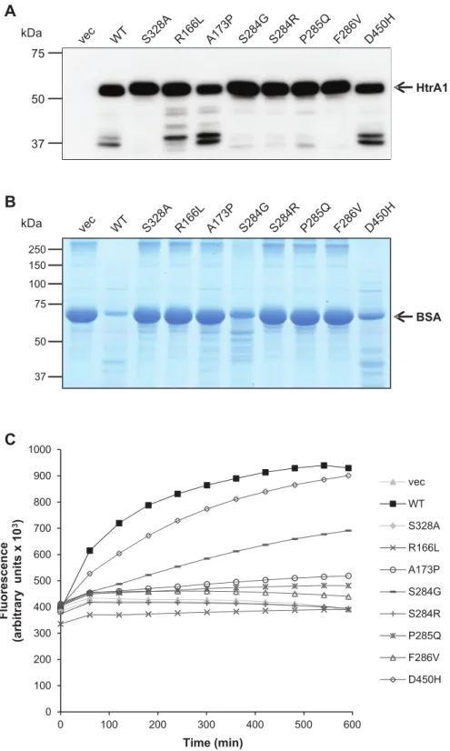
0 100 200 300 400 500 600 700 800 900 1000 0 100 200 300 400 500 600 F lu o re s c e n c e (a rb it ra ry u n it s x 1 0 3) Time (min) vec WT S328A R166L A173P S284G S284R P285Q F286V D450HFigure 4 Most heterozygous HTRA1 mutants are proteolytically inactive. (A) Mutants are effniciently expressed and secreted. Upon transient transfection in HEK293T cells, anti-myc immunoblottiing was performed on conditioned supernatants. Full-length HTRA1 migrates at 55 kDa, lower molecular weight bands represent degradation products. (B) Proteolytic activity of conditioned supernatants was analysed towards denatured BSA and evaluated by SDS-PAGE and Coomassie Blue staining. (C) Proteolytic activity of conditioned supernatants was analysed towards fluorescently labeled casein over 10 h by measuring FRET. vec = vectorE WT = wild-type.
for A173P and D450H, which were present at somewhat reduced levels, possibly due to increased autodegradation (Fig. 4A). In the BSA assay HTRA1 mutants R166L, A173P, S284R, P285Q and F286V showed a loss of activ-ity comparable to the S328A mutant (Fig. 4B). In contrast, the reduction observed for S284G and D450H was only partial. These results were fully confirmed using casein as substrate in a Fluorescent Resonance Energy Transfer (FRET) assay yielding a fluorescence increase upon casein degradation (Fig. 4C). While D450H and S284G displayed residual activity, all other mutants showed a behaviour similar to the loss-of-function controls.
Main clinical features of the unrelated
probands with heterozygous HTRA1
mutations
Main clinical and MRI features of the 11 unrelated probands with HTRA1 heterozygous mutations are sum-marized in Table 2, Figs 2, 5 and Supplementary Fig. 4. Genealogical trees are shown in Fig. 1. The clinical picture is characterized by the association of subcortical ischaemic events and cognitive decline starting in most patients in the sixth decade. None of these patients had early-onset alopecia or early-onset spondylosis. Three of them
(Patients F1-9, F3 and F6 index) suffnered from a lumbar disk prolapse but late in life. Typical neuroimaging findings include confluent or early confluent white mattier hyperin-tensities on MRI T 2-weighted and FLAIR images, with
fre-quent involvement of the external and internal capsules, sometimes associated with multiple lacunar infarcts and microbleeds (Figs 2, 5 and Supplementary Fig. 4). Anterior temporal regions and U fibres were spared. Dilated perivascular spaces with a typical ‘status cribrosum’ characterized by an innumerable number of dilated Virchow-Robin spaces and resulting in a cribriform change in basal ganglia were observed in most patients. Altogether, the clinical and MRI features of these HTRA1 heterozygous patients are similar to the ones of sporadic SVD cases.
Discussion
We report herein a family including three affnected individ-uals with a cerebral SVD of previously unknown aetiology, which segregates as an autosomal dominant disorder. Linkage analysis in combination with whole exome sequen-cing performed in this family (F1) as well as high through-put sequencing in a panel of 201 unrelated familial SVD cases (in whom NOTCH3 mutations were previously
Table 2 Main clinical and neuroimaging features of the 11 probands carrying deleterious heterozygous HTRA1 mutations F1 R166L F2A173P F3S284R F4P285Q F5F286V F6 c.973-1G 4 A F7 D450H F8S284G F133A123S F134R133G F186S121R
Sex Male Female Female Male Male Female Male Female Male Female Male
Age at onset (years) 66 65 - 50 49 66 68 62 - - 56
Age at time of study (years)
69 72 49 55 55 66 72 62 50 58 66
No. of affnected relativesa 4 1 2 1 1 1 1 2 4 1 3
Hypertension No Yes Yes No No Yes Yes No Yes No No
Symptoms at disease onset (or symptoms that led to MRI)b
Cognitive
decline Balanceimpairment (Headaches) Stroke Stroke Stroke TIA Stroke (Seizures) (Headaches) Balanceimpairment History of TIA or
ischaemic stroke Yes No No Yes Yes Yes Yes Yes No No No
Cognitive impairment Yes Yes No No Yes N.A. Yes No No No Yes
Dementia No No No No No No No No No No Yes
Gait disturbance Yes Yes No Yes Yes N.A. Yes No No No Yes
MRI features
Age at MRI (years) 69 72 49 50 55 66 70 62 50 58 62
WMH Yes Yes Yes Yes Yes Yes Yes Yes Yes Yes Yes
early confluent - - - Yes - - - - Yes Yes Yes
confluent Yes Yes Yes - Yes Yes Yes Yes - -
-Status cribrosumc Yes Yes N.A. Yes N.A. Yes N.A. Yes Yes No Yes
Lacunes Yes Yes Yes Yes Yes Yes Yes Yes Yes No Yes
Microbleedsd No N.A. No N.A. N.A. Yes N.A. Yes No N.A. No
aAffnected relatives, based on family history and/or clinicalcharts (see Fig. 1). bSymptoms which led to MRI are possibly not related to the disease. cThe presence of ‘status cribrosum’ was evaluated when T
2sequence or/and T1sequence with thin-slice were available. dThe presence of microbleeds was evaluated when gradient-echo sequences were available.
excluded) strongly suggest that (i) the causative mutation in Family F1 is a heterozygous mutation located in HTRA1; ; ; E
and (ii) roughly 5% of familial SVD of unknown aetiology
are associated with heterozygous HTRA1 mutations.
Clinical features of this autosomal dominant SVD diffner
from those of CARASIL by a later age at onset of stroke
and cognitive decline (20–30 years in age at onset) and the
absence of the typical CARASIL extra-neurological
symp-toms (Maeda et al., 1976E Nozaki et al., 2014). Indeed,
none of our patients presented with early-onset alopecia
or early-onset spondylosis. Three of them were reported
to have osteoporosis and lumbago, but those
manifest-ations occurred late in life. The phenotype of this condition
is very close to those observed in sporadic SVD cases.
Indeed, the occurence of lacunar ischaemic events
between 55 and 72 years in half of the cases and the
pro-gressive gait disturbance and subcortical cognitive
impair-ment from the age of 60 are comparable with the
phenotype of age and hypertension related SVD. The
MRI pattiern is also very similar except for the presence
of a status cribrosum, which seems to be more frequent
in this condition, and for the location of microbleeds
which involves preferentially the deep and juxtacortical
hemispheric areas.
Several observations strongly support the pathogenic role of the heterozygous HTRA1 mutations in the 11 probands reported herein. First, all these mutations are absent from the 1000 Genomes (n = 379 Europeans) and EVS databases as well as from our panel of French controls. Second, there is a highly significant diffnerence between the frequency of
deleterious HTRA1 variants in our panel of familial SVD
probands and that in control databases, including the EVS
database (n = 4300 European-AmericansE P = 4.2 10 6; ; ; E
OR = 15.4E 95% CI 4.9–45.5) and 1000 Genomes database
(n = 379 EuropeansE P = 2.1 10 5). Third, all 11
muta-tions are predicted to be deleterious by at least two in silico
prediction tools. Fourth, a complete loss of activity was
observed for five of the seven variants tested. An additional variant located in a consensus splice site (c.973-1G 4 A), is
predicted to lead to an in-frame deletion of exon 5, which
Figure 5 Status cribrosum and white mattier hyperintensity characteristics in HTRA1 mutated probands. Status cribrosum is well
visible on T2-weighted images in Patients F133-7, F8-11, F6-5, and F1-9. white mattier hyperintensitiess on FLAIR images were extensive in all cases
includes the codon 328 encoding a serine required for enzyme activityE this variant is also most likely a loss-of-function mutant. The residual activity of the variants S284G and D450H might be the result of a pathomechan-ism diffnerent from intrinsic proteolytic activity alteration
such as protein destabilization or mislocalization. In any
case, both our genetic data and the results of in silico pre-diction tools analyses strongly argue for their pathogenic
potential. Additional work on large series of patients is
required to elucidate how these mutants might alter
HTRA1 function.
The most surprising finding of this study is the
deleteri-ous role of heterozygdeleteri-ous mutations in HTRA1, a gene
pre-viously reported to be involved in a very rare autosomal
recessive SVD (Hara et al., 2009). Indeed, CARASIL was
initially reported in siblings from a consanguineous family
with unaffnected parents. So far 12 mutated CARASIL
families have been reported, including 10 consanguineous
families (Nozaki et al., 2014). The largest series includes six
families from JapanE all of these were consanguineous
families and parents of affnected cases were reported as
being unaffnected (Hara et al., 2009). Half of these families
harboured homozygous stop codons. Unaffnected parents
were further mentioned in two other consanguineous
families from Japan and Turkey (Nishimoto et al., 2011E
Bayrakli et al., 2012). Interestingly, however, in three add-itional families, cerebral MRI performed in either one or both parents was reported to show a leukoencephalopathy,
raising the question of the causality of the heterozygous
variants carried by these parentsE in the last family, both
parents were said to be deceased in their fiftwies with
emotional liability, but MRI was not available
(Mendioroz et al., 2010E Wang et al., 2012E Chen et al., 2013E Bianchi et al., 2014). None of our families is con-sanguineous and 8 of 11 of them show a pattiern of
inher-itance consistent with autosomal dominant inheritance.
Altogether, these data strongly suggest that in some cases
HTRA1 mutations behave as autosomal recessive
muta-tions, with heterozygous carriers being clinically unaffnected,
as observed in Japanese CARASIL patients. In other cases,
HTRA1 mutations behave as dominant mutations, with
heterozygous carriers being clinically affnectedE based on
our data, their frequency is predicted to be much higher than recessive type mutations. One of our current
hypoth-eses to reconcile those data is that some of the missense
mutations reported herein might be dominant negative mu-tations, the mutated allele being a loss of function mutant which interferes with the normal function of the remaining
wild-type allele, leading to a further decrease in enzyme
activity. In contrast, mutations leading to premature stop codons would lead to the decay of the mutated mRNA, the absence of synthesis of the protein encoded by the mutated allele and therefore to a haploinsufficiency which by itself would not be sufficient to cause SVD. This
has been reported in several other diseases, such as
hyper-tension caused by KLHL3 mutations or myopathies
associated with COL6A1 mutations (Boyden et al., 2012E
Zou et al., 2014). Interestingly, in most of these diseases
that exist both as dominant and recessive disorders, the
causative gene encodes proteins acting as dimers or multi-mers. HTRA1 encodes a serine protease whose functional
unit is a trimer stabilized by residues of the protease
domain (Clausen et al., 2002). Another non-mutually
exclusive hypothesis would be that some of these missense mutations are semi-dominant mutations. Comparative measurement of the residual HTRA1 endogenous activity of fibroblasts from missense and stop codons heterozygous carriers would be needed to investigate further this question
but this test is not currently available. Our last hypothesis
would be that in some CARASIL families the affnected
status of the heterozygous parents would have been overlooked due to the much later age at onset in heterozy-gous carriers as compared to offnsprings with biallelic mutationsE indeed, there is roughly 20–30 years diffnerence in age at onset between heterozygous and biallelic mutated patients.
The identification of the causative gene in these families now allows molecular genetic screening for this condition. Knowing that the clinical and MRI features of the patients reported herein were similar to those observed in sporadic SVD cases except for the familial nature of the disease,
molecular screening of HTRA1 would be indicated in all
cases with a familial SVD, in the absence of clinical or MRI findings suggestive of another condition. However, the fa-milial nature of the disease may be overlooked due to the late onset of clinical symptoms. Therefore, molecular
screening should be considered in sporadic cases when
the extent of microvascular lesions on MRI contrasts with the paucity of vascular risk factors.
In summary, we show herein that nearly 5% of familial SVDs referred for molecular screening are associated with
deleterious heterozygous mutations of HTRA1. Therefore,
these mutations would be so far the second cause of
auto-somal dominant SVD aftwer CADASIL. This autosomal
dominant SVD is characterized by a much later age of
onset than in CARASIL and the absence of typical
extra-neurological CARASIL features. Further assessment of
pathogenic molecular effnects of those heterozygous variants
and delineation of the complete mutation spectrum will
now be needed to develop efficient genetic diagnosis.
Acknowledgements
We thank all families for their participation in this study.
We are also indebted to F. Marchelli for excellent figures
editing.
Funding
The research was funded by INSERM, Programme
Hospitalier de Recherche Clinique AOM060037 (grant to
novel targets for hemorrhagic stroke’ (grant to E.T.L.), the
Leducq transatlantic network ‘Pathogenesis of Small Vessel
Disease of the Brain’ (grant to M.D.), the Vascular
Dementia Research Foundation and the Deutsche
Forschungsgemeinschaftw (DFG).
Supplementary material
Supplementary material is available at Brain online.
References
Bayrakli F, Balaban H, Gurelik M, Hizmetli S, Topaktas S. Mutation in HTRA1 gene in a patient with degenerated spine as a component of CARASIL syndrome. Turkish Neurosurg 2012E 24: 67–9. Beaufort N, Scharrer E, Kremmer E, Lux V, Ehrmann M, Hubert R,
et al. Cerebral small vessel disease-related protease Htra1 processes latent TGF-b binding protein 1 and facilitates TGF-b signaling. Proc Natl Acad Sci USA 2014E 111: 160496–501.
Bianchi S, Di Palma C, Gallus GN, Taglia I, Poggiani A, Rosini F, et al. Two novel HTRA1 mutations in a European CARASIL patient. Neurology 2014E 82: 898–900.
Boyden LM, Choi M, Choate KA, Nelson-Williams CJ, Farhi A, Toka HR, et al. Mutations in Kelch-like 3 and cullin 3 cause hypertension and electrolyte abnormalities. Nature 2012E 482: 98–102. Chabriat H, Joutel A, Dichgans M, Tournier-Lasserve E, Bousser MG.
Cadasil [Review]. Lancet Neurol 2009E 8: 643–53. Chen Y, He Z, Meng S, Li L, Yang H, Zhang X. A novel mutation of
the high-temperature requirement A serine peptidase 1 (HTRA1) gene in a Chinese family with cerebral autosomal recessive arterio-pathy with subcortical infarcts and leukoencephalopathy (CARASIL). J Int Med Res 2013E 41: 1445–55.
Clausen T, Southan C, Ehrmann M. The HTRA family of proteases: implications for protein composition and cell fate. Mol Cell 2002E 10: 443–55.
Gould DB, Phalan FC, van Mil SE, Sundberg JP, Vahedi K, Massin P, et al. Role of COL4A1 in small-vessel disease and hemorrhagic stroke. N Engl J Med 2006E 354: 1489–96.
Hara K, Shiga A, Fukutake T, Nozaki H, Miyashita A, Yokoseki A, et al. Association of HTRA1 mutations and familial ischemic cere-bral small-vessel disease. N Engl J Med 2009E 360: 1729–39. Joutel A, Corpechot C, Ducros A, Vahedi K, Chabriat H, Mouton P,
et al. Notch3 mutations in CADASIL, a hereditary adult-onset con-dition causing stroke and dementia. Nature 1996E 383: 707–10. Mendioroz M, Fernandez-Cadenas I, Del Rio-Espinola A, Rovira A,
Sole´ E, Fernandez-Figueras MT, et al. A missense HTRA1 mutation expands CARASIL syndrome to the Caucasian population. Neurology 2010E 75: 2033–5.
Maeda S, Nakayama H, Isaka K, Aihara Y, Nemoto S. Familial unusual encephalopathy of Binswanger’s type without hypertension. Folia Psychiatr Neurol Jpn 1976E 30: 165–77.
Nishimoto Y, Shibata M, Nihonmatsu M, Nozaki H, Shiga A, Shirata A. A novel mutation in the HTRA1 gene causes CARASIL without alopecia. Neurology 2011E 76: 1353–5.
Nozaki H, Nishizawa M, Onodera O. Features of cerebral autosomal recessive arteriopathy with subcortical infarcts and leukoencephalo-pathy. Stroke 2014E 45: 3447–53.
Pantoni L. Cerebral small vessel disease: from pathogenesis to clinical characteristics to therapeutic challenges. Lancet Neurol 2010E 9: 689–701.
Revesz T, Holton JL, Lashley T, Plant G, Frangione B, Rostagno A, et al. Genetics and molecular pathogenesis of sporadic and heredi-tary cerebral amyloid angiopathies. Acta Neuropathol 2009E 118: 115–30.
Wang XL, Li CF, Guo HW, Cao BZ. A Novel mutation in the HTRA1 gene identified in Chinese CARASIL pedigree. CNS Neurosc Ther 2012E 18: 867–9.
Wardlaw JM, Smith C, Dichgans M. Mechanisms of sporadic cerebral small vessel disease: insights from neuroimaging. Lancet Neurol 2013E 12: 483–97.
Zou Y, Zwolanek D, Izu Y, Ganhdy S, Schreiber G, Brockmann K, et al. Recessive and dominant mutations in COL12A1 cause a novel EDS/myopathy overlap syndrome in humans and mice. Hum Mol Genet 2014E 23: 2339–52.

![[PDF] Cours Arduino en pdf avec travaux dirigés | Cours Arduino](data:image/gif;base64,R0lGODlhAQABAIAAAP///wAAACH5BAEAAAAALAAAAAABAAEAAAICRAEAOw==)