HAL Id: inserm-00741102
https://www.hal.inserm.fr/inserm-00741102
Submitted on 11 Oct 2012
HAL is a multi-disciplinary open access
archive for the deposit and dissemination of
sci-entific research documents, whether they are
pub-lished or not. The documents may come from
teaching and research institutions in France or
abroad, or from public or private research centers.
L’archive ouverte pluridisciplinaire HAL, est
destinée au dépôt et à la diffusion de documents
scientifiques de niveau recherche, publiés ou non,
émanant des établissements d’enseignement et de
recherche français ou étrangers, des laboratoires
publics ou privés.
Bevacizumab potentiates chemotherapeutic effect on
T-leukemia/lymphoma cells by direct action on tumor
endothelial cells.
Li Wang, Wen-Yu Shi, Fan Yang, Wei Tang, Guillaume Gapihan, Mariana
Varna, Zhi-Xiang Shen, Sai-Juan Chen, Christophe Leboeuf, Anne Janin, et
al.
To cite this version:
Li Wang, Wen-Yu Shi, Fan Yang, Wei Tang, Guillaume Gapihan, et al.. Bevacizumab potentiates
chemotherapeutic effect on T-leukemia/lymphoma cells by direct action on tumor endothelial cells..
Haematologica, Ferrata Storti Foundation, 2011, 96 (6), pp.927-31. �10.3324/haematol.2010.037689�.
�inserm-00741102�
Introduction
Over the past decade, biologically targeted therapies have significantly improved the outcome of patients with hemato-logic malignancies. Rituximab (Rituxan; IDEC-C2B8), a recombinant humanized monoclonal antibody against CD20, successfully treats B-cell lymphoma and has recently been applied in CD20-positive pre-B leukemia.1,2 For T-cell
leukemia/lymphoma, which are relatively resistant to con-ventional chemotherapy and have a poor prognosis,3
bio-ther-apeutic targets are being investigated.
Angiogenesis is linked to clinically aggressive tumor sub-types and advanced-stage disease.4 Vascular endothelial
growth factor-A (VEGF-A) is a main stimulator of tumor-ini-tiated angiogenesis.5 Increased VEGF-A levels in blood and
tissue are associated with adverse prognosis in leukemia and lymphoma patients.6,7 In angioimmunoblastic T-cell
lym-phoma (AITL), we have shown that VEGF-A was over-expressed on endothelial cells, and that this overexpression
was related to tumor infiltration and poor survival. This demonstrated that endothelial cells, like lymphoma cells, con-tribute to tumor progression in AITL.8
Tumor progression is a complex process that requires tumor-endothelial cell interaction.9Cell adhesion molecules,
such as intracellular cell adhesion molecule-1 (ICAM-1) and its ligand leukocyte function-associated antigen-1 (LFA-1), are major mediators of tumor-endothelial crosstalk.10ICAM-1 is
over-expressed on lymphoma cells,11and its blockade
signifi-cantly inhibits lymphoma cell aggregation in vitro and lym-phoma growth in vivo.11,12
Bevacizumab (AvastinTM), a recombinant humanized
mon-oclonal antibody directed against VEGF-A, can reduce tumor angiogenesis and inhibit tumor growth, either alone or in combination with chemotherapy.5It has recently been
intro-duced in hematology and its therapeutic effect has been demonstrated in acute myeloid leukemia and AITL.13,14Here
we studied in vivo and in vitro the modalities of its therapeutic efficiency on T-leukemia/lymphoma.
Bevacizumab potentiates chemotherapeutic effect on T-leukemia/lymphoma
cells by direct action on tumor endothelial cells
Li Wang,1,2* Wen-Yu Shi,1* Fan Yang,1,2* Wei Tang,1*, Guillaume Gapihan,2,3,4,5Mariana Varna,2,3,4,5Zhi-Xiang Shen,1
Sai-Juan Chen,1,2Christophe Leboeuf,2,3,4,5Anne Janin,2,3,4,5and Wei-Li Zhao1,2
1State Key Laboratory of Medical Genomics, Shanghai Institute of Hematology, Shanghai Rui Jin Hospital, Shanghai Jiao Tong
University School of Medicine, Shanghai, China; 2Pôle de Recherches Sino-Français en Science du Vivant et Génomique,
Laboratory of Molecular Pathology, Shanghai, China; 3Inserm, U728, Paris, France; 4Université Paris 7, Paris, France; and 5AP-HP,
Hôpital Saint-Louis, Service de Pathologie, Paris, France
*LW, WYS, FY and WT contributed equally to this work. WLZ and AJ contributed equally and should both be considered senior authors.
Funding: this work was supported, in part, by the Chinese National High Tech Program [863:2006AA02A301], the National Natural Science Foundation of China [30750335], the Shanghai Commission of Science and Technology [08410708800], the Shanghai Rising Star Program [09QH1401700], the Program for New Century Excellent Talents in University, the Fok Ying Tung Education Foundation [111035], “Shu Guang” project supported by Shanghai Municipal Education Commission and Shanghai Education Development Foundation, and by the Co-PI Program of Samuel Waxman Cancer Research Foundation, Shanghai Rui Jin Hospital/Shanghai Jiao Tong University School of Medicine, Inserm, and Université Paris 7-Denis Diderot, and by the Institut National du Cancer, Programme Tumorothèque.
Manuscript received on November 18, 2010. Revised version arrived on January 23, 2011. Manuscript accepted on January 27, 2011.
Correspondence: Wei-Li ZHAO, State Key Laboratory of Medical Genomics, Shanghai Institute of Hematology, Shanghai Rui Jin Hospital, Shanghai Jiao Tong University School of Medicine, 197 Rui Jin Er Road, Shanghai 200025, China. Phone: international +0086.21.64370045.
Fax: international +0086.21.64743206. E-mail: weili_zhao_sih@yahoo.com; and Anne Janin, Inserm, U728, Hôpital Saint-Louis, 1 Avenue Claude Vellefaux, Paris, F-75010, France. Phone: international +33.1.42499933. Fax: international +33.1.42494922. E-mail: anne.janin728@gmail.com Vascular endothelial growth factor-A, an angiogenesis
stimula-tor expressed on both tumor endothelial and malignant T cells, is involved in tumor progression in T-leukemia/lymphoma. Here, we assessed the impact of therapeutic vascular endothe-lial growth factor-A blockade on tumor-endotheendothe-lial cell inter-action and on tumor progression. In a murine xenograft T-leukemia/lymphoma model, combined bevacizumab (mono-clonal antibody against vascular endothelial growth factor-A) with doxorubicin, compared with doxorubicin alone, signifi-cantly delayed tumor growth and induced prevalence of tumor cell apoptosis over mitosis. More importantly, the com-bined treatment induced endothelial cell swelling, microvessel occlusions, and tumor necrosis. In vitro, co-culture of endothe-lial cells with T-leukemia/lymphoma cells showed that dox-orubicin induced expression of intracellular cell adhesion mol-ecule-1, provided endothelial and malignant T cells were in direct contact. This was abrogated by bevacizumab treatment
with doxorubicin. Taken together, bevacizumab enhances the chemotherapeutic effect on T-leukemia/lymphoma cells. Directly targeting tumor endothelial cells might be a promis-ing therapeutic strategy to counteract tumor progression in T-cell malignancies.
Key words: T-cell leukemia/lymphoma, bevacizumab, and endothelial cells.
Citation: Wang L, Shi W-Y, Yang F, Tang W, Gapihan G, Varna M, Shen Z-X, Chen S-J, Leboeuf C, Janin A, and Zhao W-L. Bevacizumab potentiates chemotherapeutic effect on T-leukemia/lymphoma cells by direct action on tumor endothelial cells. Haematologica 2011;96(06):927-931.
doi:10.3324/haematol.2010.037689
©2011 Ferrata Storti Foundation. This is an open-access paper.
Design and Methods
Cells and reagents
T-leukemia/lymphoma cell lines Jurkat, HUT78, and endothelial cell line EAhy.926 (American Tissue Culture Collection, Bethesda, MD, USA) were maintained in RPMI-1640 medium supplemented with 10% heat-inactivated fetal bovine serum in a humidified atmosphere of 95% air and 5% CO2at 37°C. Bevacizumab was
obtained commercially (Hoffmann-la Roche, Basel, Switzerland).
Murine model
Four-week old nude mice (Shanghai Laboratory Animal Center, China), maintained in pathogen-free conditions, were subcuta-neously injected with 4×10⁷Jurkat cells (Day 0). Treatments were
performed on Days 21, 24, 28 and 31 in 3 groups of 10 mice. The untreated group only received RPMI-1640, the doxorubicin group received intraperitoneal injection of doxorubicin (6mg/kg), and the combined treatment group received intraperitoneal injection of doxorubicin (6mg/kg) and bevacizumab (10mg/kg). All animal procedures were approved by the Investigational Review Board of Shanghai Jiao Tong University School of Medicine. Tumor volume was calculated by the formula: 0.5×a×b2in centimeters, where ‘a’
is the length and ‘b’ is the width.
For each group, mice were sacrificed at two time points: Day 27 and Day 35. Subcutaneous tumors were dissected and cut into three parts. These were: (i) immediately snap frozen; (ii) formalde-hyde-fixed and paraffin-embedded; (iii) glutaraldeformalde-hyde-fixed and epoxy-resin-embedded for ultrastructural study.
Pathological analysis
Cell mitosis and apoptosisPathological study focused on cell proliferation and cell death on both tumor and endothelial cells. Mitotic and apoptotic cells were identified by electron microscopy (Hitachi-7560, Tokyo, Japan), or using Ki67 antibody (MIB-1, Abcam, Cambridge, UK) and TUNEL assay8on tumor sections. Cells were counted on 5 consecutive
microscopic fields by two pathologists who had not been informed of treatment modalities of the mice. A ProvisAX70 microscope (Olympus, Tokyo, Japan), with wide-field eyepiece number 26.5 was used. At x400 magnification, this wide-field eye-piece provided a field size of 0.344mm2. Results were expressed as
the mean number of cells/field at x400 magnification.
Tumor microvessel
Tumor microvessels were analyzed ultrastructurally, focusing on tumor-endothelial cell relationship. Microvessel density was assessed on 5 microscopic fields at x400 magnification on tumor sections immunostained with rat anti-mouse CD31 antibody (MEC13.3, BDpharmingen, Franklin Lakes, NJ, USA).
Tumor necrosis
Necrosis area was assessed on hematoxylin and eosin stained tumor sections. Whole slides were scanned and analyzed with a virtual slide system (Olympus). The ratio between tumor necrosis area and whole tumor area gave the relative necrosis area
ICAM-1 expression
ICAM-1 expression of human tumor cells and mouse endothe-lial cells was detected by immunostaining with goat anti-human ICAM-1 antibody (polyclonal, R&D Systems, Minneapolis, MN, USA) and goat anti-mouse ICAM-1 antibody (polyclonal, R&D Systems) on 5-mm frozen sections, respectively.
Enzyme-linked immunosorbent assay (ELISA)
5×10⁵endothelial cells were cultured in 6-well plates and grown
in confluence; 1×10⁶T-leukemia/lymphoma cells were added after
24 h. The T-leukemia/lymphoma and endothelial cells were: (i) cultured in the same vial; (ii) in Millicell Hanging Cell Culture System (Millipore Corporation, Billerica, MA, USA) with 1-mm pore filter only allowing fluid exchange. Cell co-cultures were treated with doxorubicin (200 ng/mL), or doxorubicin (200 ng/mL) combined with bevacizumab (100 mg/mL), or untreated. ELISA was performed in triplicate using ICAM-1 Kit (R&D Systems) on 100 mL supernatant, according to the manufacturer’s instructions.
Flow cytometry
5×10⁵endothelial cells and 1×10⁶T-leukemia/lymphoma cells
were co-cultured and treated as described above. Cells were disso-ciated with EDTA, incubated with Alexa 488-conjugated mouse anti-human ICAM-1 (84H10, Chemicon, Temecula, CA, USA) and allophycocyanin-conjugated mouse anti-human CD3 antibodies (BW264/56, Miltenyi Biotec, Auburn, CA, USA). Flow cytometry was used to measure fluorescent intensity (BD, Franklin, NJ, USA).
Confocal microscopy
5×10⁵endothelial cells and 1×10⁶T-leukemia/lymphoma cells
co-cultured in the same vials were immunolabeled using mouse anti-human ICAM-1 (84H10, Chemicon) and rabbit anti-human LFA-1 (polyclonal, Abcam, Cambridge, UK) as primary antibodies, and FITC-conjugated anti-mouse IgG or TexasRed-conjugated anti-rabbit IgG as secondary antibodies. Nuclei were counter-stained with DAPI. Z-stacked images were captured by confocal microscope (Zeiss-LSM-5, Welwyn Garden City, UK) and ana-lyzed by IMARIS software (Bitplane, Zurich, Switzerland).
Cell adherence assay
Co-cultured endothelial cells and T-leukemia/lymphoma cells were treated as described above. Each well was then filled with culture medium, covered with parafilm, and inverted for 30 min to remove unattached cells. Remaining adherent lymphoma cells were counted on five microscopic fields per well, and observed on inverted phase-contrast microscope (Nikon-TE-300, Japan). All assays were performed in triplicate.
Statistical analyses
Results were expressed as the mean±SD. Statistical significance of differences between treatment groups was measured by two-tailed t-test. P<0.05 was considered to be significant. All statistical analyses were performed on SAS8.2 (SAS-Institute-Inc., Cary, NC, USA).
Results
Bevacizumab targeted tumor cells and inhibited
tumor growth
To assess the therapeutic efficiency of bevacizumab in
vivo, a xenograft murine model was established. Jurkat and
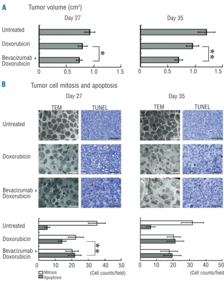
HUT-78 cells were both tested; only Jurkat cells developed a tumor at the site of subcutaneous injections. Compared with doxorubicin alone, combined bevacizumab and dox-orubicin significantly reduced tumor size, at Day 27 and Day 35 (P=0.0446 and P=0.0030, respectively, Figure 1A). To find how bevacizumab enhanced doxorubicin effect on T-leukemia/lymphoma cells, mitotic and apoptotic cells were assessed. In the untreated group, mitotic and apoptotic cells were respectively 35.3±5.1 and 5.2±1.6 at Day 27, with few changes at Day 35 (mitotic cells: 31.9±6.8 and apoptotic cells: 6.4±2.7). With doxorubicin,
mitotic cell numbers were significantly reduced (Day 27: 22.5±4.6 and Day 35: 20.40±4.13, P<0.0001), and apoptot-ic cell numbers signifapoptot-icantly increased (Day 27: 14.2±2.3 and Day 35: 21.4±5.2, P<0.0001). Compared to doxoru-bicin alone, combined bevacizumab with doxorudoxoru-bicin induced two important changes at Day 27: (i) a significant increase in apoptotic cells (21.9±3.9, P<0.0001); (ii) a prevalence of apoptosis over mitosis. This was linked to a significant increase of apoptosis, since mitosis cells were similar between the two groups (Figure 1B).
Bevacizumab targeted endothelial cells
and induced tumor necrosis
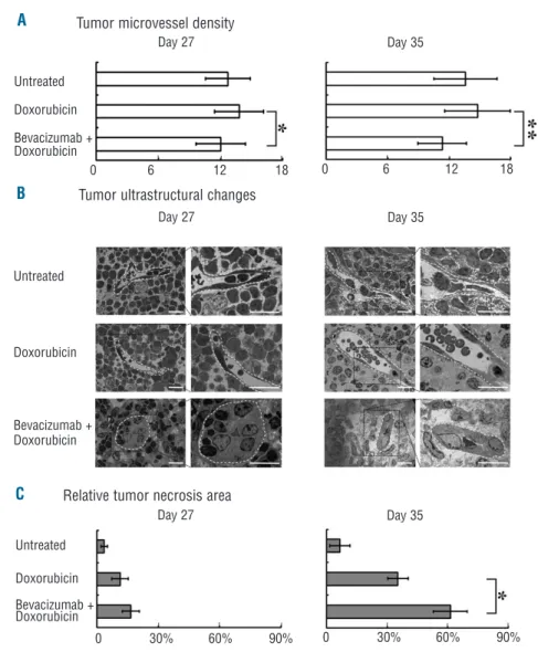
Compared with the untreated group, the microvessel density remained unchanged in the doxorubicin-treated group, but was significantly reduced, at Day 27 and Day 35, when bevacizumab was associated with doxorubicin (P=0.0218 and P=0.0004, respectively, Figure 2A).
Ultrastructural study at Day 27 showed that endothelial cells were flat with normal nuclei in the untreated group and in the doxorubicin-treated group. A striking feature in the combined treatment group was the swelling of endothelial cells in all microvessels studied. This reduced the lumen area of microvessels. Complete occlusion of some microvessels was observed with fibrin clot alone or
fibrin plus tumor cells, some of them apoptotic. At Day 35, endothelial cells remained swelling, and relative tumor necrosis area was significantly increased in the combined treatment group swollen, when compared with the dox-orubicin group (P=0.0103, Figure 2B and C).
Bevacizumab interrupted tumor-endothelial cell
interaction through ICAM-1 downregulation
Immunostainings of xenograft tumor sections showed that the numbers of human tumor cells and of murine microvessels expressing ICAM-1 were significantly increased with doxorubicin, compared with the untreated group. Combined bevacizumab with doxorubicin conse-quently reduced these numbers (Online Supplementary
Figure S1A).
Since such an effect of bevacizumab had not been pre-viously reported, we further analyzed ICAM-1 expres-sion in vitro. When Jurkat and HUT-78 cells were co-cul-tured with endothelial cells in the same vial, doxorubicin treatment induced a significant ICAM-1 overexpression, which was inhibited by addition of bevacizumab. This effect is absent when using a 1-mm pore filter allowing cytokine exchange, but not cytoplasmic contact, to sepa-rate tumor and endothelial cells (Online Supplementary
Figure S1B, left panel). Therefore, ICAM-1 reduction by
Figure 1. Bevacizumab targeted tumor cells and inhibited tumor growth. (A) Combined bevacizumab and doxorubicin significantly reduced tumor size at Day 27 and Day 35, compared with doxoru-bicin alone. Untreated mice only received intraperitoneal injection of RPMI1640. * P<0.05, ** P<0.01. (B) Transmission electron microscopy (TEM, Hitachi H7650, bar=10μm) and terminal deoxytransferase-catalyzed DNA nick-end labeling (TUNEL, bar=50mm) analy-ses showed that combined bevacizumab with doxorubicin induced a prevalence of apoptosis over mitosis in tumor cells as early as Day 27. Mitosis, Apoptosis, **P<0.01.
A
B
Tumor volume (cm3)
Tumor cell mitosis and apoptosis
Day 27 Day 35
Day 27
TEM TUNEL TEM TUNEL
Day 35 Untreated Doxorubicin Bevacizumab + Doxorubicin Untreated Doxorubicin Bevacizumab + Doxorubicin Untreated Doxorubicin Bevacizumab + Doxorubicin 0 0.5 1.0 1.5 0 10 20 30 40 50 Mitosis Apoptosis
(Cell counts/field) (Cell counts/field)
0 10 20 30 40 50
bevacizumab was only observed when direct cytoplasmic contact occurred.
Moreover, flow cytometry analysis was carried out to detect the cell surface ICAM-1 of T-leukemia/lymphoma cells (CD3+) and endothelial cells (CD3-). Compared with
the untreated group, membrane ICAM-1 of endothelial cells significantly increased after doxorubicin treatment (P<0.05), which was also abrogated by addition of beva-cizumab. The ICAM-1 of T-leukemia/lymphoma cells had a similar trend (Online Supplementary Figure S1B, right
panel).
In adhesion assays, combined bevacizumab with dox-orubicin significantly reduced T-leukemia/lymphoma cell adhesion to the endothelial monolayer when compared to doxorubicin alone (Online Supplementary Figure S1C).
On a confocal microscope, untreated cells were individ-ualized, expressing LFA-1 but not ICAM-1. Doxorubicin induced: (i) close association of endothelial with tumor cells; (ii) strong ICAM-1 expression on endothelial cells. In particular, ICAM-1 and LFA-1 were co-expressed in areas where endothelial and tumor cells were in close contact. Addition of bevacizumab abrogated ICAM-1 expression by endothelial cells, and their close association with tumor cells (Online Supplementary Figure S1D).
Discussion
Addition of bevacizumab to doxorubicin, in our xenograft murine model, significantly delayed tumor growth. First, bevacizumab could induce lymphoma cell apoptosis, as observed in another xenograft solid tumor model.15More importantly, it has a direct effect on tumor
vessels. Systematic ultrastructural analysis of tumor sec-tions revealed that bevacizumab induced endothelial cell swelling. This striking morphological change diminished the area of microvessel lumen and could explain the decreased tumor blood flow observed with computed tomography perfusion scan in the bevacizumab-treated tumor.16In our model, bevacizumab and doxorubicin also
induced microvessel occlusions, contributed to reduced microvessel density, and increased tumor necrosis area. This agrees with clinical data showing that: (i) anti-angio-genesis reduces the risk of chemotherapy resistance by targeting endothelial cells rather than tumor cells;17 (ii)
combining bevacizumab with chemotherapy results in greater anti-tumor effects than either treatment alone.17, 18
Tumor invasion requires interaction between invading tumor cells and tumor microvessels.19 ICAM-1 plays an
important role in cell adhesion.10 In our model,
doxoru-L. Wang et al.
Figure 2. Bevacizumab targeted endothelial cells and induced tumor necrosis. (A) Combined bevacizumab with doxorubicin significantly reduced tumor microvessel density at Day 27 and Day 35. *P<0.05, **P<0.01. (B) Ultrastructural analysis showed at low magnification, numerous apoptotic cells in combined treatment group at Day 27 and in doxorubicin group at Day 35. At high magnification, microvessels in un-treated mice had flat endothelium and circulating intact tumor cells in their lumen. Microvessels in the doxorubicin group had flat endothelial cells and apoptotic bodies in their lumen at Day 35. With the combined treatment, endothelial cells were swollen with volu-minous nucleus and cytoplasm, reduc-ing the lumen. Complete occlusion of some microvessel lumen was observed with fibrin clot alone or fibrin plus tumor cells. Microvessels were indicated by white broken lines, bar=20 mm. (C) Compared with doxorubicin alone, com-bined bevacizumab with doxorubicin sig-nificantly increased the relative tumor necrosis area at Day 35. * P<0.05.
A
B
C
Tumor microvessel density
Tumor ultrastructural changes
Relative tumor necrosis area
Day 27 Day 35 Day 27 Day 35 Day 27 Day 35 Untreated Doxorubicin Bevacizumab + Doxorubicin Untreated Doxorubicin Bevacizumab + Doxorubicin Untreated Doxorubicin Bevacizumab + Doxorubicin 0 6 12 18 0 30% 60% 90% 0 30% 60% 90% 0 6 12 18
bicin significantly enhanced ICAM-1 expression on both human tumor cells and murine endothelial cells. In tumor-endothelial cell co-cultures, we showed that doxorubicin significantly enhanced ICAM-1 expression only when direct cytoplasmic contact occurred, mimicking the situa-tion in vivo. The need for cytoplasmic contact between tumor and endothelial cells for upregulation of ICAM-1 expression has also been demonstrated in breast cancer cell lines.20Using flow cytometry and confocal analyses,
we further confirmed that doxorubicin favored: (i) adhe-sion of multiple T-lymphoma/leukemia cells to endothe-lial cells; (ii) ICAM-1 upregulation in endotheendothe-lial cells; (iii) concomitant expression of LFA-1 and ICAM-1 in the adhe-sive tumor and endothelial cells, respectively. The fact that the addition of bevacizumab, an anti-VEGF antibody, abrogated this ICAM-1 expression underlines the role of VEGF in doxorubicin-induced ICAM-1 expression in T-leukemia/lymphoma. The inter-relationship between dox-orubicin and VEGF has also been shown in breast, lung, and melanoma cancer, the addition of doxorubicin increasing VEGF expression.21 As far as we know, our
study is the first to show that anti-VEGF therapy could inhibit doxorubicin-induced ICAM-1 expression, leading to interruption of the ICAM-LFA-1 ligation and a decrease in T-leukemia/lymphoma-endothelial adhesion. Very
recently, bevacizumab has been reported to block lym-phocyte adhesion to the endothelium.22
The ICAM-1/LFA-1 pathway also plays a key role in trans-endothelial migration and extravasation.10
Interrupting ICAM-1-LFA-1 ligation inhibits melanoma cell extravasation and metastases in a xenografted murine model.23 By modulating tumor-endothelial interaction
through the ICAM-1/LFA-1 pathway, bevacizumab could possibly interfere with the metastatic potential of T-leukemia/lymphoma.
Taken together, bevacizumab enhanced the chemother-apeutic effect in T- leukemia/lymphoma. Targeting tumor endothelial cells might be a promising therapeutic strategy to counteract tumor progression in T-cell malignancies.
Authorship and Disclosures
The information provided by the authors about contributions from persons listed as authors and in acknowledgments is avail-able with the full text of this paper at www.haematologica.org.
Financial and other disclosures provided by the authors using the ICMJE (www.icmje.org) Uniform Format for Disclosure of Competing Interests are also available at www.haematologica.org.
References
1. Cheson BD, Leonard JP. Monoclonal anti-body therapy for B-cell non-Hodgkin's lymphoma. N Engl J Med. 2008;359(6):613-26.
2. Thomas DA, O'Brien S, Kantarjian HM. Monoclonal antibody therapy with ritux-imab for acute lymphoblastic leukemia. Hematol Oncol Clin North Am. 2009;23(5): 949-71, v.
3. Zhao WL. Targeted therapy in T-cell malig-nancies: dysregulation of the cellular signal-ing pathways. Leukemia. 2010;24(1):13-21. 4. Ferrara N, Gerber HP, LeCouter J. The biol-ogy of VEGF and its receptors. Nat Med. 2003;9(6):669-76.
5. Grothey A, Galanis E. Targeting angiogene-sis: progress with anti-VEGF treatment with large molecules. Nat Rev Clin Oncol. 2009;6(9):507-18.
6. Avramis IA, Panosyan EH, Dorey F, Holcenberg JS, Avramis VI. Correlation between high vascular endothelial growth factor-A serum levels and treatment out-come in patients with standard-risk acute lymphoblastic leukemia: a report from Children's Oncology Group Study CCG-1962. Clin Cancer Res. 2006;12(23):6978-84. 7. Paydas S, Ergin M, Erdogan S, Seydaoglu G. Prognostic significance of EBV-LMP1 and VEGF-A expressions in non-Hodgkin's lymphomas. Leuk Res. 2008;32(9):1424-30. 8. Zhao WL, Mourah S, Mounier N, Leboeuf C, Daneshpouy ME, Legres L, et al. Vascular endothelial growth factor-A is expressed both on lymphoma cells and endothelial cells in angioimmunoblastic T-cell lym-phoma and related to lymlym-phoma progres-sion. Lab Invest. 2004;84(11):1512-9.
9. Tei K, Kawakami-Kimura N, Taguchi O, Kumamoto K, Higashiyama S, Taniguchi N, et al. Roles of cell adhesion molecules in tumor angiogenesis induced by cotrans-plantation of cancer and endothelial cells to nude rats. Cancer Res. 2002;62(21):6289-96.
10. Millan J, Hewlett L, Glyn M, Toomre D, Clark P, Ridley AJ. Lymphocyte transcellu-lar migration occurs through recruitment of endothelial ICAM-1 to caveola- and F-actin-rich domains. Nat Cell Biol. 2006;8(2): 113-23.
11. Boyd AW, Dunn SM, Fecondo JV, Culvenor JG, Duhrsen U, Burns GF, et al. Regulation of expression of a human intercellular adhesion molecule (ICAM-1) during lym-phohematopoietic differentiation. Blood. 1989;73(7):1896-903.
12. Brooks KJ, Coleman EJ, Vitetta ES. The antitumor activity of an CD54 anti-body in SCID mice xenografted with human breast, prostate, non-small cell lung, and pancreatic tumor cell lines. Int J Cancer. 2008;123(10):2438-45.
13. Karp JE, Gojo I, Pili R, Gocke CD, Greer J, Guo C, et al. Targeting vascular endothelial growth factor for relapsed and refractory adult acute myelogenous leukemias: therapy with sequential 1betadarabino -furanosylcytosine, mitoxantrone, and beva-cizumab. Clin Cancer Res. 2004;10(11): 3577-85.
14. Aguiar Bujanda D. Complete response of relapsed angioimmunoblastic T-cell lym-phoma following therapy with bevacizum-ab. Ann Oncol. 2008;19(2):396-7. 15. Schicher N, Paulitschke V, Swoboda A,
Kunstfeld R, Loewe R, Pilarski P, et al. Erlotinib and bevacizumab have synergistic
activity against melanoma. Clin Cancer Res. 2009;15(10):3495-502.
16. Zhu AX, Holalkere NS, Muzikansky A, Horgan K, Sahani DV. Early antiangiogenic activity of bevacizumab evaluated by com-puted tomography perfusion scan in patients with advanced hepatocellular car-cinoma. Oncologist. 2008;13(2):120-5. 17. Kerbel R, Folkman J. Clinical translation of
angiogenesis inhibitors. Nat Rev Cancer. 2002;2(10):727-39.
18. Gerber HP, Ferrara N. Pharmacology and pharmacodynamics of bevacizumab as monotherapy or in combination with cyto-toxic therapy in preclinical studies. Cancer Res. 2005;65(3):671-80.
19. Steeg PS. Tumor metastasis: mechanistic insights and clinical challenges. Nat Med. 2006;12(8):895-904.
20. Haddad O, Chotard-Ghodsnia R, Verdier C, Duperray A. Tumor cell/endothelial cell tight contact upregulates endothelial adhe-sion molecule expresadhe-sion mediated by NFkappaB: differential role of the shear stress. Exp Cell Res. 2010;316(4):615-26. 21. Levina V, Su Y, Nolen B, Liu X, Gordin Y,
Lee M, et al. Chemotherapeutic drugs and human tumor cells cytokine network. Int J Cancer. 2008;123(9):2031-40.
22. Lee SC, Xu X, Lim YW, Iau P, Sukri N, Lim SE, et al. Chemotherapy-induced tumor gene expression changes in human breast cancers. Pharmacogenet Genomics. 2009; 19(3):181-92.
23. Zhang J, Silva T, Yarovinsky T, Manes TD, Tavakoli S, Nie L, et al. VEGF blockade inhibits lymphocyte recruitment and ame-liorates immune-mediated vascular remod-eling. Circ Res. 2010;107(3):408-17.