HAL Id: inria-00450907
https://hal.inria.fr/inria-00450907v2
Submitted on 3 Feb 2010
HAL is a multi-disciplinary open access
archive for the deposit and dissemination of
sci-entific research documents, whether they are
pub-lished or not. The documents may come from
teaching and research institutions in France or
abroad, or from public or private research centers.
L’archive ouverte pluridisciplinaire HAL, est
destinée au dépôt et à la diffusion de documents
scientifiques de niveau recherche, publiés ou non,
émanant des établissements d’enseignement et de
recherche français ou étrangers, des laboratoires
publics ou privés.
Ontology-Guided Mesh Segmentation
Sahar Hassan, Franck Hétroy, Olivier Palombi
To cite this version:
Sahar Hassan, Franck Hétroy, Olivier Palombi. Ontology-Guided Mesh Segmentation. FOCUS K3D
Conference on Semantic 3D Media and Content, FOCUS K3D, Feb 2010, Sophia Antipolis, France.
pp.5. �inria-00450907v2�
>SUBMITTED TO THE FOCUS K3D CONFERENCE ON SEMANTIC 3D MEDIA AND CONTENT< 1
Ontology-Guided Mesh Segmentation
Sahar Hassan1,2, Franck H´etroy1,2, Olivier Palombi1,2,3
1Universit´e de Grenoble & CNRS, Laboratoire Jean Kuntzmann, Grenoble, France 2INRIA Grenoble - Rhne-Alpes, Grenoble, France
3Laboratoire d’anatomie, CHU de Grenoble, France
Abstract—In this paper we present a mesh
seg-mentation technique guided by an a priori knowl-edge. This knowledge is organized in an ontology. The work is done in an anatomical context, the segmentation aims at identifying different parts of an anatomical organ using their geometric shape. The ontology was extended to include an approximation of the geometric shape of some anatomical parts. The ontology then provides the parameters needed to segment the input mesh corresponding to an organ. The segmentation is then repeated following the hierarchy of the ontology to label the different parts and sub-parts of the starting organ.
Index Terms—mesh segmentation; semantic knowledge; ontology; labeling
I. INTRODUCTION
The context of the work described in this paper is medical anatomy. As input, we start with 3D meshes of anatomical organs. These meshes are classically obtained using reconstruction techniques from medical images. Our goal is to add useful information to these meshes, for later processing such as anatomy teaching or hidden organ reconstruction (e.g., the ligaments between bones). Specifically, we want to segment an input mesh into relevant anatomical regions, and to label these regions.
According to Dameron [7], there exist two main approaches to represent medical knowledge:
• Image-based approaches: classic atlases, informatic
atlases, and probabilistic atlases. These atlases pro-vide a model for some (not all) organs, and the labeling of these organs is usually manual.
• Concept-based approaches: ontologies. An
ontol-ogy is by definition a formal representation of a set of concepts within a domain and the relation-ships between those concepts. Ontologies are more and more used to model semantic information for different domains: anatomy, industry, the web, etc. In this work, we use an anatomy ontology to guide an automatic segmentation of 3D mesh models of anatom-ical organs, and to label the resulting regions. Since the organization of the information in the ontology is hierarchical, so can be the segmentation.
II. RELATED WORK
A. Ontologies
As mentioned earlier, ontologies are becoming com-mon representations of knowledge in different do-mains, including anatomy. The Foundational Model of Anatomy (FMA) [14] is one of the most mature and completed anatomical ontologies. However, it is mainly designed to model abstract anatomical information, and geometric information (which is mandatory for us to help the segmentation) cannot be easily recovered. It is also hardly extensible, so adding geometric information is not possible. Thus, this ontology cannot be used in our context.
MyCorporisFabrica [13], a new born ontology, gives us the extensibility and the easiness of use that we cannot find in FMA. In section III-A we explain how we extended this ontology to include the desired geometric information. As most ontologies, MyCorporisFabrica is composed of entities, their attributes and relations. En-tities are different parts of the human body. Each entity can have several attributes chosen from an extensible list of attributes: color, stiffness, etc. Any entity is in connection with others thanks to different relations also chosen from an extensible list of relation types as: “part of”, “is a”, “participates in”, etc.
B. Mesh segmentation
A lot of mesh segmentation techniques exist in the literature; a well done state of the art can for instance be found in [15]. A comparison between different techniques can be found in [2] or [5]. However, most of the proposed methods either only use geometric information, or rely on user intervention.
To our knowledge very few algorithms use semantic information to automatically create the segmentation. In [11], the mesh is firstly segmented using Plumber [10], then a semantic dimension is added to the segmentation by labeling found segments depending on their geomet-ric attributes. In [3], the user of a Shape Annotator can choose between different segmentation algorithms, or even combine them, to segment a mesh. Then the user can link segments to an ontology. The ontology is to be loaded according to the type of the input mesh.
Even if those methods make a very interesting link between ontology and geometry, they only create it after the segmentation. So the semantics are not used to guide the segmentation process.
A nice use of the semantics is proposed in [12], even though not in the purpose of segmentation but to choose the best view point. After applying a segmentation technique, the semantics are translated as weights given to each feature type. Feature types change according to the chosen segmentation technique.
The algorithm recently proposed in [16] is the only work that we could find which actually uses semantic information to create the segmentation. Figure 1 shows an outline of this algorithm. The semantic information, describing useful shape adjectives, is translated into objective functions. In our work we use three of these functions:
• narrow(S) = 0.5(S.scaleS.scale2+S.scale1 3) • compact(S) = 1 − narrow(S) • f lat(S) = 0.5(S.scaleS.scale31 +
S.scale3
S.scale2)
with S.scale1, S.scale2, S.scale3 the square
roots of the eigenvalues of the covariance matrix of segment S.
Fig. 1. A scheme of the algorithm of [16].
The algorithm proceeds as follows:
• Step1: multidimensional scaling: this step is
neces-sary in the case of articulated objects.
• Step2: k-means clustering: provides an initial
seg-mentation of the mesh. The centers of the resulting clusters correspond by construction to centers of a Vorono¨ı partitioning.
• Step3: labeling: here comes the role of the objective
functions. Those functions, applied to initial seg-mentation, give us the cost of assigning each label to each segment. The labeling problem is now an optimal assignment problem.
• Step4: optimization: again the objective functions
are used as an optimization criterion. The authors use the Generalized Pattern Search algorithm to solve this optimization stage. It is to be noticed that the objective functions used during the labeling step could be different from those used during the optimization.
We explain in section III-B, how we adapt this algo-rithm to use the ontology for segmentation.
III. OURMETHOD
The present work includes two principal contribu-tions, the first concerning the ontology and the other concerning the mesh segmentation.
A scheme illustrating our method is shown on fig-ure 2. As can be seen we have two inputs:
Fig. 2. A scheme of our method.
• A mesh corresponding to an anatomical entity
(organ or part of an organ), this entity is called here after parent entity.
• The ontology MyCorporisFabrica which is used to
guide the segmentation.
To be able to use the ontology for segmentation, we pro-pose to add the geometric shape of anatomical entities to the ontology. We then use this information to create the segmentation and enhance it.
A. Adding geometric information to the ontology
We suggest an explicit geometric description for each anatomical entity when it is possible. When discussing the idea with an anatomy specialist, we found together that most of the anatomical part can be described via a simple geometric primitive, or a combination of primi-tives. We were able to limit the choice of primitives to: plane, sphere, cylinder and cone.
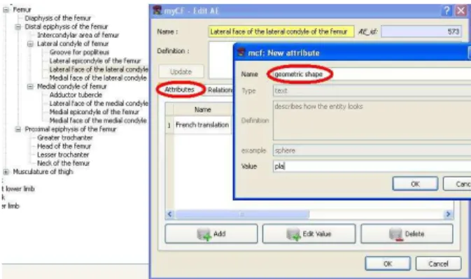
Fig. 3. Adding a geometric attribute.
As shown on figure 3, in order to include the geo-metric primitive in the ontology, we added the attribute “geometric shape” to the list of attributes. If some anatomical entity can be described via a simple primi-tive, all we need to do is to add the “geometric shape” attribute to this entity and give it the appropriate value (plane, sphere, cylinder or cone). On figure 3, we are adding the geometric attribute plane to the anatomical entity Lateral face of the lateral condyle of the femur.
(a) [1] (b) [8] (c) [17] (d) Our method.
Fig. 4. Comparison of segmentation results on a femur model.
Another interesting type of geometric information is implicitly included in the ontology, and more precisely in entities’ names. For example, the word “face” in the name of an entity gives us directly an idea about the shape of this entity (face→plane).
B. Using the ontology for the segmentation
Our second contribution is the use of the ontology (to which we added geometric information) to guide the segmentation. We extended the algorithm proposed in [16] to use the ontology. As mentioned earlier, the inputs of our method are: a mesh corresponding to parent
entity, and the ontology. Our goal is to identify different
parts of the parent entity on the mesh. We can see on figure 2 that the ontology enhances the segmentation process in three places:
1) Segmentation parameters: Number of segments
and their shape. As for the number of segments, it can be found in the ontology as the number of anatomical entities related to the parent entity by the relation
part of, we call those entities child entities. The shape
of each child entity, can be found in the geometric shape attribute of each entity. Each geometric shape is translated into an objective function.
• Plane is translated using the objective function
f lat(S) (because, by definition, a plane is a flat
surface).
• Cylinder is translated using the objective function
narrow(S) (because there is one preferential
di-rection on a cylinder, corresponding to its axis).
• Sphere is translated using the objective function
compact(S) (because there is no preferential
di-rection on a sphere).
The case of cone is to be treated in a near future. Anyway, we do not consider these functions the best way to represent the geometric primitives. They are only approximations of the error of fitting the primitive in question to a set of points (vertices of segment S).
2) Using localization information: The ontology
in-cludes implicit information that can be very useful for the segmentation. We actually can find in the names of anatomical entities a hint about their location. Key words such as: proximal, distal, lateral, medial,...etc.
gives us directly an idea about the relative position of the anatomical entity. We use this information in our segmentation technique in two steps:
• Labeling step: to label two parts with the same
geometric shape. For example, both lateral and medial condyle of the femur have a spherical shape, the choice of the label is then made according to the location (medial/lateral) w.r.t. the global frame (which is usually given by the input medical images).
• Optimization step: during the optimization step, at
each iteration segments may change their relative places. We make a check after each change to make sure that those new locations satisfy the localization information, otherwise, the suggested segmentation is not considered.
3) Using adjacency relation: We find in the ontology
the relation “adjacent to”, this relation tells us who are the neighbors of each segment. Similarly to the localization, this neighborhood information is used in the labeling step and the optimization step: a check on the neighborhood is done at each iteration, if the suggested segmentation does not satisfy the adjacency, it is rejected.
4) Relative size: We try to associate to each child entity a percentage of its size according to the size of
the parent entity. We were able to use this percentage in one of our examples (see figure 4-(d)). However, determining such a percentage needs further treatment. We may use a statistical study to determine the relative size of each entity according to its parent entity. This percentage (segment size/total size) is calculated at each iteration of the optimization step, and is then used twice:
• First, if the found percentage is different from the
one provided by the ontology (with a tolerance of
±5%), the suggested segmentation is rejected.
• Second, the difference between the found
percent-age and the registered one (in the ontology) is used as a weight for the corresponding term in the objective function. Thus, the more the sizes are respected, the less the cost is.
Since we need to calculate the percentage at each iteration, we choose to use the size of the bounding box of each segment rather than calculating the exact size,
or the size of its convex hull which are much more time consuming.
5) Hierarchy: The segmentation process is repeated
following the hierarchy found in the ontology using the “part of ” relation. Each found child entity, will be in turn the input of the segmentation process and we continue in descending the hierarchy until the finest level of details, i.e. anatomical entities with no children.
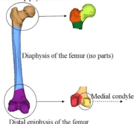
Fig. 5. Hierarchical segmentation.
Figure 5 shows the use of the hierarchy information during the segmentation of the femur. The segmentation is not done for all anatomical entities (for example: proximal epiphysis of the femur) for reasons that are explained later on.
IV. FIRSTRESULTS
Even though this work is still in progress, the first results that we obtain are very encouraging. On figure 4 we see the segmentation of the femur using our method and three other methods. In order to make a rather fair comparison, we provide these methods with the parameters that our method finds in the ontology. For the algorithm of [1], we gave number of segments and chose the primitives according to the geometric shapes of the anatomical entities (sphere, cylinder). For the other two algorithms ([8], [17]), we could only provide the number of segments. Our result represents a better match with anatomical entities. Specifically, [1] includes part of the proximal epiphysis in the segment corresponding to the diaphysis; [8] does not segment between the diaphysis and the distal epiphysis, and [17] includes part of the diaphysis in the segment corresponding to the distal epiphysis. These results were to be expected since our method benefits from more information than the other methods (localization information, adjacency relations, relative sizes).
V. FUTUREWORK
Even though the first results are very encouraging, there is room for many improvements. For the moment, we plan to improve the algorithm in the following axes:
• Initial segmentation: As mentioned earlier, a
k-means clustering is used to give the initial seg-mentation, which is then labeled and optimized. Anyway, the k-means clustering fails sometimes in giving an initial segmentation that respects the localization and adjacency relation of anatomical entities.
Fig. 6. Initial segmentation of the proximal epiphysis using k-means clustering.
For exemple, figure 6 shows the initial segmenta-tion of the proximal epiphysis of the femur using the k-means clustering. This anatomical entity is made of four parts: head of the femur, neck, lesser trochanter, and greater trochanter. The adjacency relation tells us that: the neck is adjacent to the other three parts, and those parts are mutually in adjacent. Looking at the initial segmentation we notice that no labeling satisfying the adjacency relation can be found. This current limitation ex-plains why we could not continue the segmentation hierarchically (Figure 5).
For such cases, we think of using the localization information with the adjacency relation to make the initial segmentation instead of using the k-means clustering.
• Adding a weight to the geometric shape: The
geometric shape property tries to approximate the shape of an anatomical entity by a geometric prim-itive. However, this approximation can be more or less accurate according to the entity in question. We think that adding a weight to measure the accuracy of this approximation can be useful during the segmentation optimization step.
• Boundary optimization: A post processing step of
boundary optimization can be added to give better matches between segments and anatomical entities. Such an optimization can be guided by curvature as recently proposed in [9].
VI. CONCLUSION
Using the ontology to guide the segmentation seems very promising and very natural, since when we segment a mesh, we always have a presumption about the result according to our human perception of the mesh (hand, humanoid, animal, ...etc.). The algorithm we propose makes a direct use of the a priori knowledge to guide the segmentation. Even though we presented it in an anatomical context, it can be easily generalized by changing the used ontology.
Mesh segmentation is a wide research domain with a wide variety of applications. Anyway, the evaluation of a mesh segmentation algorithm is still an open issue [4], [5]. In our case we plan to validate our method with the help of an anatomist, regarding to what extent the found segments match anatomical entities.
VII. ACKNOWLEDGEMENT
This work is partially supported by the ANR (Agence Nationale de la Recherche) through MADRAS project (ANR-07-MDCO-015).
REFERENCES
[1] M Attene, B Falcidieno, M Spagnuolo. Hierarchical mesh segmentation based on fitting primitives. Vis. Comput., 22(3):181-193, 2006.
[2] M Attene, S Katz, M Mortara, G Patan`e, M Spag-nuolo, A Tal. Mesh segmentation - A comparative study. SMI ’06: Proceedings of the IEEE Interna-tional Conference on Shape Modeling and Applica-tions, 2006.
[3] M Attene, F Robbiano, M Spagnuolo, B Falcidieno. Characterization of 3D shape parts for semantic an-notation. Computer-Aided Design, 41(10):756-763, 2009.
[4] H Benhabiles, JP Vandeborre, G Lavou´e, M Daoudi. A framework for the objective evaluation of seg-mentation algorithms using a ground-truth of human segmented 3D-models. SMI ’09: Proceedings of the IEEE International Conference on Shape Modeling and Applications, 2009.
[5] X Chen, A Golovinskiy, T Funkhouser. A bench-mark for 3D mesh segmentation. ACM Transactions on Graphics (Proc. SIGGRAPH), 28(3), 2009. [6] D Cohen-Steiner, P Alliez, M Desbrun. Variational
shape approximation. ACM Transactions on Graph-ics (Proc. SIGGRAPH), 23(3), 2004.
[7] O Dameron. Mod´elisation, repr´esentation et partage de connaissances anatomiques sur le cortex c´er´ebral
(Modeling, representing and sharing anatomical knowledge about the cerebral cortex). PhD Thesis,
Universit´e de Rennes 1, december 2003.
[8] G Lavou´e, F Dupont, A Baskurt. A new CAD mesh segmentation method, based on curvature tensor analysis. Computer-Aided Design, 37(10):975-987, 2005.
[9] L Kaplansky, A Tal. Mesh segmentation refinement. Computer Graphics Forum (Proc. Pacific Graphics), 28(7):1995-2003, 2009.
[10] M Mortara, G Patan`e, M Spagnuolo, B Falcidieno, J Rossignac. Plumber: a method for a multi-scale decomposition of 3D shapes into tubular primitives and bodies. SM ’04: Proceedings of the ninth ACM symposium on Solid modeling and applications, p. 339-344, 2004.
[11] M Mortara, G Patan`e, M Spagnuolo. From geo-metric to semantic human body models. Computers and Graphics, 30(2):185-196, 2006.
[12] M Mortara, M Spagnuolo. Semantics-driven best view of 3D shapes. Computers and Graphics, 33(3):280-290, 2009.
[13] O Palombi, G Bousquet, D Jospin, S Hassan, L Rev´eret, F Faure. My Corporis Fabrica: a unified ontological, geometrical and mechanical view of human anatomy. Lecture Notes in Computer Science 5903 (Proc. 3D Physiological Human Workshop), p. 207-219, Springer, 2009.
[14] C Rosse, JLV Mejino. A reference ontology for biomedical informatics: the Foundational Model of Anatomy. J. Biomed. Inform. 36:478-500, 2003. [15] A Shamir. A survey on mesh segmentation
tech-niques. Computer Graphics Forum, 27(6):1539-1556, 2008.
[16] P Simari, D Nowrouzezahrai, E Kalogerakis, K Singh. Multi-objective shape segmentation and la-beling. Computer Graphics Forum (Proc. Symp. on Geometry Processing), 28(5):1415-1425, 2009. [17] J Tierny, J-P Vandeborre, M Daoudi. Topology
driven 3D mesh hierarchical segmentation. SMI ’07: Proceedings of the IEEE International Conference on Shape Modeling and Applications, p. 215-220, 2007. [18] D M Yan, Y Liu, WP Wang. Quadric surface ex-traction by variational shape approximation. Lecture Notes in Computer Science 4077 (Proc. Geometric Modeling and Processing conference (GMP’06)), p. 73-86, Springer, 2006.