HAL Id: inserm-00640122
https://www.hal.inserm.fr/inserm-00640122
Submitted on 3 Oct 2014
HAL is a multi-disciplinary open access archive for the deposit and dissemination of sci-entific research documents, whether they are pub-lished or not. The documents may come from teaching and research institutions in France or abroad, or from public or private research centers.
L’archive ouverte pluridisciplinaire HAL, est destinée au dépôt et à la diffusion de documents scientifiques de niveau recherche, publiés ou non, émanant des établissements d’enseignement et de recherche français ou étrangers, des laboratoires publics ou privés.
CRB1 mutations in inherited retinal dystrophies.
Kinga Bujakowska, Isabelle Audo, Saddek Mohand-Saïd, Marie-Elise
Lancelot, Aline Antonio, Aurore Germain, Thierry Léveillard, Mélanie
Letexier, Jean-Paul Saraiva, Christine Lonjou, et al.
To cite this version:
Kinga Bujakowska, Isabelle Audo, Saddek Mohand-Saïd, Marie-Elise Lancelot, Aline Antonio, et al.. CRB1 mutations in inherited retinal dystrophies.. Human Mutation, Wiley, 2012, 33 (2), pp.306-15. �10.1002/humu.21653�. �inserm-00640122�
CRB1 mutations in inherited retinal dystrophies
Kinga Bujakowska1,2,3, Isabelle Audo1,2,3,4,5, Saddek Mohand-Saïd1,2,3,4, Marie-Elise Lancelot1,2,3, Aline Antonio1,2,3,4, Aurore Germain1,2,3, Thierry Léveillard1,2,3, Mélanie Letexier6, Jean-Paul Saraiva6, Christine Lonjou7, Wassila Carpentier7, José-Alain Sahel1,2,3,4,5,8, Shomi S. Bhattacharya1,2,3,5,9, Christina Zeitz1,2,3.
1
INSERM, U968, Paris, F-75012, France; 2CNRS, UMR_7210. Paris, F-75012, France; 3UPMC Univ Paris 06, UMR_S 968, Department of Genetics, Institut de la Vision, Paris, F-75012, France; 4Centre Hospitalier National d’Ophtalmologie des Quinze-Vingts, INSERM-DHOS CIC 503, Paris, F-75012, France; 5UCL-Institute of Ophthalmology, London, UK; 6IntegraGen SA, Genopole CAMPUS 1 bat G8 FR-91030 Evry, France; 7Plateforme Post-génomique P3S, Hôpital Pitié Salpêtrière, Paris, France; 8Fondation Ophtalmologique Adolphe de Rothschild, Paris, France; 9
Department of Cellular Therapy and Regenerative Medicine, Andalusian Centre for Molecular Biology and Regenerative Medicine (CABIMER), Isla Cartuja, Seville, Spain
Corresponding authors: Isabelle Audo and Christina Zeitz Institut de la Vision
Department of Genetics 17, Rue Moreau, 75012 Paris France
Email address: isabelle.audo@inserm.fr, christina.zeitz@inserm.fr
Financial Support: Foundation Fighting Blindness (I.A. FFB Grant No: CD-CL-0808-0466-CHNO and the CIC503 recognized as an FFB center, FFB Grant No:
C-CMM-0907-0428-INSERM04), Agence Nationale de la Recherche (SSB), Fondation Voir et Entendre (CZ), GIS-maladies rares (CZ), Ville de Paris and Région Ile de France, National Institutes of Health (USA) (KB NIH, Grant No: 1R01EY020902 - 01A1).
Abstract (max 200 words)
Mutations in the CRB1 gene are associated with variable phenotypes of severe retinal dystrophies, ranging from Leber Congenital Amaurosis (LCA) to rod-cone dystrophy (also called retinitis pigmentosa (RP)). Moreover, retinal dystrophies resulting from
CRB1 mutations may be accompanied by specific fundus features: preservation of the
para-arteriolar retinal pigment epithelium (PPRPE) and retinal telangiectasia with exudation (also referred to as Coats-like vasculopathy). In this publication we report seven novel mutations and classify over 150 reported CRB1 sequence variants that were found in more that 240 patients. The data from previous reports was used to analyse a potential correlation between CRB1 variants and the clinical features of respective patients. This meta-analysis suggests that the differential phenotype of patients with CRB1 mutations is due to additional modifying factors rather than particular mutant allele combination.
Background
Mutations in the CRB1 gene (MIM#: 604210) are associated with variable phenotypes of severe retinal dystrophies, ranging from Leber Congenital Amaurosis (LCA) to rod-cone dystrophy (also called retinitis pigmentosa (RP)) (Benayoun, et al., 2009; Bernal, et al., 2003; Booij, et al., 2005; Clark, et al., 2010; Coppieters, et al., 2010; den Hollander, et al., 2004; den Hollander, et al., 2001a; den Hollander, et al., 2007; den Hollander, et al., 1999; Galvin, et al., 2005; Gerber, et al., 2002; Hanein, et al., 2004; Henderson, et al., 2010; Henderson, et al., 2007; Jacobson, et al., 2003; Khaliq, et al., 2003; Li, et al., 2011; Lotery, et al., 2001a; Lotery, et al., 2001b; Riveiro-Alvarez, et al., 2008; Seong, et al., 2008; Simonelli, et al., 2007; Tosi, et al., 2009; Vallespin, et al., 2007; Walia, et al., 2010; Yzer, et al., 2006a; Yzer, et al., 2006b; Zernant, et al., 2005). LCA is a group of the most severe and the earliest occurring retinal dystrophies resulting in congenital blindness (den Hollander, et al., 2008). The onset of the disease occurs at birth and the characteristic features include non-recordable electroretinogram (ERG), nystagmus, sluggish or absent pupillary responses and oculo-digital reflexes, a distinctive eye-rubbing also called the Franschetti sign (den Hollander, et al., 2008; Franceschetti and Dieterle, 1954; Leber, 1869). RP is a clinically heterogeneous disorder characterised by a progressive degeneration of the photoreceptors and leading to a visual impairment of variable severity that can end in complete blindness. The disease onset is highly variable: it may commence in the first decade of life or much later. There is a considerable clinical overlap between LCA and early-onset RP and in some cases/reports the diagnosis is ambiguous. Early-onset RP, however, is considered as a relatively milder form, where patients do not have a congenital onset of visual impairment.
LCA and RP resulting from CRB1 mutations may be accompanied by specific fundus features: preservation of the para-arteriolar retinal pigment epithelium (PPRPE) (Bernal, et al., 2003; den Hollander, et al., 2004; den Hollander, et al., 1999; Heckenlively, 1982; Henderson, et al., 2010; Khaliq, et al., 2003; Simonelli, et al., 2007; Yzer, et al., 2006b) and retinal telangiectasia with exudation (also referred to as Coats-like vasculopathy) (Coppieters, et al., 2010; den Hollander, et al., 2004; den Hollander, et al., 2001a; Henderson, et al., 2010; Yzer, et al., 2006b). PPRPE is characterized by a relative preservation of retinal pigment epithelium (RPE) adjacent to retinal arterioles despite a panretinal RPE degeneration (Heckenlively, 1982). This is, however, not consistent in CRB1-associated RP and the absence of PPRPE in a severe RP should not exclude CRB1 as a potential causal gene (Lotery, et al., 2001b). Retinal telangiectasia is a condition of abnormally permeable blood vessels, leading to exudation and retinal detachment (Cahill, et al., 2001). Some patients with CRB1 mutations show macular atrophy (Henderson, et al., 2010), similar features were found for other LCA causing genes (GUCY2D MIM#:600179, AIPL1 MIM#:604392 and RPGRIP1 MIM#:605446), which lead to classification of LCA into cone-rod LCA and rod-cone LCA (Hanein, et al., 2004). Patients with CRB1 mutations belong to both categories. Predisposition of the CRB1 patients to keratoconus (McKibbin, et al., 2010; McMahon, et al., 2009) and implication for pigmented paravenous chorioretinal atrophy (McKay, et al., 2005) and nanophthalmos (Zenteno, et al., 2011) have also been reported.
CRB1 is a human homologue of the Drosophila melanogaster gene coding for
protein crumbs (crb) and it is expressed in the retina and the brain (den Hollander, et al., 1999). CRB1 consists of 12 exons and exhibits alternative splicing at the 3’ end, yielding two proteins of 1376 and 1406 amino acids (den Hollander, et al., 2001b).
Both proteins contain 19 EGF-like domains, three laminin AG-like domains and a signal peptide sequence. In addition, the longer isoform contains transmembrane and cytoplasmic domains (den Hollander, et al., 2001b; Gosens, et al., 2008). The cytoplasmic domain includes conserved FERM and PDZ binding motifs, through which CRB1 participates in the formation of adherens junction and links to the actin cytoskeleton (Gosens, et al., 2008).
In Drosophila, crb determines the polarity of the embryonic epithelium and peripheral neurons; it is important for the maintenance of zonula adherens (ZA) and it is localized in the apical membrane (Tepass, et al., 1990). In the mouse retina, Crb1 is present in the apical membranes of the epithelial cells, in Muller cells and in photoreceptor inner segments, where it concentrates in the vicinity of the outer limiting membrane (den Hollander, et al., 2002; Mehalow, et al., 2003; Pellikka, et al., 2002; van de Pavert, et al., 2004). A similar distribution was found in the human retina (van de Pavert, et al., 2004). Crumbs and its mouse homolog Crb1 is involved in the photoreceptor morphogenesis (Pellikka, et al., 2002; Tepass, et al., 1990). Analysis of the naturally occurring Crb1rd8 mouse mutant, suggests a developmental defect of the retina, where disruption of the outer limiting membrane and formation of retinal folds (pseudorosettes) are observed (Mehalow, et al., 2003). Disorganization of the retinal layers was also noted in other Crb1 mouse models (van de Pavert, et al., 2004; van de Pavert, et al., 2007). These findings are in accordance with clinical features of the patients carrying CRB1 mutations, whose retinas are thickened and show an altered laminar organization, resembling an immature normal retina (Jacobson, et al., 2003). The latter further supports the importance of CRB1 in the development of the retina.
This study presents an overview of the previously published CRB1 variants and novel mutations identified in a French cohort of simplex and autosomal recessive RP (arRP) patients. Based on the available genetic and phenotypic data from the literature and on our original findings, we classify all variants into one of the three groups (likely pathogenic, unclassified variants and unlikely pathogenic, Supp. Tables S1-S3). We discuss the clinical variability of patients harboring CRB1 mutations and analyse the phenotype-genotype correlation of likely pathogenic changes. Identification of novel mutations in the French cohort is described (Supp. Methods and Results) and precise clinical characterisation is given.
Novel CRB1 Variants
Eleven unrelated patients with ar or isolated RP in the French cohort carried likely pathogenic variants of CRB1 (Table1). Seven mutations were novel: three missense changes (p.Ser740Phe, p.Tyr1198Cys and p.Cys1223Ser), one nonsense mutation (p.Cys423*), one in-frame deletion (p.Asn789del) and two frameshift deletions (p.Leu655Trpfs*10, p.Ser1220Asnfs*62) (Table 1). Mutations identified in this study were not present in the SNP databases nor listed as non-pathogenic variants in the literature. None of the novel mutations was present in at least 362 control alleles and the mutations co-segregated in available family members (Supp. Figure S1). In all but one patient (547) two mutated CRB1 alleles were found.
The three novel missense mutations are in the conserved domains of the CRB1 protein. The p.Ser740Phe exchange replaces a highly conserved serine in the second laminin AG-like domain, the p.Tyr1198Cys mutation replaces a conserved tyrosine with a cysteine in the 16th calcium binding EGF-like domain and the p.Cys1223Ser is a replacement of a conserved cysteine with a serine in the 17th calcium binding
EGF-like domain (Figure 1). The in-frame deletion p.Asn789del is also located in the second laminin AG-like domain. Other novel mutations (p.Cys423*, p.Leu655Trpfs*10, p.Ser1220Asnfs*62) result in premature stop codons, which most likely lead to nonsense mediated decay (Chang, et al., 2007) and therefore these alleles are considered as null alleles. Five novel mutations are within exons 7 and 9, which are the most frequently mutated (Figure 1).
Clinical Characterisation of Patients with CRB1 Mutations
Clinical findings of French patients with CRB1 mutations are summarized in Tables 2 and 3. The average age at time of diagnosis was 17. Visual acuity was decreased in all patients ranging from 20/50 to light perception with no clear correlation with age or duration of the disease. Hyperopia was noted for 6/11 patients including three for whom spherical equivalent was equal or above +5 diopters. Night blindness was present in all patients but three, for whom a decrease of central vision and photophobia dominated. None of the patients had nystagmus. Most patients (9/11) had a clear lens; in the remaining two, one had undergone cataract surgery and one had significant lens opacities. These two patients were over 40 years of age. Two patterns of fundus pigmentary changes were present in this cohort: 7/11 had typical bone spicule-shaped pigment migration within the peripheral retina whereas 4/11 had widespread clumped pigmentary changes of nummular appearance at the level of the retinal pigment epithelium (Figure 2). Clumped pigmentation is therefore highly suggestive of CRB1 mutations but it is not specific since it has also been associated with mutations in NR2E3 (Schorderet and Escher, 2009; Sharon, et al., 2003), NRL (Nishiguchi, et al., 2004) or TULP1 (Mataftsi, et al., 2007). None of the patients displayed preservation of the para-arteriolar retinal pigment epithelium as previously
described in association with CRB1 mutations (Bernal, et al., 2003; den Hollander, et al., 2004; den Hollander, et al., 1999; Heckenlively, 1982; Henderson, et al., 2010; Khaliq, et al., 2003; Simonelli, et al., 2007; Yzer, et al., 2006b). In addition, none of the patients displayed Coats-like changes in the periphery. All patients had macular involvement. Six of the patients displayed cystoid macular edema whereas the other five had macular thinning with loss of the outer retinal layers and corresponding loss of autofluorescence (Figure 2). Color vision was normal in four patients or showed either tritan deficit or a dyschromatopsia with no clear axis when visual acuity allowed color vision testing. Full field electroretinogram showed severe generalized retinal dysfunction with no detectable responses in all patients except three for whom some residual rod and cone function was detectable. Among those three, the best responses on ERG were obtained in the youngest patients. Residual responses on ERG were correlated with better preservation of the visual field.
All patients displayed severe retinal involvement with early macular changes, half of them had cystoid macular edema, a higher percentage than the usually reported prevalence of about 30% in overall RP (Hajali, et al., 2008). This higher prevalence could at least be in part related to vascular abnormalities with Coats-like changes encountered in patients with CRB1 mutations (Coppieters, et al., 2010; den Hollander, et al., 2004; den Hollander, et al., 2001a; Henderson, et al., 2010; Yzer, et al., 2006b). Alternatively, these changes could be related to abnormal laminar structure associated with CRB1-mutations (Jacobson, et al., 2003). None of our patients developed Coats-like changes or para-arteriolar retinal pigment epithelium suggesting that these changes are not consistant in CRB1-related RP (Lotery, et al., 2001b). Four subjects displayed clumped retinopathies reinforcing that CRB1 should be considered as a
potential causal gene for this specific phenotype along with NR2E3 (Sharon, et al., 2003) or NRL (Nishiguchi, et al., 2004).
CRB1 Variants and Their Classification
Over 240 patients with CRB1 mutations and more than 150 gene variants have been described in the literature (Benayoun, et al., 2009; Bernal, et al., 2003; Booij, et al., 2005; Clark, et al., 2010; Coppieters, et al., 2010; den Hollander, et al., 2004; den Hollander, et al., 2001a; den Hollander, et al., 2007; den Hollander, et al., 1999; Galvin, et al., 2005; Gerber, et al., 2002; Hanein, et al., 2004; Henderson, et al., 2010; Henderson, et al., 2007; Jacobson, et al., 2003; Khaliq, et al., 2003; Li, et al., 2011; Lotery, et al., 2001a; Lotery, et al., 2001b; Riveiro-Alvarez, et al., 2008; Seong, et al., 2008; Simonelli, et al., 2007; Tosi, et al., 2009; Vallespin, et al., 2007; Yzer, et al., 2006a; Yzer, et al., 2006b; Zenteno, et al., 2011; Zernant, et al., 2005). The most frequently occurring of the known mutations is the p.Cys948Tyr in exon 9 (96 alleles reported, 24% of known CRB1 mutations) (Bernal, et al., 2003; Booij, et al., 2005; Clark, et al., 2010; Coppieters, et al., 2010; den Hollander, et al., 2004; den Hollander, et al., 2001a; den Hollander, et al., 2007; den Hollander, et al., 1999; Galvin, et al., 2005; Hanein, et al., 2004; Henderson, et al., 2010; Henderson, et al., 2007; Jacobson, et al., 2003; Lotery, et al., 2001a; Riveiro-Alvarez, et al., 2008; Tosi, et al., 2009; Vallespin, et al., 2007; Yzer, et al., 2006a; Zernant, et al., 2005). In general most of the mutations are in exons 9 (41%) and 7 (27%), therefore as a screening strategy these exons can be tested in the first instance (Figure 1, Supp. Table S1). Exons 7 and 9 encode second and third laminin AG-like domains respectively, implying that these domains are particularly important for CRB1 function. Missense mutations constitute
66% of all known mutations, the remaining being frameshift, truncation and splice site mutations.
We have attempted to classify all the reported mutations in three groups: 1) likely pathogenic, 2) unclassified variants, 3) unlikely pathogenic. This classification was based on the genetic data available from the literature, amino acid conservation and bioinformatic pathogenicity prediction tools (Supp. Tables S1-S3). An important criterion was the presence of two mutant alleles and co-segregation in the family. Approximately 30% of cases were reported with only one mutant allele, assuming that the second mutation is within the intronic region. For these patients however, one cannot exclude the possibility that there is another molecular cause of the pathology. The lack of the second mutant CRB1 allele is sometimes explained by a digenic inheritance, however so far it has not been proven by co-segregation analysis (Li, et al., 2011; Vallespin, et al., 2007).
Pathogenicity is easier to asses in deletions and frameshift variants than in the case of missense changes, hence the importance of the bioinformatic analysis of the pathogenicity, amino acid conservation and functional analysis of the variants. On this basis we have not considered two changes identified in our cohort as pathogenic (p.Gly959Ser and p.Ala1354Thr) (den Hollander, et al., 2004; den Hollander, et al., 2001a)). The respective patients did not carry a second CRB1 mutation and we did not consider the p.Gly959Ser and p.Ala1354Thr substitutions as likely pathogenic, based on poor conservation of the residues and low pathogenicity predictions using online bioinformatic tools: PolyPhen-2 and SIFT (Supp. Tables S2 and S3). One report suggests involvement of CRB1 in autosomal dominant pigmented paravenous chorioretinal atrophy (McKay, et al., 2005), though the reported mutation
p.Val162Met has a questionable pathogenicity, since valine is not conserved and methionine is present in this position in other mammals (Supp. Table S2).
Prevalence
In the investigated cohort, at least 2.5% of arRP patients carry CRB1 gene defects, which lies within the previously published range of 0-6.5% (Bernal, et al., 2003; den Hollander, et al., 2004; Vallespin, et al., 2007), or 2.7% after cohort averaging (Table 4). The high preponderance of novel CRB1 mutations in our cohort suggests, however, that probably more arRP patients carry CRB1 pathogenic defects, which are novel and therefore undetectable by arRP microarray. Much higher prevalence is observed in LCA/EORD cohorts and RP with additional features like PPRPE and retinal telangiectasia, representing 10.1%, 74.1%, 53.3% respectively in averaged cohorts (Table 4) (Bernal, et al., 2003; Coppieters, et al., 2010; den Hollander, et al., 2004; den Hollander, et al., 2001a; den Hollander, et al., 2007; den Hollander, et al., 1999; Hanein, et al., 2004; Henderson, et al., 2010; Henderson, et al., 2007; Lotery, et al., 2001a; Seong, et al., 2008; Simonelli, et al., 2007; Vallespin, et al., 2007; Walia, et al.).
Genotype-Phenotype Correlation
We were not able to establish a clear genotype/phenotype correlation for our cohort, which might be due to the small number of patients with CRB1 mutations and their variable phenotype. In addition, the nature of existing published data makes it difficult to correlate the recurring CRB1 mutations with different phenotypes for a number of reasons. First, the phenotyping of patients is complex and distinguishing between early-onset RP and LCA is often arbitrary and depends on the guidelines of a
particular clinical center. Second, precise clinical data is often omitted in the publications and therefore it is difficult to adjust for these diagnostic differences in a cross-paper analysis. Despite these inconsistencies, we attempted to analyse data from previous reports in order to find the relationship between the CRB1 variants and the clinical features of respective patients. In this meta-analysis we used 171 patients, who carried two likely pathogenic mutations in trans (Benayoun, et al., 2009; Bernal, et al., 2003; Booij, et al., 2005; Clark, et al., 2010; Coppieters, et al., 2010; den Hollander, et al., 2004; den Hollander, et al., 2001a; den Hollander, et al., 2007; den Hollander, et al., 1999; Galvin, et al., 2005; Hanein, et al., 2004; Henderson, et al., 2010; Henderson, et al., 2007; Jacobson, et al., 2003; Khaliq, et al., 2003; Li, et al., 2011; Lotery, et al., 2001a; Lotery, et al., 2001b; McKibbin, et al., 2010; Riveiro-Alvarez, et al., 2008; Seong, et al., 2008; Simonelli, et al., 2007; Tosi, et al., 2009; Vallespin, et al., 2007; Yzer, et al., 2006a). Combination of two mutant alleles was analysed in relation to clinical characteristics of the published cases. Based on the reports we distinguished the following phenotypes: LCA, early onset retinal degeneration (EORD), RP, presence of PPRPE and Coats-like vasculopathy. The mutations were classed as null mutations (all mutations leading to a premature stop codon) or as variants leading to an altered protein (missense and in frame deletions). The likely pathogenic mutations were plotted on a graph, where affected codons on allele 1 and allele 2 served as coordinates (codon 0 was assigned to null mutations). The results show that we cannot assign a specific allele combination to a particular phenotype, e.g. homozygous null alleles or homozygous p.Cys948Tyr alleles are found in LCA, EORD and RP patients (Figure 3 A). Null alleles are however more frequent in LCA cohorts (Figure 3 B) as previously suggested (den Hollander, et al., 2004). The presence/absence of PPRPE or Coats-like vasculopathy did not reveal a
particular mutation pattern (Figure 3 C). These findings suggest the involvement of additional modifying factors (genetic and/or environmental), which are responsible for the modulation of the phenotype in patients harboring CRB1 mutations.
Future Directions
The above analysis of the phenotype-genotype correlation suggests that the disease severities associated with CRB1 mutations are in fact a continuum of the same clinical entity with possible additional modifying factors influencing disease onset and progression. There is increasing evidence of the involvement of multiple alleles in the patient’s phenotype, as has been shown for the Bardet-Biedl patients (Katsanis, et al., 2001) and more recently for a PRPH2-associated macular dystrophy family, where the phenotype has been modulated by additional heterozygous mutations in
ABCA4 (MIM#: 601691) and ROM1 (MIM#: 180721) (Poloschek, et al., 2010). It is
likely that the new next generation sequencing (NGS) technology will help to shed light on the potential genetic modifiers that influence disease phenotype. One has, however, to analyse the data with caution since NGS will reveal large numbers of polymorphic changes, which do not modulate the disease. The potential new modifying changes will have to be confirmed by appropriate genetic and functional analysis. The certainty of the molecular cause of a disease is particularly important in the era of gene therapy trials. Genetic treatment of recessive disorders should not be undertaken before obtaining proof that both alleles of a given gene are dysfunctional. In-depth genetic analysis, as presented here, is necessary to provide a basis for conducting such therapies.
Acknowledgments
The authors would like to thank patients and families for participation in this study, Dominique Santiard-Baron, Christine Chaumeil and clinical staff for their help in clinical data and DNA collection, Sandro Banfi, Robert Henderson and Qingjiong Zhang for additional information on genotype-phenotype correlations of previously published mutations and Robert Gillan for help with the manuscript. The project was financially supported by the Foundation Fighting Blindness (I.A. FFB Grant No: CD-CL-0808-0466-CHNO and the CIC503 recognized as an FFB center, FFB Grant No: C-CMM-0907-0428-INSERM04), Agence Nationale de la Recherche (SSB), Fondation Voir et Entendre (CZ), GIS-maladies rares (CZ), Ville de Paris and Région Ile de France, National Institutes of Health (USA) (KB NIH, Grant No: 1R01EY020902 - 01A1).
References
Adzhubei IA, Schmidt S, Peshkin L, Ramensky VE, Gerasimova A, Bork P, Kondrashov AS, Sunyaev SR. 2010. A method and server for predicting damaging missense mutations. Nat Methods 7:248-9.
Audo I, Sahel JA, Mohand-Said S, Lancelot ME, Antonio A, Moskova-Doumanova V, Nandrot EF, Doumanov J, Barragan I, Antinolo G and others. 2010. EYS is a major gene for rod-cone dystrophies in France. Hum Mutat 31:E1406-35.
Benayoun L, Spiegel R, Auslender N, Abbasi AH, Rizel L, Hujeirat Y, Salama I, Garzozi HJ, Allon-Shalev S, Ben-Yosef T. 2009. Genetic heterogeneity in two consanguineous families segregating early onset retinal degeneration: the pitfalls of homozygosity mapping. Am J Med Genet A 149A:650-6.
Bernal S, Calaf M, Garcia-Hoyos M, Garcia-Sandoval B, Rosell J, Adan A, Ayuso C, Baiget M. 2003. Study of the involvement of the RGR, CRPB1, and CRB1 genes in the pathogenesis of autosomal recessive retinitis pigmentosa. J Med Genet 40:e89.
Booij JC, Florijn RJ, ten Brink JB, Loves W, Meire F, van Schooneveld MJ, de Jong PT, Bergen AA. 2005. Identification of mutations in the AIPL1, CRB1, GUCY2D, RPE65, and RPGRIP1 genes in patients with juvenile retinitis pigmentosa. J Med Genet 42:e67.
Cahill M, O'Keefe M, Acheson R, Mulvihill A, Wallace D, Mooney D. 2001. Classification of the spectrum of Coats' disease as subtypes of idiopathic retinal telangiectasis with exudation. Acta Ophthalmol Scand 79:596-602.
Chang YF, Imam JS, Wilkinson MF. 2007. The nonsense-mediated decay RNA surveillance pathway. Annu Rev Biochem 76:51-74.
Clark GR, Crowe P, Muszynska D, O'Prey D, O'Neill J, Alexander S, Willoughby CE, McKay GJ, Silvestri G, Simpson DA. 2010. Development of a diagnostic genetic test for simplex and autosomal recessive retinitis pigmentosa. Ophthalmology 117:2169-77 e3.
Coppieters F, Casteels I, Meire F, De Jaegere S, Hooghe S, van Regemorter N, Van Esch H, Matuleviciene A, Nunes L, Meersschaut V and others. 2010. Genetic screening of LCA in Belgium: predominance of CEP290 and identification of potential modifier alleles in AHI1 of CEP290-related phenotypes. Hum Mutat 31:E1709-66.
den Hollander AI, Davis J, van der Velde-Visser SD, Zonneveld MN, Pierrottet CO, Koenekoop RK, Kellner U, van den Born LI, Heckenlively JR, Hoyng CB and others. 2004. CRB1 mutation spectrum in inherited retinal dystrophies. Hum Mutat 24:355-69.
den Hollander AI, Ghiani M, de Kok YJ, Wijnholds J, Ballabio A, Cremers FP, Broccoli V. 2002. Isolation of Crb1, a mouse homologue of Drosophila crumbs, and analysis of its expression pattern in eye and brain. Mech Dev 110:203-7. den Hollander AI, Heckenlively JR, van den Born LI, de Kok YJ, van der
Velde-Visser SD, Kellner U, Jurklies B, van Schooneveld MJ, Blankenagel A, Rohrschneider K and others. 2001a. Leber congenital amaurosis and retinitis pigmentosa with Coats-like exudative vasculopathy are associated with mutations in the crumbs homologue 1 (CRB1) gene. Am J Hum Genet 69:198-203.
den Hollander AI, Johnson K, de Kok YJ, Klebes A, Brunner HG, Knust E, Cremers FP. 2001b. CRB1 has a cytoplasmic domain that is functionally conserved between human and Drosophila. Hum Mol Genet 10:2767-73.
den Hollander AI, Lopez I, Yzer S, Zonneveld MN, Janssen IM, Strom TM, Hehir-Kwa JY, Veltman JA, Arends ML, Meitinger T and others. 2007. Identification of novel mutations in patients with Leber congenital amaurosis and juvenile RP by genome-wide homozygosity mapping with SNP microarrays. Invest Ophthalmol Vis Sci 48:5690-8.
den Hollander AI, Roepman R, Koenekoop RK, Cremers FP. 2008. Leber congenital amaurosis: genes, proteins and disease mechanisms. Prog Retin Eye Res 27:391-419.
den Hollander AI, ten Brink JB, de Kok YJ, van Soest S, van den Born LI, van Driel MA, van de Pol DJ, Payne AM, Bhattacharya SS, Kellner U and others. 1999. Mutations in a human homologue of Drosophila crumbs cause retinitis pigmentosa (RP12). Nat Genet 23:217-21.
Franceschetti A, Dieterle P. 1954. [Diagnostic and prognostic importance of the electroretinogram in tapetoretinal degeneration with reduction of the visual field and hemeralopia]. Confin Neurol 14:184-6.
Galvin JA, Fishman GA, Stone EM, Koenekoop RK. 2005. Evaluation of genotype-phenotype associations in leber congenital amaurosis. Retina 25:919-29.
Gerber S, Perrault I, Hanein S, Shalev S, Zlotogora J, Barbet F, Ducroq D, Dufier J, Munnich A, Rozet J and others. 2002. A novel mutation disrupting the cytoplasmic domain of CRB1 in a large consanguineous family of Palestinian origin affected with Leber congenital amaurosis. Ophthalmic Genet 23:225-35. Gosens I, den Hollander AI, Cremers FP, Roepman R. 2008. Composition and
function of the Crumbs protein complex in the mammalian retina. Exp Eye Res 86:713-26.
Hajali M, Fishman GA, Anderson RJ. 2008. The prevalence of cystoid macular oedema in retinitis pigmentosa patients determined by optical coherence tomography. Br J Ophthalmol 92:1065-8.
Hanein S, Perrault I, Gerber S, Tanguy G, Barbet F, Ducroq D, Calvas P, Dollfus H, Hamel C, Lopponen T and others. 2004. Leber congenital amaurosis: comprehensive survey of the genetic heterogeneity, refinement of the clinical definition, and genotype-phenotype correlations as a strategy for molecular diagnosis. Hum Mutat 23:306-17.
Heckenlively JR. 1982. Preserved para-arteriole retinal pigment epithelium (PPRPE) in retinitis pigmentosa. Br J Ophthalmol 66:26-30.
Henderson RH, Mackay DS, Li Z, Moradi P, Sergouniotis P, Russell-Eggitt I, Thompson DA, Robson AG, Holder GE, Webster AR and others. 2010. Phenotypic variability in patients with retinal dystrophies due to mutations in CRB1. Br J Ophthalmol.
Henderson RH, Waseem N, Searle R, van der Spuy J, Russell-Eggitt I, Bhattacharya SS, Thompson DA, Holder GE, Cheetham ME, Webster AR and others. 2007. An assessment of the apex microarray technology in genotyping patients with Leber congenital amaurosis and early-onset severe retinal dystrophy. Invest Ophthalmol Vis Sci 48:5684-9.
Jacobson SG, Cideciyan AV, Aleman TS, Pianta MJ, Sumaroka A, Schwartz SB, Smilko EE, Milam AH, Sheffield VC, Stone EM. 2003. Crumbs homolog 1 (CRB1) mutations result in a thick human retina with abnormal lamination. Hum Mol Genet 12:1073-8.
Katsanis N, Ansley SJ, Badano JL, Eichers ER, Lewis RA, Hoskins BE, Scambler PJ, Davidson WS, Beales PL, Lupski JR. 2001. Triallelic inheritance in Bardet-Biedl syndrome, a Mendelian recessive disorder. Science 293:2256-9.
Khaliq S, Abid A, Hameed A, Anwar K, Mohyuddin A, Azmat Z, Shami SA, Ismail M, Mehdi SQ. 2003. Mutation screening of Pakistani families with congenital eye disorders. Exp Eye Res 76:343-8.
Leber T. 1869. Ueber Retinitis pigmentosa und angeborene Amaurose. Graefe's Archive For Clinical And Experimental Ophthalmology 15:1-25.
Li L, Xiao X, Li S, Jia X, Wang P, Guo X, Jiao X, Zhang Q, Hejtmancik JF. 2011. Detection of variants in 15 genes in 87 unrelated chinese patients with leber congenital amaurosis. PLoS One 6:e19458.
Lotery AJ, Jacobson SG, Fishman GA, Weleber RG, Fulton AB, Namperumalsamy P, Heon E, Levin AV, Grover S, Rosenow JR and others. 2001a. Mutations in the CRB1 gene cause Leber congenital amaurosis. Arch Ophthalmol 119:415-20. Lotery AJ, Malik A, Shami SA, Sindhi M, Chohan B, Maqbool C, Moore PA, Denton
MJ, Stone EM. 2001b. CRB1 mutations may result in retinitis pigmentosa without para-arteriolar RPE preservation. Ophthalmic Genet 22:163-9.
Mataftsi A, Schorderet DF, Chachoua L, Boussalah M, Nouri MT, Barthelmes D, Borruat FX, Munier FL. 2007. Novel TULP1 mutation causing leber congenital amaurosis or early onset retinal degeneration. Invest Ophthalmol Vis Sci 48:5160-7.
McKay GJ, Clarke S, Davis JA, Simpson DA, Silvestri G. 2005. Pigmented paravenous chorioretinal atrophy is associated with a mutation within the crumbs homolog 1 (CRB1) gene. Invest Ophthalmol Vis Sci 46:322-8.
McKibbin M, Ali M, Mohamed MD, Booth AP, Bishop F, Pal B, Springell K, Raashid Y, Jafri H, Inglehearn CF. 2010. Genotype-phenotype correlation for leber congenital amaurosis in Northern Pakistan. Arch Ophthalmol 128:107-13. McMahon TT, Kim LS, Fishman GA, Stone EM, Zhao XC, Yee RW, Malicki J.
2009. CRB1 gene mutations are associated with keratoconus in patients with leber congenital amaurosis. Invest Ophthalmol Vis Sci 50:3185-7.
Mehalow AK, Kameya S, Smith RS, Hawes NL, Denegre JM, Young JA, Bechtold L, Haider NB, Tepass U, Heckenlively JR and others. 2003. CRB1 is essential for external limiting membrane integrity and photoreceptor morphogenesis in the mammalian retina. Hum Mol Genet 12:2179-89.
Ng PC, Henikoff S. 2003. SIFT: Predicting amino acid changes that affect protein function. Nucleic Acids Res 31:3812-4.
Nishiguchi KM, Friedman JS, Sandberg MA, Swaroop A, Berson EL, Dryja TP. 2004. Recessive NRL mutations in patients with clumped pigmentary retinal degeneration and relative preservation of blue cone function. Proc Natl Acad Sci U S A 101:17819-24.
Pellikka M, Tanentzapf G, Pinto M, Smith C, McGlade CJ, Ready DF, Tepass U. 2002. Crumbs, the Drosophila homologue of human CRB1/RP12, is essential for photoreceptor morphogenesis. Nature 416:143-9.
Poloschek CM, Bach M, Lagreze WA, Glaus E, Lemke JR, Berger W, Neidhardt J. 2010. ABCA4 and ROM1: implications for modification of the PRPH2-associated macular dystrophy phenotype. Invest Ophthalmol Vis Sci 51:4253-65.
Riveiro-Alvarez R, Vallespin E, Wilke R, Garcia-Sandoval B, Cantalapiedra D, Aguirre-Lamban J, Avila-Fernandez A, Gimenez A, Trujillo-Tiebas MJ, Ayuso C. 2008. Molecular analysis of ABCA4 and CRB1 genes in a Spanish family segregating both Stargardt disease and autosomal recessive retinitis pigmentosa. Mol Vis 14:262-7.
Schorderet DF, Escher P. 2009. NR2E3 mutations in enhanced S-cone sensitivity syndrome (ESCS), Goldmann-Favre syndrome (GFS), clumped pigmentary retinal degeneration (CPRD), and retinitis pigmentosa (RP). Hum Mutat 30:1475-85. Seelow D, Schuelke M, Hildebrandt F, Nurnberg P. 2009. HomozygosityMapper--an
interactive approach to homozygosity mapping. Nucleic Acids Res 37:W593-9. Seong MW, Kim SY, Yu YS, Hwang JM, Kim JY, Park SS. 2008. Molecular
characterization of Leber congenital amaurosis in Koreans. Mol Vis 14:1429-36. Sharon D, Sandberg MA, Caruso RC, Berson EL, Dryja TP. 2003. Shared mutations
in NR2E3 in enhanced S-cone syndrome, Goldmann-Favre syndrome, and many cases of clumped pigmentary retinal degeneration. Arch Ophthalmol 121:1316-23. Simonelli F, Ziviello C, Testa F, Rossi S, Fazzi E, Bianchi PE, Fossarello M, Signorini S, Bertone C, Galantuomo S and others. 2007. Clinical and molecular genetics of Leber's congenital amaurosis: a multicenter study of Italian patients. Invest Ophthalmol Vis Sci 48:4284-90.
Tepass U, Theres C, Knust E. 1990. crumbs encodes an EGF-like protein expressed on apical membranes of Drosophila epithelial cells and required for organization of epithelia. Cell 61:787-99.
Tosi J, Tsui I, Lima LH, Wang NK, Tsang SH. 2009. Case report: autofluorescence imaging and phenotypic variance in a sibling pair with early-onset retinal dystrophy due to defective CRB1 function. Curr Eye Res 34:395-400.
Vallespin E, Cantalapiedra D, Riveiro-Alvarez R, Wilke R, Aguirre-Lamban J, Avila-Fernandez A, Lopez-Martinez MA, Gimenez A, Trujillo-Tiebas MJ, Ramos C and others. 2007. Mutation screening of 299 Spanish families with retinal dystrophies by Leber congenital amaurosis genotyping microarray. Invest Ophthalmol Vis Sci 48:5653-61.
van de Pavert SA, Kantardzhieva A, Malysheva A, Meuleman J, Versteeg I, Levelt C, Klooster J, Geiger S, Seeliger MW, Rashbass P and others. 2004. Crumbs homologue 1 is required for maintenance of photoreceptor cell polarization and adhesion during light exposure. J Cell Sci 117:4169-77.
van de Pavert SA, Meuleman J, Malysheva A, Aartsen WM, Versteeg I, Tonagel F, Kamphuis W, McCabe CJ, Seeliger MW, Wijnholds J. 2007. A single amino acid substitution (Cys249Trp) in Crb1 causes retinal degeneration and deregulates expression of pituitary tumor transforming gene Pttg1. J Neurosci 27:564-73. Walia S, Fishman GA, Jacobson SG, Aleman TS, Koenekoop RK, Traboulsi EI,
Weleber RG, Pennesi ME, Heon E, Drack A and others. 2010. Visual acuity in patients with Leber's congenital amaurosis and early childhood-onset retinitis pigmentosa. Ophthalmology 117:1190-8.
Yzer S, Fishman GA, Racine J, Al-Zuhaibi S, Chakor H, Dorfman A, Szlyk J, Lachapelle P, van den Born LI, Allikmets R and others. 2006a. CRB1 heterozygotes with regional retinal dysfunction: implications for genetic testing of leber congenital amaurosis. Invest Ophthalmol Vis Sci 47:3736-44.
Yzer S, Leroy BP, De Baere E, de Ravel TJ, Zonneveld MN, Voesenek K, Kellner U, Ciriano JP, de Faber JT, Rohrschneider K and others. 2006b. Microarray-based mutation detection and phenotypic characterization of patients with Leber congenital amaurosis. Invest Ophthalmol Vis Sci 47:1167-76.
Zenteno JC, Buentello-Volante B, Ayala-Ramirez R, Villanueva-Mendoza C. 2011. Homozygosity mapping identifies the Crumbs homologue 1 (Crb1) gene as responsible for a recessive syndrome of retinitis pigmentosa and nanophthalmos. Am J Med Genet A 155A:1001-6.
Zernant J, Kulm M, Dharmaraj S, den Hollander AI, Perrault I, Preising MN, Lorenz B, Kaplan J, Cremers FP, Maumenee I and others. 2005. Genotyping microarray (disease chip) for Leber congenital amaurosis: detection of modifier alleles. Invest Ophthalmol Vis Sci 46:3052-9.
Figure Legends
Figure 1. Distribution of CRB1 mutations in the gene and protein. A) Nucleotide numbering is based on cDNA sequence of CRB1 (Ref. NM_201253.2) where A of
the ATG initiation codon is 1. The stop and frameshift mutations are indicated
above the structure of the gene and the position of the missense mutations are drawn in relation to protein domains. The novel mutations are indicated in red. B) The structures of EGF-like and Ca++ binding EGF-like domains with indications of conserved residues and recurrent mutations. The highly conserved cysteine residues are in black, the conserved residues between both domains are in grey and the conserved amino acids specific to the Ca2+ binding domain are in blue. C) Evolutionary conservation of the likely pathogenic CRB1 residue changes identified in this work.
Figure 2. Fundus color photographs and Optical Coherence Tomography (OCT). A) Color fundus photograph of the left eye of 3969 showing nummular pigmentary migration in the mid periphery in addition to pigmentary changes within the macula. B) Vertical scan OCT of the left eye of 3969 showing cystic changes in the macular region. C) Color fundus photograph of the right eye of 547 showing bone spicules pigmentary migration in the periphery in addition to atrophic changes within the macula. D) Vertical scan OCT of the right eye of 547 showing atrophic changes in the macular region after resolution of episodes of cystoid changes.
Figure 3. Genotype-phenotype correlation of patients with CRB1 mutations. A) Distribution of CRB1 mutations in LCA, EORD and RP. XY axes represent allele 1 and 2 of the patients, the affected codons serve as xy coordinates, null allele coordinate is designated as 0. The size of the circles is proportional to the number of the CRB1 patients with a given genotype. B) Frequency of null and missense allele combinations in LCA, EORD and RP patients. C) Distribution of CRB1 mutations in patients with/without additional features: PPRPE and Coats-like vasculopathy.
Table 1. Patients with CRB1 mutations identified in this study
Patient
number Family
Allele 1 Allele 2
Exon Nucleotide change Protein change Exon Nucleotide
change Protein change
229 159 2 c.613_619del p.Ile205Aspfs*13 7 c.2365_2367del
AAT p.Asn789del
53 No family
members 6 c.1269C>A p.Cys423* 7 c.2506C>A p.Pro836Thr
368 249 6 c.1750G>T p.Asp584Tyr 7 c.2506C>A p.Pro836Thr
547 372 6 c.1963delC p.Leu655Trpfs*10 ?
4240a 2025 7 c.2219C>T p.Ser740Phe 7 c.2219C>T p.Ser740Phe
54 39 7 c.2222T>C p.M741T 9 c.3593A>G p.Tyr1198Cys
3969 No family
members 7 c.2506C>A p.Pro836Thr 7 c.2506C>A p.Pro836Thr
409 281 9 c.2843G>A p.Cys948Tyr 9 c.3668G>C p.Cys1223Ser
1183b 709 9 c.3659_3660delinsA p.Ser1220Asnfs*62 9 c.3659_3660del
insA p.Ser1220Asnfs*62
1731 1008 9 c.2843G>A p.Cys948Tyr 9 c.2843G>A p.Cys948Tyr
3144 1302 9 c.2843G>A p.Cys948Tyr 7 c.3307G>A p.Gly1103Arg
a
mutation in this patient was identified by NGS b
mutation in this patient was found through homozygosity mapping novel mutations are in bold
Table 2: Clinical data Patient Age at time of testing Age at time of diagnosis Sex
Relevant medical and ophthalmology history Family history Symptoms BCVA OD/OS Refraction Lens Fundus
examination OCT FAF
53 27 20 M none From Ivory Coast, 10 brothers and sisters, 1 sister affected Night blindness at 6 then photophobia then decreased vision LP 20/500 +2(-1.50)60° +1.75(-1.5)125° Clear Widespread clumped pigment migration with no pale optic disc or narrowed retinal vessels Macular thinning with loss of ONL Loss of AF at the posterior pole and periphery 54 41 25 F none From French descent One affected brother Night blindness 20/640 20/100 Prior to lens surgery: +5.50(-1)5° +5.50(-1)165° IOL Peripheral RPE changes with bone spicules, perifoveal atrophy, pale optic disc, narrowing of retinal vessels Thinning of the ONL within the macular region Loss of AF in the perifoveal region and outside the vascular arcades
229 29 20 F none From French
descent Night blindness
20/80 20/50 +2(-0.75)5° +2.50(-1.50)5° Clear Peripheral RPE changes, little bone
spicules, no pale optic disc or narrowed retinal vessels, CME CME, thinning of ONL Patchy loss of AF in the periphery; foveal modification of AF due to the CME 368 13 12 F Seizure in infancy From Turkish descent maternal grand-mother said to be blind photophobia 20/80 20/63 +6.50(-1.25)160° +6.50(-1)7° Clear Peripheral RPE changes with bone spicules, perifoveal atrophy, pale optic disc, narrowing of retinal vessels, CME CME with relative preservation of foveal architecture Patchy loss of AF outside the vascular arcades, foveal AF changes due to CME 409 43 Teenage years F none From Italian descent Night blindness then photophobia 20/160 20/100 Plano Plano Clear Peripheral bone spicules with perifoveal atrophy Thinning of the ONL Loss of AF outside the vascular arcades and in the perifoveal area
547 57 39 M
Recurrent anterior uveitis, which delayed
the diagnosis of RP From French descent, no family history of RP Night blindness then photophobia and decreased vision 20/80 20/63 +0.25(-0.50)110° -2(-1.25)65° Bilateral nuclear cataract Peripheral bone spicules with CME
Bilateral CME, perifoveal thinning Loss of AF in the perifoveal region and outside the vascular arcades 1183 38 15 F none From Tunisian descent; consanguinity among parents Night blindness and photophobia 20/640 20/640 Emetropia Clear Widespread clumped pigment migration with no pale optic disc or narrowed blood vessels; OD asteroides hyaloids Macular thinning with loss of ONL Loss of AF at the posterior pole and periphery
1731 23 17 M Deafness since age 9
From Spanish descent; parents first cousins; one brother affected Low vision since early childhood HM 20/80 Emetropia Clear Widespread clumped pigment migration with relative sparing of the macula, with no
pale optic disc or narrowed blood vessels Macular thinning with loss of ONL Loss of AF at the posterior pole and periphery
3144 20 9 F none From French
descent Night blindness since early childhood 20/80 20/80 +9(-1.50)170° +7.50 Clear
Some RPE changes in the periphery, normal disc color and no narrowing of blood vessels; CME
CME with relatively spared foveal structure Patchy loss of AF outside the vascular arcades, foveal AF changes due to CME
3969 28 12 F none From Mali
Night blindness then photophobia 20/125 20/320 +0.50(-1.50)90° +1.75(-1.25)95° Clear Widespread clumped pigment migration in the posterior pole and
periphery CME CME Thinning of ONL Diffuse patchy loss of AF within the posterior pole and periphery 4240 7 6 M none One sister affected, from Turkish descent Decreased vision 20/63 20/80 -1.50(-1.50)10° -2(-0.75)180° Clear Moderate RPE changes in the periphery CME CME with relatively spared parafoveal Patchy loss of AF outside the vascular arcade, normal AF
structure within posterior pole except AF modification due
to CME in the fovea
BCVA: best corrected visual acuity; CME: cystoid macular edema; ND: not detectable; FAF: Fundus Autofluorescence; OD: Oculis dextra (right eye); OS: Oculis Sinistra (left eye); IOL: intra ocular lens; CF: counting fingers; HM: hand motion; LP: light perception; RPE: retinal pigment epithelium; RP: retinitis pigmentosa; OHT: ocular hypertension; ONL: Outer Nuclear Layer
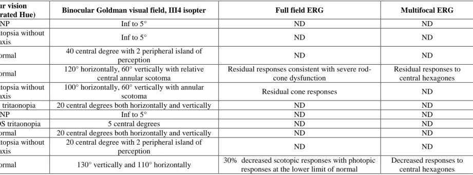
Table 3: Function data
Patient Colour vision
(15 saturated Hue) Binocular Goldman visual field, III4 isopter Full field ERG Multifocal ERG
53 NP Inf to 5° ND ND
54 Dyschromatopsia without
axis Inf to 5° ND ND
229 Normal 40 central degree with 2 peripheral island of
perception ND ND
368 Normal 120° horizontally, 60° vertically with relative central annular scotoma
Residual responses consistent with severe rod-cone dysfunction
Residual responses to central hexagones 409 Dyschromatopsia without
axis
100° horizontally, 60° vertically with annular
scotoma Residual cone responses ND
547 Bilateral tritaonopia 20 central degrees both horizontally and vertically ND ND
1183 NP Inf to 5° ND ND
1731 OD NP, OS tritaonopia 5 central degrees ND ND
03144 Normal 20 central degrees both horizontally and vertically ND ND
3969 Dyschromatopsia without axis
20 central degree with 2 peripheral island of
perception ND ND
4240 Normal 130° vertically and 110° horizontally 30% decreased scotopic responses with photopic responses at the lower limit of normal
Decreased responses to central hexagones
Table 4. Average prevalence of CRB1 mutations in retinal dystrophy patients in published reports
Dystrophy Prevalence* Patients with two CRB1 alleles
Patients with one CRB1 allele
Added
cohort size References
LCA/EORD 10.1% 109 57 1645
(Bernal, et al., 2003; Coppieters, et al., 2010; den Hollander, et al., 2004; den Hollander, et al., 2001; den Hollander, et al.,
2007; den Hollander, et al., 1999; Hanein, et al., 2004; Henderson, et al., 2010; Henderson, et al., 2007; Li, et al., 2011; Lotery, et al., 2001; Seong, et al., 2008; Simonelli, et
al., 2007; Vallespin, et al., 2007; Walia, et al.)
RP 2.7% 4 5 335 (Bernal, et al., 2003; den Hollander, et al., 2004; Vallespin, et
al., 2007)
RP+PPRPE 74.1% 18 2 27 (den Hollander, et al., 2004; den Hollander, et al., 1999)
RP+ret
telangiectasia 53.3% 8 8 30
(den Hollander, et al., 2004; den Hollander, et al., 2001; Henderson, et al., 2010)
Classic Coats
disease 0.0% 0 0 18 (den Hollander, et al., 2004)
* The average prevalence was calculated on the basis of all the published reports indicating phenotypes of patients with CRB1 mutations and the size of screened cohorts.
SUPPORTING MATERIAL Methods and Results
Clinical assessment
Patients with a provisional diagnosis of arRP were collected and clinically examined in the Clinical Investigating Centre of the Quinze-Vingts Hospital. Informed consent was obtained from each patient and normal controls after explanation of the study and its potential outcome. The study protocol adhered to the tenets of the Declaration of Helsinki and was approved by the local ethics committee. Each patient underwent full ophthalmic examination with clinical assessment as described earlier.(Audo, et al., 2010). For additional family members who could not come to our centre for examination, ophthalmic records were obtained from local ophthalmologists.
Mutation detection by arRP microarray
Total genomic DNA was extracted from peripheral blood leucocytes according to manufacturer’s recommendation (Puregen Kit, Qiagen, Courtaboeuf, France). The DNAs of 400 index patients were analyzed for known mutations by microarray analysis on a commercially available chip (arRP, ASPER Ophthalmics, Tartu, Estonia). Mutations identified by this approach were validated by direct Sanger sequencing. In cases where only one heterozygous mutation was detected, the second mutation was identified by direct sequencing of all exons and flanking intronic sequences of CRB1 (NM_201253.2; including alternative transcript AF154671.1).
Out of 400 index patients nine probands were found to have CRB1 mutations on the microarray. Two patients were homozygous and two other compound heterozygous for known mutations. Four patients were heterozygous for one known mutation and one patient showed an unexpected event in exon 6 of CRB1. Direct
sequencing of this exon identified a novel frameshift mutation (p. Leu655Trpfs*10,) in a heterozygous state. All mutations identified by microarray analysis were confirmed by direct sequencing and the second mutation was identified in four of the five patients (Table 1 main text). Using this strategy we identified five novel CRB1 mutations, two missense changes (p.Tyr1198Cys and p.Cys1223Ser), one nonsense mutation (p.Cys423*), one in-frame deletion (p.Asn789del) and one frameshift deletion mentioned above (p. Leu655Trpfs*10) (Table 1).
Homozygosity mapping
One consanguineous family (F709), excluded for known mutations by the first screening approach, was analysed using a 700K SNP microarray (HumanOmniExpress, Illumina, Eindhoven, The Netherlands). The SNP genotypes were analysed using commercially available software (GenomeStudio, Illumina, Eindhoven, The Netherlands) according to the protocols provided by Illumina. In the initial analysis, 686389 SNPs passed quality control. The homozygous regions were found through a web-based tool HomozygosityMapper (http://www.homozygositymapper.org/) (Seelow, et al., 2009).
The analysis revealed eight significant homozygous regions on chromosome 1 (16, 17 and 53 Mb), chromosome 4 (29 Mb), chromosome 6 (16 and 20 Mb) and chromosome 12 (13 and 56 Mb). These homozygous regions contained ten known retinopathy genes: (ABCA4, PRPF3, SEMA4A, CRB1, CC2D2A, BBS7, BBS12,
PROM1, BBS10, CEP290) of which CRB1 was the most promising candidate as
suggested by the patient’s phenotype. CRB1 was located in a 17 Mb homozygous region on chromosome 1, which was the 4th largest homozygous region. Direct
sequencing of CRB1 revealed a novel homozygous deletion-insertion in exon 9 (c.3659_3660delinsA, p.Ser1220Asnfs*62) (Table 1).
Next generation sequencing (NGS)
One consanguineous family was investigated by NGS using a custom-made oligonucleotide library targeting 177 known genes underlying retinal disorders (http://www.sph.uth.tmc.edu/retnet/sum-dis.htm, October 2010) and additional
candidate genes (Audo et al., 2011 “Application of next-generation-sequencing (NGS) allows novel genotype-phenotype correlations of retinal diseases”). A custom-made SureSelect oligonucleotide probe library was designed to capture the exons according to Agilent’s recommendations, using the eArray web-based probe design tool (https://earray.chem.agilent.com/earray). The following parameters were chosen for probe design: 120 bp length, 3x probe-tiling frequency, 20 bp overlap allowed in avoided region and exclusion of repetitive DNA sequences identified by implementing eArray's RepeatMasker program. A total of 27 430 probes, covering 1 177 Mb, were designed and synthesized by Agilent Technologies (Santa Clara, CA, USA). Sequence capture, enrichment, and elution were performed according to the manufacturer’s instructions (SureSelect, Agilent). Briefly, 3 µg of each genomic DNA were fragmented by sonication and purified to yield fragments of 150-200 bp. Paired-end adaptor oligonucleotides from Illumina were ligated on repaired DNA fragments, which were then purified and enriched by 6 PCR cycles. 500ng of the purified libraries were hybridized to the SureSelect oligo probe capture library for 24h. After hybridization, washing, and elution, the eluted fraction was PCR-amplified with 14 cycles, purified and quantified by qPCR to obtain sufficient DNA template for downstream applications. Each eluted-enriched DNA sample was then sequenced on
an Illumina GAIIx as paired-end 75 bp reads. Image analysis and base calling was performed using Illumina Real Time Analysis (RTA) Pipeline version 1.10 with default parameters. Sequence reads were aligned to the reference human genome (UCSC hg19) using commercially available software (CASAVA1.7, Illumina) and the ELANDv2 alignment algorithm. Genetics variation annotation was performed using the in-house pipeline, which consisted of gene annotation (RefSeq), detection of known polymorphisms (dbSNP 131, 1000 Genome) followed by a mutation characterization (exonic, intronic, silent, nonsense etc.). For each position, the exomic frequencies (homozygous and heterozygous) were determined from all the exomes already sequenced by Integragen, and the exome results provided by HapMap project. The first screening criteria applied to the index patient form the consanguineous family were absence of the variant in dbSNP databases and homozygous appearance. This initial screen resulted in three homozygous mutations, of which p.Ser740Phe exchange in CRB1 was the most convincing (Table 1 in the main test). This mutation was confirmed by Sanger sequencing and by performing cosegregation analysis in the family members (Figure 1). More details on data analysis from the NGS study of retinal genes are published elsewhere (Audo et al., 2011 “Application of next-generation-sequencing (NGS) allows novel genotype-phenotype correlations of retinal diseases”).
Sanger sequencing
For Sanger sequencing, CRB1 gene (CRB1 RefSeq NM_201253) was PCR amplified in 15 fragments using oligonucl eotides flanking the exons and a polymerase (HotFire, Solis Biodyne, Estonia) in the presence of 1.5-2.0 mM MgCl2
and at an annealing temperature of 55°C. The PCR products were enzymatically purified (ExoSAP-IT, USB Corporation, Cleveland, Ohio, USA purchased from GE Healthcare, Orsay, France) and sequenced with a commercially available sequencing mix (BigDyeTerm v1.1 CycleSeq kit, Applied Biosystems, Courtaboeuf, France). The sequenced products were purified on a presoaked Sephadex G-50 (GE Healthcare) 96-well multiscreen filter plate (Millipore, Molsheim, France), the purified product analyzed on an automated 48-capillary sequencer (ABI 3730 Genetic analyzer, Applied Biosystems) and the results interpreted by applying a software (SeqScape, Applied Biosystems). At least 362 commercially available control chromosomes were used to validate the pathogenicity of the novel sequence variants (Human random control panel 1-3, Health Protection Agency Culture Collections, Salisbury, United Kingdom).
Mutation nomenclature and assessment of the pathogenicity of mutations
Nucleotide numbering is based on cDNA sequence of CRB1 (Ref.
NM_201253.2) where A of the ATG initiation codon is 1. To evaluate the
pathogenicity of the novel changes we applied the following criteria: 1) stop/frameshift mutations are most likely disease causing; 2) cosegregation in the family; 3) absence in control samples; 4) for missense mutations and in-frame deletions, amino acid conservation was studied in the UCSC Genome Browser using 27 species belonging to different evolutionary branches (Human, Chimp, Gorilla, Rhesus, Tarsier, Mouse lemur, Bushbaby, Tree shrew, Mouse, Squirrel, Rabbit, Cow, Horse, Cat, Dog, Hedgehog, Elephant, Sloth, Wallaby, Opossum, Platypus, Chicken, Lizard, X.tropicalis, Tetraodon, Stickleback and Zebrafish); if the amino acid residue did not change throughout the species it was considered as “highly conserved”; if a change was seen in fewer than five species and not in the primates then it was
considered as “moderately conserved”; if a change was present in 5-7, it was considered as “weakly conserved”; otherwise the amino acid residue was considered as “not conserved”; 5) pathogenicity predictions with bioinformatic tools (PolyPhen-2, Polymorphism Phenotyping, http://genetics.bwh.harvard.edu/pph2/ (Adzhubei, et al.), and SIFT, Sorting Intolerant From Tolerant; http://blocks.fhcrc.org/sift/SIFT.html (Ng and Henikoff, 2003)); 6) presence of the second mutant allele. These criteria were applied to the mutations found in the patients described in this study as well as for the previously published mutations. All the variants were classified into three groups: likely pathogenic; unclassified variants, unlikely pathogenic. This classification is only indicative and has been based on the above criteria.
Supplement Figure S1. Cosegregation analysis of CRB1 mutations in nine arRP families. Circles indicate females and squares males, the filled symbols represent affected individuals and the empty symbols denote healthy family members. Arrows indicate index patients and the question mark denotes an unknown allele. Cosegregation in patients 53 and 3969 is not represented due to unavailable family members.
Supplement Table S1. Likely pathogenic mutations in CRB1 Exon Nucleotide
change
Aminoacid change Protein domain Effect/residue conservation SIFT predictions PolyPhen predictions No. of reported alleles
Phenotype remarks reference
2 c.107C>G p.Ser36* EGF1 protein
truncation, NMD
- - 2 LCA (McKibbin, et al., 2010)
2 c.111delT p.Ser38Leufs*33 EGF1 protein
truncation, NMD
- - 1 LCA unknown
second allele
(Lotery, et al., 2001a)
2 c.135C>G p.Cys45Trp EGF1 Highly conserved
(considering 23 species) Affect protein function (score 0.00) Probably Damaging (score 0.997) 1 RP unknown second allele (Clark, et al.) 2 c.257_258dupT G
p.Asn87* EGF2 protein
truncation, NMD
- - 2 LCA (Jacobson, et al., 2003; Lotery, et
al., 2001a)
2 c.258C>T p.Gln120* EGF3 protein
truncation, NMD
- - 2 LCA (Simonelli, et al., 2007)
2 c.428_432delG ATTC
p.Arg143Metfs*2 EGF3 protein
truncation, NMD
- - 1 LCA unknown
second allele
(Lotery, et al., 2001a)
2 c.430T>G p.Phe144Val EGF3 Highly conserved
in placental mammals (considering 18 species) Tolerated (score 0.50) Possibly Damaging (score 0.600) 1 LCA unknown second allele
(Lotery, et al., 2001a)
2 c.470G>C p.Cys157Ser EGF4 Highly conserved
(considering 26 species) Affect protein function (score 0.00) Probably Damaging (score 0.996)
1 EOCRD (Henderson, et al., 2010)
2 c.481dupG p.Ala161Glyfs*8 EGF4 protein
truncation, NMD
- - 5 RP, LCA,
EORP,
(Bernal, et al., 2003; Vallespin, et al., 2007b)
2 c.482C>T p.Ala161Val EGF4 Highly conserved
(considering 26 species) Affect protein function (score 0.01) Probably Damaging (score 0.995) 2 RP with PPRPE
(den Hollander, et al., 1999)
2 c.584G>T p.Cys195Phe EGF5 Highly conserved
(considering 26 species) Affect protein function Probably Damaging (score 1 RP with PPRPE
7 c.2438_2439ins >100A
insertion of >100 bp poly A, codons
812-813
LamAG 2 frameshift, NMD - - 1 LCA unknown
second allele
(Lotery, et al., 2001a)
7 c.2441_2442del p.Leu814Argfs*23 LamAG 2 protein truncation, NMD
- - 1 LCA (Coppieters, et al., 2010)
7 c.2465G>A p.Trp822* LamAG 2 protein
truncation, NMD - - 2 EORP, EORP PPRPE (Riveiro-Alvarez, et al., 2008; Vallespin, et al., 2007b)
7 c.2479G>T p.Gly827* LamAG 2 protein
truncation, NMD
- - 1 LCA (Hanein, et al., 2004)
7 c.2506C>A p.Pro836Thr LamAG 2 Highly conserved up to chicken (considering 17 species) Tolerated (score 0.60) Probably Damaging (score 0.991) 6 EORD, EOCRD, RP PPRPE
(den Hollander, et al., 2004; Henderson, et al., 2010)
This study
7 c.2509G>C p.Asp837His* LamAG 2 Weakly
conserved (considering 22 species) Tolerated (score 0.28) Possibly Damaging (score 0.604) 1 RP ret telangiectas ia two mutations on the same allele (with p.Ala1354Thr), cosegregation
(den Hollander, et al., 2001a)
7 c.2536G>A p.Gly846Arg LamAG 2 Highly conserved (considering 22 species) Tolerated (score 0.35) Probably Damaging (score 0.997) 4 EORP, RP PPRPRE
(Henderson, et al., 2010; Khaliq, et al., 2003)
7 c.2548_2551del GGCT
p.Gly850Valfs*5 LamAG 2 protein truncation, NMD
- - 2 LCA unknown
second allele
(Galvin, et al., 2005; Lotery, et al., 2001a)
7 c.2548G>A p.Gly850Ser LamAG 2 Highly conserved (considering 22 species) Tolerated (score 0.09) Probably Damaging (score 0.995) 6 LCA, RP, RP PPRPE
(Clark, et al., 2010; den Hollander, et al., 2004; Henderson, et al., 2010)
7 c.2555T>C p.Ile852Thr LamAG 2 Weakly
conserved (considering 22 species, Val in Bushbaby, Mouse, Horse) Tolerated (score 0.23) Possibly Damaging (score 0.426)
2 LCA, RP (Hanein, et al., 2004; Simonelli,
et al., 2007)
(score 0.00) 0.998)
2 c.613_619del p.Ile205Aspfs*13 EGF5 protein
truncation, NMD
- - 14 LCA
EORD
(den Hollander, et al., 2001a; Galvin, et al., 2005; Hanein, et al.,
2004; Lotery, et al., 2001a; Vallespin, et al., 2007b; Zernant,
et al., 2005) this study (CIC00229)
3 c.717_718insG Gln240Alafs*21 EGF6 protein
truncation, NMD
- - 1 LCA (Henderson, et al., 2010)
3 c.750T>G p.Cys250Trp EGF6 Highly conserved
(considering 24 species) Affect protein function (score 0.00) Probably Damaging (score 0.918) 6 LCA, EORCD, EOCRD, PPRPE, ret talangiectas ia
(den Hollander, et al., 1999; Henderson, et al., 2010; Henderson, et al., 2007)
4 c.915T>A p.Cys305* EGF8 protein
truncation, NMD
- - 1 RP no
cosegregation or phenotype information
(Vallespin, et al., 2007a)
4 c.929G>A p.Cys310Tyr EGF8 Highly conserved
(considering 22 species) Affect protein function (score 0.00) Probably Damaging (score 0.940)
1 EORD (Coppieters, et al., 2010)
4 c.936T>G p.Asn312Lys EGF8 Moderately
conserved (considering 22 species, His in Squirrel, Hedgehog, Tetraodon) Affect protein function (score 0.01) Benign (score 0.071) 1 EOCRD, ret talangiectas ia (Henderson, et al., 2010)
5 c.998G>A p.Gly333Asp EGF8 Highly conserved
(considering 21 species) Affect protein function (score 0.00) Probably Damaging (score 0.997)
2 LCA (Seong, et al., 2008)
5 c.1084C>T p.Gln362* EGF9 protein
truncation, NMD
- - 5 LCA,
EORD
(Coppieters, et al., 2010; den Hollander, et al., 2007; Yzer, et
al., 2006)
5 c.1125C>G p.Tyr375* EGF9 protein
truncation, NMD
- - 2 EORD,
nanophthal mos
(Zenteno, et al., 2011)
5 c.1148G>A p.Cys383Tyr EGF9 Highly conserved
(considering 22 species) Affect protein function (score 0.00) Probably Damaging (score 0.999)
1 LCA (Lotery, et al., 2001a)
6 c.1208C>G p.Ser403* EGF10 protein
truncation, NMD
- - 2 RP PPRPE,
RP, ret talangiectas
ia
(den Hollander, et al., 2001b; den Hollander, et al., 1999)
6 het.c.1269C>A, p.Cys423* EGF10 protein truncation,
NMD
- - 1 EORD (not found in
362 control alleles)
This study
6 c.1298A>G p.Tyr433Cys (!) EGF10 Moderately conserved (considering 24 species, Phe in Cow, Elephant) Affect protein function (score 0.04) Probably Damaging (score 0.881) 1 RP, ret talangiectas ia (!) A stop mutation was present on the same allele (p.Ser403*)
(den Hollander, et al., 2001b)
6 c.1313G>A p.Cys438Tyr EGF10 Highly conserved (considering 23 species) Affect protein function (score 0.00) Probably Damaging (score 0.998) 1 LCA PPRPE (Simonelli, et al., 2007)
6 c.1438T>C p.Cys480Arg EGF11 Highly conserved (considering 25 species) Affect protein function (score 0.00) Probably Damaging (score 0.998)
2 LCA (Galvin, et al., 2005; Lotery, et
al., 2001b)
6 c.1438T>G p.Cys480Gly EGF11 Highly conserved (considering 25 species) Affect protein function (score 0.01) Probably Damaging (score 0.997)
2 LCA (Lotery, et al., 2001b)
6 c.1576C>T p.Arg526* LamAG 1 protein
truncation, NMD
- - 2 LCA (Henderson, et al., 2010; Seong,
et al., 2008)
6 c.1604T>C p.Leu535Pro LamAG 1 Moderately
conserved
Tolerated (score 0.08)
Probably Damaging