HAL Id: hal-01844914
https://hal.archives-ouvertes.fr/hal-01844914
Submitted on 2 Jun 2020
HAL is a multi-disciplinary open access
archive for the deposit and dissemination of
sci-entific research documents, whether they are
pub-lished or not. The documents may come from
teaching and research institutions in France or
abroad, or from public or private research centers.
L’archive ouverte pluridisciplinaire HAL, est
destinée au dépôt et à la diffusion de documents
scientifiques de niveau recherche, publiés ou non,
émanant des établissements d’enseignement et de
recherche français ou étrangers, des laboratoires
publics ou privés.
Copyright
too much glucose or not enough insulin?
A. Marques, F. Dutheil, E. Durand, I. Rieu, A. Mulliez, M. L. Fantini, Yves
Boirie, F. Durif
To cite this version:
A. Marques, F. Dutheil, E. Durand, I. Rieu, A. Mulliez, et al.. Glucose dysregulation in advanced
Parkinson’s Disease: too much glucose or not enough insulin?. 4. Congress of the
European-Academy-of-Neurology (EAN), Jun 2018, Lisbon, Portugal. European Journal of Neurology, 25 (Suppl 2), 711
p., 2018, European Journal of Neurology. �hal-01844914�
Volume 25, Supplement 2, June 2018
abstracts of the 4
th
Congress of the
European academy of neurology
Lisbon, Portugal
Disclaimer:
This abstract volume has been produced using author-supplied copy. Editing has been restricted to some corrections of spelling and style where appropriate. No responsibility is assumed for any claims, instructions, methods or drug dosages contained in the abstracts: it is recommended that these are verified independently.
ISSN 1351-5101(201806)25:6+2
head office: Breite Gasse 4/7 1070 Vienna, Austria phone: +43 1 889 05 03 fax: +43 1 889 05 03 13
e-mail: headoffice@ean.org web: www.ean.org
Federica Agosta, Italy
Diana Aguiar de Sousa, Portugal Yuri Alekseenko, Belarus Pasquale Annunziata, Italy Fabio Antonaci, Italy Angelo Antonini, Italy Zohar Argov, Israel Nadine Attal, France
Shakya Bhattacharjee, United Kingdom Thomas Brandt, Germany
Geir Brathen, Norway Filipe Brogueira Rodrigues,
United Kingdom
Wallace Brownlee, United Kingdom Jean Marc Burgunder, Switzerland Pasquale Calabrese, Switzerland Stefano Cappa, Italy
Declan Chard, United Kingdom Hannah Cock, United Kingdom Carlo Colosimo, Italy Giancarlo Comi, Italy Pamela Correia, Switzerland Ana Cristina Duque Santos, Portugal Laszlo Csiba, Hungary
Anna Czlonkowska, Poland Maxwell Damian, United Kingdom Marianne de Visser, The Netherlands Vida Demarin, Hungary
Günther Deuschl, Germany Giovanni Di Liberto, Switzerland Marianne Dieterich, Germany Karin Diserens, Switzerland Raffaele Dubbioso, Italy Andreij Dubenko, Ukraine Francois Ducray, France Edvard Ehler, Czech Rpublic
Stefan Evers, Germany Franz Fazekas, Austria Antonio Federico, Italy Luigi Ferini-Strambi, Italy José Ferro, Portugal Massimo Filippi, Italy Alessandro Filla, Italy Thomas Gattringer, Austria Raquel Gil-Gouveia, Portugal Raquel Gutiérrez Zúñiga, Spain Mario Habek, Hungary Hans-Peter Hartung, Germany Wolfgang Heide, Germany Raimund Helbok, Austria Max J. Hilz, Germany Maria José Sá, Portugal Ulf Kallweit, Germany Reetta Kälviäinen, Finland Vincent Keereman, Belgium Michael Khalil, Austria Nina Khizanishvili, Germany Thomas Klopstock, Germany Christian Krarup, Denmark Jera Kruja, Albania Jan Kuks, The Netherlands Lieven Lagae, Belgium Maurizio Leone, Italy Vitalie Lisnic, Moldova
João Luís Oliveira Durães, Portugal Monica Margoni, Italy
OANA MARIA OBRISCA, Romania Hugh Markus, United Kingdom Geert Mayer, Germany Patrik Michel, Switzerland Stefan Oberndorfer, Austria David Oliver, United Kingdom
Irina Orban, Romania Serefnur Öztürk, Turkey
Eleftherios Papathanasiou, Cyprus Viktoria Papp, Denmark
Simon Podnar, Slovenia Werner Poewe, Austria Maura Pugliatti, Italy Olivier Rascol, France Radu Razvan, Romania Heinz Reichmann, Germany Roberta Rudá, Italy Marina Ruiz Piñero, Spain Simona Sacco, Italy
Anna Sauerbier, United Kingdom Philip Scheltens, The Netherlands Erich Schmutzhard, Austria Stefan Schwab, Germany Alessandro Simonati, Italy Riccardo Soffietti, Italy Aleksandra Sokołowska, Poland Israel Steiner, Israel
Walter Struhal, Austria Miguel Tábuas-Pereira, Portugal Radu Tanasescu, Romania Maarten Titulaer, The Netherlands Antonio Toscano, Italy
Klaus Toyka, Germany Marie Trad, France Iris Unterberger, Austria José Valdueza, Germany Josep Valls-Solé, Spain Domizia Vecchio, Italy David Vodusek, Slovenia Tim von Oertzen, Austria Pieter Vos, The Netherlands Editorial Board
1 SYMPOSIA 1 Saturday, 16 June
1 EAN/MDS-ES: Tauopathies: pathophysiology, clinical features and experimental therapies 3 Overarching Theme: Diagnosing genetic epilepsies 4 EAN/ESO: Cerebral small vessel disease – recent
advances and clinical implications 5 EAN/EAPC: Palliative care and neurology 7 EAN/ECTRIMS: Therapeutic challenges in
progressive Multiple Sclerosis 8 Monday, 18 June
8 The spectrum of dementias
10 Coma: Neuromodulation, imaging and neurobiology 12 Tuesday, 19 June
12 A new look in neuropathogenesis of Multiple Sclerosis 14 Advances in molecular characterisation and
personalised therapies in brain tumors 16 ORAL SESSIONS
16 Saturday, 16 June
16 Neurological manifestations of systemic diseases 19 Movement disorders 1
23 MS and related disorders 1 28 Peripheral nerve disorders 1 31 Neurogenetics 35 Neuroimmunology 38 Sunday, 17 June 38 Cerebrovascular diseases 1 42 Epilepsy 46 Neuro-oncology 49 Miscellaneous 1 52 Monday, 18 June
52 Ageing and dementia 55 Peripheral nerve disorders 2 58 Cerebrovascular diseases 2 62 Motor neurone diseases
65 Cognitive neurology/neuropsychology 69 MS and related disorders 2
73 Sleep disorders 77 Tuesday, 19 June
77 Headache and pain 80 Miscellaneous 2
83 Muscle and neuromuscular junction disease 87 Movement disorders 2
90 ePOSTER 90 Saturday, 16 June
90 Ageing and Dementia 97 Cerebrovascular diseases 1 102 Cognitive neurology/neuropsychology 108 Epilepsy 1
112 Headache and pain 1 117 Miscellaneous 1 122 Movement disorders 1 129 MS and related disorders 1 137 MS and related disorders 2
141 Muscle and neuromuscular junction disease 1 147 Neurological manifestations of systemic diseases 153 Sunday, 17 June
153 Cerebrovascular diseases 2
157 Child neurology/developmental neurology 161 Clinical neurophysiology
165 Epilepsy 2
170 Infectious diseases; Sleep disorders 175 Movement disorders 2
181 Movement disorders 3 187 MS and related disorders 3 192 Neurogenetics 1
198 Neuro-oncology 205 Neurorehabilitation 210 Peripheral nerve disorders 217 Monday, 18 June
217 Cerebrovascular diseases 3 224 Epilepsy 3
229 Headache and pain 2 233 Miscellaneous 2 240 Motor neurone diseases;
Cognitive neurology/neuropsychology 246 Movement disorders 4
252 MS and related disorders 4
257 Muscle and neuromuscular junction disease 2 262 Neurogenetics 2
267 Neuro-ophthalmology/neuro-otology 271 Sleep disorders
Volume 25, Supplement 2, June 2018
Abstracts of the 4th Congress of the European Academy of Neurology, Lisbon, Portugal, June 2018
277 ePRESENTATION 277 Saturday, 16 June
277 Ageing and dementia 1 282 Cerebrovascular diseases 1 286 Cerebrovascular diseases 2
290 Cognitive neurology/neuropsychology 1 293 Epilepsy 1
297 Headache and pain 1 302 Motor neurone diseases 1 306 Movement disorders 1 310 Movement disorders 2 314 Movement disorders 3 318 MS and related disorders 1 324 MS and related disorders 2 329 MS and related disorders 3
336 Muscle and neuromuscular junction disease 1 341 Neurogenetics 1
345 Neuroimaging 1 349 Neuroimmunology 1
353 Neurological manifestations of systemic diseases 358 Neuro-ophthalmology/neuro-otology;
Spinal cord and root disorders 363 Neurorehabilitation 1 367 Peripheral nerve disorders 1 372 Peripheral nerve disorders 2 378 Sunday, 17 June
378 Ageing and dementia 2
383 Autonomic nervous system; Sleep disorders 386 Cerebrovascular diseases 3
391 Cerebrovascular diseases 4
395 Child neurology/developmental neurology 399 Clinical neurophysiology
404 Epilepsy 2
407 Headache and pain 2 410 Infectious diseases 415 Movement disorders 4 419 Movement disorders 5 424 Movement disorders 6 429 MS and related disorders 4 434 MS and related disorders 5 440 MS and related disorders 6 445 Neurogenetics 2
450 Neurogenetics 3 454 Neuroimaging 2
459 Neurological manifestations of systemic diseases; Neuro-oncology
463 Neurorehabilitation 2 467 Peripheral nerve disorders 3
473 Peripheral nerve disorders; Muscle and neuromuscular junction diseases
477 Monday, 18 June
477 Ageing and dementia 3 481 Autonomic nervous system 485 Cerebrovascular diseases 5
491 Cognitive neurology/neuropsychology 2 495 Epilepsy 3
500 Headache and pain 3 505 Miscellaneous
509 Motor neurone diseases 2 513 Movement disorders 7 518 Movement disorders 8 523 Movement disorders 9 527 MS and related disorders 7 531 MS and related disorders 8 536 MS and related disorders 9
540 Muscle and neuromuscular junction disease 2 544 Neuroepidemiology
549 Neurogenetics 4 553 Neuroimmunology 2
557 Neuro-ophthalmology/ neuro-otology 562 Neurorehabilitation 3
566 Peripheral nerve disorders 5 570 Sleep disorders
574 POSTER ON DISPLAY 629 FOCUSED WORKSHOPS 629 Saturday, 16 June
629 Brain health in Multiple Sclerosis: a catalyst for a new approach to management
631 Biomarkers of neuromuscular disorders
632 The importance of brain network reorganisation in old age and for successful rehabilitation after stroke 634 Ability to drive in neurological disorders
636 Overarching Theme: Familial amyloid polyneuropathy: phenotype, genetics, treatment 638 Sunday, 17 June
638 EAN/MDS-ES: Management of Parkinson’s disease in non-routine circumstances
640 Epilepsy surgery in 2018: Has anything changed in the last decades?
641 Impact of technology in neuro-rehabilitation 642 Overarching theme: Novel genetic techniques in
neurological practice – time for responsible clinical and research implementation
644 Lost in space – clinical and neurobiological aspects of topographagnosia
646 Monday, 18 June
646 CGRP Inhibition for Migraine Prevention – hype or hope?
647 Overarching Theme: Neurogenetic avenues and new directions on risk factors in dementia?
648 Autoimmune encephalitis in the Intensive Care Unit (ICU)
650 EAN/MDS-ES: Evidence-based medicine and beyond in neuropsychiatric complications of Parkinson’s diseases
652 Guillain-Barré Syndrome: new developments in immunology, electrophysiology and treatment 654 Precision medicine in stroke
655 SPECIAL SESSIONS 655 Saturday, 16 June
655 EAN/MDS-ES: European Basal Ganglia Club 656 EAN/EFAS: Autonomic dysfunction as early markers
in rare and neurodegenerative diseases
658 EAN and European involvement in Rare Neurologic Diseases (European Reference Networks and other activities)
660 Sunday, 17 June
660 Neurological disorders of famous composers 662 EAN Resident and Research Fellow Section Round
Table Discussion:
Learn about clinical work and research (clinical and laboratory) around Europe
664 EAN Corresponding Members of the Mediterranean Area - Clinical Neuroimmunology
666 Monday, 18 June
666 History of Neurology: Lessons from Portuguese Clinical Neurosciences
667 EAN/EBC/EFNA: The Value of Treatment (VoT) of brain disorders
669 Tuesday, 19 June
669 Population, migration and neurological disorders 670 TOURNAMENTS
670 Sunday, 17 June 670 Tournament Basic 675 Monday, 18 June
675 Tournament Clinical
Symposium
Saturday, June 16 2018
EAN/MDS-ES: Tauopathies:
pathophysiology, clinical features and
experimental therapies
SYMP01_1
Tau protein in normal and pathological
brain
T.F. Outeiro
Göttingen, Germany
Abstract: Tau protein is traditionally known as a microtubule binding protein, but its precise function is still unclear. Under certain conditions we do not fully understand, Tau can become hyperphosphorylated, leading to its accumulation in pathological protein inclusions that occur in various neurodegenerative disorders such as Alzheimer’s disease, frontotemporal dementia, and parkinsonism. In an effort to understand the biology and pathobiology of Tau, we have investigated its interactions with other proteins associated with neurodegenerative diseases.
We used a variety of cell and molecular approaches to identify Tau-interactors, and performed additional studies in animal models of tauopathy.
We found that Tau interacts various aggregation-prone proteins, such as alpha-synuclein and huntingtin. In addition, we found that Tau also interacts with PTEN (phosphatase and tensin homologue on chromosome 10), and that it can regulate brain insulin signalling. This highlights a novel function of Tau that raise the hypothesis that loss of Tau function favours brain insulin resistance, a key process in cognitive and metabolic impairments in Alzheimer’s disease patients. In total, understanding the role of Tau in normal and pathological conditions is essential for the development of novel therapeutic strategies for tauopathies.
Disclosure: Nothing to disclose
SYMP01_2
Progressive supranuclear palsy and
corticobasal degeneration: one disease or
two
C. Colosimo
Terni, Italy
Abstract: Progressive supranuclear palsy (PSP) and corticobasal degeneration (CBD) were first described in the sixties as new and distinctive clinocopathological entities, both of which were later classified on molecular grounds as 4-repeat (4R) tauopathies. In the last two decades the overlap between these two diseases has become indeed even stronger. It is now well accepted that up to a quarter of cases having pathologically-proven PSP may in life present with the classical clinical picture of CBD, also known as corticobasal syndrome. On the other hand, smaller number of people with pathologically definite CBD may present in life with a clinical syndrome resembling typical PSP. This matter has been fully recognised by the most recent consensus criteria for CBD published in 2013, and by those for PSP published last year. In the Movement Disorder Society–endorsed PSP criteria, in particular, this has been fully recognized introducing the new diagnostic category of 4R tauopathy, which characterises cases in whom is impossible to predict during life the final pathological diagnosis of PSP or CBD. The addition of diagnostic biomarkers would hopefully increase diagnostic specificity for these individuals in the near future.
SYMP01_4
Emerging disease-modifying treatments
for tauopathies
G.U. Hoeglinger
Munich, Germany
Abstract: The currently proposed therapies for tauopathies are oriented towards symptomatic improvements and have limited efficacy. No officially approved drugs are available for disease modification, i.e. slowing of disease progression. Recent advances in the understanding of the pathological changes in tauopathies have allowed to develop novel rational therapeutic strategies, aimed to interfere at the core pathological disease mechanisms. These approaches include antisense oligonucleotides, aggregation inhibitors, microtubule stabilizers, kinase inhibitors, OGA inhibitors, and tau antibodies. This talk will provide a timely overview on the current developments in this rapidly evolving field of research.
Overarching Theme:
Diagnosing genetic epilepsies
SYMP02_1
Is epilepsy a genetic disease?
S.D. Shorvon
London, United Kingdom
Abstract: Of course epilepsy has a genetic component, almost all conditions and diseases and most human physical and mental characteristics do. However, the genetic influences are often complex involving epigenetic, epistatic and non-genetic components. Genetic influences are most prominent in the early epileptic encephalopathies and in the epilepsies due to rare metabolic and congenital disorders, but in the great majority of other cases, including the idiopathic epilepsies, the genetic influence is not clear cut and often small. It is intellectually lazy to relabel these epilepsies ‘genetic’, as has been the recent ILAE proposal. In this talk, a brief overview of the epilepsies in which there are a strong genetic contribution and the genetic mechanisms involved will be given, followed by a short description of some of the issues relating to the epilepsies with weak genetic influence and epilepsy pharmacogenetics. The importance and social implications of accurate terminology will be stressed.
Disclosure: Nothing to disclose
SYMP02_2
Molecular diagnosis in genetic epilepsies:
why we need to test
S. Weckhuysen
Antwerpen-Wilrijk, Belgium
Abstract: Recent advances in genetic techniques have led to the discovery of an increasing amount of epilepsy genes. While most progress has been made in the field of severe childhood epilepsies, several gene findings have now also proven a role of genetics in the etiology of focal epilepsy syndromes with later onset age. The identification of a genetic cause will in the first place reduce the need for further (sometimes invasive) diagnostic testing, and guide counseling about prognosis and recurrence risk. Most importantly however, for a selection of genetic epilepsies, establishing a genetic diagnosis also has important treatment implications. An increasing amount of case reports and small cohort studies report on the benefits of selected treatments in specific genetic epilepsies. The sometimes conflicting results in clinical practice do however also illustrate the difficulties encountered when designing “precision medicine” trials for often rare genetic epilepsies. Disclosure: Nothing to disclose
EAN/ESO: Cerebral small vessel
disease - recent advances and clinical
implications
SYMP03_2
New insights into what causes SVD- and
treatment implications
H. Markus
London, United Kingdom
Abstract: Cerebral small vessel disease (SVD) is one of the most important cerebrovascular conditions worldwide, causing a quarter of all strokes and being the most common pathology underlying vascular dementia. Despite its importance there are few treatments. A major factor underlying this is a lack of understanding of the disease mechanisms. Recent insights particularly from imaging and genetics have provided new insights into disease mechanisms, and potential therapeutic possibilities. These have highlighted the importance of endothelial dysfunction, and blood brain barrier breakdown. The talk will provide an overview of these advances, and what our current thinking of disease pathogenesis is. It will look forward to how these advances may helkp development of future treatments. Disclosure: Nothing to disclose
EAN/EAPC: Palliative care and
neurology
SYMP04_1
Palliative care from a neurologist’s
perspective: the evidence
D. Oliver
Canterbury, United Kingdom
Abstract: Awareness of the role of palliative care for patients with neurological disease is increasing with the aim of providing a holistic approach – considering physical, emotional, social and spiritual aspects of patients and their families. There is increasing evidence that this approach may improve the quality of life, help in symptom management and may even increase survival.
Studies have shown that as specialist palliative care service for neurological patients may lead to the improvement in quality of life and the symptoms of pain, breathlessness, bowel symptoms and sleep and the mortality is not increased. Palliative care for MS patients improved symptoms and caregiver burden and was cost effective. Other studies have shown that a specialist multidisciplinary team approach, including palliative care, for people with amyotrophic lateral sclerosis improved the quality of life of patient and their families and an increase in survival was shown – a median survival improvement from 11 months to 19 months.
The role of palliative care has been strengthened by the publication of the Consensus document on palliative care from the European Academy of Neurology and the European Association for Palliative Care. This report recommends the increased involvement of palliative care, with all neurology services providing a palliative care approach – listening to patients and their families and assessing and managing these issues – in collaboration with more specialist palliative care services for more complex areas. In this way the quality of life, and the care at the end of life, for patients and families can be improved.
Disclosure: Nothing to disclose.
SYMP04_2
Guidelines in progress across Europe
R. Voltz
Cologne, Germany
Abstract: Recently, The Lancet Neurology highlighted the topic of palliative care in neurology in an Editorial (2017;16:489). This was prompted by the European Association for Neuro-Oncology guideline on palliative care in glioblastoma (Lancet Oncology 2017;18:e330-340). An EAN Task Force is currently working on a European Guideline on Palliative Care and Multiple Sclerosis. Earlier, a consensus review on the general principles in palliative care for patients with chronic and progressive neurological disease was published, jointly developed by the EAN and the European Association for Palliative Care (EAPC, Eur J Neurol 2016;23:30-38).
With this growing interest and number of guidelines, the question arises how general and disease-specific guidelines can complement each other. A solution could be based on the palliative care framework of the German Guideline Program in Oncology (www.leitlinienprogramm-onkologie. de/english-language/) which encompasses general palliative care principles that are independent from the underlying diagnoses (ie, a horizontal guideline), together with guidelines on disease-specific aspects. Specific guidelines should refer to the principles outlined in the horizontal guideline and provide only guidance on disease-specific aspects.
To achieve the high-quality evidence for such guidelines and to guide patient care, increased funding for research in this area is clearly needed. We hope that funders and reviewers of proposals in the future acknowledge the clinical relevance of this new and important topic in neurology.
SYMP04_3
What can a neurologist learn from
palliative care specialists?
S. Veronese
Turin, Italy
Abstract: Palliative medicine and neurology are medical specialties that share many issues in clinical practice and in the process of care for patients affected by incurable, progressive and potentially life-threatening diseases. Since the 90’s the AAN published several statements and clinical recommendations on ethics, aimed at encouraging neurologists to learn and put in practice the principles of palliative care, recognising how most of their patients will not be curable and will die as a direct consequence of the neurological disorder that they have diagnosed.
According to the recently published EAN / EAPC consensus review on palliative care in neurology, the main areas of improvement for neurologists facing the challenge of caring for incurable patients are: communication, proactive needs assessment, accurate symptom control, prevention of crisis, care of psychosocial and spiritual existential issues, awareness of EoL choices, role of advanced directives, recognition of deterioration and dying phase, bereavement for the relatives.
These issues now need to be developed and implemented. There are tools that may help, such as structured communication models for breaking bad news (SPIKES), Patients’ reported outcome measures tools (IPOS_NEURO), prognostic indicator tools for palliative care involvement (GSF toolkit, SPICT, NECPAL), end of life pathways, prevention of crisis interventions (Just in case kit).
By close collaboration, and mutual education, neurologists and palliative care specialists can develop a better understanding of the role of palliative care can offer and how to effectively collaborate so that the management of neurological patients may improve throughout the disease progression, including the end of life.
EAN/ECTRIMS: Therapeutic
challenges in progressive Multiple
Sclerosis
SYMP09_2
Understanding progression: the
contribution of clinical trials and natural
history studies
J. Sastre-Garriga
Barcelona, Spain
In view of the new therapeutic options that will be soon available for patients with primary and secondary progressive multiple sclerosis, a better understanding of factors related to clinical worsening is clearly needed. Seminal studies in the late ‘80s showed that lesion loads in patients with primary progressive multiple sclerosis were lower in comparison with relapse-onset patients with comparable disability, indicating that lesion-related magnetic resonance markers might be less useful in this disease phenotype. However, studies in the early phases of primary progressive multiple sclerosis as well as natural history studies have shown that magnetic resonance imaging parameters are useful tools to monitor and predict disease evolution. Recent clinical trials have reinforced this idea, greatly adding to our knowledge with regards to the importance of inflammatory (presence of new lesions and lesions with gadolinium enhancement) and neuro degene-rative magnetic resonance imaging markers (brain and spinal cord atrophy). In this talk a review of natural history and clinical trial data focusing on the potential use of magnetic resonance imaging as a valid surrogate will be presented.
SYMP09_3
Present and future treatment options in
progressive MS
T. Derfuss
Basel, Switzerland
Abstract: Despite considerable progress in the treatment of relapsing-remitting MS there is still a tremendous need for an effective treatment of progressive forms of MS. Results from clinical trials during the last years indicate that an immunosuppressive/-modulatory treatment during the progressive stages has only a modest impact on disability progression. The S1P modulator siponimod has demonstrated a significant but modest reduction in disability progression along with an improvement in MRI parameters in secondary progressive MS. The monoclonal, B cell depleting anti-CD20 antibody ocrelizumab has shown comparable results in a phase III trial of primary progressive MS. These trial results indicate that the inflammatory component during later stages of MS has only a partial and probably minor influence on disability progression. Another problem of treating progressive MS comes from the notion that the inflammation might be compartimentalized in the brain. Treatments that do not affect inflammatory cells within the brain including brain-resident microglia and infiltrated lymphocytes and macrophages will have no impact even on inflammation related disability progression. This is exemplified by the negative results of the clinical trial of natalizumab in secondary progressive MS. Therefore there is a great interest in and demand for neuro-/myelin protective/regenerating therapies. Indeed clinical trials during recent years give a glimpse of hope. Neuroprotective strategies with sodium channel blockers like phenytoin and myelin regenerative/protective therapies like Biotin, anti-LINGO, and clemastine showed promising results in phase II trials. However, improvement in these trials was often limited to surrogate markers without a significant effect on clinical outcomes, with the only exception of biotin that showed an improvement of clinical disability in a trial of progressive MS. There is still an urgent need for more trials in progressive MS but it seems that we are at the beginning of a new era where anti-inflammatory treatments might be combined with neuro-/myelin protective/regenerating therapies to improve the disability and prognosis of patients with progressive MS.
Monday, June 18 2018
The spectrum of dementias
SYMP05_1
Alzheimer’s disease
R. Schmidt
Graz, Austria
Abstract: Over the last 10 years research in Alzheimer´s disease (AD) has focused on revisions of diagnostic criteria. The main goal of these revisions is to diagnose AD earlier, incorporate the entire clinical spectrum of the disease including atypical forms, and to prove that a pathophysiological process of AD creates the basis of symptomatology. Assessment of AD biomarkers is considered instrumental for diagnosis-making. The most recent development is a unifying update of the 2011 NIA-AA criteria considerd to be a “research framework” because its intended use is for observational and interventional research, not routine clinical care. AD diagnosis is purely based on biomarker positivity irrespective of clinical symptoms with biomarkers being grouped into those of beta-amyloid deposition, pathologic tau, and neurodegeneration [AT(N)]. The lecture will present these new criteria and discuss possible consequences and shortcomings. The authors themselves appreciate the concern that this biomarker-based research framework has the potential to be misused. Overall, at the preclinical and prodromal stage more than 30 groups can be separated based on their biomarker pattern and clinical presentation. Each group may have differing risk for conversion to dementia or cognitive decline and it will last decades to know the respective figures of conversion and time to conversion for trial planning. The hope of the authors is that the new NIA-AA research frame work will enable a more precise approach to interventional trials where specific pathways can be targeted in the disease process and in the appropriate people is shared by the lecturer. Nonetheless, a major point of discussion is that AD may be become a laboratory-based diagnosis and interventional research may be inclined to treat the proteinopathy alone but not the clinical syndrome.
Disclosure: Nothing to disclose.
SYMP05_2
Vascular dementia
J.M.M.C. Ferro
Lisbon, Portugal
Abstract: Vascular dementia and vascular cognitive impairment are second to Alzheimer’s pathology as a cause of cognitive decline and associated dependency. Vascular dementia is more frequent in low and middle income countries and in those with a high prevalence of stroke and of uncontrolled vascular risk factors. Cerebrovascular pathology also often combines with degenerative pathology. Vascular dementia can results from multiple pathophysiological mechanisms. The most relevant subtype of vascular dementia is that due to subcortical small vessel disease, which has a human genetic model (CADASIL), a characteristic clinical (subcortical vascular dementia) and imaging (lacunes, micro-bleeds and white matter lesions) phenotype.
We will present an update review on vascular dementia and on the contributory role of vascular pathology to the cognitive decline in the elderly. We will emphasize the clinical, neuroimaging, biochemical and genetic biomarkers of vascular dementia. Current management will be addressed, with focus on methodological issues of experimental studies and new targets for prevention and treatment
SYMP05_3
Frontal lobe dementias
J.M. Schott
London, United Kingdom
Abstract: The frontal lobe dementias represent a diverse groups of disorders with a variety of phenotypes, underlying pathologies and in a substantial proportion of cases, genetic underpinnings.
In this session I will provide a clinical overview of the canonical clinical syndromes - behavioural variant frontotemporal dementia, progressive non-fluent aphasia, and semantic dementia - using video cases and covering contemporary clinical diagnostic criteria, also considering overlap syndromes e.g. with corticobasal syndrome and motor neuron diseases.
I will outline the various pathologies underpinning these disorders and autosomal dominant genetic causes, and the extent to which they relate both to one another and to the various clinical syndromes; current and emerging disease biomarkers; and current and future prospects for treatment. Disclosure: I report no disclosures relevant to this talk, but have received research funding and PET tracer from Eli Lilly, have consulted for Roche, Eli Lilly, Biogen and Merck, received royalties from Oxford University Press and Henry Stewart Talks, given education lectures sponsored by Eli Lilly and Biogen, and serve on a Data Safety Monitoring Committee for Axon Neuroscience SE.
SYMP05_4
Lewy Body dementia
E. Lemstra
Leiden, Netherlands
Abstract: Dementia with Lewy bodies and Parkinson’s disease dementia, together called Lewy body dementias, are the second most common type of degenerative dementia in the elderly after Alzheimer’s disease. However, Lewy body dementias receive relatively little attention and patients are often misdiagnosed. Patients with Lewy body dementia not only suffer from dementia but face other challenging symptoms such as hallucinations, parkinsonism, autonomic dysfunction and sleep disorders. Accurate diagnosis is crucial because these patients need a specific treatment approach. Much has been gained in the past decade in the improvement of diagnostic accuracy and the understanding of pathophysiological mechanisms. Recently, diagnostic criteria have been revised now incorporating distinctive biomarkers. During this symposium these new criteria will be reviewed. Lewy body dementias are alpha-synucleinopathies but concomitant Alzheimer-pathology in varying degree often occurs. Alzheimer-pathology in Lewy body dementias probably influences disease manifestation. The issue of overlapping pathologies and the relationship to clinical phenotypes will be discussed. Furthermore, large genetic studies on Lewy body dementias have been performed recently, shedding new light on pathophysiological mechanisms in these diseases. The main findings will be presented in this symposium.
Coma: Neuromodulation, imaging and
neurobiology
SYMP06_1
Mechanisms of impaired consciousness
D. Kondziella
Copenhagen, Denmark
Abstract: According to neurological doctrine, consciousness is lost or impaired following strategic lesions in the brainstem, bilateral damage to the cerebral hemispheres or global metabolic dysfunction; and patients may recover from coma by successively passing through stages of limited consciousness to full consciousness with or without remaining cognitive deficits. However, functional neuroimaging, elaborate EEG paradigms and standardised clinical bedside techniques have paved the way for a more nuanced understanding of the many facets of disorders of consciousness, and we as clinical neurologists are increasingly thinking in terms of neuronal connectivity and brain networks as opposed to isolated pathological lesions. Yet, it is still not appreciated widely enough that patients exist who are clearly conscious but have lost all means of communicating it to the outside world because they no longer have any motor output at all. In this lecture, the pathophysiological mechanisms of impaired consciousness are discussed, highlighting the origin of specific clinical signs and syndromes, the recognition of which are crucial to discerning the state of consciousness of a given patient. Although this talk is tailored to the needs of the general neurologist at the bedside, we will make the case that key concepts of clinical consciousness research have been described by novelists and poets a long time before they entered the minds of neurologists, and that important principles to the understanding of the origin of human consciousness and its disorders may be derived from unsuspected fields such as comparative biology and phylogenetics.
Disclosure: Nothing to disclose.
SYMP06_2
Where structural changes predict
outcome
L. Puybasset
Paris, France
Abstract: We developed a score derived from MRI to predict 1-year outcome of patients unresponsive to simple orders after traumatic brain injury, aneurysmal subarachnoid haemorrhage, and cardiac arrest in the day 7 - day 45 period post brain injury. Recent studies reported late awakeners cases, even in cardiac arrest and, in contrary, that around 10% of patients with acute brain injury remain with permanent disorders of consciousness. The need of reliable prognosis tool at the early phase, while the patient is still in the ICU, is critical. ComaScore, based on the quantitative analysis of diffusion tensor imaging, was developed from a derivation cohort of 506 patients. It is much more performing than existing tools (IMPACT, OHCA) in this respect. In absence of reliable tool for prognostication, the reality is that patients’ management and care titration highly depend on the hospital/service where the patient is cared, leading to unequal access to care and decision regarding care withdrawal. The implementation in clinical routine may be fast because (i) only nearly conventional MRI sequences are required and (ii) a prototype of the medical device (https://comaweb.org) is already functioning (iii) this web-application has been used for data collection by 10 French centers since 2015. Growing social and economic challenges of intensive care must encourage health agencies to establish standards for care management of comatose patients. We believe that the use of comaScore is a pillar of these standards.
SYMP06_3
Cognitive-motor dissociation: cave!
K. Diserens
Lausanne, Switzerland
Abstract: Disorders of consciousness (DOC) are a common consequence of severe brain injuries. Physicians in neuro-intensive care units are faced with the challenge of providing a diagnosis and a prognostic orientation, the latter eventually leading to complex therapeutic and ethical decisions. Bedside clinical examination of non-communicating DOC patients is based on validated coma scales scoring essentially motor efference and verbal interaction to evaluate consciousness. In the very acute phase after stop of sedation this evaluation may be hampered by several factors (e.g. neurological deficits, concomitant medical conditions, fluctuation in arousal, assessor variability). The use of the Motor Behaviour Tool (MBT, Pignat et al. 2016), a supplemental tool to the robust JFK Coma Recovery Scale-Revised (CRS-r) (Giacino et al., 2004), helps the examiners to identify several pitfalls alerting them to the high risk of misdiagnosis especially in case of Cognitive Motor Dissociation (CMD) patients (Schiff N et al., 2018). Results from clinical observations of a consequent sample of DOC patients in the very acute phase will be presented. Finally, the controversy of the methodological approach to improve diagnostic accuracy of CMD will be discussed.
Disclosure: Nothing to disclose.
SYMP06_4
Neuromodulation: outlook into the future
S. Laureys
Liege, Belgium
Abstract: Neuromodulation techniques aimed at normalising the neurophysiologic disturbance produced by brain lesions or dysfunction. They have been studied for years in attempts to modulate brain activity to treat several neurological diseases. Non-invasive brain stimulations offer an opportunity to improve the recovery of severely brain injured patients with disorders of consciousness (DOC), a population that lacks of effective treatment options, especially at the chronic stage.
In this presentation, we will expose the neural mechanisms of neuromodulation and how these novels techniques can, from a mechanistically point of view, improve the recovery of severely brain injured patients. We will also describe non-invasive techniques, namely transcranial direct current stimulation (tDCS) and transcranial magnetic stimulation (TMS), as therapeutic (and diagnosis) options for patients with DOC. The first studies on tDCS, targeting the left prefrontal cortex, have shown encouraging results, with significant behavioral improvements, in both acute and chronic patients. More recent studies targeting other brain regions (e.g., posterior parietal or motor cortex) or aiming at better understand the mechanisms of action of tDCS (using neuroimaging techniques) in severely brain injured patients have also been performed, confirming the clinical potential of tDCS in this population of patients. Sor far, prefrontal tDCS has shown to reproductively improve patients’ signs of consciousness following one, five and twenty days of stimulation. TMS has also been investigating with excellent results for diagnosis purpose but less so for therapeutic treatments. TMS has also been investigating with excellent results for diagnosis purpose, when combined with EEG, it allows to differentiate between unresponsive/ vegetative and minimally conscious patients at the single level. The therapeutic effects of repetitive TMS are encouraging but it still needs more validation. Lastly, we will discuss the therapeutical (e.g., benefit/risk ratio) and ethical issues (e.g., end-of-life decision) that arisen with such a challenging population.
Even if more work has to be done to strengthen our understanding of the mechanisms and potential treatments to promote the recovery of consciousness in patients with DOC, the field of neuromodulation seems to be a promising therapeutic option to improve patients’ rehabilitation. Disclosure: Nothing to disclose
Tuesday, June 19 2018
A new look in neuropathogenesis of
Multiple Sclerosis
SYMP07_1
Role of environmental factors for MS
R. Gold
Bochum, Germany
Abstract: The genetic aspects of Multiple Sclerosis have been recognised early, based on observations in monozygotic twins and in afflicted families. Yet, even with the most sophisticated multicentre genetic studies these factors come to a maximum of 35% contribution for susceptibility to MS. At the same time, prevalence data of MS in Northern and Central European countries have risen 2-3 times, coming to a gross figure of 1:400 MS risk in these populations. Although there has been progress in MRI techniques and updates in MS diagnostic criteria, these figures by far exceed the expected range.
The most fundamental changes in modern civilisation have occurred with respect to nutrition and environment. There is overall decrease of fiber rich food such as fruit and vegetables, paralleled by intake of high-sugar and long-chain fatty enriched ‘fast food’. In the recent years, sophisticated transgenic models have been established and studied which allude to these factors. They also allow to study the respective microbiota, which can be easily transferred into patients with specific disease courses of MS. In addition, the influence of salt and spices has been assessed as additional confounding factors. Exposure to cigarette smoking further modulates the risk factors. In this presentation, the available molecular, cellular and clinical data will be discussed which further support the modulation of susceptibility for multiple sclerosis.
Disclosure: Nothing to disclose.
SYMP07_3
Challenges for modern MS therapy on
back-ground of pathogenesis
L. Massacesi
Florence, Italy
Abstract: Pathogenesis of Multiple Sclerosis (MS) varies over time in the same individual from purely immune-mediated to degenerative, requiring different therapeutic strategies according to the disease phase. Initially the immune system damages the central nervous system (CNS) from the periphery, then it gradually compartimentalises in the CNS, damaging from inside. To this inflammatory mechanism gradually over the years overlap toxic-degenerative mechansms of neuron disruption due to reparing/scaring processes, progressively contributing to deplete neuron reserve of patients and determining irreversible disability. Then, the only effective therapeutc strategy in MS is preventing this depletion. This can be done with the available treatments active on the immune system, optimally in the very early stages of the disease, preventing new waves of inflammation from outside the CNS. However only a few of these treatments can reach the foci of inflammation compartmentalized in the CNS crossing the blood brain barrier (BBB). Then, these are the treatments that should be more effective against the compartmentalized immune response. Available markers of compartmentalization are both clinical and paraclinical. The clinical are time/ space dissemination changes (from relapsing remitting and multifocal to progressive and pauci-focal) and interindividual variability of the course (from highly heterogeneous to homogeneous). The paraclinical ones are intrathecal Ig production and visualization by MRi of leptomeningeal infiltrates. Protection of the neurons and of myelin (and myelin regeneration) –when available- should also optimally be pursued early in the course of the disease, when neuron damage is still minimal, but particularly early in the course of any new lesion.
Disclosure: LM: declares honoraria for speaking in scientific meetings or for participation in advisory boards, by Genzyme, Biogen, Mylan and Roche; travel support for scientific meetings from Merck-Serono, Biogen, Teva, Genzyme and Novartis.
SYMP07_4
The contribution of MRI for better
under-standing of MS
D. Chard
London, United Kingdom
Abstract: Our understanding of the neuropathogenesis of MS has changed recently with, for example, an increasing recognition of the extent and clinical relevance of grey matter pathology, and emerging evidence that factors outside the brain may substantially influence pathology within it. In this session we will consider how MRI has contributed to this, and how it has helped to bridge the gap between neuropathological studies and clinical outcomes. We will also look at new and emerging MRI techniques for assessing MS pathology in life, and their potential role in the diagnosis of MS and in treatment trials.
Disclosure: I have received research support from the MS Society of Great Britain and Northern Ireland, and the National Institute for Health Research University College London Hospitals Biomedical Research Centre. Within the past two years I have also received honoraria (paid to my employer) from Excemed for faculty-led education work and had meeting expenses covered by ECTRIMS, Novartis, and the Société des Neurosciences.
Advances in molecular characterisation
and personalised therapies in brain
tumors
SYMP08_1
Targeting the immune cells or the glial
cells in glioblastoma: the new question
M. Weller
Zurich, Switzerland
Abstract: The current standard of care for glioblastoma of neurosurgical resection as feasible followed by involved-field radiotherapy and concomitant and maintenance temozolomide chemotherapy prolongs survival to a median of 16 months in clinical trial populations, but survival with glioblastoma is still in the range of one year on a population level. Immunosuppression is one of the hallmarks of the glioblastoma microenvironment, prompting the clinical development of various immunotherapeutic strategies that are currently being studied in clinical trials of phase I, II or III. Efforts focusing on the antagonism of glioma-associated immunosuppression alone, e.g., blocking the transforming growth factor (TGF)-β pathway or programmed cell death ligand (PD-L)-1, have not been successful. Similarly, counteracting inhibitory signalling to T-cells at the target cell level via cytotoxic T lymphocyte-associated protein (CTLA)-4 or programmed death (PD)-1 using various neutralizing antibodies has not been demonstrated to improve outcome yet. Various vaccination approaches have also been tested, including dendritic cell-based vaccines, using either crude tumor lysates (DCVax) or tailored mRNA or peptide stimulation (ICT-107), or defined peptides alone, like the epidermal growth factor receptor (EGFR) variant III vaccine, rindopepimut. Efficacy for any of theses vaccines remains to be demonstrated. Thus, it appears that alleviating the immunosuppression generated by glioma cells remains a prime goal for better treatment successes in glioblastoma, but attention should at the same time be directed to render immune effectors more active against target cells, and more resistant to inhibition, e.g., by generating CAR T-cells refractory to immune inhibitory signalling.
Disclosure: MW has received research grants from Abbvie, Acceleron, Actelion, Bayer, Merck, Sharp & Dohme (MSD), Merck (EMD), Novocure, OGD2, Piqur, Roche and Tragara, and honoraria for lectures or advisory board participation or consulting from Abbvie, BMS, Celgene, Celldex, Merck, Sharp & Dohme (MSD), Merck (EMD), Novocure, Orbus, Pfizer, Progenics, Roche, Teva and Tocagen.
SYMP08_2
Subependymal giant cell astrocytoma
(SEGA): a model of targeting tumor
growth and seizures
F. Ducray
Lyons, France
Abstract: Seizures are a major problem in brain tumor patients. This presentation will focus on how advances in molecular characterisation and personalised therapies in brain tumors may help managing epilepsy. For this purpose this presentation will review the pathophysiology of brain tumor related epilepsy and analyze the impact on epilepsy management of advances in the molecular characterization of subependymal giant cell astrocytoma (SEGA) but also IDH-mutant gliomas and glioneuronal tumors (including DNET and gangliogliomas).
SYMP08_3
Specific inhibitors of molecular pathways
in brain metastases: from improved
response to improved survival
R. Soffietti, R. Rudà, A. Pellerino
Turin, Italy
Abstract: Few clinical trials of systemic agents have been conducted to date in patients with brain metastases, and this population has frequently been excluded from clinical trials of emerging investigational drugs. Historically, the use of systemic therapy in patients with brain metastases has been limited by the presence of the blood brain barrier (BBB), that limites the access of hydrophilic and/or large agents into the CNS. However, the BBB is disrupted in macroscopic brain metastases resulting in an increased exposure to systemic drugs. Recent advances in understanding the molecular basis of tumor growth in many solid tumors have allowed the development of agents targeting molecular pathways both in the extracranial and intracranial disease. Encouraging results have emerged for tyrosine kinase inhibitors and monoclonal antibodies in subgroups of patient with brain metastases. Regarding brain and leptomeningeal metastases from non-small cell lung cancer (NSCLC), EGFR and ALK inhibitors yield a high rate of durable responses. In this regard, the capacity of the first two generations of compounds (gefitinib, erlotinib, afatinib, crizotinib, ceritinib) to cross an intact BBB was very limited, while it is now greatly improved with the next generation of compounds (osimertinib, alectinib, brigatinib). HER2 positive breast cancer brain metastases can now be targeted by specific inhibitors (lapatinib, neratinib), and the same is true for brain metastases from BRAF-mutated melanoma (vemurafeninb, dabrafenib). One of the major limitations with targeted therapies employing small molecules is the risk of the emergence of a resistance, due to the development of novel mutations.
Disclosure: Nothing to disclose
SYMP08_4
New molecular subtypes of
medulloblastomas and ependymomas:
different outcome and treatment options
S. Pfister
Heidelberg, Germany
Abstract: Medulloblastoma (MB) are two of the most common malignant pediatric brain tumors. Both of them display extensive inter-tumoral heterogeneity, which probably reflects different cells-of-origin. These molecular subgroups and subtypes, which are often associated with specific genetic hits, need to be taken into account for patient stratification as well as the interpretation of clinical trial results. The talk will address the biological and clinical differences of medulloblastoma and ependymoma subgroups as well as their reliable detection in a clinical setting and potential implications for novel therapies. Disclosure: Nothing to disclose.
Oral Sessions
Saturday, 16 June 2018
Neurological manifestations of
systemic diseases
O101
EXPLORE: a prospective, multinational
natural history study of patients with
acute hepatic porphyria with recurrent
attacks
L. Gouya1, J. Bloomer2, M. Balwani3, D. Rees4,
D. Bissell5, H. Bonkovsky6, P. Ventura7, E. Sardh8,
P. Harper8, R. Desnick3, S. Rock9, Q. Dinh9, A. Chan9,
W. Querbes9, C. Penz9, A. Simon9, K. Anderson11,
J.-C. Deybach1
1Centre Francais des Porphyries, Paris, France, 2University
of Alabama, Birmingham, USA, 3Mt. Sinai Icahn School of
Medicine, New York, USA, 4King’s College Hospital, London,
United Kingdom, 5University of California , San Francisco,
USA, 6Wake Forest University, Winston-Salem, USA,
7Università degli Studi di Modena e Reggio Emilia,
Emilia-Romagna, Italy, 8Karolinska University Hospital, Solna,
Sweden, 10Alnylam, Cambridge, USA, 11University of Texas,
Medical Branch, Galveston, USA
Background and aims: Acute Hepatic Porphyrias (AHPs) are rare, genetic diseases caused by mutations in the heme pathway. Central to AHPs is the upregulation of aminolevulinic acid synthase1 (ALAS1), the first, rate-limiting enzyme which causes accumulation of neurotoxic heme intermediates, aminolevulinic acid (ALA) and porphobilinogen (PBG). This results in life-threatening attacks and chronic, debilitating manifestations due to injury to the nervous systems, including neurovisceral pain, fatigue, and motor weakness.
Methods: EXPLORE is the first observational study characterizing clinical management of patients with AHPs with ≥3 attacks/year, including patients receiving prophylactic treatment to prevent attacks. We will be presenting updated ≥12 month data.
Results: 112 patients enrolled from 13 countries. Patients reported a mean of 9.3 attacks in 12 months prior to the study, with pain (99%), mood/sleep, and digestive (each 96%) symptoms being the most common. Annualized attack rate on study was 4.9 attacks/person, of which 77% required treatment. For those on hemin prophylactically, mean attack rate/person-year=4.0. Chronic symptoms were reported by 65% of patients, with pain (63%), mood/sleep (44%), and digestive (36%) manifestations most frequent. The EQ5D quality of life score was 66 (1-100); 35% had some difficulty walking and 57% had difficulty with usual activities or anxiety/depression. Mean
ALA and PBG levels at screening (during non-attack) were increased to 8Xs and 20Xs ULN, respectively.
Conclusion: EXPLORE demonstrates that patients suffer from chronic symptoms in addition to frequent attacks that decrease quality of life. Given morbidity and mortality, there remains an unmet need for novel therapies to prevent attacks and treat chronic symptoms.
Disclosure: This is supported by Alnylam.
O102
Clinical characterisation of Wilson’s
Disease patients: a retrospective study at
a tertiary-care centre in Lisbon
J. Rosa, A. Sousa, P. Brás, M. Machado, M. Dias, M. Manita
Centro Hospitalar de Lisboa Central, Neurology, Lisbon, Portugal
Background and aims: Wilson’s Disease (WD) is an autosomal recessive metabolic disorder caused by ATP7B gene mutations, producing toxic copper accumulation, mainly in the liver and the brain. We aim to characterise the population of patients with WD followed at our centre and to identify possible factors that may correlate with neurological involvement in WD.
Methods: We identified all patients with the diagnosis of WD listed in our centre’s database between 2009 and 2017. We reviewed case records and collected clinical, laboratorial, genetic and imaging data.
Results: We identified 24 patients, 17 (81%) of them were females. Median age at diagnosis was 17 years (SD±14). ATP7B gene sequencing result reported c.2123T>C as the most frequent mutation. Mixed hepatic and neurological presentation was the most common form (45.8%, 11 cases). Pure hepatic and neurological presentations were found in 10 (41.7%) and 3 (12.5%) patients, respectively. Patients with neurological involvement were older at diagnosis than patients with only hepatic involvement (29 vs. 17 years). Rigidity, bradykinesia and tremor were the most reported neurological signs, with bradykinesia being more frequent in the younger patients. Normal liver transaminase levels at diagnosis correlated with presence of neurological disease (p=0.000034). Six patients with neurological symptoms presented brain MRI changes compatible with WD. Follow-up reported improvement with treatment in 8/11 (73%) patients with neurological symptoms.
Conclusion: Initial assessment of liver transaminase levels may help to identify WD patients who are more likely to develop in time neurological symptoms, alerting to the need of regular neurological evaluations.
O103
Impact of Patisiran, an investigational
RNAi Therapeutic, on nutritional status in
patients with hereditary
Transthyretin-Mediated Amyloidosis
L. Obici1, T. Coelho2, D. Adams3, A. Gonzalez-Duarte4,
W. O’Riordan5, C.-C. Yang6, T. Yamashita7, A. Kristen8,
I. Tournev9, H. Schmidt10, J. Berk11, K.-P. Lin12,
P. Gandhi13, M. Sweetser13, J. Chen13, S. Goyal13,
J. Gollob13, O. Suhr14
1Amyloidosis Research and Treatment Center, Fondazione
IRCCS Policlinico San Matteo, Pavia, Italy, 2Hospital Santo
António, Centre for the Study of Amyloidoses, Porto,
Portugal, 3CHU Bicêtre, Neurologie Adulte - NNERF
(French Reference Center for FAP and other Rare Peripheral
Neuropathies), Le Kremlin-Bicêtre, France, 4National
Institute of Medical Sciences and Nutrition - Salvador
Zubiran(INCMNSZ), Mexico D.F., Mexico, 5eStudy Site,
Lamesa, USA, 6National Taiwan University Hospital, Taipei,
Taiwan, Chinese Taipei, 7Kumamoto University Hospital ,
Kumamoto, Japan, 8Heidelberg University Hospital,
Heidelberg, Germany, 9Sofia Medical University, Department
of Neurology, University Hospital Alexandrovska, Department of Cognitive Science and Psychology, New
Bulgarian University, Sofia, Bulgaria, 10Universitätsklinikum
Münster, Münster, Germany, 11Boston University, Boston,
USA, 12Taipei Veterans General Hospital, Taipei, Taiwan,
Chinese Taipei, 13Alnylam Pharmaceuticals, Cambridge,
USA, 14Umeå, Sweden
Background and aims: Patients with hereditary transthyretin mediated amyloidosis (hATTR), a multi-systemic, rapidly-progressive, life-threatening disease, often have poor nutritional status and overall weight loss due in part to severe gastrointestinal manifestations as well as cardiac disease. In the phase 3 APOLLO study, Patisiran demonstrated significant improvements in neuropathy (mNIS+7) and quality of life (QOL) compared to placebo in hATTR amyloidosis patients with polyneuropathy, and was generally well-tolerated. We present the impact of Patisiran on nutritional status, as measured by modified body mass index (mBMI) the APOLLO study.
Methods: APOLLO was a multi-center, international, randomized (2:1), double-blind study of Patisiran 0.3mg/kg or placebo IV q3W in hATTR amyloidosis patients with polyneuropathy (NCT01960348). Primary endpoint was change from baseline at 18-months in mNIS+7. One of the secondary endpoints was change in mBMI, defined as the product of BMI and albumin levels.
Results: APOLLO enrolled 225 patients: mean age 60.5 years (24-83), 74% males and 43% V30M. At baseline, mBMI was similar in the Patisiran and placebo groups. In the placebo group, mBMI declined by a LS mean of 119.4 kg/m2 x g/L over 18-months relative to baseline, whereas LS mean decline was only 3.7 with Patisiran. This improvement compared to placebo was seen as early as 3-months of treatment with Patisiran.
Conclusion: Patients treated with Patisiran maintained their nutritional status over 18 months. The favorable effect of
Patisiran on mBMI relative to placebo indicates stabilization of mBMI decline in hATTR amyloidosis patients, therefore improving overall nutritional status.
Disclosure: This research was supported by Alnylam Pharmaceuticals.
O104
Impact of prior TTR stabilizer use in
patients with hereditary
Transthyretin-Mediated Amyloidosis in the APOLLO
phase-3 study of Patisiran
T. Coelho1, D. Adams2, A. Gonzalez-Duarte3,
W. O’Riordan4, C.-C. Yang5, T. Yamashita6, A. Kristen7,
I. Tournev8, H. Schmidt9, J. Berk10, K.-P. Lin11,
P. Gandhi12, M. Sweetser13, J. Chen12, S. Goyal12,
J. Gollob12, O. Suhr14
1Porto, Portugal, 2CHU Bicêtre, Neurologie Adulte - NNERF
(French Reference Center for FAP and other Rare Peripheral
Neuropathies), Le Kremlin-Bicêtre, France, 3National
Institute of Medical Sciences and Nutrition - Salvador
Zubiran(INCMNSZ) , Mexico D.F., Mexico, 4eStudy Site,
Lamesa, USA, 5National Taiwan University Hospital , Taipei,
Taiwan, Chinese Taipei, 6Kumamoto University Hospital ,
Kumamoto, Japan, 7Heidelberg University Hospital ,
heidelberg, Germany, 8Sofia Medical University, Department
of Neurology, University Hospital Alexandrovska, Department of Cognitive Science and Psychology, New
Bulgarian University, Sofia, Bulgaria, 9Universitätsklinikum
Münster, Münster, Germany, 10Boston University, Boston,
USA, 11Taipei Veterans General Hospital , Taipei, Taiwan,
Chinese Taipei, 12Alnylam Pharmaceuticals, Cambridge,
USA, 13Biogen Idec, Cambridge, USA, 14Umeå, Sweden
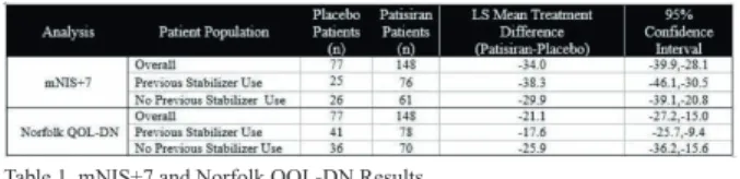
Background and aims: Hereditary transthyretin-mediated (hATTR) amyloidosis is a multi-systemic, life-threatening disease caused by transthyretin (TTR) mutations resulting in TTR protein destabilization forming multi-organ amyloid fibril deposits. TTR tetramer stabilizers have been used in hATTR amyloidosis patients; however, some studies have shown disease progression is still observed. In the APOLLO study, Patisiran, an investigational RNAi therapeutic, resulted in significant improvement in neuropathy (mNIS+7) and Norfolk Quality of Life Diabetic Neuropathy (Norfolk QOL-DN) compared to placebo in hATTR amyloidosis patients and was generally well-tolerated. We evaluated the impact of prior treatment with TTR tetramer stabilizers on Patisiran efficacy from the APOLLO study. Methods: APOLLO was a Phase 3, randomized (2:1), double-blind, study of patisiran 0.3mg/kg or placebo IV q3W (NCT01960348) in hATTR amyloidosis patients with polyneuropathy. Primary endpoint was change from baseline at 18-months in mNIS+7. TTR tetramer stabilizer discontinuation was required 14 or 3 days prior to study entry for tafamidis or diflunisal, respectively.
Results: APOLLO enrolled 225 patients: mean age 60.5 years (24-83); 74% males; 43% V30M. Prior to study entry, 53% of patients had previously received a TTR stabilizer (Tafamidis: n=74(33%); Diflunisal; n=45(20%). An
improvement in mNIS+7 and Norfolk QOL-DN was seen in patients with or without prior stabilizer use (Table 1) at 18-months. Additional efficacy and safety data to be presented.
Table 1. mNIS+7 and Norfolk QOL-DN Results
Conclusion: Patisiran demonstrated significant benefit relative to placebo in mNIS+7 and Norfolk QOL-DN in patients with or without prior TTR tetramer stabilizer use, thus providing evidence that hATTR amyloidosis patients with prior stabilizer use may benefit from Patisiran. Disclosure: This research was supported by Alnylam Pharmaceuticals.
O105
Phase 1/2, randomized, placebo controlled
and open-label extension studies of
Givosiran an investigational RNA
interference (RNAi) therapeutic, in patients
with Acute Intermittent Porphyria
E. Sardh1, J. Phillips2, P. Harper3, M. Balwani4, P. Stein5,
D. Rees6, J. Bloomer7, D. Bissell8, R.J. Desnick4,
C. Parker2, H. Bonkovsky9, N. Al-Tawil10, S. Rock11,
Q. Dinh11, C. Penz11, A. Chan11, W. Querbes11,
A. Simon11, K. Anderson12
1Karolinska University Hospital, Solna, Sweden, 2University of Utah, Salt Lake City, USA, 3Karolinska University Hospital,
Solona, Sweden, 4Mt. Sinai Icahn School of Medicine, New
York, USA, 5King’s College Hospital, London, United
Kingdom, 6King’s College Hospital, London, United Kingdom,
7University of Alabama, Birmingham, USA, 8University of
California, San Francisco, USA, 9Wake Forest University,
Winston-Salem, USA, 10Karolinska Trial Alliance Phase Unit,
Solna, Sweden, 11Alnylam, Cambridge, USA, 12University of Texas, Medical Branch, Galveston, USA
Background and aims: Acute hepatic porphyrias (AHPs) are rare genetic diseases resulting from loss-of-function mutations that cause upregulation of Aminolevulinic Acid Synthase 1 (ALAS1), the first and rate-limiting enzyme in the heme pathway. The resulting accumulation of neurotoxic intermediates Aminolevulinic Acid (ALA) and porphobilinogen (PBG) leads to neurovisceral attacks and chronic symptoms. Common neurological symptoms include pain, fatigue, muscle weakness, peripheral neuropathy, and neuropsychological manifestations. Givosiran acts via RNA interference (RNAi) to inhibit liver ALAS1 synthesis. Methods: A Phase 1/2 (ClinicalTrials.gov Identifier: NCT02452372), multinational study evaluated the safety, tolerability, pharmacokinetics, and pharmacodynamics of subcutaneously administered Givosiran (2.5mg/kg monthly). Impact on clinical activity, including chronic symptoms, annualized attack rates, and hemin use were explored. Patients completing the study were eligible for the open label extension (OLE) study (NCT0294983).
Results: Givosiran was generally well tolerated with no clinically significant laboratory abnormalities related to study drug. One unexpected serious adverse event (SAE; hypersensitivity) related to Givosiran occurred. Urinary ALA and PBG were reduced by 77% and 76% versus baseline, respectively. Additionally, Givosiran decreased mean annualized attack rate versus placebo by 73% and decreased annualized hemin doses versus run-in period by 73%. The OLE (n=8) data showed maintenance of clinical activity as observed in Phase 1.
Conclusion: Givosiran was generally well-tolerated and resulted in rapid and durable lowering of neurotoxic intermediates. ALA and PBG lowering were associated with marked reductions in both the annualised attack rate and hemin use. Complete Phase 1/2 and interim OLE data will be presented.