HAL Id: hal-00741034
https://hal.archives-ouvertes.fr/hal-00741034
Submitted on 11 Oct 2012
HAL is a multi-disciplinary open access
archive for the deposit and dissemination of
sci-entific research documents, whether they are
pub-lished or not. The documents may come from
teaching and research institutions in France or
abroad, or from public or private research centers.
L’archive ouverte pluridisciplinaire HAL, est
destinée au dépôt et à la diffusion de documents
scientifiques de niveau recherche, publiés ou non,
émanant des établissements d’enseignement et de
recherche français ou étrangers, des laboratoires
publics ou privés.
Acousto-optical coherence tomography with a digital
holographic detection scheme
Emilie Benoit a La Guillaume, Salma Farahi, Emmanuel Bossy, Michel Gross,
François Ramaz
To cite this version:
Emilie Benoit a La Guillaume, Salma Farahi, Emmanuel Bossy, Michel Gross, François Ramaz.
Acousto-optical coherence tomography with a digital holographic detection scheme. Optics Letters,
Optical Society of America - OSA Publishing, 2012, 37 (15), pp.3216-3218. �10.1364/OL.37.003216�.
�hal-00741034�
Acousto-Optical Coherence Tomography with a digital holographic
detection scheme
Emilie Benoit a la Guillaume,1,∗
Salma Farahi,1
Emmanuel Bossy,1
Michel Gross,2
and Francois Ramaz1
1
Institut Langevin, ESPCI ParisTech, CNRS UMR 7587, INSERM U979, Universite Paris VI - Pierre et Marie Curie,´
10 rue Vauquelin 75231 Paris Cedex 05, France 2
Laboratoire Charles Coulomb, CNRS UMR 5221, Universite Montpellier II, 34095 Montpellier, France´
∗Corresponding author: emilie.benoit@espci.fr
Compiled October 11, 2012
Acousto-Optical Coherence Tomography (AOCT) consists in using random phase jumps on ultrasound and light to achieve a millimeter resolution when imaging thick scattering media. We combined this technique with heterodyne off-axis digital holography. Two-dimensional images of absorbing objects embedded in scattering phantoms are obtained with a good signal to noise ratio. We study the impact of the phase modulation characteristics on the amplitude of the acousto-optic signal and on the contrast and apparent size of the absorbing inclusion. c 2012 Optical Society of America
OCIS codes: 110.0113, 110.7170, 170.3880, 170.3660, 090.2880, 090.1995.
Medical imaging is a contrast issue. Different types of waves (X-rays, ultrasound, etc.) are used depending on which organ is observed. Imaging with light, e.g. for breast cancer screening, raises the problem of detect-ing a millimeter-sized absorbdetect-ing object in a several-centimeters thick scattering medium. When using light only, like in Diffuse Optical Tomography [1], the resolu-tion of a breast tissues image is usually around 10 mm so that doctors can hardly detect an emerging tumor. Even if another imaging technique is used for screening, like radiography, the optical information remains pre-cious since it supplements the knowledge of the tissues with rich physiological details [2]. The idea to use acous-tic waves to achieve a millimeter resolution on opacous-tical information [3] gave rise to Ultrasound modulated Opti-cal Tomography (UOT).
UOT is based on the acousto-optic (AO) effect which enables to tag scattered light by shifting its frequency ωL
of the acoustic frequency ωU S [4]. The detection of this
weak signal at ωL± ωU S provides spatially resolved
op-tical information by scanning the acoustic source within the medium. The size of the tagging zone is determined by the shape of the acoustic wave beam, which depends
on ωU S, on the aperture and on the focal length of the
transducer. A focusing ultrasound transducer typically yields 1-2 mm lateral resolution in the focal plane when used at several MHz. However, localization of the AO ef-fect is about 10 times less accurate along the propagation direction of the ultrasound. As a consequence, the opti-cal and acoustic waves have to be temporally reshaped in order to improve the axial resolution.
Wang and Ku [5] proposed to encode each axial po-sition with a frequency-swept acoustic wave but the recording time was greater than the speckle decorrela-tion time, excluding any in vivo experiment. As com-monly used in standard echography, acoustic bursts are a solution to get axial resolution that involves times
com-PBS Ti:Sapphire Laser HWP +1 UT Phantom AOM 1 MOPA FI FI λ = 780 nm HWP A O M 2 +1 τ-shifted random phase modulation fAOM1sine Random phase modulation
Π 0 Π 0 τ Π 0 fUSsine fAOM2sine fL CMOS camera fC x z y y z x θ≈1°
Fig. 1. (Color on-line) Experimental set-up. HWP: half-wave plate, PBS: polarizing beam splitter, AOM1, AOM2: acousto-optic modulators, FI: Faraday isolator, UT: ultrasound transducer, fC: camera framerate, fL: laser frequency. In the tank, the scheme axes are modified in order to give a view of the inside.
patible with speckle decorrelation and medical standards on ultrasound exposure. The burst technique has bene-fited photo-refractive UOT [6,7] and has also been imple-mented with digital holographic detection by Atlan et al. [8]. Nevertheless, detecting s signals with a camera whose minimum acquisition time is in the order of hundreds of µs is quite inefficient. Lesaffre et al. solution [9], known as AOCT, is interesting because it returns to continuous acoustic and optical waves, performing a good resolu-tion though, thanks to a random phase modularesolu-tion on ultrasound and light. Up to now, AOCT technique has only been implemented in a photo-refractive holographic scheme. In this Letter, we demonstrate the potential of AOCT when combined with digital holography. We first experimentally study the influence of the random pat-tern characteristics on 1D AO images. Then the
resolu-tion of the technique is tested by performing 2D images on thick tissue-like phantoms containing small absorbing objects. We finally compare two AO profiles of the same object respectively obtained with AOCT and the burst technique.
Figure 1 depicts schematically the experimental set-up. A single-frequency 300-mW CW Ti:Sapphire laser (Coherent, MBR 110) with a coherence length of 300 m is tuned at 780 nm in order to work in the optical therapeutic window. A polarising beam splitter (PBS) divides light into an object beam and a reference beam. The object beam, after being amplified by a 2.5 W semi-conductor amplifier (MOPA, Sacher Lasertechnik) illu-minates a scattering sample immersed in a transparent water tank and insonified by a single element
acous-tic transducer (Panametrics A395S, fU S=2.3 MHz,
fo-cal length=78 mm, diameter=38 mm). The transmit-ted light passes through a 6-mm diameter circular di-aphragm and interferes with the reference beam on a 12-bit, 1024x1024 pixels fast CMOS camera (Photron
FastCam SA4) which records images at frame rate fC
(3.6 kHz maximum frame rate at full resolution). Two AO modulators (AOM 1, 2) are placed in each arm of the interferometric set-up and shift the frequency of light of fAOM 1,2. By choosing fAOM 2−fAOM 1= fU S+fC/2, we exclusively select the tagged photons because the other contributions vary too fast to be caught by the camera [10] and we perform a two-phases demodulation in order to reduce the mean intensity term [11]. The off-axis con-figuration adds a spatial separation of modulated and non-modulated light in the Fourier domain [12].
To get axial resolution in AOCT, the same random phase modulation Φ(t) is applied to the optical refer-ence wave and to the acoustic wave via two synchronized waveform generators. The reference beam is delayed of a time τ compared to the acoustic signal in order to se-lect a probed zone localized at a given y0position within the sample (y0= vU Sτ ). The interference cross term de-tected by the camera is the same as in standard AO imaging apart from the phase modulation factor. It can
be written as: ET ER∗ exp[jφ(t − y/vU S)]exp[−jφ(t −
τ )] + c.c., where ET and ERare the complex amplitudes
of the tagged photons field and the reference field, vU Sis the velocity of ultrasound (in water vU S≃ 1500 m.s−1), and c.c. is the complex conjugate. Lesaffre et al. [13] did the theoretical study of the tagged photons field in AOCT in the case of a {0; π} random phase jump modu-lation. They demonstrated that the tagged photons field is proportional to the correlation product between the two random phase modulations, both in amplitude and spatial extent.
In our configuration of holographic detection, the same phase modulation, based on {0; π} phase jumps occur-ring every δt, is implemented. The random pattern is recorded in the memory of a waveform generator (Tek-tronix AFG 3252) which limits the signal length to 524 µs. As a consequence, the pattern has to be repeated several times when the camera acquisition time exceeds
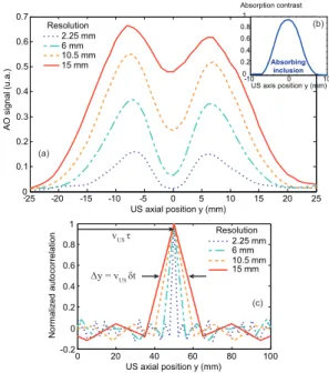
524 µs. The first experiment aims at studying the depen-dence of the AO signal on the modulation characteristic time δt. The results are compiled in figures 2 and 3.
-25 -20 -15 -10 -5 0 5 10 15 20 25 0 0.1 0.2 0.3 0.4 0.5 0.6 0.7 US axial position y (mm) AO s ig n a l (u .a .) 2.25 mm 6 mm 10.5 mm 15 mm Resolution -10 0 10 0 0.2 0.4 0.6 0.8 1 US axis position y (mm) Absorption contrast Absorbing inclusion 0 20 40 60 80 100 -0.2 0 0.2 0.4 0.6 0.8 US axial position y (mm) N o rma lize d a u to c o rre la ti o n 2.25 mm 6 mm 10.5 mm 15 mm Resolution vUS τ Δy = vUS δt 1 (a) (b) (c)
Fig. 2.(Color online) (a) AO profiles along y axis of a scattering phantom (thickness=3 cm, µ′s = 10 cm−1), containing a black inked cylinder of 10 × 6 × 6 mm3
(x, y, z). The same AO profile is represented with different resolution values. fC=500 Hz. (b) Shape of the absorbing inclusion, extracted from the AO profile with a 2.25 mm resolution. (c) Numerical simulation of the normalized autocorrelation function of the phase modulation sequences used to obtain the AO profiles in (a).
Figure 2(a) shows axial AO profiles obtained in a 3-cm thick (along z axis) 10%-Intralipid and agar gel containing an absorbing cylinder for several resolution
values. The transport mean free path l∗ of this
phan-tom is about 1 mm. The phase modulation pattern has an initial length of 200 µs (100 jumps, δt = 2 µs) and is stretched or compressed by changing the reading pe-riod of the waveform generator in order to monitor the characteristic time and thus the axial resolution. The typical shape of the autocorrelation function of the
ran-dom phase jump sequence is a triangle of ∆y = vU Sδt
in width at half maximum, as shown in figure 2(c). The theoretical minimum for ∆y is one period of the acoustic
wave, i.e. 0.65 mm at fU S=2.3 MHz. When δt increases
from 1.5 to 10 µs, the peak width of the autocorrelation function becomes larger so that the absorbing inclusion appears in the AO profile with a contrast decreasing from 95 to 54%. Figure 3 shows the evolution of the ampli-tude of the AO signal and the inclusion contrast and size as a function of the resolution. These curves are ex-tracted from the experimental measurements. In order to calculate the characteristics of the inclusion revealed by the AO profiles (contrast and size), each profile is fitted with a Gaussian envelope used as an approxima-tion of the scattering photons intensity profile along the acoustic axis. The difference between the envelope and the measured profile reveals the inclusion shape as seen
in figure 2(b). 0 5 10 15 0 0.1 0.2 0.3 0.4 0.5 0.6 0.7
AO signal amplitude (a.u.)
0 2 4 6 8 10 12 14 0 0.2 0.4 0.6 0.8 1 US axis resolution (mm) Inclusion contrast (a) (b) US axis resolution (mm) 0 5 10 15 6 7 8 9 10 US axis resolution (mm) Inclusion FWHM (mm) (c)
Fig. 3.Characteristics of the AO profile as functions of the reso-lution: (a) amplitude, (b) contrast, (c) full width at half maximum of the inclusion (FWHM).
Figure 3 demonstrates that the AO signal amplitude and the contrast of the inclusion vary in opposite direc-tions as funcdirec-tions of ∆y, which is expected since the peak of the autocorrelation function of the random phase pat-tern grows in amplitude and widens when ∆y increases. Thus, the difficulty to perform high contrast AO profiles lies in dealing with weak signals. As long as the resolu-tion is less than the absorbing object size, the contrast and the FWHM of the inclusion are nearly constant and give an estimation of the inclusion diameter. If we look at the first points on graph 3(c), the extracted diameter of the cylinder is 7.5 mm which is 25% as large as the real one (6 mm). This difference can be explained by the inhomogeneity of the photons distribution in the pres-ence of an absorbing object. Indeed, the probability of photon absorption is larger in the neighbourhood of the inclusion than in the rest of the scattering medium.
Position transverse X (mm) Position axiale Z (mm) −20 −10 0 10 20 −25 −20 −15 −10 −5 0 5 10 15 20 25 0 0.02 0.04 0.06 0.08 0.1 0.12 0.14 0.16 0.18 0.2 US transverse position x (mm) US axis position y (mm) −20 −10 0 10 20 −25 −20 −15 −10 −5 0 5 10 15 20 25 0 0.02 0.04 0.06 0.08 0.1 0.12 0.14 0.16 0.18 0.2 AO signal (a.u.) US transverse position x (mm) US axis position y (mm) −15 −10 −5 0 5 10 15 −15 −10 −5 0 5 10 15 0.01 0.02 0.03 0.04 0.05 0.06 0.07 0.08 AO signal (a.u.) (b) (a) 0
Fig. 4. (Color online) (a) AO image of the same phantom
as in figure 2, performed with ∆y = 3 mm axial resolution, fC=500 Hz. (b) AO image of a xy section of a scattering phan-tom (thickness=4 cm, µ′
s= 10 cm−1) with 2 absorbing inclusions of 3 × 3 × 5 mm3
(x, y, z), performed with ∆y = 2.25 mm axial resolution, fC=500 Hz.
An xy AO image of the same sample, presented in figure 4(a), is obtained by scanning both the US trans-ducer along x and the delay τ between the two random phase patterns. The quality of this imaging technique has also been tested by performing a 2D AO image of a 4-cm thick 10%-Intralipid and agar scattering phantom
(l∗ = 1 mm) containing two black-inked absorbing
in-clusions separated by 3.5 mm (figure 4(b)). The random pattern jump time is δt = 1.5 µs, giving a spatial resolu-tion of 2.25 mm. The two inclusions are clearly separated and their size is well retrieved.
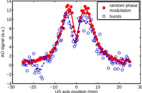
The advantage of using random phase jumps instead of bursts to achieve axial resolution is the noise reduc-tion on the AO signal. In figure 5, the same scattering phantom is scanned along the acoustic axis with both
−30 −20 −10 0 10 20 30 −4 −2 0 2 4 6 8 10 12 14 US axis position (mm) AO signal (a.u.) random phase modulation bursts
Fig. 5. AO profile along the acoustic axis of a 10%-Intralipid and agar scattering phantom (thickness=2 cm, µ′
s = 10 cm−1) with a black-inked absorbing inclusion of 3 × 3 × 6 mm3
(x, y, z), performed with ∆y = 2.6 mm axial resolution thanks to random phase jumps (filled dots) or bursts (empty dots), fC=1 kHz.
techniques in the same conditions of acoustic tagging. As a consequence, the signal amplitude, the inclusion contrast and the FWHM are equal within less than 10% on the two profiles. However, the signal-to-noise ratio on the AO profile is 3 times worse with the burst technique because the useful signal represents only a short frac-tion of the acquisifrac-tion time and is thus surrounded by a parasitic signal.
In conclusion, we have successfully implemented a ran-dom phase modulation on US and light to perform dig-ital holographic UOT with a millimeter resolution. The experiments have been run with continuous acoustic and optical waves but the next step is to apply AOCT with a 1-ms-pulse laser in order to keep a high level of signal while observing medical safety standards.
The authors gratefully acknowledge the Agence
Na-tionale de la Recherche for financial support (project
ICLM-ANR-2011-BS04-017-01). References
1. A. P. Gibson, J. C. Hebden, and S. R. Arridge, Phys. Med. Biol. 50, R1 (2005).
2. P. Taroni, A. Pifferi, E. Salvagnini, L. Spinelli, A. Torri-celli, and R. Cubeddu, Opt. Express 17, 15932 (2009). 3. F. A. Marks, H. W. Tomlinson, and G. W. Brooksby,
Proc. Soc. Photo-Opt. Instrum. Eng 1888, 500 (1993). 4. W. Leutz, and G. Maret, Physica B 204, 14 (1995). 5. L.-H. Wang and G. Ku, Opt. Lett. 23, 975 (1998). 6. T. W. Murray, L. Sui, G. Maguluri, R. A. Roy, A. Nieva,
F. Blonigen, and C. A. DiMarzio, Opt. Lett. 29, 2509 (2004).
7. S. Farahi, G. Montemezzani, A. A. Grabar, J.-P. Huig-nard, and F. Ramaz, Opt. Lett. 35, 1798 (2010). 8. M. Atlan, B. C. Forget, F. Ramaz, A. C. Boccara, and
M. Gross, Opt. Lett. 30, 1360 (2005).
9. M. Lesaffre, S. Farahi, M. Gross, P. Delaye, A. C. Boc-cara, and F. Ramaz, Opt. Express 17, 18211 (2009). 10. F. Le Clerc, L. Collot, and M. Gross, Opt. Lett. 25, 716
(2000).
11. I. Yamaguchi, and T. Zhang, Opt. Lett. 22, 1268 (1997). 12. M. Gross and M. Atlan, Opt. Lett. 32, 909 (2007). 13. M. Lesaffre, S. Farahi, A. C. Boccara, F. Ramaz and M.
Informational Fourth Page References
1. A. P. Gibson, J. C. Hebden, and S. R. Arridge, “Recent advances in diffuse optical imaging,” Phys. Med. Biol.
50,R1 (2005).
2. P. Taroni, A. Pifferi, E. Salvagnini, L. Spinelli, A. Torri-celli, and R. Cubeddu, “Seven-wavelength time-resolved optical mammography extending beyond 1000 nm for breast collagen quantification,” Opt. Express 17, 15932– 15946 (2009).
3. F. A. Marks, H. W. Tomlinson, and G. W. Brooksby, “Comprehensive approach to breast cancer detection us-ing light: photon localization by ultrasound modulation and tissue characterization by spectral discrimination,” Proc. Soc. Photo-Opt. Instrum. Eng 1888, 500 (1993). 4. W. Leutz, and G. Maret, “Ultrasonic modulation of
mul-tiply scattered light,” Physica B 204, 14–19 (1995). 5. L.-H. Wang and G. Ku, “Frequency-swept
ultrasound-modulated optical tomography of scattering media,” Opt. Lett. 23, 975–977 (1998).
6. T. W. Murray, L. Sui, G. Maguluri, R. A. Roy, A. Nieva, F. Blonigen, and C. A. DiMarzio, “Detection of ultrasound-modulated photons in diffuse media us-ing the photorefractive effect,” Opt. Lett. 29, 2509–2511 (2004).
7. S. Farahi, G. Montemezzani, A. A. Grabar, J.-P. Huig-nard, and F. Ramaz, “Photorefractive acousto-optic imaging in thick scattering media at 790 nm with a Sn2P2S6: T e crystal,” Opt. Lett. 35, 1798–1800 (2010).
8. M. Atlan, B. C. Forget, F. Ramaz, A. C. Boccara, and M. Gross, “Pulsed acousto-optic imaging in dynamic scatte-ring media with heterodyne parallel speckle detection,” Opt. Lett. 30, 1360-1362 (2005).
9. M. Lesaffre, S. Farahi, M. Gross, P. Delaye, A. C. Boc-cara, and F. Ramaz, “Acousto-optical coherence tomog-raphy using random phase jumps on ultrasound and light,” Opt. Express 17, 18211–18218 (2009).
10. F. Le Clerc, L. Collot, and M. Gross, “Numerical het-erodyne holography with two-dimensional photodetector arrays,” Opt. Lett. 25, 716–718 (2000).
11. I. Yamaguchi, and T. Zhang, “Phase-shifting digital holography,” Opt. Lett. 22, 1268–1270 (1997).
12. M. Gross and M. Atlan, “Digital holography with ulti-mate sensitivity,” Opt. Lett. 32, 909–911 (2007). 13. M. Lesaffre, S. Farahi, A. C. Boccara, F. Ramaz and M.
Gross, “Theoretical study of acousto-optical coherence tomography using random phase jumps on ultrasound and light,” JOSA A, 28, 1436–1444 (2011).


