RESEARCH OUTPUTS / RÉSULTATS DE RECHERCHE
Author(s) - Auteur(s) :
Publication date - Date de publication :
Permanent link - Permalien :
Rights / License - Licence de droit d’auteur :
Bibliothèque Universitaire Moretus Plantin
Institutional Repository - Research Portal
Dépôt Institutionnel - Portail de la Recherche
researchportal.unamur.be
University of Namur
Inhibition of mitochondrial genome expression triggers the activation of CHOP-10 by a
cell signaling dependent on the integrated stress response but not the mitochondrial
unfolded protein response
Michel, Sebastien; Canonne, Morgane; Arnould, Thierry; Renard, Patricia
Published in:
Mitochondrion
DOI:
10.1016/j.mito.2015.01.005
Publication date:
2015
Document Version
Publisher's PDF, also known as Version of record
Link to publication
Citation for pulished version (HARVARD):
Michel, S, Canonne, M, Arnould, T & Renard, P 2015, 'Inhibition of mitochondrial genome expression triggers
the activation of CHOP-10 by a cell signaling dependent on the integrated stress response but not the
mitochondrial unfolded protein response', Mitochondrion, vol. 21, pp. 58-68.
https://doi.org/10.1016/j.mito.2015.01.005
General rights
Copyright and moral rights for the publications made accessible in the public portal are retained by the authors and/or other copyright owners and it is a condition of accessing publications that users recognise and abide by the legal requirements associated with these rights. • Users may download and print one copy of any publication from the public portal for the purpose of private study or research. • You may not further distribute the material or use it for any profit-making activity or commercial gain
• You may freely distribute the URL identifying the publication in the public portal ?
Take down policy
If you believe that this document breaches copyright please contact us providing details, and we will remove access to the work immediately and investigate your claim.
Inhibition of mitochondrial genome expression triggers the activation
of CHOP-10 by a cell signaling dependent on the integrated stress
response but not the mitochondrial unfolded protein response
Sebastien Michel, Morgane Canonne, Thierry Arnould, Patricia Renard
⁎
Laboratory of Biochemistry and Cell Biology (URBC), NAmur Research Institute for LIfe Sciences (NARILIS), University of Namur (UNamur), 61 rue de Bruxelles, 5000 Namur, Belgium
a b s t r a c t
a r t i c l e i n f o
Article history:
Received 1 September 2014
Received in revised form 10 January 2015 Accepted 20 January 2015
Available online 30 January 2015
Keywords:
Mitochondrial dysfunction
Mitochondria unfolded protein response (mtUPR)
Integrated stress response (ISR) C/EBP homologous protein 10 (CHOP-10) mtDNA depletion
Doxycycline
Mitochondria-to-nucleus communication, known as retrograde signaling, is important to adjust the nuclear gene expression in response to organelle dysfunction. Among the transcription factors described to respond to mito-chondrial stress, CHOP-10 is activated by respiratory chain inhibition, mitomito-chondrial accumulation of unfolded proteins and mtDNA mutations. In this study, we show that altered/impaired expression of mtDNA induces CHOP-10 expression in a signaling pathway that depends on the eIF2α/ATF4 axis of the integrated stress response rather than on the mitochondrial unfolded protein response.
© 2015 Elsevier B.V. and Mitochondria Research Society. All rights reserved.
1. Introduction
Vestige of itsα-proteobacterial origin, mitochondria keep DNA (mtDNA) encoding 13 polypeptides, all subunits of the electron trans-port chain (ETC), as well as 2 ribosomal RNAs (rRNAs) and 22 transfer RNAs (tRNAs) that are required for their translation. Still, this only represents 1% of the mitochondrial proteome, estimated at up to 1500 proteins that are therefore predominantly encoded by the nuclear DNA (Calvo and Mootha, 2010). Altered expression of mitochondrial proteins is increasingly associated with many and various human
diseases (Boczonadi and Horvath, 2014). If mutations within genes encoding components or assembly factors of the ETC are the most obvi-ous cause of mitochondrial dysfunction and pathology, the better understanding of mtDNA maintenance and expression has shed light on new mutations associated with mitochondrial diseases. Indeed, those not only arise from alterations in the mtDNA but also in nuclear genes encoding proteins required for regulation of mtDNA expression. On one hand, mutations or deletions within one of the 2 rRNAs or 22 tRNAs encoding genes will affect the translation of the 13 mtDNA-encoded subunits of the ETC. Such alterations lead to mitochondrial dis-eases such as MELAS (Mitochondrial Encephalopathy, Lactic Acidosis, and Stroke-like episodes), MERRF (Myoclonic Epilepsy with Ragged Red Fibers) and KSS (Kearns–Sayre Syndrome) mainly associated with mutations within tRNAleu(A3243G), tRNAlys(A8344G) or
large-scale mtDNA deletion, respectively (Goto et al., 1990; Maceluch and Niedziela, 2006; Shoffner et al., 1990). On the other hand, the proteins required to support mtDNA expression are encoded by the nuclear genome (Vafai and Mootha, 2012). Therefore, it is not surprising that an increasing number of human mitochondrial diseases have been linked with nuclear genes encoding proteins involved in mitochondrial protein synthesis such as tRNA-modifying enzymes, aminoacyl-tRNA synthetases, ribosomal proteins, elongation, mRNA stability and termi-nation factors as well as translation activators (Pearce et al., 2013). Altered expression of mtDNA results in mitochondrial dysfunction and perturbs cellular homeostasis as mitochondria is important not only for ATP production by oxidative phosphorylation but also for lipid
Abbreviations: AARE, amino acid responsive element; ATF4, activating transcription fac-tor4;CHOP-10, C/EBPhomologousprotein 10; CK2,casein kinase 2; CREB, cAMP-responsive element binding protein; COX, cytochrome C oxidase; eIF2α, eukaryotic translation initia-tion factor 2A; ERAD, endoplasmic-reticulum-associated protein degradainitia-tion; ERSE, endo-plasmic reticulum stress element; erUPR, unfolded protein response of the endoendo-plasmic reticulum; ETC, electron transport chain; HERP, homocysteine-inducible, endoplasmic re-ticulum stress-inducible, ubiquitin-like domain member 1; EtBr, ethidium bromide; FCS, fetal calf serum; GCN2, general control non-derepressible-2; HRI, heme-regulated inhibitor; HSPD1, heat shock 60 kDa protein 1; ISR, integrated stress response; JNK, c-Jun N-terminal Kinase; KSS, Kearns-Sayre Syndrome; MELAS, Mitochondrial Encephalopathy, Lactic Acidosis, and Stroke-like episodes; MERRF, Myoclonic Epilepsy with Ragged Red Fibers; mtUPR, mitochondrial unfolded protein response; mtDNA, mitochondrial DNA; NFAT, nuclear factor of activated T cells; NFκB, Nuclear Factor kappa B; OTC, ornithine transcarbamylase; PERK, PKR-like ER-kinase; PKR, protein kinase double-stranded RNA-dependent; TOM40, translocase of the outer membrane; Trib3, tribble 3.
⁎ Corresponding author.
E-mail address:[email protected](P. Renard).
http://dx.doi.org/10.1016/j.mito.2015.01.005
1567-7249/© 2015 Elsevier B.V. and Mitochondria Research Society. All rights reserved.
Contents lists available atScienceDirect
Mitochondrion
metabolism, synthesis of iron–sulfur cluster, calcium and ROS signaling as well as regulation of programmed cell death. Hence, general feature of mitochondrial diseases include reduced ATP production, decreased membrane potential, altered calcium homeostasis and ROS production (James et al., 1996; Lenaz et al., 2004; Moudy et al., 1995; Vives-Bauza et al., 2006).
Coordinated expression of both genomes and communication be-tween mitochondria and nucleus is thus critical to maintain proper cell function. Transcriptional regulators are key players in mitochondria-to-nucleus communication as they orchestrate the expression of nuclear genes in response to both external challenges and organelle dysfunction to regulate processes such as mitochondrial biogenesis, cell proliferation, metabolism and apoptosis, as reviewed inRyan and Hoogenraad, (2007). Some of them have very well described signaling pathways activated in response to mitochondrial stress such as among other NFAT (nuclear fac-tor of activated T cells), NFκB (nuclear factor kappa B) and CREB (cAMP-responsive element binding protein) triggered by increased cytosolic cal-cium concentration because of altered mitochondrial buffering function (Arnould et al., 2002; Biswas et al., 1999, 2003). First and mainly reported in the context of endoplasmic reticulum stress, the transcription factor CHOP-10 (C/EBP homologous protein 10, also known as DDIT3 or GADD153) has also been reported to respond to mitochondrial dysfunc-tion. Indeed, increased expression of the transcription factor has been demonstrated in the context of disrupted mitochondrial protein homeo-stasis (proteohomeo-stasis) (Moisoi et al., 2009; Zhao et al., 2002), inhibition of mitochondrial ETC (Ishikawa et al., 2009; Vankoningsloo et al., 2006) and mutation within mtDNA (Cortopassi et al., 2006; Fujita et al., 2007). However, events and stress signaling pathways associated with mito-chondrial dysfunction-induced CHOP-10 overexpression are not clear. Hoogenraad and colleagues have demonstrated that CHOP-10 is regu-lated by AP-1 in the context of the mitochondrial unfolded protein re-sponse (mtUPR), a specific quality control process that aims to resolve proteotoxic stress by coordinating expression of mitochondrial chaper-ones and proteases such as HSPD1 (also known as chaperonin 60 or HSP60) and ClpP, respectively (Horibe and Hoogenraad, 2007). Briefly, in this model, overexpression of a truncated version of the enzyme ornithine transcarbamylase (OTCΔ) results in its accumulation as an unfolded proteins within mitochondrial matrix that leads to JNK (c-Jun N-terminal Kinase) activation and phosphorylation of the AP-1 family member c-Jun. Once translocated into the nucleus, this transcrip-tion factor increases CHOP-10 expression that in turn triggers expres-sion of stress responsive genes (Horibe and Hoogenraad, 2007). Moreover, the impairment of the stoichiometric equilibrium between nuclear and mtDNA-encoded proteins is another condition known to induce mtUPR as suggested in rho0cells and demonstrated for murine cells incubated in the presence of doxycycline, an antibiotic of the tetra-cycline family that inhibits mitochondrial translation (Houtkooper et al., 2013; Martinus et al., 1996). So far, the global mechanism of mtUPR is still missing in mammals as cell signaling activated by accumulation of unfolded proteins has been mainly described in Caenorhabditis elegans (Pellegrino et al., 2013).
However, others have shown that increased expression of CHOP-10 associated with mitochondrial respiration defect relies more on the in-tegrated stress response (ISR) than mtUPR (Cortopassi et al., 2006; Fujita et al., 2007; Ishikawa et al., 2009; Moisoi et al., 2009). ISR is a sig-naling pathway converging to eIF2α phosphorylation and characterized by a global reduction in protein synthesis, together with a selective overexpression of stress-responsive genes (Fujita et al., 2007; Silva et al., 2009). ISR sensors consist of a set of four different kinases: PERK (PKR-like ER-kinase), GCN2 (general control non-derepressible-2), HRI (heme-regulated inhibitor) and PKR (protein kinase double-stranded RNA-dependent). These four kinases are activated in response to various cues such as endoplasmic reticulum stress, amino acid depletion, viral infection, oxidative stress, heme deprivation, UV irradi-ation and proteasome inhibition (Donnelly et al., 2013; Harding et al., 2000, 2003). Even if eIF2α phosphorylation reduces cytosolic protein
synthesis, mRNAs containing short upstream open reading frames are preferentially translated, such as ATF4, ATF3 and CHOP-10 (Wek et al., 2006). The latter is a pleiotropic transcription factor that is associated with cell death/survival under ER stress as it can either trigger or prevent apoptosis (Fujihara et al., 2009; Zinszner et al., 1998). In addi-tion to its expression level, CHOP-10 can be regulated by post-transcriptional modifications such as phosphorylation by p38 or CK2 (casein kinase 2), resulting in an enhanced or attenuated transcriptional activity, respectively (Ubeda and Habener, 2003; Wang and Ron, 1996). Together with other transcription factors of the bZIP family, ATF4 and CHOP-10 regulate the expression of specific stress genes such as the ERAD (endoplasmic-reticulum-associated protein degradation)-associated protein HERP (homocysteine-inducible, endoplasmic reticu-lum stress-inducible, ubiquitin-like domain member 1) and the pseudo-kinase Trib3 (tribble 3) (Harding et al., 2003; Kilberg et al., 2009).
In this study, as CHOP-10 can be activated by both mtUPR and ISR, we analyzed the regulation of CHOP-10 expression in different cell models in which mtDNA expression is impaired, such as mitochondrial genome depletion and translation inhibition, to better understand sig-naling pathways triggered by mitochondrial dysfunction that could converge to the activation of this transcription factor. The investigation of these stress signaling pathways might help to better characterize mitochondrial diseases associated with mtDNA depletion or impaired organelle translation.
2. Materials and methods
2.1. Cell culture, transfection and reagents
The HeLa adenocarcinoma and Hep3B hepatocellular carcinoma cell lines were purchased from ATCC (CCL-2 and HB-8064, respectively). HeLa rho0cells were a kind gift of Professor R. Wiesner (University of Co-logne, Germany). HeLa cells were grown in Minimum Essential Media (MEM, Gibco) supplemented with pyruvate (11360-039, Gibco), non-essential amino acid (11140-035, Gibco) and 10% of fetal calf serum (FCS) (Gibco). HeLa rho0cells were grown in 4.5 g/l glucose-containing
Dulbecco's modified Eagle Medium (DMEM, Gibco) supplemented with pyruvate, non-essential amino acid, 10% of fetal calf serum and 50μg/ml uridine. Hep3B cells were cultured in Roswell Park Memorial Institute medium (RPMI 1640 Invitrogen, 21875) containing 10% of FCS. Fresh human hepatocytes (HEP220) were obtained from Biopredic inter-national and kept in medium for long-term culture (MIL600C) supple-mented with additive (ADD27111C). All cell lines were kept at 37 °C under a humidified atmosphere containing 5% CO2. Stock solution of
doxycycline (Sigma, D9891-1G) was prepared in deionized water at a concentration of 15 mg/ml,filter sterilized and used at a final concentra-tion of 15 or 30μg/ml as indicated.
For reporter gene experiments, HeLa cells have been transfected with SuperFect transfection reagent from Qiagen (301305). Briefly, cells were seeded one day before transfection in 12-well plates (50,000 cells/well). Cells were transiently transfected with 0.6μg of a Firefly luciferase reporter plasmid and 0.4 μg of a plasmid containing a cDNA encoding Renilla luciferase under the control of the TK promoter (pRenillaLuc-TK), used for normalization. The DNA/SuperFect ratio was 1:2 and cells were incubated with complexes for 3 h in the absence of serum before refreshing with new medium, containing doxycycline or not, for 48 h.
For silencing experiments, On-target plus siRNA smartpools purchased from Dharmacon were used: ATF4 (L-005125-00-0005), PERK (L-004883-00-0005), GCN2 (L-005314-00-0005), HRI (L-005007-00-0005), PKR (L-003527-00-0005). Non-targeting On-target plus siRNA (D-001810-10-05) have been used as negative control. 200,000 cells were seeded in 6-well plates in complete growth medium. The next day, cells were transfected with 50 nM siRNA diluted in opti-MEM (Gibco, 31985-070) and using Dharmafect 1 (T-2001-01) as transfection reagent, following the manufacturer's recommendation. After 24 h of
incubation with the complexes under standard culture condition, the me-dium was replaced with complete culture meme-dium for another 24 h. Cells were then incubated with doxycycline or control medium for the indicat-ed time.
2.2. Luciferase reporter assay
The pCHOP-10_luc constructs, a kind gift of Dr. Vaandrager (Utrecht University, Nederland's), consist of the pGL3 basic plasmid with the −442 to +91 human CHOP-10 promoter region fused to the luciferase gene. The reporter plasmids contain either wild-type or mutated sequences within the AARE (amino acid responsive element), AP-1 or ERSE (endoplasmic reticulum stress element) binding sites as fully described invan der Sanden et al. (2004). Reporter gene assay was per-formed using the dual-luciferase reporter assay from Promega (E1910) and following manufacturer's recommendation. Luminescence was measured using Lucetta luminometer (Lonza, AAL-1001) and Firefly activity was normalized to Renilla activity.
2.3. Real-time RT-qPCR
Cells were scraped in RLT lysis buffer and total RNA was prepared using the RNeasy mini kit from Qiagen (74104). An optional on-column DNase (79254, Qiagen) digestion of 15 min incubation step was performed to prevent any co-purification of DNA along with RNA. Complementary DNA was synthesized from 2μg of total RNA and with oligoT primers using the Transcriptor First Strand cDNA Synthesis kit from Roche (04896866001). Real-time PCR analysis was performed on the StepOneplus system from Applied Biosystem using FastStart Univer-sal SYBR Green Master mix (04913914001, Roche) and specific primers at afinal concentration of 300 nM in a volume of 25 μl. Primers were de-signed, using the online primer blast software, to evaluate mRNA abun-dance of the following targets: CHOP-10 forward: 5′-GCAAGAGGTCCT GTCTTCAGATG-3′ and reverse: 5′-CTCAGTCAGCCAAGCCAGAGA-3′; HSPD1 forward: 5′-TGATGCTATGGCTGGAGATTT-3′ and reverse: 5′-ACACCAGCAGCATCCAATAA-3′; ClpP forward: 5′-GTTGCCAGCCTTGTTA TCG-3′ and reverse: 5′-TGCATCGTGTCGTAGATGG-3′; Trib3 forward: 5′-CGTGATCTCAAGCTGTGTCG-3′ and reverse: 5′-AGCTTCTTCCTCTCAC GGTC-3′; ATF3 forward: 5′-GGAGCCTGGAGCAAAATGATG-3′ and re-verse: 5′-AGGGCGTCAGGTTAGCAAAA-3′; HERP forward: 5′-ATTTAG ACCGAGGCCGGTTC-3′ and reverse: 5′-CAGGATCAGTGCCTTCCTGTA-3′; PPIE forward: 5′-TCATGCTGCGTTCATTCCTT-3′ and reverse: 5′-GCTC AGATTCATTCATGTTGTCG-3′. PCR program started with 5 min at 95 °C followed by 40 cycles at 95 °C for 15 s and 60 °C for 1 min. A melt curve was added at the end of the PCR to check intended product ampli-fication. PPIE was used as a reference gene for normalization and mRNA expression level was quantified using the threshold cycle method as de-scribed in (Livak and Schmittgen, 2001).
2.4. Western blotting analysis
For protein extraction, cells were kept on ice and scraped in lysis buffer (20 mM Tris, pH 7.4; 150 mM KCl, 1 mM EDTA and 1% Triton X-100) complemented with protease inhibitor cocktail (complete from Roche) and phosphatase inhibitor mixture (1 mM NaVO3, 10 mM
p-nitrophenyl phosphate, 10 mMβ-glycerophosphate and 5 mM NaF). Clear lysates were retrieved from the supernatant of cell lysates after a 10 min centrifugation at 13,000 rpm at 4 °C and protein concentration was determined using the Pierce 660 nm Protein Assay (Thermo scientif-ic, 22660).
For western blotting analysis, 15 to 20μg of proteins were loaded on 4–15% Mini-PROTEAN TGX precast gel (Bio-rad #456-1086) or 3–8% Tris–Acetate gel (NuPAGE, Life technologies #EA0375BOX) and re-solved by electrophoresis in Tris–Glycine or Tris–Acetate running buffer, respectively. Proteins were transferred onto a low-fluorescence PVDF membrane (Immobilon FL, Merck-Millipore), blocked in Li-cor Odyssey
blocking buffer (927–40,000) diluted in PBS and incubated with appro-priate primary and secondary antibodies diluted in Odyssey blocking buffer supplemented with 1% Tween. Proteins were revealed using the infraredfluorescence Odyssey scanner (Li-cor). Protein abundance was evaluated using the following primary antibodies: COXI (Ab14705, Abcam), COXII (AbAA0258, Abcam), COXIV (A21348, Life Technolo-gies), CHOP-10 (ALX-804-551-C100, Enzo Life Sciences), P-eIF2α Ser51 (3795S, Cell signaling), eIF2α (9722S, Cell signaling), HSPD1 (SAB1405973-50UG, Sigma), TOM40 (sc-11414, Santa-cruz),α-tubulin (T5168, Sigma) and corresponding secondary antibodies coupled to infrared dyes (IRdye, Li-cor).
2.5. Statistical analysis
Data from at least three independent experiments are presented as means ± S.D. and have been analyzed using the Sigma-stat software with the appropriate statistical test such as ANOVA-I or ANOVA-II followed by a Holm–Sidak post-test. Differences between means were only considered as statistically significant when p b 0.05 or less. For some experiments, when normality test failed, data have been trans-formed into logarithmic values.
3. Results
3.1. Mitochondrial dysfunction triggers CHOP-10 overexpression
As few studies have described the induction of CHOP-10 upon differ-ent kinds of mitochondrial dysfunction (Cortopassi et al., 2006; Fujita et al., 2007; Ishikawa et al., 2009; Moisoi et al., 2009), wefirst assessed CHOP-10 mRNA abundance in two different models of impaired mtDNA expression. First, we used rho0cells, an extensively described and useful
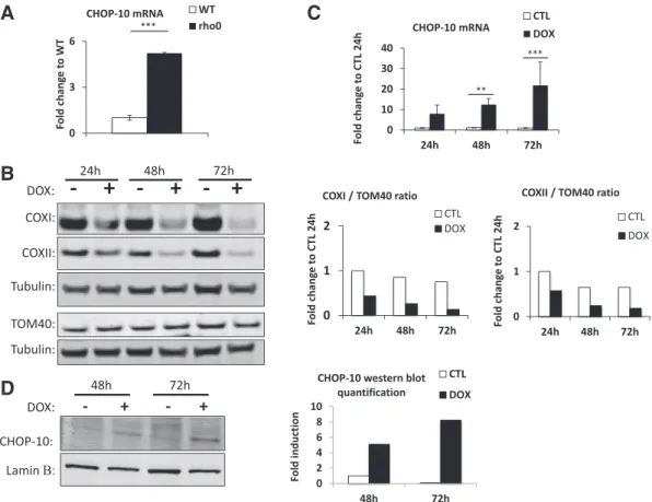
model to study OXPHOS defect and retrograde signaling induced by mtDNA depletion (Chandel and Schumacker, 1999; Mercy et al., 2005; Mineri et al., 2009; Miranda et al., 1999). The mRNA abundance of 10 assessed by RT-qPCR indicates that the abundance of CHOP-10 mRNA is significantly increased in rho0HeLa cells when compared
with their wild-type counterpart (Fig. 1A). This result has been con-firmed in HeLa cells incubated with ethidium bromide (EtBr), an inter-calating agent known to inhibit mtDNA transcription and replication and to be responsible for a progressive dilution and depletion of mito-chondrial genome as cells divide (Seidel-Rogol and Shadel, 2002). A 15 day-treatment with 50 ng/ml ethidium bromide strongly decreases the relative copy number of mtDNA (Fig. S1A) that is associated with an increase in CHOP-10 mRNA abundance suggesting that CHOP-10 induction in mtDNA-depleted cells is not just a clone effect or a result of long term cell adjustment but an early response observed in cells fac-ing a reduction of mtDNA content (Fig. S1B).
As a second cell model of impaired mitochondrial genome expres-sion, we used doxycycline. In bacteria, doxycycline is well known to interfere with the binding of the amino-acylated tRNA to the mRNA, thus inhibiting translation (Chopra et al., 1992). In line with the α-proteobacteria origin of mitochondria, such antibiotic has been de-scribed to inhibit mitochondrial translation (van den Bogert et al., 1986). As decreased abundance of mtDNA-encoded proteins is there-fore expected in doxycycline-treated cells, wefirst assessed the steady state level of COXI and COXII, by western blot, in HeLa cells incubated for 24, 48, or 72 h in the presence of 15μg/ml of the antibiotic. A de-crease in the abundance of COXI and COXII is observed as early as after 24 h of incubation, and is even more pronounced after 48 and 72 h, while the level of the nuclear-encoded protein TOM40 stays un-changed (Fig. 1B). As for the rho0cell model, the mitochondrial impair-ment provoked by doxycycline is accompanied by a time-dependent increase in CHOP-10 mRNA abundance that reaches a 20 fold-induction after 72 h of treatment (Fig. 1C). The induction of CHOP-10 expression in response to doxycycline was reflected at the protein level as illustrated atFig. 1D. To rule out unspecific effect of doxycycline,
we also assessed CHOP-10 mRNA abundance in HeLa cells incubated for 24 h with chloramphenicol, another antibiotic described to inhibit mito-chondrial translation in mammals (Balbi, 2004). A strong CHOP-10 induction was also obtained for cells incubated with the antibiotic (Fig. S2A).
3.2. Impairment of mtDNA expression does not trigger the mitochondrial unfolded protein response but activates the integrated stress response
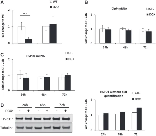
CHOP-10 is known to be associated with the unfolded protein re-sponse of the endoplasmic reticulum (erUPR) (Schroder and Kaufman, 2005). However, more than 10 years ago, Hoogenraad and colleagues already reported that CHOP-10 is also activated in a mitochondria-specific unfolded protein response (mtUPR) (Zhao et al., 2002). Still, if some mechanisms of mtUPR have been recently described in C. elegans (Pellegrino et al., 2013), the mammalian counterpart is less defined. In addition, among the different gene markers that are potentially overexpressed in response to mtUPR in mammals such as the protease YME1L1, MPPβ and the endonuclease G (Aldridge et al., 2007), only genes encoding ClpP and HSPD1 have been consistently assessed in subsequent studies dealing with mito-nuclear protein imbalance (Houtkooper et al., 2013; Mouchiroud et al., 2013). As disrupting mtDNA expression leads to depletion of proteins encoded by the mito-chondrial genome, we thus studied the putative activation of mtUPR by assessing the relative mRNA abundance of ClpP and HSPD1 using RT-qPCR. Unexpectedly, ClpP expression is strongly repressed in rho0
HeLa cells when compared with wild-type counterpart while there is
no significant difference for HSPD1 between mtDNA-depleted and wild-type HeLa cell lines (Fig. 2A). Similarly, there is no induction of the mtUPR-responsive ClpP and HSPD1 genes in EtBr-treated HeLa cells (Fig. S1C). The abundance of those two mtUPR markers has also been assessed in doxycycline- (Fig. 2B and C) and chloramphenicol-(Fig. S2B) treated HeLa cells, but there is neither induction of the mito-chondrial protease nor the chaperone at the transcript level. Results ob-tained at the mRNA level have been confirmed at the protein level for HSPD1 abundance but no change was observed in doxycycline-treated HeLa cells when compared with untreated control cells (Fig. 2D).
Based on the two main markers used to characterize mtUPR activa-tion, the data shows that CHOP-10 induction is not associated with this specific stress pathway in the different models of impaired mitochon-drial DNA expression tested, corresponding to mtDNA depletion or inhi-bition of mitochondrial translation. As previously mentioned, CHOP-10 is also induced during the ISR, thus, to answer the question whether impaired mtDNA expression could trigger this stress response, the mRNA abundance of Trib3, ATF3 and HERP was assessed by RT-qPCR (Harding et al., 2003; Kilberg et al., 2009). As illustrated inFig. 3A, the transcript abundance of these three ISR markers is increased in rho0
when compared with wild-type HeLa cells (Fig. 3A) and in EtBr-treated HeLa cells (Fig. S1D). Trib3 and ATF3 are also overexpressed in HeLa cells incubated with doxycycline in a time–dependent manner and HERP mRNA is also significantly more abundant in cells treated with doxycycline for the three time points studied (Fig. 3B). As for CHOP-10, the mRNA abundance of these three genes is also increased in HeLa cells incubated for 24 h with chloramphenicol (Fig. S2C).
A
C
B
D
Fig. 1. Disruption of mtDNA expression increases CHOP-10 expression in HeLa cells. CHOP-10 has been studied in 2 different models of impaired protein synthesis within mitochondria. (A) CHOP-10 mRNA abundance has been evaluated by RT-qPCR in HeLa cells totally depleted of mtDNA (rho0) and compared to wild-type (WT) cells. (B) Doxycycline-mediated inhibition of mitochondrial translation efficiency was monitored by western blot analysis of the abundance of COXI and COXII, 2 mtDNA-encoded proteins, and TOM40 used as a control for nuclear-encoded proteins. HeLa cells have been incubated with control or 15μg/ml doxycycline (DOX) containing medium during 24, 48 or 72 h. Immunodetection of tubulin was used as loading control. To highlight the mito-nuclear protein imbalance triggered by doxycycline, on the right of the graph is presented the western blot quantifications and expressed as the ratio of COXI or COXII to TOM40, all normalized to their respective tubulin. (C) CHOP-10 mRNA abundance has been studied by RT-qPCR in HeLa cells incubated with control medium (CTL) or with medium containing 15μg/ml doxycycline (DOX) for 24, 48 or 72 h as well as (D) at the protein level by western blot analysis for the last two time points. Western blot quantification using the Odyssey software is represented at the right for CHOP-10 abundance normalized to laminβ used as the loading control. Results represent means ± 1 SD for 3 independent experiments and are expressed as fold change compared to WT (A) and CTL 24 h (C). **: pb 0.01 and ***: p b 0.001.
A
C
D
B
Fig. 2. Impairing mitochondrial protein synthesis in HeLa cells does not increase expression of genes associated with the mitochondrial unfolded protein response. (A) mRNA abundance of the two mtUPR-associated protein markers ClpP and HSPD1 was evaluated by RT-qPCR in rho0
HeLa cells compared to wild-type counterpart. The abundance of ClpP (B) and HSPD1 (C) mRNA was assessed by RT-qPCR in HeLa cells incubated in control medium (CTL) or in the presence of 15μg/ml doxycycline (DOX) for 24, 48 or 72 h. Results are presented as means ± 1 SD for three independent experiments and are expressed as fold change to WT (A) or CTL 24 h (B, C) and ***: pb 0.001. (D) Protein level of HSPD1 was determined by western blot analysis in HeLa cells incubated with or without 15μg/ml doxycycline for 24, 48 or 72 h. Tubulin is used as a loading control. The quantification of HSPD1 protein abundance normal-ized to tubulin abundance is shown at the right of the chart.
A
B
Fig. 3. Interfering with mtDNA expression in HeLa cells triggers the integrated stress response. mRNA abundance of ISR-related gene markers Trib3, ATF3 and HERP was assessed by RT-qPCR in HeLa cells totally depleted of mtDNA (A) and in HeLa cells incubated with control medium or supplemented with 15μg/ml doxycycline for 24, 48 and 72 h (B). Results are expressed as fold change to WT cells (A) or control 24 h (B) and represent means for 3 independent experiments. *: pb 0.05, **: p b 0.01 and ***: p b 0.001.
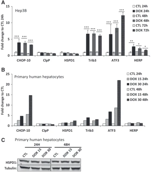
Despite the use of different models in which mtDNA expression is altered, we were thus unable to evidence an induction of mtUPR markers. In view of these unexpected results and even if they have also been confirmed in breast cancer MCF7 cells (Fig. S3A and B), we next questioned the cell models studied. Indeed, although cancer cell lines represent a very useful tool, their nuclear gene expression back-ground is often altered when compared with their healthy counterpart. Thus, we next compared the effect of doxycycline treatment in trans-formed versus primary human hepatocytes to validate the results obtained. The hepatocellular carcinoma-derived Hep3B cells were incu-bated with 15μg/ml doxycycline for 24, 48 or 72 h. The doxycycline ef-ficiency was first checked by western blot analysis of COXI and COXII protein abundance. In these conditions, comparable results to HeLa cells were obtained: the COXI/TOM40 and COXII/TOM40 ratio decreases after 24 h of incubation of Hep3B cells with the antibiotics (Fig. S4A). The mRNA abundance of CHOP-10, ClpP, HSPD1, Trib3, ATF3 and HERP was then assessed in the same experimental conditions (Fig. 4A). First, CHOP-10 expression is induced already after 24 h of treatment of Hep3B cells. Second, the levels of ClpP and HSPD1 mRNA were either unchanged or decreased regardless of the duration of incubation with
doxycycline. Third, the abundance of Trib3, ATF3 and HERP is increased in doxycycline-treated Hep3B cells after a 24, 48 or 72 h-treatment when compared with untreated cells. A comparable experiment was conducted in primary human hepatocytes that have been incubated with doxycycline at 15 or 30μg/ml during 24 or 48 h and results are illustrated inFig. 4B. As for cancer cells lines, CHOP-10 mRNA abun-dance increases in a time and concentration–dependent manner in treated hepatocytes when compared with untreated cells. While no dif-ference between control and doxycycline-treated hepatocytes is detect-able regarding ClpP mRNA, there is a slight but time- and concentration-dependent increase in HSPD1 mRNA (up to 2 fold-increase in cells incubated for 48 h with doxycycline at 30 μg/ml). However, the up-regulation of HSPD1 could not be reflected at the protein level as illustrated atFig. 4C. Finally, Trib3 and ATF3 mRNA are both induced in hepatocytes treated with doxycycline whereas HERP transcript abun-dance is only barely increased when compared with control cells. Even if the intensity of induction of the different ISR markers in response to doxycycline varies depending on the cell type, these results show that ISR activation in response to mitochondrial translation inhibition is not only restricted to cancer cells but is also observed in primary
A
B
C
Fig. 4. Doxycycline triggers expression of genes associated with the integrated stress response but not the mitochondrial unfolded response in both cancer and primary hepatocytes. (A) mRNA abundance of CHOP-10, mtUPR-related ClpP and HSPD1 and ISR-induced Trib3, ATF3 and HERP was evaluated by RT-qPCR in Hep3B cells. Cells were incubated with control (CTL) or 15μg/ml doxycycline-supplemented (DOX) medium for 24, 48 or 72 h. Results are expressed as fold change to CTL 24 h as means ± 1 SD for 3 independent experiments. *; **; ***: pb 0.05; 0.01; and 0.001, respectively. (B) Human primary hepatocytes were incubated 24 or 48 h with control (CTL) or doxycycline containing (15 or 30 μg/ml, DOX) medium. Results are expressed as fold change to corresponding CTL and represent means of 2 independent experiments. (C) HSPD1 protein abundance assessed by western blot in human primary hepatocytes incubated with doxycycline (15 or 30μg/ml) during 24 or 48 h.
human cells such as hepatocytes. However, it is important to point out that COXI and COXII abundance is kept unchanged in doxycycline-treated hepatocytes (Fig. S4B), which could explain the non-induction of mtUPR markers in this cell type. Most importantly, this result ques-tions the triggering event of ISR activation, since the latter is observed without mito-nuclear imbalance. It can be hypothesized that either the mito-nuclear protein imbalance is not at the origin of the stress response or it involves an alternative stress-signaling mechanism in primary hepatocytes that also leads to ISR activation. Altogether, the ex-periments performed in Hep3B cells and primary human hepatocytes incubated with doxycycline confirmed the results obtained with HeLa cells showing the induction of ISR-related targets such as CHOP-10, Trib3 and ATF3.
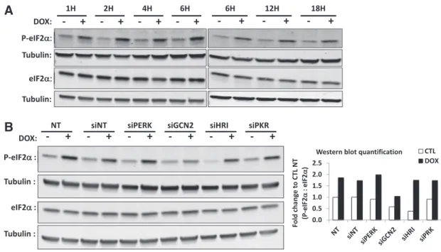
3.3. Doxycycline-induced eIF2α phosphorylation is mediated by GCN2 To confirm these results and the activation of ISR, we also analyzed the phosphorylation status of eIF2α in a time-course experiment. As shown inFig. 5A, eIF2α is phosphorylated in cells incubated with doxy-cycline as soon as after 1 h of treatment and the post-translational modification is still observed for longer incubation time up to 18 h of treatment. We next aimed to identify the kinase upstream eIF2α phos-phorylation. So far, four serine–threonine kinases have been described to phosphorylate eIF2α: PERK, GCN2, PKR and HRI (Donnelly et al., 2013). First, we checked the silencing efficiency of siRNA for the kinases. After 48 h of transfection, the protein level of the siRNA-targeted PERK, GCN2 and PKR in HeLa cells is reduced below 20% when compared to control cells, without affecting the level of the other kinases (Fig. S5A-B). Although, we were not able to assess the silencing ef fi-ciency of HRI at the protein level, due to the poor quality of the antibod-ies, a strong decrease (97% decrease) was observed on the abundance of the HRI transcript, a data compatible with a down-regulation of the kinase at the protein level (Fig. S5C). In these experimental conditions of silencing, the level of eIF2α phosphorylation in doxycycline-treated HeLa cells was assessed by western blot (Fig. 5B). In the absence of doxycycline, the level of eIF2α phosphorylation is quite comparable
between the different conditions, excepted in the presence of siRNA against GCN2 and HRI that reduces the phosphorylation of eIF2α. In doxycycline-treated HeLa cells, the level of eIF2α phosphorylation increases in all conditions except when GCN2 abundance is reduced, a result suggesting that the kinase that phosphorylates eIF2α in cells treated with doxycycline is GCN2.
3.4. ATF4 triggers CHOP-10 induction in doxycycline-treated HeLa cells To further understand CHOP-10 transcriptional regulation upon mi-tochondrial dysfunction, we next determined the putative contribution of three well-described cis-acting elements within its promoter: the ER stress responsive element (ERSE,−104/−75, (Ubeda and Habener, 2000)), the AP-1 binding site (−250/−225, (Guyton et al., 1996)) and the amino acid responsive element (AARE,−313/−295, (Bruhat et al., 1997)). A reporter plasmid containing the luciferase gene under the control of the CHOP-10 promoter region, spanning from nucleotides −442 to +91, that is either wild-type or mutated for one of these three regulatory sequences was used (van der Sanden et al., 2004) and is illus-trated on the left ofFig. 6A. First, a 48 h-incubation of HeLa cells with doxycycline increases reporter activity in cells transiently transfected with the plasmid containing the wild-type sequence of the CHOP-10 promoter, a data in agreement with the results shown for the endoge-nous mRNA and protein levels (Fig. 1). Second, mutation of either the ERSE or the AP-1 binding site has no drastic effect on the reporter activ-ity. By contrast, the luciferase activity induced in response to doxycy-cline is strongly reduced when the AARE binding site is mutated (Fig. 6A), suggesting the requirement for ATF4 activation in the doxycycline-induced CHOP-10 expression, as ATF4 is well known to bind to the AARE (Averous et al., 2004). In addition, increased ATF4 abundance is consistent with the doxycycline-induced phosphorylation of eIF2α (Fig. 5) as eIF2α is known to trigger preferential ATF4 translation (Vattem and Wek, 2004). Indeed, ATF4 protein abundance increases in a time–dependent manner in HeLa cells treated with 15 μg/ml doxycycline while being undetectable in control cells (Fig. 6B).
B
A
Fig. 5. Doxycycline-induced eIF2α phosphorylation in HeLa cells is dependent on the GCN2 kinase. (A) The level of eIF2α phosphorylation has been evaluated during a time-course ex-periment in HeLa cells incubated with or without doxycycline at 15μg/ml during 1 up to 18 h. Both phosphorylated and total eIF2α abundance were assessed by western blot and immunodetection of tubulin was used as a loading control. (B) HeLa cells were not transfected (NT) or transfected with siRNA against each of the four kinases described to phosphorylate eIF2α: PERK (siPERK), GCN2 (siGCN2), HRI (siHRI) and PKR (siPKR), as well as with non-targeting siRNA (siNT). After 48 h of transfection, cells were incubated with control or 15 μg/ml doxycycline-containing medium for 1 h. Phosphorylated and total eIF2α abundance was assessed by western blot and immunodetection of tubulin was used as a loading control. On the right is shown the quantification of P-eIF2α represented as the ratio of P-eIF2α to eIF2α, both normalized to their respective loading control. Results are finally expressed as fold change to non-transfected HeLa cells incubated with control medium (CTL NT). The result is the representative of three independent experiments.
The functional involvement of ATF4 in doxycycline-induced CHOP-10 expression wasfinally confirmed thanks to ATF4 silencing by siRNA. While transfection with non-targeting siRNA slightly increases CHOP-10 expression, silencing of ATF4 strongly decreases the abun-dance of CHOP-10 in HeLa cells treated for 24 h with doxycycline (Fig. 6C). We further checked the involvement of ATF4 in the expression of other ISR target genes such as Trib3. As shown inFig. 6D, Trib3 induc-tion is completely prevented when cells are transfected with siRNA against ATF4 while still robustly overexpressed in the non-targeting siRNA condition.
4. Discussion
The integrated stress response (ISR) is a stress response pathway based on four kinases that regulate eIF2α phosphorylation and gene ex-pression in response to broad types of cues, such as extensively de-scribed for endoplasmic reticulum stress and amino acid starvation (Donnelly et al., 2013). In this study, we have shown that disrupting mtDNA expression activates the ISR (Fig. 7). Indeed, either depletion or inhibition of mtDNA expression in HeLa cells increases expression of ISR-responsive genes such as the transcription factors CHOP-10 and ATF3, the pseudo kinase Trib3 and the ERAD-associated protein HERP (Figs. 1A, C, D,3A and B). CHOP-10 and Trib3 inductions are dependent on the central transcriptional regulator ATF4 as demonstrated by
silencing experiments (Fig. 6C and D). Consistent with the observed eIF2α phosphorylation, the protein abundance of ATF4 is increased when cells are incubated with doxycycline, an antibiotic that inhibits mitochondrial translation (Figs. 5A and6B). Increased expression of these targets has also been confirmed in human primary hepatocytes (Fig. 4B) as well as in Hep3B (Fig. 4A) and MCF7 cells (Fig. S3).
Surprisingly, in doxycycline-treated hepatocytes, the protein abun-dance of COXI and COXII (Fig. S4B), both mtDNA-encoded proteins, is not decreased as observed in HeLa (Fig. 1B) and Hep3B (Fig. S4A) cells, suggesting that depletion of mtDNA-encoded ETC subunits is not the triggering event of ISR activation per se. A very recent study showed that ATF4, ATF3 and CHOP-10 are overexpressed in cells incubated with tetracyclines (Bruning et al., 2014). Doxycycline belongs to this class of antibiotics that, in addition to mitochondrial translation inhibitor, are also known as bivalent ions chelators. Bruning and colleagues attributed some of the observed effects to magnesium depletion (Bruning et al., 2014). However, mitochondrial translation impairment with other clas-ses of antibiotics such as actinonin, the peptide deformylase inhibitor, in mouse embryonicfibroblasts (MEFs) also rapidly (and in a sustained manner) induces CHOP-10, Trib3 and ATF3 overexpression (Richter et al., 2013). Together with the results obtained in rho0cells (Figs. 1A
and3A) as well as for cells treated with chloramphenicol (Fig. S2) or ethidium bromide (Fig. S1), these results suggest that the ISR is trig-gered by the doxycycline-induced disrupted mtDNA expression and not
A
B
C
D
Fig. 6. ATF4 triggers CHOP-10 induction in doxycycline-treated HeLa cells. (A) Reporter plasmid assay using a fragment of the CHOP-10 promoter sequence (−442 to +91) upstream of the Firefly luciferase gene. 24 h post-transfection with the reporter and normalization plasmids, HeLa cells were incubated with or without (control: CTL) 15 μg/ml doxycycline containing medium (DOX) for 48 h before assessing reporter activity. Promoter sequence of the reporter plasmid contains either wild-type (WT) or mutated sequence for one of the three following cis-acting elements: ER stress response element (ERSE), AP-1 binding site (AP-1) or the amino acid responsive element (AARE) site as illustrated on the left panel. Firefly luciferase activity was normalized to Renilla luciferase activity and expressed as fold change to respective control. (B) ATF4 protein level evaluated by western blot. HeLa cells were incubated with medium containing or not with 15μg/ml doxycycline (DOX) for 6, 12 or 18 h. Immunodetection of tubulin was used as the loading control. The graph on the right represents ATF4 abundance quan-tification normalized to tubulin and expressed as fold change compared to HeLa cells incubated for 6 h in control medium. (C) Impact of ATF4 silencing on increased doxycycline-induced CHOP-10 and Trib3 mRNA abundance. HeLa cells were either not transfected (NT), transfected with non-targeting siRNA (siNT) or siRNA against ATF4 (siATF4) during 24 h before medium renewal and incubation for 48 h with control (CTL) or 15μg/ml doxycycline supplemented medium (DOX). Results are expressed as fold change to no-transfected (NT) CTL and represent means ± 1 S.D. of three independent experiments. *: pb 0.05 and ***: p b 0.001.
an unspecific effect of the antibiotic. We have also shown that the GCN2 kinase is required for eIF2α phosphorylation in response to mitochon-drial translation inhibition in HeLa cells incubated with doxycycline (Fig. 5B). The mechanism of GCN2 activation in response to impaired mitochondrial translation is still unknown but, as suggested by results in primary human hepatocytes, it is unlikely to be triggered by the mito-nuclear protein imbalance. This idea is reinforced by the fact that eIF2α phosphorylation is rapidly observed in response to doxycy-cline treatment (after 1 h of doxycydoxycy-cline incubation) while the mito-nuclear imbalance observed in HeLa cells requires at least 24 h of incubation with doxycycline (data not shown). The best-described mechanism of GCN2 activation is related to amino acid depletion and requires the binding of an unloaded tRNA to the His-tRNA synthetase-like and CTD (C-terminal dimerization and ribosome binding site) do-mains of the kinase (Castilho et al., 2014). On the other hand, although the molecular mechanism is missing, GCN2 activation is also reported under proteasome inhibition in several studies (Acharya et al., 2009; Jiang and Wek, 2005; Neznanov et al., 2008). Interestingly, the concen-tration of 15 amino acids and the proteasomal activity are both de-creased in 143B cybrid cells harboring a 7.5 kb mtDNA deletion and in lymphoblasts from patients carrying a 5 kb mtDNA deletion (Alemi et al., 2007). In addition, in the yeast Saccharomyces cerevisiae, oxidative stress, such as incubation with peroxide hydrogen, also activates GCN2 by a still unclear mechanism requiring the His-RS-like domain of the ki-nase (Mascarenhas et al., 2008). Besides, induction of the GCN2-eIF2α axis is associated with mitochondrial ETC defect in the C. elegans clk-1 (required for ubiquinone synthesis) mutant strain, which can be prevented by NAC treatment (Baker et al., 2012). Thus, it would be interesting to further investigate both the involvement of amino acid depletion, proteasome inhibition, as well as ROS production in GCN2 activation upon doxycycline treatment.
In mammals, the mitochondria-specific unfolded protein response (mtUPR) is currently accepted as a quality control relying on a retrograde signaling to coordinate expression of specific genes, such as the chaper-one HSPD1 and the protease ClpP, to resolve organelle proteotoxic stress (Pellegrino et al., 2013). Different cell models have been described to induce mtUPR: either the accumulation of protein aggregates in the
mitochondrial matrix, caused by the OTCΔ overexpression (Rath et al., 2012; Zhao et al., 2002), or an imbalance between mitochondrial and nuclear-encoded proteins, as shown in mouse hepatocytes treated with doxycycline (Houtkooper et al., 2013) or in mtDNA depleted (rho0) rat
hepatoma cells (Martinus et al., 1996). Even if it is known that sensitivity and response to drugs might differ between rodent and human hepato-cytes (Black et al., 2012; Kotokorpi et al., 2007; Seok et al., 2013), in our hands, similar stresses increase neither HSPD1 nor ClpP expression despite testing four different models, of which one in three cancer cell lines as well as in primary human hepatocytes (Figs. 2A, B, C, D, S1C, S2B). However, in the later case, the non-induction of mtUPR markers might be the result of the absence of mito-nuclear protein imbal-ance as suggested by the steady-state protein level of COXI and COXII (Fig. S4B).
In the mtUPR context provoked by OTCΔ overexpression, CHOP-10 induction would be regulated by the AP-1 transcription factor after c-jun phosphorylation by JNK and/or PKR (Horibe and Hoogenraad, 2007; Rath et al., 2012). In the present study, even if CHOP-10 is overexpressed, reporter plasmid experiments clearly demonstrate that, when mitochondrial translation is inhibited by doxycycline, the AARE binding site is required for increased transcription of the luciferase reporter gene under the control of a region of the CHOP-10 promoter whereas mutation within the AP-1 binding site has no effect (Fig. 6A). In addition, we confirmed ATF4 requirement as silencing of this transcription factor prevents CHOP-10 induction by doxycycline.
Actually, a careful analysis of the literature reveals previous reports in line with our results showing no induction of mtUPR upon mitochon-drial dysfunction. First, a study from Piechota and colleagues also ana-lyzed the impact of mitochondrial genome expression inhibitors in HeLa cells and showed that a six-day-treatment with ethidium bromide or thiamphenicol (a derivative of chloramphenicol) does not increase HSPD1 abundance (Piechota et al., 2006). Second, a recent in vivo study conducted on mice KO for DARS2 (mitochondrial aspartyl-tRNA synthetase) in cardiac and skeletal muscle tissues highlights a tissue-specific response to mitochondrial translation impairment: no mtUPR marker is observed in skeletal muscle, while a weak induction of HSP60 and no modification of ClpP abundance are observed in cardiomyocytes (Dogan et al., 2014). Third, in the extensive expression profiling by Cortopassi and colleagues of mitochondrial diseases associated with mtDNA mutations/deletions, ClpP is not induced and is even repressed in two conditions of mtDNA deletion when compared with their respec-tive controls (Cortopassi et al., 2006). Yet, among shared overexpressed genes between the different models of mitochondrial dysfunction, these authors highlighted ISR targets such as CHOP-10, ATF4, ATF3 and Trib3. Four, in addition to organelle dysfunction associated with altered mtDNA expression, mitochondrial proteotoxic stress in mice knock out for the mitochondrial intermembrane space protease HtrA2 also points CHOP-10, ATF3 and HERP as overexpressed in brain cells, whereas ClpP and HSPD1 expression stays unchanged (Moisoi et al., 2009). Finally, a transcriptomic analysis following the selective inhibition of heat shock protein-90 (Hsp90) in mitochondria by GamitrinibTPP(GA mitochondrial matrix inhibitor G-TPP) revealed increased mRNA abundance of CHOP-10, Trib3, ATF3 and HERP together with phosphorylation of eIF2α (Siegelin et al., 2011). Altogether these results strongly suggest that ISR might be a general response to mitochondrial dysfunction while mtUPR would be restricted to more specific conditions affecting mitochondrial function.
Coordination between nuclear and mitochondrial genomes is neces-sary to ensure proper organelle function and cell integrity. The central ISR event eIF2α phosphorylation might be crucial for such coordination in response to mitochondrial dysfunction (Fig. 7). Interestingly, in C. elegans the GCN2-eIF2α axis acts in parallel with mtUPR to keep mitochondrial proteostasis and function. Indeed, GCN2 silencing in-creases mitochondrial stress and transcriptional activity of the HSP60-GFP reporter plasmid (Baker et al., 2012). Global attenuation of protein synthesis might also prevent excessive importation and accumulation
Fig. 7. Hypothetical model for stress response to mitochondrial dysfunction. In response to mitochondrial dysfunction such as mtDNA depletion and inhibition of mitochondrial translation, the GCN2 kinase is activated. In turn, the kinase phosphorylates eIF2α that has at least 2 consequences. While the cytosolic translation is globally attenuated, mRNAs containing an uORF are preferentially translated such as the transcription factors ATF4 and CHOP-10. The profit of this GCN2-eIF2α-ATF4 pathway might be stress attenua-tion by relieving the load of protein imported into mitochondria but also by increasing the expression of stress responsive genes. Dashed lines represent a hypothetical link.
of unfolded proteins within mitochondria and thus detrimental over-whelming of resident chaperones and proteases.
This scenario is already well described for the endoplasmic reticu-lum. Indeed, it has been shown that eIF2α phosphorylation and protein synthesis shutdown reduce ER-stress, promoting cell survival (Han et al., 2013; Harding et al., 2000). Likewise, reduced protein translation in mouseβ-cells is required to keep organelle in shape as cells lacking eIF2α have swollen mitochondria with disrupted cristae (Back et al., 2009). Reduced cytosolic protein synthesis also prevents mitochondrial degeneration in a yeast model of aging-related degenerative disease (Wang et al., 2008). PINK1 is a serine/threonine kinase involved in the mitochondrial quality control mitophagy. In a Drosophila melanogaster model of familial, early onset of Parkinson's disease, reduction of protein translation suppresses PINK1 mutant phenotype whereas increased translation through S6K activation worsen it (Liu and Lu, 2010). Finally, phosphorylation of eIF2α also protects from oxidative stress triggered by the mitochondrial complex I inhibitor paraquat, both in C. elegans and mammalian cells (Rainbolt et al., 2013).
The phosphorylation of eIF2α and global attenuation of protein syn-thesis represent thus an interesting mechanism to prevent/attenuate mitochondrial damage and dysfunction. Knowing that impaired mito-chondrial translation is increasingly associated with mitomito-chondrial dis-eases such as Leigh syndrome and Perrault syndrome (Boczonadi and Horvath, 2014; Pearce et al., 2013), further investigation of the function-al impact of the GCN2/P-eIF2α/ATF4 axis is now necessary to better understand the biological processes controlled by this cell signaling path-way associated with mitochondria dysfunction and related pathologies. Acknowledgments
S. Michel is recipient of a doctoral fellowship from the Fonds pour la Recherche dans l'Industrie et l'Agriculture (FRIA, Belgium). This work was supported by the Belgian Association for Muscular Diseases (ABMM, Belgium, grant 2013-08). The authors especially thank Prof. Wiesner for the gift of the mtDNA-depleted HeLa rho0cells.
Appendix A. Supplementary data
Supplementary data to this article can be found online athttp://dx. doi.org/10.1016/j.mito.2015.01.005.
References
Acharya, P., Engel, J.C., Correia, M.A., 2009.Hepatic CYP3A suppression by high concentrations of proteasomal inhibitors: a consequence of endoplasmic reticulum (ER) stress induc-tion, activation of RNA-dependent protein kinase-like ER-bound eukaryotic initiation factor 2alpha (eIF2alpha)-kinase (PERK) and general control nonderepressible-2 eIF2alpha kinase (GCN2), and global translational shutoff. Mol. Pharmacol. 76, 503–515. Aldridge, J.E., Horibe, T., Hoogenraad, N.J., 2007.Discovery of genes activated by the mitochondrial unfolded protein response (mtUPR) and cognate promoter elements. PLoS One 2, e874.
Alemi, M., Prigione, A., Wong, A., Schoenfeld, R., DiMauro, S., Hirano, M., et al., 2007. Mi-tochondrial DNA deletions inhibit proteasomal activity and stimulate an autophagic transcript. Free Radic. Biol. Med. 42, 32–43.
Arnould, T., Vankoningsloo, S., Renard, P., Houbion, A., Ninane, N., Demazy, C., et al., 2002. CREB activation induced by mitochondrial dysfunction is a new signaling pathway that impairs cell proliferation. EMBO J. 21, 53–63.
Averous, J., Bruhat, A., Jousse, C., Carraro, V., Thiel, G., Fafournoux, P., 2004.Induction of CHOP expression by amino acid limitation requires both ATF4 expression and ATF2 phosphorylation. J. Biol. Chem. 279, 5288–5297.
Back, S.H., Scheuner, D., Han, J., Song, B., Ribick, M., Wang, J., et al., 2009.Translation atten-uation through eIF2alpha phosphorylation prevents oxidative stress and maintains the differentiated state in beta cells. Cell Metab. 10, 13–26.
Baker, B.M., Nargund, A.M., Sun, T., Haynes, C.M., 2012.Protective coupling of mitochon-drial function and protein synthesis via the eIF2alpha kinase GCN-2. PLoS Genet. 8, e1002760.
Balbi, H.J., 2004.Chloramphenicol: a review. Pediatr. Rev. 25, 284–288.
Biswas, G., Adebanjo, O.A., Freedman, B.D., Anandatheerthavarada, H.K., Vijayasarathy, C., Zaidi, M., et al., 1999.Retrograde Ca2+ signaling in C2C12 skeletal myocytes in re-sponse to mitochondrial genetic and metabolic stress: a novel mode of inter-organelle crosstalk. EMBO J. 18, 522–533.
Biswas, G., Anandatheerthavarada, H.K., Zaidi, M., Avadhani, N.G., 2003.Mitochondria to nucleus stress signaling: a distinctive mechanism of NFkappaB/Rel activation through calcineurin-mediated inactivation of IkappaBbeta. J. Cell Biol. 161, 507–519. Black, M.B., Budinsky, R.A., Dombkowski, A., Cukovic, D., LeCluyse, E.L., Ferguson, S.S., et
al., 2012.Cross-species comparisons of transcriptomic alterations in human and rat primary hepatocytes exposed to 2,3,7,8-tetrachlorodibenzo-p-dioxin. Toxicol. Sci. 127, 199–215.
Boczonadi, V., Horvath, R., 2014.Mitochondria: impaired mitochondrial translation in human disease. Int. J. Biochem. Cell Biol. 48, 77–84.
Bruhat, A., Jousse, C., Wang, X.Z., Ron, D., Ferrara, M., Fafournoux, P., 1997.Amino acid lim-itation induces expression of CHOP, a CCAAT/enhancer binding protein-related gene, at both transcriptional and post-transcriptional levels. J. Biol. Chem. 272, 17588–17593.
Bruning, A., Brem, G.J., Vogel, M., Mylonas, I., 2014.Tetracyclines cause cell stress-dependent ATF4 activation and mTOR inhibition. Exp. Cell Res. 320, 281–289. Calvo, S.E., Mootha, V.K., 2010.The mitochondrial proteome and human disease. Annu.
Rev. Genomics Hum. Genet. 11, 25–44.
Castilho, B.A., Shanmugam, R., Silva, R.C., Ramesh, R., Himme, B.M., Sattlegger, E., 2014. Keeping the eIF2 alpha kinase Gcn2 in check. Biochim. Biophys. Acta 1843, 1948–1968.
Chandel, N.S., Schumacker, P.T., 1999.Cells depleted of mitochondrial DNA (rho0) yield insight into physiological mechanisms. FEBS Lett. 454, 173–176.
Chopra, I., Hawkey, P.M., Hinton, M., 1992.Tetracyclines, molecular and clinical aspects. J. Antimicrob. Chemother. 29, 245–277.
Cortopassi, G., Danielson, S., Alemi, M., Zhan, S.S., Tong, W., Carelli, V., et al., 2006. Mito-chondrial disease activates transcripts of the unfolded protein response and cell cycle and inhibits vesicular secretion and oligodendrocyte-specific transcripts. Mito-chondrion 6, 161–175.
Dogan, S.A., Pujol, C., Maiti, P., Kukat, A., Wang, S., Hermans, S., et al., 2014.Tissue-specific loss of DARS2 activates stress responses independently of respiratory chain deficien-cy in the heart. Cell Metab. 19, 458–469.
Donnelly, N., Gorman, A.M., Gupta, S., Samali, A., 2013.The eIF2alpha kinases: their struc-tures and functions. Cell. Mol. Life Sci. 70, 3493–3511.
Fujihara, M., Bartels, E., Nielsen, L.B., Handa, J.T., 2009.A human apoB100 transgenic mouse expresses human apoB100 in the RPE and develops features of early AMD. Exp. Eye Res. 88, 1115–1123.
Fujita, Y., Ito, M., Nozawa, Y., Yoneda, M., Oshida, Y., Tanaka, M., 2007.CHOP (C/EBP ho-mologous protein) and ASNS (asparagine synthetase) induction in cybrid cells har-boring MELAS and NARP mitochondrial DNA mutations. Mitochondrion 7, 80–88. Goto, Y., Nonaka, I., Horai, S., 1990.A mutation in the tRNA(Leu)(UUR) gene associated
with the MELAS subgroup of mitochondrial encephalomyopathies. Nature 348, 651–653.
Guyton, K.Z., Xu, Q., Holbrook, N.J., 1996.Induction of the mammalian stress response gene GADD153 by oxidative stress: role of AP-1 element. Biochem. J. 314 (Pt 2), 547–554.
Han, J., Back, S.H., Hur, J., Lin, Y.H., Gildersleeve, R., Shan, J., et al., 2013.ER-stress-induced transcriptional regulation increases protein synthesis leading to cell death. Nat. Cell Biol. 15, 481–490.
Harding, H.P., Zhang, Y., Bertolotti, A., Zeng, H., Ron, D., 2000.Perk is essential for transla-tional regulation and cell survival during the unfolded protein response. Mol. Cell 5, 897–904.
Harding, H.P., Zhang, Y., Zeng, H., Novoa, I., Lu, P.D., Calfon, M., et al., 2003.An integrated stress response regulates amino acid metabolism and resistance to oxidative stress. Mol. Cell 11, 619–633.
Horibe, T., Hoogenraad, N.J., 2007.The chop gene contains an element for the positive regulation of the mitochondrial unfolded protein response. PLoS One 2, e835. Houtkooper, R.H., Mouchiroud, L., Ryu, D., Moullan, N., Katsyuba, E., Knott, G., et al., 2013.
Mitonuclear protein imbalance as a conserved longevity mechanism. Nature 497, 451–457.
Ishikawa, F., Akimoto, T., Yamamoto, H., Araki, Y., Yoshie, T., Mori, K., et al., 2009.Gene ex-pression profiling identifies a role for CHOP during inhibition of the mitochondrial re-spiratory chain. J. Biochem. 146, 123–132.
James, A.M., Wei, Y.H., Pang, C.Y., Murphy, M.P., 1996.Altered mitochondrial function in fibroblasts containing MELAS or MERRF mitochondrial DNA mutations. Biochem. J. 318 (Pt 2), 401–407.
Jiang, H.Y., Wek, R.C., 2005.Phosphorylation of the alpha-subunit of the eukaryotic initiation factor-2 (eIF2alpha) reduces protein synthesis and enhances apoptosis in response to proteasome inhibition. J. Biol. Chem. 280, 14189–14202.
Kilberg, M.S., Shan, J., Su, N., 2009.ATF4-dependent transcription mediates signaling of amino acid limitation. Trends Endocrinol. Metab. 20, 436–443.
Kotokorpi, P., Ellis, E., Parini, P., Nilsson, L.M., Strom, S., Steffensen, K.R., et al., 2007. Phys-iological differences between human and rat primary hepatocytes in response to liver X receptor activation by 3-[3-[N-(2-chloro-3-trifluoromethylbenzyl)-(2,2-diphenylethyl)amino]propyloxy]phe nylacetic acid hydrochloride (GW3965). Mol. Pharmacol. 72, 947–955.
Lenaz, G., Baracca, A., Carelli, V., D'Aurelio, M., Sgarbi, G., Solaini, G., 2004.Bioenergetics of mitochondrial diseases associated with mtDNA mutations. Biochim. Biophys. Acta 1658, 89–94.
Liu, S., Lu, B., 2010.Reduction of protein translation and activation of autophagy protect against PINK1 pathogenesis in Drosophila melanogaster. PLoS Genet. 6, e1001237. Livak, K.J., Schmittgen, T.D., 2001.Analysis of relative gene expression data using
real-time quantitative PCR and the 2(-Delta Delta C(T)) Method. Methods 25, 402–408.
Maceluch, J.A., Niedziela, M., 2006.The clinical diagnosis and molecular genetics of Kearns–Sayre syndrome: a complex mitochondrial encephalomyopathy. Pediatr. Endocrinol. Rev. 4, 117–137.
Martinus, R.D., Garth, G.P., Webster, T.L., Cartwright, P., Naylor, D.J., Hoj, P.B., et al., 1996. Selective induction of mitochondrial chaperones in response to loss of the mitochon-drial genome. Eur. J. Biochem. 240, 98–103.
Mascarenhas, C., Edwards-Ingram, L.C., Zeef, L., Shenton, D., Ashe, M.P., Grant, C.M., et al., 2008.Gcn4 is required for the response to peroxide stress in the yeast Saccharomyces cerevisiae. Mol. Biol. Cell 19, 2995–3007.
Mercy, L., Pauw, A., Payen, L., Tejerina, S., Houbion, A., Demazy, C., et al., 2005. Mitochon-drial biogenesis in mtDNA-depleted cells involves a Ca2 + -dependent pathway and a reduced mitochondrial protein import. FEBS J. 272, 5031–5055.
Mineri, R., Pavelka, N., Fernandez-Vizarra, E., Ricciardi-Castagnoli, P., Zeviani, M., Tiranti, V., 2009.How do human cells react to the absence of mitochondrial DNA? PLoS One 4, e5713.
Miranda, S., Foncea, R., Guerrero, J., Leighton, F., 1999.Oxidative stress and upregulation of mitochondrial biogenesis genes in mitochondrial DNA-depleted HeLa cells. Biochem. Biophys. Res. Commun. 258, 44–49.
Moisoi, N., Klupsch, K., Fedele, V., East, P., Sharma, S., Renton, A., et al., 2009.Mitochondrial dysfunction triggered by loss of HtrA2 results in the activation of a brain-specific transcriptional stress response. Cell Death Differ. 16, 449–464.
Mouchiroud, L., Houtkooper, R.H., Moullan, N., Katsyuba, E., Ryu, D., Canto, C., et al., 2013. The NAD(+)/sirtuin pathway modulates longevity through activation of mitochon-drial UPR and FOXO signaling. Cell 154, 430–441.
Moudy, A.M., Handran, S.D., Goldberg, M.P., Ruffin, N., Karl, I., Kranz-Eble, P., et al., 1995. Abnormal calcium homeostasis and mitochondrial polarization in a human encepha-lomyopathy. Proc. Natl. Acad. Sci. U. S. A. 92, 729–733.
Neznanov, N., Dragunsky, E.M., Chumakov, K.M., Neznanova, L., Wek, R.C., Gudkov, A.V., et al., 2008.Different effect of proteasome inhibition on vesicular stomatitis virus and poliovirus replication. PLoS One 3, e1887.
Pearce, S., Nezich, C.L., Spinazzola, A., 2013.Mitochondrial diseases: translation matters. Mol. Cell. Neurosci. 55, 1–12.
Pellegrino, M.W., Nargund, A.M., Haynes, C.M., 2013.Signaling the mitochondrial unfolded protein response. Biochim. Biophys. Acta 1833, 410–416.
Piechota, J., Szczesny, R., Wolanin, K., Chlebowski, A., Bartnik, E., 2006.Nuclear and mito-chondrial genome responses in HeLa cells treated with inhibitors of mitomito-chondrial DNA expression. Acta Biochim. Pol. 53, 485–495.
Rainbolt, T.K., Atanassova, N., Genereux, J.C., Wiseman, R.L., 2013.Stress-regulated trans-lational attenuation adapts mitochondrial protein import through Tim17A degrada-tion. Cell Metab. 18, 908–919.
Rath, E., Berger, E., Messlik, A., Nunes, T., Liu, B., Kim, S.C., et al., 2012.Induction of dsRNA-activated protein kinase links mitochondrial unfolded protein response to the patho-genesis of intestinal inflammation. Gut 61, 1269–1278.
Richter, U., Lahtinen, T., Marttinen, P., Myohanen, M., Greco, D., Cannino, G., et al., 2013.A mitochondrial ribosomal and RNA decay pathway blocks cell proliferation. Curr. Biol. 23, 535–541.
Ryan, M.T., Hoogenraad, N.J., 2007.Mitochondrial-nuclear communications. Annu. Rev. Biochem. 76, 701–722.
Schroder, M., Kaufman, R.J., 2005.The mammalian unfolded protein response. Annu. Rev. Biochem. 74, 739–789.
Seidel-Rogol, B.L., Shadel, G.S., 2002.Modulation of mitochondrial transcription in re-sponse to mtDNA depletion and repletion in HeLa cells. Nucleic Acids Res. 30, 1929–1934.
Seok, J., Warren, H.S., Cuenca, A.G., Mindrinos, M.N., Baker, H.V., Xu, W., et al., 2013. Geno-mic responses in mouse models poorly miGeno-mic human inflammatory diseases. Proc. Natl. Acad. Sci. U. S. A. 110, 3507–3512.
Shoffner, J.M., Lott, M.T., Lezza, A.M., Seibel, P., Ballinger, S.W., Wallace, D.C., et al., 1990. Myoclonic epilepsy and ragged-redfiber disease (MERRF) is associated with a mito-chondrial DNA tRNA(Lys) mutation. Cell 61, 931–937.
Siegelin, M.D., Dohi, T., Raskett, C.M., Orlowski, G.M., Powers, C.M., Gilbert, C.A., et al., 2011.Exploiting the mitochondrial unfolded protein response for cancer therapy in mice and human cells. J. Clin. Invest. 121, 1349–1360.
Silva, J.M., Wong, A., Carelli, V., Cortopassi, G.A., 2009.Inhibition of mitochondrial function induces an integrated stress response in oligodendroglia. Neurobiol. Dis. 34, 357–365. Ubeda, M., Habener, J.F., 2000.CHOP gene expression in response to endoplasmic-reticular stress requires NFY interaction with different domains of a conserved DNA-binding element. Nucleic Acids Res. 28, 4987–4997.
Ubeda, M., Habener, J.F., 2003.CHOP transcription factor phosphorylation by casein kinase 2 inhibits transcriptional activation. J. Biol. Chem. 278, 40514–40520. Vafai, S.B., Mootha, V.K., 2012.Mitochondrial disorders as windows into an ancient
organ-elle. Nature 491, 374–383.
van den Bogert, C., van Kernebeek, G., de Leij, L., Kroon, A.M., 1986.Inhibition of mito-chondrial protein synthesis leads to proliferation arrest in the G1-phase of the cell cycle. Cancer Lett. 32, 41–51.
van der Sanden, M.H., Meems, H., Houweling, M., Helms, J.B., Vaandrager, A.B., 2004. In-duction of CCAAT/enhancer-binding protein (C/EBP)-homologous protein/growth ar-rest and DNA damage-inducible protein 153 expression during inhibition of phosphatidylcholine synthesis is mediated via activation of a C/EBP-activating tran-scription factor-responsive element. J. Biol. Chem. 279, 52007–52015.
Vankoningsloo, S., De Pauw, A., Houbion, A., Tejerina, S., Demazy, C., de Longueville, F., et al., 2006.CREB activation induced by mitochondrial dysfunction triggers triglyceride accumulation in 3T3-L1 preadipocytes. J. Cell Sci. 119, 1266–1282.
Vattem, K.M., Wek, R.C., 2004.Reinitiation involving upstream ORFs regulates ATF4 mRNA translation in mammalian cells. Proc. Natl. Acad. Sci. U. S. A. 101, 11269–11274. Vives-Bauza, C., Gonzalo, R., Manfredi, G., Garcia-Arumi, E., Andreu, A.L., 2006.Enhanced
ROS production and antioxidant defenses in cybrids harbouring mutations in mtDNA. Neurosci. Lett. 391, 136–141.
Wang, X.Z., Ron, D., 1996.Stress-induced phosphorylation and activation of the transcrip-tion factor CHOP (GADD153) by p38 MAP kinase. Science 272, 1347–1349. Wang, X., Zuo, X., Kucejova, B., Chen, X.J., 2008.Reduced cytosolic protein synthesis
sup-presses mitochondrial degeneration. Nat. Cell Biol. 10, 1090–1097.
Wek, R.C., Jiang, H.Y., Anthony, T.G., 2006.Coping with stress: eIF2 kinases and transla-tional control. Biochem. Soc. Trans. 34, 7–11.
Zhao, Q., Wang, J., Levichkin, I.V., Stasinopoulos, S., Ryan, M.T., Hoogenraad, N.J., 2002.A mitochondrial specific stress response in mammalian cells. EMBO J. 21, 4411–4419. Zinszner, H., Kuroda, M., Wang, X., Batchvarova, N., Lightfoot, R.T., Remotti, H., et al., 1998.
CHOP is implicated in programmed cell death in response to impaired function of the endoplasmic reticulum. Genes Dev. 12, 982–995.