Clinical course and risk factors for mortality of adult
inpatients with COVID-19 in Wuhan, China: a retrospective
cohort study
Fei Zhou*, Ting Yu*, Ronghui Du*, Guohui Fan*, Ying Liu*, Zhibo Liu*, Jie Xiang*, Yeming Wang, Bin Song, Xiaoying Gu, Lulu Guan, Yuan Wei, Hui Li, Xudong Wu, Jiuyang Xu, Shengjin Tu, Yi Zhang, Hua Chen, Bin Cao
Summary
Background Since December, 2019, Wuhan, China, has experienced an outbreak of coronavirus disease 2019 (COVID-19), caused by the severe acute respiratory syndrome coronavirus 2 (SARS-CoV-2). Epidemiological and clinical characteristics of patients with COVID-19 have been reported but risk factors for mortality and a detailed clinical course of illness, including viral shedding, have not been well described.
Methods In this retrospective, multicentre cohort study, we included all adult inpatients (≥18 years old) with laboratory-confirmed COVID-19 from Jinyintan Hospital and Wuhan Pulmonary Hospital (Wuhan, China) who had been discharged or had died by Jan 31, 2020. Demographic, clinical, treatment, and laboratory data, including serial samples for viral RNA detection, were extracted from electronic medical records and compared between survivors and non-survivors. We used univariable and multivariable logistic regression methods to explore the risk factors associated with in-hospital death.
Findings 191 patients (135 from Jinyintan Hospital and 56 from Wuhan Pulmonary Hospital) were included in this study, of whom 137 were discharged and 54 died in hospital. 91 (48%) patients had a comorbidity, with hypertension being the most common (58 [30%] patients), followed by diabetes (36 [19%] patients) and coronary heart disease (15 [8%] patients). Multivariable regression showed increasing odds of in-hospital death associated with older age (odds ratio 1·10, 95% CI 1·03–1·17, per year increase; p=0·0043), higher Sequential Organ Failure Assessment (SOFA) score (5·65, 2·61–12·23; p<0·0001), and d-dimer greater than 1 µg/mL (18·42, 2·64–128·55; p=0·0033) on admission. Median duration of viral shedding was 20·0 days (IQR 17·0–24·0) in survivors, but SARS-CoV-2 was detectable until death in non-survivors. The longest observed duration of viral shedding in survivors was 37 days.
Interpretation The potential risk factors of older age, high SOFA score, and d-dimer greater than 1 µg/mL could help clinicians to identify patients with poor prognosis at an early stage. Prolonged viral shedding provides the rationale for a strategy of isolation of infected patients and optimal antiviral interventions in the future.
Funding Chinese Academy of Medical Sciences Innovation Fund for Medical Sciences; National Science Grant for Distinguished Young Scholars; National Key Research and Development Program of China; The Beijing Science and Technology Project; and Major Projects of National Science and Technology on New Drug Creation and Development. Copyright © 2020 Elsevier Ltd. All rights reserved.
Introduction
In December, 2019, Wuhan city, the capital of Hubei province in China, became the centre of an outbreak of pneumonia of unknown cause. By Jan 7, 2020, Chinese scientists had isolated a novel coronavirus, severe acute respiratory syndrome coronavirus 2 (SARS-CoV-2; previ-ously known as 2019-nCoV), from these patients with virus-infected pneumonia,1,2 which was later designated coronavirus disease 2019 (COVID-19) in February, 2020, by WHO.3
Although the outbreak is likely to have started from a zoonotic transmission event associated with a large seafood market that also traded in live wild animals, it soon became clear that efficient person-to-person trans-mission was also occurring.4 The clinical spectrum of SARS-CoV-2 infection appears to be wide, encompassing
asymptomatic infection, mild upper respiratory tract illness, and severe viral pneumonia with respiratory failure and even death, with many patients being hospitalised with pneumonia in Wuhan.5–7 Although some case series have been published, many patients in these series remained hospitalised at time of publication. To our knowledge, no previous studies have been done among patients with definite outcomes. The estimation of risk factors for severe disease and death in these earlier case series are therefore not very robust. Additionally, details of the clinical and virological course of illness have not yet been well described.
Here, we present details of all patients admitted to the two designated hospitals in Wuhan—Jinyintan Hospital and Wuhan Pulmonary Hospital—with labo-ratory-confirmed COVID-19 and a definite clinical
Published Online
March 9, 2020 https://doi.org/10.1016/ S0140-6736(20)30566-3
This online publication has been corrected. The corrected version first appeared at thelancet.com on March 12, 2020 *Contributed equally Department of Pulmonary and Critical Care Medicine, Center of Respiratory Medicine, National Clinical Research Center for Respiratory Diseases, Institute of Respiratory Medicine, Chinese Academy of Medical Sciences, Peking Union Medical College, Beijing, China
(F Zhou MD, G Fan MS, Z Liu MD, Y Wang MD, X Gu PhD, H Li MD, Y Zhang MD, Prof B Cao MD);
Department of Tuberculosis and Respiratory Disease
(T Yu MD,Y Liu MD, B Song MS, Y Wei MS, S Tu MD, Prof H Chen MD) and
Department of Clinical Laboratory (J Xiang MS), and GCP Center (X Wu MS), Jinyintan Hospital, Wuhan, China; Department of Pulmonary and Critical Care Medicine, Wuhan Pulmonary Hospital, Wuhan, China
(R Du MD, L Guan MD);
Department of Pulmonary and Critical Care Medicine
(Prof B Cao) and Institute of
Clinical Medical Sciences (G Fan,
X Gu), China-Japan Friendship
Hospital, Beijing, China; Department of Respiratory Medicine, Capital Medical University, Beijing, China
(Y Wang, Prof B Cao); and
Tsinghua University School of Medicine, Beijing, China
(J Xu MD, Prof B Cao) Correspondence to: Prof Bin Cao, Department of Pulmonary and Critical Care Medicine, China-Japan Friendship Hospital, Beijing 100029, China
caobin_ben@163.com
Prof Hua Chen, Department of Tuberculosis and Respiratory Disease, Jinyintan Hospital, Dongxihu District, Wuhan 430023, China
2716637947@qq.com
outcome (death or discharge) as of Jan 31, 2020. We aim to explore risk factors of in-hospital death for patients and describe the clinical course of symptoms, viral shedding, and tem poral changes of laboratory findings during hospitalisation.
Methods
Study design and participants
This retrospective cohort study included two cohorts of adult inpatients (≥18 years old) from Jinyintan Hospital and Wuhan Pulmonary Hospital (Wuhan, China). All adult patients who were diagnosed with COVID-19 according to WHO interim guidance were screened, and those who died or were discharged between Dec 29, 2019 (ie, when the first patients were admitted), and Jan 31, 2020, were included in our study.Since these two hospitals were the only desig-nated hospitals for transfer of patients with COVID-19 from other hospitals in Wuhan until Feb 1, 2020, our study enrolled all adult inpatients who were hospitalised for COVID-19 and had a definite outcome (dead or discharged) at the early stage of the outbreak.
Before Jan 11, 2020, SARS-CoV-2 RNA detection results were not available in the electronic medical records, from which data for this study were obtained retrospectively; therefore, this study includes 29 of the 41 patients originally reported on.5
The study was approved by the Research Ethics Commission of Jinyintan Hospital (KY-2020–01.01) and the requirement for informed consent was waived by the Ethics Commission as described previously.5
Data collection
Epidemiological, demographic, clinical, laboratory, treat-ment, and outcome data were extracted from electronic medical records using a standardised data collection
form, which was a modified version of the WHO/ International Severe Acute Respiratory and Emerging Infection Consortium case record form for severe acute respiratory infections. All data were checked by two physicians (FZ and ZL) and a third researcher (GF) adjudicated any difference in interpretation between the two primary reviewers.
Laboratory procedures
Methods for laboratory confirmation of SARS-CoV-2 infection have been described elsewhere.5 Briefly, four institutions—the Chinese Center for Disease Control and Prevention, the Chinese Academy of Medical Science, the Academy of Military Medical Sciences, and the Wuhan Institute of Virology, Chinese Academy of Sciences—were responsible for SARS-CoV-2 detection in respiratory specimens by next-generation sequencing or real-time RT-PCR methods. From Jan 11, 2020, SARS-CoV-2 RNA were detected by local Centers for Disease Control and Prevention, local health institutions, and Jingyintan Hospital and Wuhan Pulmonary Hospital. Throat-swab specimens were obtained for SARS-CoV-2 PCR re-examination every other day after clinical remission of symptoms, including fever, cough, and dyspnoea, but only qualitative data were available. The criteria for discharge were absence of fever for at least 3 days, substantial improvement in both lungs in chest CT, clinical remission of respiratory symptoms, and two throat-swab samples negative for SARS-CoV-2 RNA obtained at least 24 h apart.
Routine blood examinations were complete blood count, coagulation profile, serum biochemical tests (including renal and liver function, creatine kinase, lactate dehydrogenase, and electrolytes), myocardial enzymes, interleukin-6 (IL-6), serum ferritin, and
Research in context
Evidence before this study
We searched PubMed on Feb 23, 2020, for articles that documented the risk factors of mortality and viral shedding in patients with coronavirus disease 2019 (COVID-19), resulting from infection with severe acute respiratory syndrome coronavirus 2 (SARS-CoV-2), using the search terms (“novel coronavirus” OR “SARS-CoV-2” OR “COVID-19”) AND (“death” OR “mortality” OR “viral shedding”) with no language or time restrictions. Age, comorbidities, lymphocytopenia and elevated alanine aminotransferase, d-dimer, creatine kinase, high-sensitivity cardiac troponin I, prothrombin time, and disease severity were reported to be associated with intensive care unit admission. However, no published works were found about the risk factors of mortality for adult patients with COVID-19. One study compared the sensitivity of SARS-CoV-2 RNA detection in throat and nasopharyngeal swab in 17 patients with COVID-19.
Added value of this study
In this retrospective cohort study of adult inpatients in two hospitals in Wuhan, China, we found increasing odds of in-hospital death associated with older age (odds ratio 1·10, 95% CI 1·03–1·17; p=0·0043), higher Sequential Organ Failure Assessment (SOFA) score (5·65, 2·61–12·23; p<0·0001), and d-dimer levels greater than 1·0 µg/mL (18·42, 2·64–128·55; p=0·0033) on admission. Duration of viral shedding ranged between 8 and 37 days. The median duration of viral shedding was 20·0 days (IQR 17·0–24·0) in survivors, but continued until death in fatal cases.
Implications of all the available evidence
Older age, elevated d-dimer levels, and high SOFA score could help clinicians to identify at an early stage those patients with COVID-19 who have poor prognosis. Prolonged viral shedding provides the rationale for a strategy of isolation of infected patients and optimal antiviral interventions in the future.
procalcitonin. Chest radiographs or CT scan were also done for all inpatients. Frequency of examinations was determined by the treating physician.
Definitions
Fever was defined as axillary temperature of at least 37·3°C. Sepsis and septic shock were defined according to the 2016 Third International Consensus Definition for Sepsis and Septic Shock.5 Secondary infection was diagnosed when patients showed clinical symptoms or signs of pneumonia or bacteraemia and a positive culture of a new pathogen was obtained from lower respiratory tract specimens (qualified sputum, endotracheal aspirate, or bronchoalveolar lavage fluid) or blood samples after admission.5 Ventilator-associated pneumonia was diagnosed according to the guidelines for treatment of hospital-acquired and ventilator-associated pneumonia.8 Acute kidney injury was diagnosed according to the KDIGO clinical practice guidelines9 and acute respiratory distress syndrome (ARDS) was diagnosed according to the Berlin Definition.10 Acute cardiac injury was diag-nosed if serum levels of cardiac biomarkers (eg, high-sensitivity cardiac troponin I) were above the 99th percentile upper reference limit, or if new abnormalities were shown in electrocardiography and echocardiography.5 The illness severity of COVID-19 was defined according to the Chinese management guideline for COVID-19 (version 6.0).11 Coagulopathy was defined as a 3-second extension of pro thrombin time or a 5-second extension of activated partial thromboplastin time. Hypoproteinaemia was defined as blood albumin of less than 25 g/L. Exposure history was defined as exposure to people with confirmed SARS-CoV-2 infection or to the Wuhan seafood market.
Statistical analysis
Continuous and categorical variables were presented as median (IQR) and n (%), respectively. We used the Mann-Whitney U test, χ² test, or Fisher’s exact test to compare differences between survivors and non-survivors where appropriate. To explore the risk factors associated with in-hospital death, univariable and multivariable logistic regression models were used. Considering the total number of deaths (n=54) in our study and to avoid overfitting in the model, five variables were chosen for multivariable analysis on the basis of previous findings and clinical constraints. Previous studies have shown blood levels of d-dimer and Sequential Organ Failure Assessment (SOFA) scores to be higher in critically ill or fatal cases, whereas lymphopenia and cardiovascular disease have been less commonly observed in non-critical or surviving patients with SARS-COV-2 infection.5,6,12 Similar risk factors, including older age, have been reported associated with adverse clinical outcomes in adults with SARS and Middle East respiratory syndrome (MERS).3,13 Some laboratory find ings, including alanine amino transferase (ALT), lactate dehydrogenase, high-sensitivity cardiac
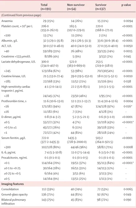
Total
(n=191) Non-survivor (n=54) Survivor (n=137) p value Demographics and clinical characteristics
Age, years 56·0 (46·0–67·0) 69·0 (63·0–76·0) 52·0 (45·0–58·0) <0·0001 Sex ·· ·· ·· 0·15 Female 72 (38%) 16 (30%) 56 (41%) ·· Male 119 (62%) 38 (70%) 81 (59%) ·· Exposure history 73 (38%) 14 (26%) 59 (43%) 0·028 Current smoker 11 (6%) 5 (9%) 6 (4%) 0·21 Comorbidity 91 (48%) 36 (67%) 55 (40%) 0·0010 Hypertension 58 (30%) 26 (48%) 32 (23%) 0·0008 Diabetes 36 (19%) 17 (31%) 19 (14%) 0·0051
Coronary heart disease 15 (8%) 13 (24%) 2 (1%) <0·0001
Chronic obstructive lung
disease 6 (3%) 4 (7%) 2 (1%) 0·047
Carcinoma 2 (1%) 0 2 (1%) 0·37
Chronic kidney disease 2 (1%) 2 (4%) 0 0·024
Other 22 (12%) 11 (20%) 11 (8%) 0·016
Respiratory rate
>24 breaths per min 56 (29%) 34 (63%) 22 (16%) <0·0001
Pulse ≥125 beats per min 2 (1%) 2 (4%) 0 0·024
Systolic blood pressure
<90 mm Hg 1 (1%) 0 1 (1%) 0·53 Fever (temperature ≥37·3°C) 180 (94%) 51 (94%) 129 (94%) 0·94 Cough 151 (79%) 39 (72%) 112 (82%) 0·15 Sputum 44 (23%) 14 (26%) 30 (22%) 0·55 Myalgia 29 (15%) 8 (15%) 21 (15%) 0·93 Fatigue 44 (23%) 15 (28%) 29 (21%) 0·33 Diarrhoea 9 (5%) 2 (4%) 7 (5%) 0·67 Nausea or vomiting 7 (4%) 3 (6%) 4 (3%) 0·40 SOFA score 2·0 (1·0–4·0) 4·5 (4·0–6·0) 1·0 (1·0–2·0) <0·0001 qSOFA score 1·0 (0·0–1·0) 1·0 (1·0–1·0) 0·0 (0·0–1·0) <0·0001 CURB-65 score 0·0 (0·0–2·0) 2·0 (1·0–3·0) 0·0 (0·0–1·0) <0·0001 0–1 141/188 (75%) 16 (30%) 125/134 (93%) <0·0001* 2 32/188 (17%) 23 (43%) 9/134 (7%) ·· 3–5 15/188 (8%) 15 (28%) 0/134 ··
Disease severity status ·· ·· ·· <0·0001
General 72 (38%) 0 72 (53%) ··
Severe 66 (35%) 12 (22%) 54 (39%) ··
Critical 53 (28%) 42 (78%) 11 (8%) ··
Time from illness onset to
hospital admission, days 11·0 (8·0–14·0) 11·0 (8·0–15·0) 11·0 (8·0–13·0) 0·53 Laboratory findings
White blood cell count,
× 10⁹ per L 6·2 (4·5–9·5) 9·8 (6·9–13·9) 5·2 (4·3–7·7) <0·0001 <4 32 (17%) 5 (9%) 27 (20%) <0·0001* 4–10 119 (62%) 24 (44%) 95 (69%) ·· >10 40 (21%) 25 (46%) 15 (11%) ·· Lymphocyte count, × 10⁹ per L 1·0 (0·6–1·3) 0·6 (0·5–0·8) 1·1 (0·8–1·5) <0·0001 <0·8 77 (40%) 41 (76%) 36 (26%) <0·0001 Haemoglobin, g/L 128·0 (119·0–140·0) (115·0–138·0)126·0 (120·0–140·0)128·0 0·30 (Table 1 continues on next page)
troponin I, creatine kinase, d-dimer, serum ferritin, and IL-6, might be unavailable in emergency circumstances. Therefore, we chose lymphocyte count, d-dimer, SOFA score, coronary heart disease, and age as the five variables for our multivariable logistic regression model.
We excluded variables from the univariable analysis if their between-group differences were not significant, if their accuracy was unconfirmed (eg, exposure, which was self-reported), if the number of events was too small to calculate odds ratios, and if they had colinearity with the SOFA score.
We compared patient characteristics between the two hospitals and used a generalised linear model to adjust for possible differences in patients’ characteristics and treatment between the two study centres.
A two-sided α of less than 0·05 was considered statis-tically significant. Statistical analyses were done using the SAS software (version 9.4), unless otherwise indicated.
Role of the funding source
The funder of the study had no role in study design, data collection, data analysis, data interpretation, or writing of the report. The corresponding authors (BC and HC) had full access to all the data in the study and had final responsibility for the decision to submit for publication.
Results
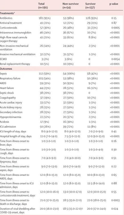
813 adult patientswere hospitalised in Jinyintan Hospital or Wuhan Pulmonary Hospital with COVID-19 before Jan 31, 2020. After excluding 613 patients that were still hospitalised or not confirmed by SARS-CoV-2 RNA detection as of Jan 31, 2020, and nine inpatients without available key information in their medical records, we included 191 inpatients (135 from Jinyintan Hospital and 56 from Wuhan Pulmonary Hospital) in the final analysis. 54 patients died during hospitalisation and 137 were discharged. The median age of the 191 patients was 56·0 years (IQR 46·0–67·0), ranging from 18 years to 87 years, and most patients were male (table 1). Co morbidities were present in nearly half of patients, with hypertension being the most common comorbidity, followed by diabetes and coronary heart disease (table 1). The most common symptoms on admission were fever and cough, followed by sputum production and fatigue (table 1). Lymphocytopenia occurred in 77 (40%) patients. 181 (95%) patients received antibiotics and 41 (21%) received antivirals (lopinavir/ritonavir; table 2). Systematic corticosteroid and intravenous immuno-globulin use differed significantly between non-survivors and survivors (table 2). The comparison of charac-teristics, treatment, and outcomes of patients from the two hospitals are shown in the appendix (pp 2–4).
The median time from illness onset (ie, before admission) to discharge was 22·0 days (IQR 18·0–25·0), whereas the median time to death was 18·5 days (15·0–22·0; table 2). 32 patients required invasive mechanical ventilation, of whom 31 (97%) died. The median time from illness onset to invasive mechanical ventilation was 14·5 days (12·0–19·0). Extracorporeal membrane oxygenation was used in three patients, none of whom survived. Sepsis was the most frequently observed complication, followed by respiratory failure, ARDS, heart failure, and septic shock (table 2). Half of non-survivors experienced a secondary infection, and ventilator-associated pneumonia occurred in ten (31%) of 32 patients requiring invasive mechanical ventilation. The frequency of complications were higher in non-survivors than survivors (table 2).
Total
(n=191) Non-survivor (n=54) Survivor (n=137) p value (Continued from previous page)
Anaemia 29 (15%) 14 (26%) 15 (11%) 0·0094
Platelet count, × 10⁹ per L 206·0
(155·0–262·0) (107·0–229·0)165·5 (168·0–271·0)220·0 <0·0001 <100 13 (7%) 11 (20%) 2 (1%) <0·0001 Albumin, g/L 32·3 (29·1–35·8) 29·1 (26·5–31·3) 33·6 (30·6–36·4) <0·0001 ALT, U/L 30·0 (17·0–46·0) 40·0 (24·0–51·0) 27·0 (15·0–40·0) 0·0050 >40 59/189 (31%) 26 (48%) 33/135 (24%) 0·0015 Creatinine >133 μmol/L 8/186 (4%) 5 (9%) 3/132 (2%) 0·045
Lactate dehydrogenase, U/L 300·0
(234·0–407·0) (363·0–669·0)521·0 (219·0–318·0)253·5 <0·0001
>245 123/184 (67%) 53 (98%) 70/130(54%) <0·0001
Creatine kinase, U/L 21·5 (13·0–72·4) 39·0 (19·5–151·0) 18·0 (12·5–52·1) 0·0010
>185 22/168 (13%) 11/52 (21%) 11/116 (9%) 0·038 High-sensitivity cardiac troponin I, pg/mL 4·1 (2·0–14·1) 22·2 (5·6–83·1) 3·0 (1·1–5·5) <0·0001 >28 24/145 (17%) 23/50 (46%) 1/95 (1%) <0·0001 Prothrombin time, s 11·6 (10·6–13·0) 12·1 (11·2–13·7) 11·4 (10·4–12·6) 0·0004 <16 171/182 (94%) 47 (87%) 124/128 (97%) 0·016* ≥16 11/182 (6%) 7 (13%) 4/128 (3%) ·· D-dimer, μg/mL 0·8 (0·4–3·2) 5·2 (1·5–21·1) 0·6 (0·3–1·0) <0·0001 ≤0·5 55/172 (32%) 4 (7%) 51/118 (43%) <0·0001* >0·5 to ≤1 45/172 (26%) 6 (11%) 39/118 (33%) ·· >1 72/172 (42%) 44 (81%) 28/118 (24%) ·· Serum ferritin, μg/L 722·0 (377·2–1435·3) (728·9–2000·0)1435·3 (264·0–921·5)503·2 <0·0001 >300 102/128 (80%) 44/46 (96%) 58/82 (71%) 0·0008 IL-6, pg/mL 7·4 (5·3–10·8) 11·0 (7·5–14·4) 6·3 (5·0–7·9) <0·0001 Procalcitonin, ng/mL 0·1 (0·1–0·1) 0·1 (0·1–0·5) 0·1 (0·1–0·1) <0·0001 <0·1 114/164 (70%) 19/51 (37%) 95/113 (84%) <0·0001* ≥0·1 to <0·25 30/164 (18%) 16/51 (31%) 14/113 (12%) ·· ≥0·25 to <0·5 6/164 (4%) 3/51 (6%) 3/113 (3%) ·· ≥0·5 14/164 (9%) 13/51 (25%) 1/113 (1%) ·· Imaging features Consolidation 112 (59%) 40 (74%) 72 (53%) 0·0065 Ground-glass opacity 136 (71%) 44 (81%) 92 (67%) 0·049 Bilateral pulmonary infiltration 143 (75%) 45 (83%) 98 (72%) 0·090
Data are median (IQR), n (%), or n/N (%). p values were calculated by Mann-Whitney U test, χ² test, or Fisher’s exact test, as appropriate. SOFA=Sequential Organ Failure Assessment. qSOFA=Quick SOFA. ALT=alanine aminotransferase. IL-6=interleukin-6. *χ² test comparing all subcategories.
Table 1: Demographic, clinical, laboratory, and radiographic findings of patients on admission
In univariable analysis, odds of in-hospital death was higher in patients with diabetes or coronary heart disease (table 3). Age, lymphopenia, leucocytosis, and elevated ALT, lactate dehydrogenase, high-sensitivity cardiac troponin I, creatine kinase, d-dimer, serum ferritin, IL-6, prothrombin time, creatinine, and procalcitonin were also associated with death (table 3).
We included 171 patients with complete data for all variables (53 non-survivors and 118 survivors) in the multivariable logistic regression model. We found that older age, higher SOFA score, and d-dimer greater than 1 μg/mL at admission were associated with increased odds of death (table 3). When adjusting for study centre, our generalised linear model showed similar results (appendix p 5).
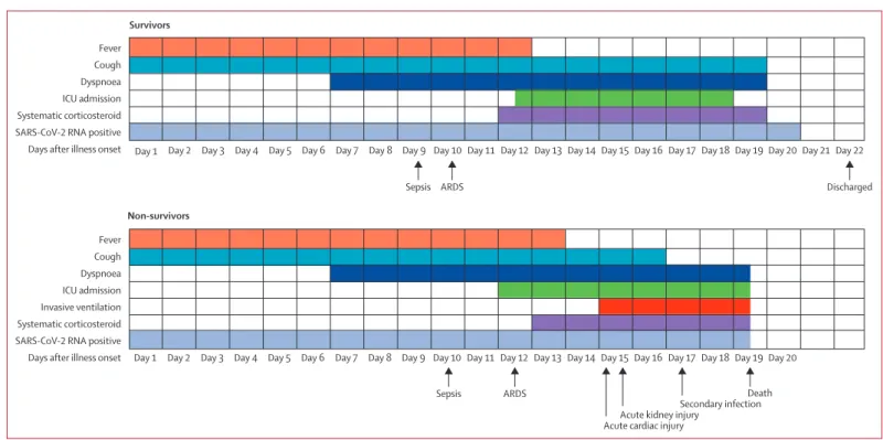
For survivors, the median duration of viral shedding was 20·0 days (IQR 17·0–24·0) from illness onset, but the virus was continuously detectable until death in non-survivors (table 2; figure 1). The shortest observed duration of viral shedding among survivors was 8 days, whereas the longest was 37 days. Among 29 patients who received lopinavir/ritonavir and were discharged, the median time from illness onset to initiation of antiviral treatment was 14·0 days (IQR 10·0–17·0) and the median duration of viral shedding was 22·0 days (18·0–24·0). The median duration of viral shedding was 19·0 days (17·0–22·0) in patients with severe disease status and 24·0 days (22·0–30·0) in patients with critical disease status.
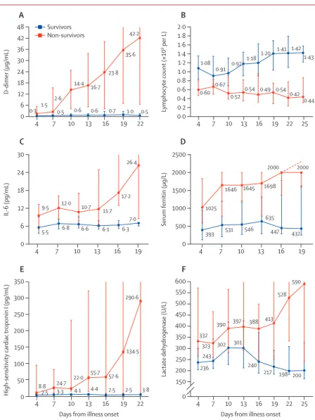
Major laboratory markers were tracked from illness onset (figure 2). Baseline lymphocyte count was signifi-cantly higher in survivors than non-survivors; in survivors, lymphocyte count was lowest on day 7 after illness onset and improved during hospitalisation, whereas severe lymphopenia was observed until death in non-survivors. Levels of d-dimer, high-sensitivity cardiac troponin I, serum ferritin, lactate dehydrogenase, and IL-6 were clearly elevated in non-survivors compared with survivors throughout the clinical course, and increased with illness deterioration (figure 2). In non-survivors, high-sensitivity cardiac troponin I increased rapidly from day 16 after disease onset, whereas lactate dehydrogenase increased for both survivors and non-survivors in the early stage of illness, but decreased from day 13 for survivors.
Median time from illness onset to dyspnoea was similar in survivors and non-survivors, with a median duration of dyspnoea of 13·0 days (9·0–16·5) for survivors (table 2; figure 1). In survivors, the median duration of fever was 12·0 days (8·0–13·0) and cough persisted for 19·0 days (IQR 12·0–23·0; figure 1). 62 (45%) survivors still had cough on discharge and 39 (72%) non-survivors still had cough at the time of death. The dynamic profiles of fever, cough, and dyspnoea are shown in the appendix (p 6). Sepsis developed at a median of 9·0 days (7·0–13·0) after illness onset among all patients, followed by ARDS (12·0 days [8·0–15·0]), acute cardiac injury (15·0 days [10·0–17·0]), acute kidney injury (15·0 days [13·0–19·5]), and secondary infection (17·0 days [13·0–19·0]). The
initiation time and duration of systematic corticosteroid use was also similar between the two groups. Among non-survivors, the median time from illness onset was 10·0 days (7·0–14·0) to sepsis, 12·0 days (8·0–15·0) to ARDS, 14·5 days (9·5–17·0) to acute cardiac injury, and
Total
(n=191) Non-survivor (n=54) Survivor (n=137) p value Treatments*
Antibiotics 181 (95%) 53 (98%) 128 (93%) 0·15
Antiviral treatment 41 (21%) 12 (22%) 29 (21%) 0·87
Corticosteroids 57 (30%) 26 (48%) 31 (23%) 0·0005
Intravenous immunoglobin 46 (24%) 36 (67%) 10 (7%) <0·0001
High-flow nasal cannula
oxygen therapy 41 (21%) 33 (61%) 8 (6%) <0·0001
Non-invasive mechanical
ventilation 26 (14%) 24 (44%) 2 (1%) <0·0001
Invasive mechanical ventilation 32 (17%) 31 (57%) 1 (1%) <0·0001
ECMO 3 (2%) 3 (6%) 0 0·0054
Renal replacement therapy 10 (5%) 10 (19%) 0 <0·0001
Outcomes Sepsis 112 (59%) 54 (100%) 58 (42%) <0·0001 Respiratory failure 103 (54%) 53 (98%) 50 (36%) <0·0001 ARDS 59 (31%) 50 (93%) 9 (7%) <0·0001 Heart failure 44 (23%) 28 (52%) 16 (12%) <0·0001 Septic shock 38 (20%) 38 (70%) 0 <0·0001 Coagulopathy 37 (19%) 27 (50%) 10 (7%) <0·0001
Acute cardiac injury 33 (17%) 32 (59%) 1 (1%) <0·0001
Acute kidney injury 28 (15%) 27 (50%) 1 (1%) <0·0001
Secondary infection 28 (15%) 27 (50%) 1 (1%) <0·0001
Hypoproteinaemia 22 (12%) 20 (37%) 2 (1%) <0·0001
Acidosis 17 (9%) 16 (30%) 1 (1%) <0·0001
ICU admission 50 (26%) 39 (72%) 11 (8%) <0·0001
ICU length of stay, days 8·0 (4·0–12·0) 8·0 (4·0–12·0) 7·0 (2·0–9·0) 0·41 Hospital length of stay, days 11·0 (7·0–14·0) 7·5 (5·0–11·0) 12·0 (9·0–15·0) <0·0001 Time from illness onset to
fever, days 1·0 (1·0–1·0) 1·0 (1·0–1·0) 1·0 (1·0–1·0) 0·16
Time from illness onset to
cough, days 1·0 (1·0–3·0) 1·0 (1·0–1·0) 1·0 (1·0–4·0) 0·30
Time from illness onset to
dyspnoea, days 7·0 (4·0–9·0) 7·0 (4·0–10·0) 7·0 (4·0–9·0) 0·51 Time from illness onset to
sepsis, days 9·0 (7·0–13·0) 10·0 (7·0–14·0) 9·0 (7·0–12·0) 0·22 Time from illness onset to
ARDS, days 12·0 (8·0–15·0) 12·0 (8·0–15·0) 10·0 (8·0–13·0) 0·65 Time from illness onset to ICU
admission, days 12·0 (8·0–15·0) 12·0 (8·0–15·0) 11·5 (8·0–14·0) 0·88 Time from illness onset to
corticosteroids treatment, days 12·0 (10·0–16·0) 13·0 (10·0–17·0) 12·0 (10·0–15·0) 0·55 Time from illness onset to
death or discharge, days 21·0 (17·0–25·0) 18·5 (15·0–22·0) 22·0 (18·0–25·0) 0·0003 Duration of viral shedding after
COVID-19 onset, days 20·0 (16·0–23·0) 18·5 (15·0–22·0)† 20·0 (17·0–24·0) 0·024 Data are median (IQR) or n (%). p values were calculated by Mann-Whitney U test, χ² test, or Fisher’s exact test, as appropriate. ECMO=extracorporeal membrane oxygenation. ARDS=acute respiratory distress syndrome. ICU=intensive care unit. COVID-19=coronavirus disease 2019. *Ordered by escalating scale of respiratory support. †Detectable until death.
17·0 days (13·0–19·0) to secondary infection (figure 1; table 2). Among survivors, secondary infection, acute kidney injury, and acute cardiac injury were observed in one patient each, occurring 9 days (acute kidney injury), 14 days (secondary infection), and 21 days (acute cardiac injury) after illness onset.
The median time from dyspnoea to intubation was 10·0 days (IQR 5·0–12·5) for patients who received invasive mechanical ventilation and the time from inva-sive mechanical ventilation to occurrence of ventilator-associated pneumonia was 8·0 days (2·0–9·0; figure 1).
Discussion
This retrospective cohort study identified several risk factors for death in adults in Wuhan who were
hospitalised with COVID-19. In particular, older age, d-dimer levels greater than 1 μg/mL, and higher SOFA score on admission were associated with higher odds of in-hospital death. Additionally, elevated levels of blood IL-6, high-sensitivity cardiac troponin I, and lactate dehydrogenase and lymphopenia were more commonly seen in severe COVID-19 illness. Sustained viral detection in throat samples was observed in both survivors and non-survivors.
Previously, older age has been reported as an impor-tant independent predictor of mortality in SARS and MERS.14,15 The current study confirmed that increased age was associated with death in patients with COVID-19. Previous studies in macaques inoculated with SARS-CoV found that older macaques had stronger host innate responses to virus infection than younger adults, with an Univariable OR
(95% CI) p value Multivariable OR (95% CI) p value Demographics and clinical characteristics
Age, years* 1·14 (1·09–1·18) <0·0001 (1·03–1·17)1·10 0·0043 Female sex (vs male) (0·31–1·20)0·61 0·15 ·· ·· Current smoker (vs non-smoker) 2·23 (0·65–7·63) 0·20 ·· ··
Comorbidity present (vs not present) Chronic obstructive lung disease 5·40 (0·96–30·40) 0·056 ·· ·· Coronary heart disease (4·64–98·76)21·40 <0·0001 (0·26–17·79)2·14 0·48 Diabetes 2·85 (1·35–6·05) 0·0062 ·· ·· Hypertension 3·05 (1·57–5·92) 0·0010 ·· ··
Respiratory rate, breaths per min
≤24 1 (ref) ·· ·· ·· >24 8·89 (4·34–18·19) <0·0001 ·· ·· SOFA score 6·14 (3·48–10·85) <0·0001 (2·61–12·23)5·65 <0·0001 qSOFA score 12·00 (5·06–28·43) <0·0001 ·· ·· Laboratory findings
White blood cell count, × 10⁹ per L
<4 0·73 (0·26–2·10) 0·56 ·· ·· 4–10 1 (ref) ·· ·· ·· >10 6·60 (3·02–14·41) <0·0001 ·· ·· Lymphocyte count, × 10⁹ per L* 0·02 (0·01–0·08) <0·0001 (0·02–1·62)0·19 0·13 ALT, U/L ≤40 1 (ref) ·· ·· ·· >40 2·87 (1·48–5·57) 0·0018 ·· ··
(Table 3 continues in next column)
Univariable OR
(95% CI) p value Multivariable OR (95% CI) p value (Continued from previous column)
Creatinine, μmol/L
≤133 1 (ref) ·· ·· ··
>133 4·39
(1·01–19·06) 0·048 ·· ··
Lactate dehydrogenase, U/L
≤245 1 (ref) ·· ·· ··
>245 45·43
(6·10–338·44) 0·0002 ·· ··
Creatine kinase, U/L
≤185 1 (ref) ·· ·· ··
>185 2·56
(1·03–6·36) 0·043 ·· ··
High-sensitivity cardiac troponin I, pg/mL
≤28 1 (ref) ·· ·· ·· >28 80·07 (10·34–620·36) <0·0001 ·· ·· D-dimer, μg/mL ≤0·5 1 (ref) ·· 1 (ref) ·· > 0·5 1·96 (0·52–7·43) 0·32 (0·21–21·39)2·14 0·52 > 1 20·04 (6·52–61·56) <0·0001 (2·64–128·55)18·42 0·0033 Prothrombin time, s <16 1 (ref) ·· ·· ·· ≥16 4·62 (1·29–16·50) 0·019 ·· ·· Serum ferritin, μg/L ≤300 1 (ref) ·· ·· ·· >300 9·10 (2·04–40·58) 0·0038 ·· ·· IL-6, pg/mL* 1·12 (1·03–1·23) 0·0080 ·· ·· Procalcitonin, ng/mL* (1·81–104·40)13·75 0·011 ·· ··
OR=odds ratio. S OFA=Sequential Organ Failure Assessment. qSOFA=Quick SOFA. ALT=alanine aminotransferase. IL-6=interleukin-6. *Per 1 unit increase. Table 3: Risk factors associated with in-hospital death
increase in differential expression of genes associated with inflammation, whereas expression of type I inter-feron beta was reduced.16 The age-dependent defects in T-cell and B-cell function and the excess production of type 2 cytokines could lead to a deficiency in control of viral replication and more prolonged proinflammatory responses, potentially leading to poor outcome.17
SOFA score is a good diagnostic marker for sepsis and septic shock, and reflects the state and degree of multi-organ dysfunction.18,19 Although bacterial infec-tions are usually regarded as a leading cause of sepsis, viral infection can also cause sepsis syndrome. Previ-ously, we determined that sepsis occurred in nearly 40% of adults with community-acquired pneumonia due to viral infection.20 In the current study, we found that more than half of patients developed sepsis. Additionally, we found that more than 70% of patients had white blood cell count below 10·0 × 10⁹ per Lor procalcitonin below 0·25 ng/mL, and no bacterial pathogens were detected in these patients on admission. Sepsis was a common complication, which might be directly caused by SARS-CoV-2 infection, but further research is needed to investigate the pathogenesis of sepsis in COVID-19 illness.
Cardiac complications, including new or worsening heart failure, new or worsening arrhythmia, or myocardial infarction are common in patients with pneumonia. Cardiac arrest occurs in about 3% of inpatients with pneumonia.21 Risk factors of cardiac events after
pneumonia include older age, pre-existing cardiovascular diseases, and greater severity of pneumonia at presen-tation.22 Coronary heart disease has also been found to be associated with acute cardiac events and poor outcomes in influenza and other respiratory viral infections.22–24 In this study, increased high-sensitivity cardiac troponin I during hospitalisation was found in more than half of those who died. The first autopsy of a 53-year-old woman with chronic renal failure in Jinyintan Hospital showed acute myocardial infarction (data not published; personal communication with a pathologist from the Chinese Academy of Science). About 90% of inpatients with pneumonia had increased coagulation activity, marked by increased d-dimer concentrations.25 In this study, we found d-dimer greater than 1 µg/mL is associated with fatal outcome of COVID-19. High levels of d-dimer have a reported asso ciation with 28-day mortality in patients with infection or sepsis identified in the emergency department.26 Contri butory mechanisms include systemic pro-inflammatory cytokine responses that are mediators of atherosclerosis directly contributing to plaque rupture through local inflammation, induction of procoagulant factors, and haemodynamic changes, which predispose to ischaemia and thrombosis.27–29 In addition, angiotensin converting enzyme 2, the receptor for SARS-CoV-2, is expressed on myocytes and vascular endothelial cells,30,31 so there is at least theoretical potential possibility of direct cardiac involvement by the virus. Of note, interstitial mononuclear inflammatory infiltrates in heart tissue has
Figure 1: Clinical courses of major symptoms and outcomes and duration of viral shedding from illness onset in patients hospitalised with COVID-19
Figure shows median duration of symptoms and onset of complications and outcomes. ICU=intensive care unit. SARS-CoV-2=severe acute respiratory syndrome coronavirus 2. ARDS=acute respiratory distress syndrome. COVID-19=coronavirus disease 2019.
Fever Cough Dyspnoea ICU admission Systematic corticosteroid SARS-CoV-2 RNA positive
Days after illness onset Day 1
Survivors
Day 2 Day 3 Day 4 Day 5 Day 6 Day 7 Day 8 Day 9 Day 10 Day 11 Day 12 Day 13 Day 14 Day 15 Day 16 Day 17 Day 18 Day 19 Day 20 Day 21 Day 22
Fever Cough Dyspnoea ICU admission Invasive ventilation Systematic corticosteroid SARS-CoV-2 RNA positive
Days after illness onset Day 1
Non-survivors
Day 2 Day 3 Day 4 Day 5 Day 6 Day 7 Day 8 Day 9 Day 10 Day 11 Day 12 Day 13 Day 14 Day 15 Day 16 Day 17 Day 18 Day 19 Day 20
Sepsis ARDS Discharged
Sepsis ARDS
Acute kidney injury
Death Secondary infection Acute cardiac injury
been documented in fatal cases of COVID-19, although viral detection studies were not reported.32
The level and duration of infectious virus replication are important factors in assessing the risk of trans mission and guiding decisions regarding isolation of patients. Because coronavirus RNA detection is more sensitive than virus isolation, most studies have used qualitative or quantitative viral RNA tests as a potential marker for infectious coronavirus. For SARS-CoV, viral RNA was detected in respiratory specimens from about a third of patients as long as 4 weeks after disease onset.33 Similarly, the dura tion of MERS-CoV RNA detection in lower respiratory specimans persisted for at least 3 weeks,34,35 whereas the duration of SARS-CoV-2 RNA detection has not been well characterised.
In the current study, we found that the detectable SARS-CoV-2 RNA persisted for a median of 20 days in survivors and that it was sustained until death in non-survivors. This has important implications for both patient isolation decision making and guidance around the length of antiviral treatment. In severe influenza virus infection, prolonged viral shedding was associated with fatal outcome and delayed antiviral treatment was an independent risk factor for prolonged virus detection.36 Similarly, effective antiviral treatment might improve outcomes in COVID-19, although we did not observe shortening of viral shedding duration after lopinavir/ritonavir treatment in the cur-rent study. Ran domised clinical trials for lopinavir/ ritonavir (ChiCTR2000029308) and intravenous remdesivir (NCT04257656, NCT04252664) in treatment of COVID-19 are currently in progress.
Our study has some limitations. First, due to the retrospective study design, not all laboratory tests were done in all patients, including lactate dehydrogenase, IL-6, and serum ferritin. Therefore, their role might be underestimated in predicting in-hospital death. Second, patients were sometimes transferred late in their illness to the two included hospitals. Lack of effective antivirals, inadequate adherence to standard supportive therapy, and high-dose corticosteroid use might have also contributed to the poor clinical outcomes in some patients. Third, the estimated duration of viral shedding is limited by the frequency of respiratory specimen collection, lack of quantitative viral RNA detection, and relatively low positive rate of SARS-CoV-2 RNA detection in throat-swabs.37 Fourth, by excluding patients still in hospital as of Jan 31, 2020, and thus relatively more severe disease at an earlier stage, the case fatality ratio in our study cannot reflect the true mortality of COVID-19. Last but not least, interpretation of our findings might be limited by the sample size. However, by including all adult patients in the two designated hospitals for COVID-19, we believe our study population is representative of cases diagnosed and treated in Wuhan.
To the best of our knowledge, this is the largest retro-spective cohort study among patients with COVID-19 who have experienced a definite outcome. We found that older age, higher SOFA score, and elevated d-dimer at admission were risk factors for death of adult patients with COVID-19. The prolonged viral shedding provides the rationale for testing novel coronavirus antiviral inter-ventions in efforts to improve outcomes.
Contributors
BC and HC had the idea for and designed the study and had full access to all of the data in the study and take responsibility for the integrity of the data and the accuracy of the data analysis. BC, FZ, GF, ZL, JXi, and YL drafted the paper. BC, FZ, RD, GF, ZL, XG, HL, and YWa did the analysis, and all authors critically revised the manuscript for important intellectual content and gave final approval for the version to be published. FZ, RD, GF, ZL, YL, BS, LG, YWe, XW, JXu, ST, and YZ collected the data. All authors agree to be accountable for all aspects of the work in ensuring that questions related to the accuracy or integrity of any part of the work are appropriately investigated and resolved.
Figure 2: Temporal changes in laboratory markers from illness onset in patients hospitalised with COVID-19
Figure shows temporal changes in d-dimer (A), lymphocytes (B), IL-6 (C), serum ferritin (D), high-sensitivity cardiac troponin I (E), and lactate dehydrogenase (F). Differences between survivors and non-survivors were significant for all timepoints shown, except for day 4 after illness onset for d-dimer, IL-6, and high-sensitivity cardiac troponin I. For serum ferritin (D), the median values after day 16 exceeded the upper limit of detection, as indicated by the dashed line. COVID-19=coronavirus disease 2019. IL-6=interleukin-6.
4 7 10 13 16 19 22 4 7 10 13 16 19 22 25
0
High-sensitivity cardiac
troponin I (pg/mL)
Days from illness onset
0 50 200 150 250 100 300 150 350 200 400 250 450 300 500 550 350 600 E F Lactate dehydrogenase (U/L ) 4 7 10 13 16 19 4 7 10 13 16 19 0 IL -6 (pg/mL) 0 6 500 12 1000 18 24 1500 30 2000 2500 C D Serum ferritin (µg/L)
Days from illness onset
4 7 10 13 16 19 22 4 7 10 13 16 19 22 25 0 D-dimer (µg/mL) 0·0 6 0·40·2 0·6 12 0·8 18 1·0 24 1·2 30 1·4 36 1·6 42 1·8 48 2·0 A B Lymphocyte count (×10 9 per L) 2·5 5·5 6·8 6·6 6·1 7·0 6·3 236 332 323 243 390 302 397 388 198 590 200 301 413 528 217 240 8·8 9·5 1·5 0·5 2·6 14·4 16·7 0·6 0·7 1·0 0·5 0·60 0·67 0·52 0·54 0·49 0·540·42 0·44 1·08 0·91 0·97 1·18 1·20 1·41 1·42 1·43 42·2 35·6 23·8 0·6 0·3 12·0 10·7 11·7 17·2 26·4 1025 1646 1645 1698 2000 2000 432 635 447 546 531 393 22·0 55·7 57·6 134·5 290·6 24·7 4·1 4·4 2·5 2·5 3·8 3·3 Survivors Non-survivors
Declaration of interests
We declare no competing interests. Acknowledgments
This study received the following funding: Chinese Academy of Medical Sciences Innovation Fund for Medical Sciences (CIFMS
2018-I2M-1–003); National Science Grant for Distinguished Young Scholars (81425001/H0104); National Key Research and Development Program of China (2018YFC1200100, 2018YFC1200102); The Beijing Science and Technology Project (Z19110700660000); Major Projects of National Science and Technology on New Drug Creation and Development (2020ZX09201001). We acknowledge all health-care workers involved in the diagnosis and treatment of patients in Wuhan; and we thank Chen Wang (Peking Union Medical College, Beijing, China), Peter W Horby (University of Oxford, Oxford, UK), and Frederick G Hayden (University of Virginia School of Medicine, Charlottesville, VA, USA) for guidance in study design and interpretation of results, and review of the manuscript. References
1 Phelan AL, Katz R, Gostin LO. The novel coronavirus originating in Wuhan, China: challenges for global health governance. JAMA 2020; published online Jan 30. DOI:10.1001/jama.2020.1097. 2 Gorbalenya AE, Baker SC, Baric RS, et al. Severe acute respiratory
syndrome-related coronavirus: the species and its viruses—a statement of the Coronavirus Study Group. bioRxiv 2020; published online Feb 11. DOI:10.1101/2020.02.07.937862 (preprint).
3 Chan JWM, Ng CK, Chan YH, et al. Short term outcome and risk factors for adverse clinical outcomes in adults with severe acute respiratory syndrome (SARS). Thorax 2003; 58: 686–89.
4 Li Q, Guan X, Wu P, et al. Early transmission dynamics in Wuhan, China, of novel coronavirus-infected pneumonia. N Engl J Med 2020; published online Jan 29. DOI:10.1056/NEJMoa2001316. 5 Huang C, Wang Y, Li X, et al. Clinical features of patients infected
with 2019 novel coronavirus in Wuhan, China. Lancet 2020; 395: 497–506.
6 Wang D, Hu B, Hu C, et al. Clinical characteristics of 138 hospitalized patients with 2019 novel coronavirus-infected pneumonia in Wuhan, China. JAMA 2020; published online Feb 7. DOI:10.1001/jama.2020.1585.
7 Chen N, Zhou M, Dong X, et al. Epidemiological and clinical characteristics of 99 cases of 2019 novel coronavirus pneumonia in Wuhan, China: a descriptive study. Lancet 2020; 395: 507–13. 8 Kalil AC, Metersky ML, Klompas M, et al. Management of adults with
hospital-acquired and ventilator-associated pneumonia: 2016 clinical practice guidelines by the Infectious Diseases Society of America and the American Thoracic Society. Clin Infect Dis 2016; 63: e61–111. 9 Khwaja A. KDIGO clinical practice guidelines for acute kidney
injury. Nephron Clin Pract 2012; 120: c179–84.
10 Ranieri VM, Rubenfeld GD, Thompson BT, et al. Acute respiratory distress syndrome: the Berlin Definition. JAMA 2012; 307: 2526–33. 11 National Health Commission of the People’s Republic of China.
Chinese management guideline for COVID-19 (version 6.0). Feb 19, 2020. http://www.nhc.gov.cn/yzygj/s7653p/202002/8334a832 6dd94d329df351d7da8aefc2/files/b218cfeb1bc54639af227f922bf6b817. pdf (accessed Feb 19, 2020; in Chinese).
12 Yang X, Yu Y, Xu J, et al. Clinical course and outcomes of critically ill patients with SARS-CoV-2 pneumonia in Wuhan, China: a single-centered, retrospective, observational study.
Lancet Respir Med 2020; published online Feb 24. https://doi.org/
10.1016/S2213-2600(20)30079-5.
13 Arabi YM, Balkhy HH, Hayden FG, et al. Middle East respiratory syndrome. N Engl J Med 2017; 376: 584–94.
14 Choi KW, Chau TN, Tsang O, et al. Outcomes and prognostic factors in 267 patients with severe acute respiratory syndrome in Hong Kong. Ann Intern Med 2003; 139: 715–23.
15 Hong K-H, Choi J-P, Hong S-H, et al. Predictors of mortality in Middle East respiratory syndrome (MERS). Thorax 2018; 73: 286–89.
16 Smits SL, de Lang A, van den Brand JMA, et al. Exacerbated innate host response to SARS-CoV in aged non-human primates.
PLoS Pathog 2010; 6: e1000756-e.
17 Opal SM, Girard TD, Ely EW. The immunopathogenesis of sepsis in elderly patients. Clin Infect Dis 2005; 41 (suppl 7): S504–12. 18 Singer M, Deutschman CS, Seymour CW, et al. The Third
International Consensus Definitions for Sepsis and Septic Shock (Sepsis-3). JAMA 2016; 315: 801–10.
19 Ferreira FL, Bota DP, Bross A, Mélot C, Vincent JL. Serial evaluation of the SOFA score to predict outcome in critically ill patients. JAMA 2001; 286: 1754–58.
20 Zhou F, Wang Y, Liu Y, et al. Disease severity and clinical outcomes of community-acquired pneumonia caused by non-influenza respiratory viruses in adults: a multicentre prospective registry study from the CAP-China Network. Eur Respir J 2019; 54: 1802406. 21 Marrie TJ, Shariatzadeh MR. Community-acquired pneumonia
requiring admission to an intensive care unit: a descriptive study.
Medicine (Baltimore) 2007; 86: 103–11.
22 Corrales-Medina VF, Musher DM, Shachkina S, Chirinos JA. Acute pneumonia and the cardiovascular system. Lancet 2013; 381: 496–505.
23 Udell JA, Zawi R, Bhatt DL, et al. Association between influenza vaccination and cardiovascular outcomes in high-risk patients: a meta-analysis. JAMA 2013; 310: 1711–20.
24 Blackburn R, Zhao H, Pebody R, Hayward A, Warren-Gash C. Laboratory-confirmed respiratory infections as predictors of hospital admission for myocardial infarction and stroke: time-series analysis of English data for 2004–2015. Clin Infect Dis 2018; 67: 8–17. 25 Milbrandt EB, Reade MC, Lee M, et al. Prevalence and significance
of coagulation abnormalities in community-acquired pneumonia.
Mol Med 2009; 15: 438–45.
26 Rodelo JR, De la Rosa G, Valencia ML, et al. D-dimer is a significant prognostic factor in patients with suspected infection and sepsis.
Am J Emerg Med 2012; 30: 1991–99.
27 Smeeth L, Thomas SL, Hall AJ, Hubbard R, Farrington P, Vallance P. Risk of myocardial infarction and stroke after acute infection or vaccination. N Engl J Med 2004; 351: 2611–18. 28 Corrales-Medina VF, Musher DM, Wells GA, Chirinos JA, Chen L,
Fine MJ. Cardiac complications in patients with community-acquired pneumonia: incidence, timing, risk factors, and association with short-term mortality. Circulation 2012; 125: 773–81.
29 Davidson JA, Warren-Gash C. Cardiovascular complications of acute respiratory infections: current research and future directions.
Expert Rev Anti Infect Ther 2019; 17: 939–42.
30 Gallagher PE, Ferrario CM, Tallant EA. Regulation of ACE2 in cardiac myocytes and fibroblasts. Am J Physiol Heart Circ Physiol 2008; 295: H2373–79.
31 Mendoza-Torres E, Oyarzún A, Mondaca-Ruff D, et al. ACE2 and vasoactive peptides: novel players in cardiovascular/renal remodeling and hypertension. Ther Adv Cardiovasc Dis 2015; 9: 217–37.
32 Xu Z, Shi L, Wang Y, et al. Pathological findings of COVID-19 associated with acute respiratory distress syndrome. Lancet Respir Med 2020; published online Feb 18. https://doi.org/10.1016/
S2213-2600(20)30076-X.
33 Xu D, Zhang Z, Jin L, et al. Persistent shedding of viable SARS-CoV in urine and stool of SARS patients during the convalescent phase.
Eur J Clin Microbiol Infect Dis 2005; 24: 165–71.
34 Corman VM, Albarrak AM, Omrani AS, et al. Viral shedding and antibody response in 37 patients with Middle East Respiratory Syndrome coronavirus infection. Clin Infect Dis 2016; 62: 477–83. 35 Oh MD, Park WB, Choe PG, et al. Viral load kinetics of MERS
coronavirus infection. N Engl J Med 2016; 375: 1303–05.
36 Wang Y, Guo Q, Yan Z, et al. Factors associated with prolonged viral shedding in patients with avian influenza A(H7N9) virus infection.
J Infect Dis 2018; 217: 1708–17.
37 Zou L, Ruan F, Huang M, et al. SARS-CoV-2 viral load in upper respiratory specimens of infected patients. N Engl J Med 2020; published online Feb 19. DOI:10.1056/NEJMc2001737.


