HAL Id: hal-01926568
https://hal.archives-ouvertes.fr/hal-01926568
Submitted on 19 Nov 2018
HAL is a multi-disciplinary open access
archive for the deposit and dissemination of
sci-entific research documents, whether they are
pub-lished or not. The documents may come from
teaching and research institutions in France or
abroad, or from public or private research centers.
L’archive ouverte pluridisciplinaire HAL, est
destinée au dépôt et à la diffusion de documents
scientifiques de niveau recherche, publiés ou non,
émanant des établissements d’enseignement et de
recherche français ou étrangers, des laboratoires
publics ou privés.
The integral membrane S-locus receptor kinase of
Brassica has serinethreonine kinase activity in a
membranous environment and spontaneously forms
oligomers in planta
Jean-Loïc Giranton, Christian Dumas, J. Mark Cock, T. Gaude
To cite this version:
Jean-Loïc Giranton, Christian Dumas, J. Mark Cock, T. Gaude.
The integral membrane
S-locus receptor kinase of Brassica has serinethreonine kinase activity in a membranous
environ-ment and spontaneously forms oligomers in planta. Proceedings of the National Academy of
Sci-ences of the United States of America , National Academy of SciSci-ences, 2000, 97 (7), pp.3759-3764.
�10.1073/pnas.97.7.3759�. �hal-01926568�
The integral membrane
S-locus receptor kinase of
Brassica has serine
兾threonine kinase activity in a
membranous environment and spontaneously
forms oligomers
in planta
Jean-Loïc Giranton, Christian Dumas, J. Mark Cock, and Thierry Gaude*
Laboratoire de Reproduction et De´veloppement des Plantes, UMR 5667, Centre National de la Recherche Scientifique-Institut National de la Recherche Agronomique-Ecole Normale Supe´rieure, 46, alle´e d’Italie, 69364 Lyon Cedex 07, France
Communicated by Roland Douce, University of Grenoble, Grenoble, France, January 19, 2000 (received for review February 18, 1999)
To gain further insight into the mode of action of S-locus receptor kinase (SRK), a receptor-like kinase involved in the self-incompat-ibility response in Brassica, different recombinant SRK proteins have been expressed in a membranous environment using the insect cell兾baculovirus system. Recombinant SRK proteins exhib-ited properties close to those of the endogenous stigmatic SRK protein and were found to autophosphorylate on serine and threonine residues in insect cell microsomes. Autophosphorylation was constitutive because it did not require the presence of pollen or stigma extracts in the phosphorylation buffer. Phosphorylation was shown to occur in trans, suggesting the existence of consti-tutive homooligomers of membrane-anchored recombinant SRK. To investigate the physiological relevance of these results, we have examined the oligomeric status of SRK in planta in cross-linking experiments and by velocity sedimentation on sucrose gradients. Our data strongly suggest that SRK is associated both with other SRK molecules and other stigma proteins in nonpollinated flowers. These findings may have important implications for our under-standing of self-pollen signaling.
baculovirus兩 oligomerization 兩 sporophytic self-incompatibility
I
n the Brassica family, the cell–cell interaction that leads to the rejection of pollen at the stigmatic surface [the self-incompatibility (SI) reaction] is controlled genetically by the multiallelic S-locus (1). When the pollen parent and the stigma share common S haplotypes, pollen germination or pollen tube growth is inhibited, thereby preventing self-fertilization. Several genes have been localized at the S locus but only two, the S-locus glycoprotein (SLG) and the S-locus receptor kinase (SRK) genes, exhibit the polymorphic nature expected for genes in-volved in the SI response (2, 3). SLG encodes a secreted glycoprotein that accumulates in the cell wall of papillae (4), whereas SRK encodes a plasmalemma-anchored glycoprotein (5, 6). SRK is structurally analogous to animal receptor kinases and belongs to the plant receptor-like kinase (RLK) family (7). Although its membrane topology has not been experimentally defined, DNA sequence analysis predicts that SRK consists of three domains: an extracellular glycosylated N-terminal domain (the S-domain) that shares extensive homology with SLG, a membrane-spanning domain, and a cytoplasmic domain (3). The cytoplasmic domain has been shown to have serine兾threonine kinase activity when expressed in bacteria (8). In the S3haplo-type, SRK has been shown to encode, in addition to the integral membrane SRK protein, a soluble truncated form corresponding to the S-domain of SRK, the eSRK protein (9).
Although there is some controversy as to whether SLG is required for the SI response (10, 11), there is strong evidence that SRK is necessary. For example, SRK was shown to be mutated in two Brassica lines that exhibited a self-compatible phenotype (12, 13). Moreover, Stahl and coworkers (14) recently
have shown that the SI phenotype was altered in transgenic
Brassica napus plants expressing a kinase-defective SRK that
seems to act as a dominant negative mutant. Their results strongly indicate that SRK is a key component of the SI reaction. By analogy with animal receptor kinases, which transduce signals after interaction with peptidic extracellular ligands, it has been proposed that SRK initiates the SI reaction after the interaction of its S-domain with an as-yet-unknown self-pollen-borne ligand. Although candidates have been proposed for the male component of the SI response, there is not yet any molecular information concerning this molecule (15–17). The recognition by SRK of its putative ligand is thought to induce an activation of the kinase domain, leading to the recruitment of cytoplasmic targets that mediate the SI reaction. Two types of
Brassica proteins have been shown to interact with the
cytoplas-mic domain of SRK: two thioredoxins and an arm repeat-containing protein (ARC1) (18, 19). The kinase interaction domains of the kinase-associated protein phosphatases of
Ara-bidopsis thaliana and maize also were shown to interact with the
kinase domain of SRK (20). ARC1 is perhaps the most inter-esting of these proteins because it is expressed specifically in
Brassica stigmas and it interacts only with the
autophosphory-lated form of the SRK cytoplasmic domain.
Very little is known about the behavior of SRK in the plasma membrane, and particularly whether it can form homo- or hetero-oligomers. This is a crucial question because of the important role that oligomerization has been shown to play in the activation of receptor kinases in animals (21, 22). Here we show that recombinant integral membrane SRK protein expressed in the insect cell兾baculovirus system is able to autophosphorylate. Autophosphorylation does not require the presence of a ligand and occurs at least partially by transphosphorylation mecha-nisms, indicating that recombinant SRK oligomerizes in the membrane. Moreover, we report biochemical evidence, based on cross-linking experiments and velocity sedimentation on sucrose gradients, that oligomeric complexes containing SRK occur naturally in planta at the stigmatic surface of mature unpolli-nated Brassica flowers. Current models, based on the structure of S locus genes, propose that self-pollen signaling is mediated by a SRK receptor complex. Our results provide physical evi-dence for the existence of an SRK receptor complex in planta.
Abbreviations: Ni-NTA, nickel chelated by nitrilotetra-acetic acid; RLK, receptor-like kinase;
Sf21, cell line derived from Spodoptera frugiperda; SI, self-incompatibility; SLG, S-locus
glycoprotein; SRK, S-locus receptor kinase; HA, hemagglutinin.
*To whom reprint requests should be addressed. E-mail: thierry.gaude@ens-lyon.fr. The publication costs of this article were defrayed in part by page charge payment. This article must therefore be hereby marked “advertisement” in accordance with 18 U.S.C. §1734 solely to indicate this fact.
Article published online before print: Proc. Natl. Acad. Sci. USA, 10.1073兾pnas.070025097. Article and publication date are at www.pnas.org兾cgi兾doi兾10.1073兾pnas.070025097
PLANT
Materials and Methods
Viral and Plasmid DNA Constructs.Sequences encoding epitope tags
were inserted into the reading frame of SRK3(5) at a ClaI site
13 bp upstream of the termination codon so that the expressed proteins consisted of the reconstructed SRK polypeptide directly followed by either a hexahistidine sequence or the hemagglutinin (HA) epitope (YPYDVPDYA; ref. 23). The kinase-negative form of SRK was created by converting Lys-553 to an arginine codon by using site-specific mutagenesis PCR. SRK constructs were cloned into pBacPAK (CLONTECH) downstream of the polyhedrin promoter and cotransfected with Bsu30I-linearized BacPAK6 baculovius DNA into Spodoptera frugiperda Sf21 insect cells (CLONTECH).
Expression of Recombinant SRK and Protein Extraction.Amplified
recombinant baculoviruses were used to infect exponentially growing cells at a multiplicity of infection of either 10 for single infections or five of each virus for coinfections as described (24). To extract proteins under denaturing conditions, washed insect cell pellets were resuspended at 1.5⫻ 106cells per 100l
in SDS兾PAGE loading buffer [62.5 mM Tris, pH 6.8兾10% (vol/vol) glycerol兾3% (mass/vol) SDS兾2.5% (mass/vol) DTT] and boiled for 5 min. Homogenates were cleared at 18,300 g for 20 min at 4°C, supernatants were acetone-precipitated, and protein concentration was determined (25).
To extract proteins under native conditions, washed cell pellets were solubilized in lysis buffer [50 mM Tris, pH 8兾150 mM NaCl兾1% (mass/vol) Triton X-100兾10% (mass/vol) glyc-erol], to which 1 mM PMSF, 10 g兾ml leupeptine, 25 mM benzamidine, and 30 g兾ml aprotinine were added. Lysates were incubated for 30 min at 4°C and cleared at 10,000 g for 10 min at 4°C. To test whether recombinant SRK was membrane-anchored, extraction was performed under native conditions in the presence or absence of 1% (mass兾vol) Triton X-100, and cleared lysates were centrifuged at 100,000 g for 1 h at 2°C. Proteins contained in the resulting supernatants were analyzed by immunoblotting. Immunocytofluorescence detection and de-termination of the topology of recombinant SRK are published as supplementary material on the PNAS website, www.pnas.org.
Phosphorylation in Vitro of Microsomal Membrane Preparations.
Microsomal membranes, prepared as described (26), were re-suspended at a final protein concentration of 1.6 mg兾ml in phosphorylation buffer containing 20 mM Hepes (pH 7.4), 0.1% (mass兾vol) Triton X-100, 10 mM MgCl2, 2 mM MnCl2, 0.4
Ci兾l [␥-32P]ATP (Amersham Pharmacia, specific activity:
3,000 Ci兾mmol, final concentration of radiolabeled ATP: 0.133 M), phosphatase inhibitors (10 mM NaF, 0.1 mM Na3VO4, and
1M okadaic acid), and protease inhibitors (10 g兾ml leupep-tine, 10g兾ml aprotinine, and 1 mM PMSF) and incubated for 30 min at room temperature. The reaction was stopped by adding EDTA to a final concentration of 60 mM, and proteins were denatured by boiling for 5 min in SDS兾PAGE loading buffer.
To specifically recover hexahistidine-tagged recombinant pro-teins after phosphorylation in vitro, denatured samples were desalted on handmade columns containing 300l of Bio-Gel P-6DG (Bio-Rad) equilibrated in chaotropic buffer (8 M urea兾100 mM NaH2PO4兾10 mM Tris, pH 8.0). Desalted
sam-ples then were diluted in 1 ml of chaotropic buffer and incubated at room temperature for 1 h with 50l of nickel chelated by nitrilotetra-acetic acid (Ni-NTA) agarose beads (Qiagen, Courtaboeuf, France). Bound hexahistidine-tagged proteins were recovered according to the procedure of the manufacturer.
Phosphoamino Acid and Phosphopeptide Analysis. Radiolabeled
proteins were separated as above and then electroblotted onto a poly(vinylidene difluoride) membrane. After autoradiography,
radioactive bands of interest were excised for phosphoamino acid analysis as described (27). For phosphopeptide analysis, micro-somes were radiolabeled as above, and proteins (200g) were separated by SDS兾PAGE and stained with Coomassie blue. Phosphopeptide analysis was performed as described (28). Briefly, the gel band corresponding to SRK was excised and digested with 10 g of trypsin (sequencing grade; Promega). Tryptic peptides then were separated by HPLC, and phos-phopeptides were sequenced by Edman degradation using a Procise 492 Protein Sequencer (Applied Biosystems).
Cross-Linking Analysis of SRKin Planta.Stigmas of unpollinated
flowers of the S3haplotype were ground in liquid nitrogen in a
1.5-ml tube and resuspended (4l per stigma) in solubilization buffer [25 mM Hepes, pH 7.4兾1% (mass/vol) Triton X-100兾10% (vol/vol) glycerol兾5 mM-mercaptoethanol兾1 mM EDTA兾5 mM EGTA兾2 mM benzamidine兾1 mM PMSF兾5g/ml leupep-tine兾5 g/ml aprotinine]. Homogenates were centrifuged at 12,000 g for 10 min at 2°C, and supernatants were recovered. Oligomeric complexes were cross-linked by adding glutaralde-hyde to a final concentration of 80 mM. The cross-linking reaction was performed at room temperature for 1 min and quenched by adding glycine at pH 9.0 to a final concentration of 285 mM. Samples were denatured by addition of SDS兾PAGE loading buffer and boiled for 5 min.
Velocity Sedimentation on Sucrose Gradients. Proteins were
ex-tracted from 50 stigmas of the S3haplotype as described above
but in octyl-glucoside buffer (20 mM Hepes, pH 7.4兾100 mM NaCl兾35 mM octyl-glucoside). Molecular mass markers [3 g兾ml BSA (66 kDa), 2.5 g兾ml alcohol dehydrogenase (150 kDa), 1.5g兾ml -amylase (200 kDa), Sigma] were diluted in 50 l of solubilization buffer and added to the stigma protein extract. The resulting mixture (200l) was layered at the top of a 4.0-ml gradient of 7.5–30% (mass兾vol) sucrose in solubiliza-tion buffer, and gradients were centrifuged and analyzed as described (21). Gradient fractions were immunoblotted with mAb 85–36-71 and anti-SLG3N-ter antibodies (29) that detect
either SRK3and its soluble S-domain eSRK3or SLG3,
respec-tively. To test the effect of a strong denaturing detergent on the oligomeric status of SRK3, SDS at the final concentration of
0.5% (mass兾vol) was added in the supernatant containing stigma proteins, in the molecular mass markers mix, and in the sucrose gradient.
Protein Immunoblotting.Proteins were electrophoresed through a
7.5% SDS兾PAGE gel, electroblotted, and immunodetected as described (5, 29).
Results
Expression of Recombinant SRK Proteins.Three different forms of
SRK3were expressed in insect cells (Fig. 1A). These consisted of
SRK3 bearing either the HA epitope (SRK3HA) or the
hexa-histidine tag (SRK3His), and of a kinase-defective form of
SRK3His (mSRK3His). Fig. 1B shows that infection of cells with
a baculovirus containing the SRK3HA construct resulted in the
accumulation of a 122-kDa protein, which was recognized by two different anti-SRK3antibodies (lanes 2 and 4) and the anti-HA
antibody (lane 6). Both the anti-SRK3C-dom and anti-HA
antibodies also detected a second major protein of 92 kDa (lanes 4 and 6), which probably corresponded to a truncated product of SRK lacking the N terminus. We confirmed that the 122-kDa protein corresponded to SRK by MS analysis, and we found by deglycosylation experiments that the molecular mass of the glycan side chains was similar to that observed for SRK from stigmas (data not shown). Fig. 1B (lanes 7 and 8) shows that Ni-NTA agarose beads allowed the specific recovery of SRK3His
(andmSRK
3His, data not shown). Immunocytological analysis
indicated that only a small proportion of recombinant SRK was addressed to the plasmalemma, the majority being retained in vesicles under the cell surface (see Fig. 6, which is provided as supplementary material on the PNAS web site, www.pnas.org). Treatment of microsome fractions with trypsin in the presence of different concentrations of Triton X-100 indicated that the recombinant SRK spanned the membrane and was orientated with its S domain in the lumen of the microsomal vesicles (see Fig. 6). Biochemical experiments therefore were carried out on microsomal preparations.
In Vitro Phosphorylation Assays on Microsomal Membranes. The
cytoplasmic domain of SRK has been shown to have serine兾thre-onine kinase activity when expressed in Escherichia coli (8, 30). To determine whether SRK exhibited the same catalytic prop-erty in a membranous environment, an in vitro phosphorylation assay was performed on microsomes containing SRK. Phosphor-ylated proteins were analyzed by SDS兾PAGE and detected by autoradiography. Microsomes prepared from noninfected Sf21 cells were used as a negative control. A major phosphorylated band of 122 kDa was labeled in microsomes from cells expressing SRK3HA and SRK3His but was absent from the control (Fig.
2A). To verify that this product corresponded to SRK, we purified SRK3His with Ni-NTA-agarose beads after
radiolabel-ing of microsomes. As shown in Fig. 2 A (lanes 4 and 5), the
122-kDa band was specifically recovered from microsomes con-taining SRK3His but not from SRK3HA-containing microsomes
used as a negative control. To demonstrate that the phosphor-ylation of SRK did not result from an endogenous insect cell kinase, we constructed a kinase-defective form of SRK3His in
which the invariant lysine of subdomain II (Lys-553), which is thought to be involved in the interaction with ATP (31), was replaced by an arginine. We characterized the phosphorylation status of this mutated protein (mSRK
3His) in radiolabeled
microsomes. Our results indicated that mSRK
3His was not
phosphorylated in contrast with SRK3HA used as a positive
control (Fig. 2 A, lanes 6 and 7). Taken together these results indicated that SRK was able to autophosphorylate in the mem-branous environment of insect cell microsomes. The nature of the phosphorylated residues was determined. As shown in Fig. 2B, only phosphorylated serine and threonine residues were detected, demonstrating a constitutive serine兾threonine kinase activity.
To investigate whether the kinase activity of SRK may be up-regulated by pollen components (containing the putative
S-ligand) or as a result of dimerization induced by bivalent
antibodies, microsomes containing recombinant SRK were in-cubated in the presence of various concentrations of pollen proteins or the anti-SRK3N-ter antibody. Neither treatment
resulted in an increase in the phosphorylation level of SRK (data not shown). These results suggest that either (i) SRK possesses a constitutive basal kinase activity that is not up-regulated by ligand interaction and兾or induced dimerization, or (ii) that SRK may require a cofactor, which is absent in our assay, for its complete activation.
To define more precisely the position of the phosphorylated residues, we attempted to sequence tryptic peptide fragments of radiolabeled recombinant SRK. Only one radioactive peptide (914 cps兾min) gave an unambiguous sequence:
SPY-Fig. 1. Expression of recombinant SRK3proteins in insect cells. (A) Schematic
representation of the three recombinant SRK proteins, SRK3HA, SRK3His, and
mSRK
3His. The position of the different epitopes and tags are shown. The N
and C termini of the recombinant proteins are to the left and right, respec-tively. The white rectangle represents the S-domain (S), the black vertical bar the membrane-spanning (tm) domain, the hatched rectangle indicates the cytoplasmic domain (kin) and the light and dark gray stippled rectangles indicate the HA epitope and hexahistidine tag, respectively. Epitopes recog-nized by different antibodies and the binding site for Ni-NTA are indicated. The substitution of Lys-553 with an arginine (K-⬎R) in themSRK
3His construct
is indicated by a vertical arrowhead. (B) Immunoblotting of SRK recombinant proteins. Proteins extracted from Sf21 cells infected by the parental baculo-virus (C) (lanes 1 and 3) or from Sf21 cells infected with baculobaculo-virus driving the expression of SRK3HA (HA) (lanes 2, 4, 6, and 7) or SRK3His (His) (lanes 5 and 8)
were separated by SDS兾PAGE and electroblotted. In some cases, electro-phoresis was preceded by a purification with Ni-NTA agarose beads as indicated.
Fig. 2. Recombinant SRK autophosphorylates on serine and threonine residues in a membranous environment. (A) Microsomes from uninfected Sf21 cells (C) or from Sf21 cells expressing SRK3HA (HA), SRK3His (His) or kinase
defectivemSRK
3His (mHis) were radiolabeled with [␥-32P]ATP. In some cases,
proteins (lanes 4 and 5) were purified on Ni-NTA agarose beads after radio-labeling. Proteins were separated by SDS兾PAGE and detected by autoradiog-raphy. (B) Radiolabeled SRK3His was purified on Ni-NTA agarose beads and
hydrolyzed. Free amino acids were separated in two dimensions by chroma-tography and electrophoresis as indicated, and radiolabeled amino acids were detected by autoradiography. Phosphoamino acids (P-ser, P-thr, and P-tyr for phosphoserine, phosphothreonine, and phosphotyrosine, respectively) were positioned by staining nonradiolabeled phosphoamino acids, added before separation, with nihydrin. Pi indicates inorganic phosphate.
PLANT
ELDPSSSR. This peptide corresponded to the region from Ser-792 to Arg-802 within the C-terminal region of the SRK3
kinase domain. We were unable to determine which of the four serine residues were phosphorylated but a search for consensus phosphorylation sequences with thePROSITEdatabase revealed
that Ser-792 and Ser-799 reside in typical consensus phosphor-ylation sites for casein kinase II and protein kinase C, respec-tively. These data suggest that SRK may possess a kinase activity with a specificity similar to casein kinase II and兾or protein kinase C.
To determine whether SRK autophosphorylation involved an intramolecular or an intermolecular mechanism, we performed
in vitro phosphorylation assays on microsomes that contained
both the functional SRK3HA and the kinase-defective mSRK3His. These microsomes were prepared from Sf21 cells
coinfected with the two appropriate recombinant baculoviruses. After radiolabeling,mSRK
3His protein was recovered by affinity
binding to Ni-NTA agarose beads. If autophosphorylation had occurred by an intermolecular mechanism, themSRK
3His
pro-tein should have been phosphorylated by SRK3HA. In contrast,
if autophosphorylation only occurred by an intramolecular mechanism purifiedmSRK3His should not be radiolabeled. Fig.
3 shows that several different phosphoproteins were recovered, but for the sake of simplicity, we will focus on the integral
mSRK3His of 122 kDa. The 122-kDa phosphoprotein was not
recovered if SRK3HA ormSRK3His were expressed alone (Fig.
3, lanes 1 and 2) as expected, because SRK3HA does not bind the
Ni-NTA agarose beads andmSRK3His is not able to
autophos-phorylate. However, when mSRK
3His was coexpressed with
SRK3HA, it became phosphorylated as shown by the appearance
of a new radiolabeled protein at 122 kDa (Fig. 3, lane 3). One possibility that we considered was that autophosphorylated SRK3HA might bind to the Ni-NTA agarose beads in the
presence ofmSRK3His, by virtue of an indirect interaction with
the bound mSRK3His protein. To rule out this possibility, we
added nonradiolabeledmSRK
3His just before the purification
step and showed that SRK3HA was not recovered under these
conditions (Fig. 3, lane 4). Together, these data strongly sug-gested that autophosphorylation of membrane-anchored recom-binant SRK involved an intermolecular mechanism and transient or stable oligomerization.
Oligomeric Status of SRK in Planta. To test whether oligomeric
complexes of SRK also exist in planta, proteins from unpolli-nated stigmas were solubilized in the presence of Triton X-100 and cross-linked by addition of glutaraldehyde. Proteins were analyzed by immunoblotting with mAb 85–36-71. Fig. 4A shows that comparison of glutaraldehyde-treated with untreated ex-tracts revealed two additional products with apparent molecular masses of 233 and 161 kDa. The 233-kDa product had a molecular mass that was very similar to that expected for an SRK dimer (240 kDa). This result indicated that SRK may form complexes either with other SRK molecules and兾or with other proteins in unpollinated stigmas.
To verify this result, we performed velocity sedimentation on sucrose gradients and examined the oligomeric status of SRK in the presence or absence of 0.5% SDS, which destabilizes weak oligomeric interactions. Fig. 4B indicates that the majority of SRK protein was in fractions of a molecular mass greater than 200 kDa in the absence of SDS. In contrast, in the presence of SDS, SRK was predominantly concentrated in fractions corre-sponding to 66–150 kDa. The change in the distribution of SRK in the presence of SDS suggests that SRK was mainly oligomeric under native conditions and that SRK oligomers were dissoci-ated by SDS. Interestingly, the distribution of the soluble pro-teins eSRK and SLG was not greatly affected in the presence of SDS, suggesting that these proteins are mainly monomeric. However, in the absence of SDS, some of the eSRK and SLG protein was detected in fractions corresponding to high molec-ular masses, indicating that a small proportion of each protein may be included in oligomeric complexes. As SLG and eSRK are principally monomeric, in contrast to SRK, we conclude that the 160- and 240-kDa products detected after cross-linking experi-ments corresponded to an SRK heteromer (for example, an SRK monomer associated with one molecule of SLG or eSRK) and an SRK homodimer, respectively. Apart from these molecular complexes based on noncovalent interactions, the existence of disulphide-bonded SLG homodimers has been reported (32). According to our velocity sedimentation experiments, such
Fig. 3. Intermolecular phosphorylation of recombinant SRK proteins. Mi-crosomes from Sf21 cells expressing either SRK3HA (HA) or kinase-defective
mSRK
3His (mHis) or from Sf21 cells coexpressingmSRK3His and SRK3HA (mHis⫹
HA) were radiolabeled, and proteins carrying the hexahistidine tag were purified on Ni-NTA agarose beads. For the sample shown in lane 4, extracts of unlabeled microsomes from cells expressingmSRK
3His were added before
purification on Ni-NTA agarose beads. Proteins eluted from Ni-NTA agarose beads were separated by SDS兾PAGE and analyzed by autoradiography. Trun-cated recombinant SRK3and contaminating polypeptides are indicated by
gray arrowheads, and phosphorylated SRK is indicated by an arrow.
Fig. 4. Identification of SRK oligomeric complexes in stigma extract. (A) Proteins were extracted from stigmas expressing the S3haplotype in a Triton
X-100-containing buffer and either treated (⫹) or not (⫺) with the cross-linking reagent glutaraldehyde. Proteins then were separated by SDS兾PAGE and immunoblotted with mAb 85–36-71. SRK3-containing complexes of 161
and 233 kDa are indicated by arrows. Monomeric SRK3and eSRK3are indicated
with an asterisk and an arrowhead, respectively. (B) Velocity sedimentation on sucrose gradients. Proteins were extracted in buffer containing octyl-glucoside, and stigma extracts were either supplemented with SDS at the concentration of 0.5% (mass兾vol) (⫹SDS) or not (⫺SDS). The presence of SRK3
(SRK), eSRK3(eSRK), and SLG3(SLG) in the different fractions was examined by
immunoblotting with mAb 85–36-71 and anti-SLG3antibodies. The
distribu-tion of molecular mass markers (66, 150, and 200 kDa) is indicated at the bottom of each panel.
covalent SLG complexes do not seem to implicate the major fraction of SLG molecules as the peak of SLG was found to correspond to that of the 67-kDa BSA molecular mass marker, in the presence or absence of SDS. We have confirmed that a disulphide-linked SLG complex is present in stigmas of the S3
haplotype by Western blotting of stigmatic extracts separated under nonreducing conditions (data not shown). If stigma pro-teins are extracted in the absence of DTT a high molecular mass form (approximately 110 kDa) of SLG that could correspond to a disulphide-bonded homodimer is detected. However, this 110-kDa complex is considerably less abundant than the mono-meric form of SLG in the S3haplotype.
Discussion
It is difficult to investigate the molecular mechanism by which SRK mediates the SI response in planta because of the low level of expression of SRK in Brassica. Moreover, attempts to over-express SRK in Brassica have encountered problems because of cosuppression effects (14, 33). To overcome this obstacle and to gain more insight into how SRK functions at the molecular level, we expressed recombinant SRK in the insect cell兾baculovirus system. The recombinant SRK proteins closely resembled SRK extracted from stigmas. They had a similar molecular mass, were solubilized in the presence of Triton X-100, were N-glycosylated, and were membrane-localized. However, although SRK was correctly inserted into the membrane, with the C terminus extending into the cytoplasm, the majority of the recombinant SRK was not targeted to the plasmalemma of the insect cells. This finding contrasts with results obtained for animal receptor kinases, such as epidermal growth factor receptor or insulin receptor, which have been shown to be correctly addressed to the plasmalemma when expressed in insect cells (34, 35). Neverthe-less, it is worth noting that plant K⫹channels, which are plasma
membrane proteins, also were retained mainly in the internal cellular membranes when expressed in the insect cell兾baculo-virus system (36, 37).
The observation that recombinant protein spanned the mem-brane reinforces the similarities between SRK and animal re-ceptor kinases and supports models in which SRK transduces a signal from the extracellular space to the cytoplasm.
The kinase domains of SRK (8, 30) and several other RLKs (38–41) have been shown to possess serine兾threonine kinase activity when expressed as soluble polypeptides in E. coli. Currently, the only exception among the plant RLK family is PRK1, a pollen-specific RLK from Petunia inflata, which has been shown to autophosphorylate on serine and tyrosine resi-dues (42). In the case of Arabidopsis protein kinase 1, which exhibits serine兾threonine兾tyrosine kinase activity, it has been proposed that the dual specificity may depend on the presence of consensus sequences from both serine兾threonine kinases (subdomains VIb and VIII) and tyrosine kinases (subdomain IX) (43). The catalytic domain of SRK possesses consensus motifs of serine兾threonine kinases (subdomains VI and VIII) in addition to sequences closely related to tyrosine kinases (subdomain IX). Therefore, it was particularly interesting to determine the ki-nase-specific activity of the integral SRK in a membranous environment. Under our experimental conditions, SRK was shown to autophosphorylate on serine and threonine but no tyrosine phosphorylation was detected.
Autophosphorylation of SRK in insect cell microsomes was shown to involve, at least in part, an intermolecular mechanism indicating that recombinant SRK associates in constitutive oli-gomeric complexes. We cannot exclude the possibility that such complexes form as a result of the high concentration of recom-binant SRK in the microsomal environment. However, data from cross-linking experiments and velocity sedimentation on sucrose gradients strongly suggest that SRK oligomers occur naturally in
planta. The Arabidopsis RLK CLAVATA1 (CLV1) recently has
been shown to be part of a complex including kinase-associated protein phosphatases, a Rho-related protein, and possibly more than one molecule of CLV1 (44), suggesting that signaling complexes may be a common feature of receptors of the RLK family.
In animals, two main mechanisms of receptor kinase activa-tion have been described depending on the kinase specificity of the receptor considered (22). Thus, tyrosine kinase receptors interact with their ligands to form homodimers or heterodimers with closely related proteins. This dimerization step allows transphosphorylation of their kinase domains by bringing them close together and activating the receptor. In contrast, signal transduction by serine兾threonine kinase receptors is mediated by two structurally dissimilar receptor types, the type I and type II receptors. For example, in transforming growth factor signal transduction, the type II receptor forms constitutive, auto-phoshorylated dimers that interact with the ligand and the type I receptor to form a heterooligomer (21, 45). In this complex, the type I receptor is activated, after its phosphorylation by the type II receptor, and transduces the signal (45). In our study, we have shown that recombinant SRK autophosphorylates constitutively and exists as dimers in nonpollinated stigmas. The fact that a recombinant kinase-defective SRK expressed in transgenic
Bras-sica plants acts as a dominant negative mutant further suggests
that homooligomerization of fully functional SRK is necessary for the SI reaction (14). However, the detection of SRK dimers in nonpollinated stigmas indicates that the activation of SRK during the SI response requires an additional step after the formation of SRK homooligomers.
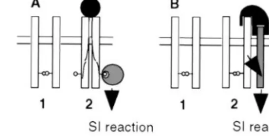
On the basis of our results, we propose that activation of SRK is not coincidental with the formation of SRK oligomers but instead results from the modification of a pre-existing SRK oligomeric complex. Two models are proposed (Fig. 5). In the first model, SRK兾ligand interaction would lead to conforma-tional changes in the kinase domain allowing the recruitment of a specific substrate(s) (Fig. 5A). This idea is supported by the fact conformational modification of the kinase domain of SRK910
from B. napus seems to be required for its interaction with thioredoxin THL1 (18). In this model, SRK would mediate both haplotype-specific recognition of the pollen ligand and the recruitment of downstream substrates. Indeed, one (or more)
Fig. 5. Two models for the molecular mechanism of signal transduction via SRK in the SI response. (A) This model proposes that SRK (open rectangle) spontaneously associates as a dimer in the plasmalemma of stigmatic papillar cells before pollination. Interaction with a self-pollen borne ligand (filled circle) induces a conformational change of SRK, which allows the recruitment of cytoplasmic targets (large hatched circles) that mediate the SI response. In the example shown here, the cytoplasmic targets recognize phosphorylated residues (small circles) on the SRK protein. (B) In the second model, SRK is also present as a constitutively formed dimer but the activation of SI response by pollen-ligand (in black) requires both binding to SRK in an allele-specific manner, and then binding to a second as-yet-unknown coreceptor (hatched rectangle), which does not need to exhibit any allelic specificity. Signal trans-duction then may involve interaction of the second receptor with cytosolic targets analogous to those described for serine兾threonine receptor kinases in animals.
PLANT
phosphoserine(s) present in the C terminus of recombinant SRK could act as docking sites for the recruitment of cytoplasmic targets. In the second model, SRK would function in a manner analogous to transforming growth factor receptor type II. The role of SRK would be to recruit (and perhaps activate) a second membrane-anchored coreceptor that could interact with the SRK兾pollen ligand complex (Fig. 5B). This model assumes that the two steps of the SI response, i.e., haplotype-specific recog-nition and signal transduction, are performed by two different receptors. To test these models, it will be necessary to charac-terize the constituents of SRK complexes in stigmas both before and after pollination with self-incompatible and cross-compatible pollen.
Studies of self-compatible mutants and transgenic Brassica plants in which the SRK kinase activity is defective have indicated that SRK is necessary for the SI response. If SRK has the same constitutive kinase activity in planta as we have observed in insect cells, we might expect the SI response to be constitutively activated and the stigma to reject both self- and cross-pollen. But as the stigma permits cross-pollination, we suggest that the SRK signal transduction pathway may be inhibited both in the absence of pollination and after cross-pollination, for example by proteins that inhibit SRK kinase activity in stigmas. Interestingly, in animals the FK 506 binding protein (FKBP12) has been shown to inhibit the signaling activity of the transforming growth factor receptor (TR) type
I兾TR type II complex, which can form even in the absence of ligand (46). In the case of SRK, proteins that interact with the SRK kinase domain, such as thioredoxins, ARC1, and kinase-associated protein phosphatases (18–20), could be involved either in transducing the signal downstream of SRK or in the inhibition of signal transduction in the absence of self-pollen. The identification of a 161-kDa SRK complex in unpollinated stigmas suggests that SRK may be associated with other proteins before pollination, and one possibility is that the formation of such complexes has a negative regulatory effect on SRK. The role of these complexes in the regulation of SRK needs to be precisely defined in the future by investigating their occurrence in cross- and self-pollinated stigmas. The approaches developed in this study, in particular the baculovirus兾insect cell system, which allows the coexpression of two different proteins, and velocity sedimentation, open possibilities to characterize physi-cal associations between SRK and other putative components (SLG, eSRK, cytosolic proteins, etc.) of the SRK receptor complex.
We are very grateful to Christophe Grangeasse, Marguerite-Marie Boutillon, Marie-Claire Ronzie`re (Institut de Biologie et de Chimie des Prote´ines de Lyon), and Je´roˆme Garin (Centre de l’Energie Atomique, Grenoble) for their help. We also thank Anne-Marie Thierry and Richard Blanc for their technical assistance. T.G. is a member of the Centre National de la Recherche Scientifique.
1. De Nettancourt, D. (1977) Incompatibility in Angiosperms (Springer, New York).
2. Nasrallah, J. B., Kao, T.-H., Goldberg, M. L. & Nasrallah, M. E. (1985) Nature
(London) 318, 617–618.
3. Stein, J. C., Howlett, B., Boyes, D. C., Nasrallah, M. E. & Nasrallah, J. B. (1991)
Proc. Natl. Acad. Sci. USA 88, 8816–8820.
4. Kandasamy, M. K., Paolillo, D. J., Faraday, C. D., Nasrallah, J. B. & Nasrallah, M. E. (1989) Dev. Biol. 134, 462–472.
5. Delorme, V., Giranton, J. L., Hatzfeld, Y., Friry, A., Heizmann, P., Ariza, M. J., Dumas, C., Gaude, T. & Cock, J. M. (1995) Plant J. 7, 429–440.
6. Stein, J. C., Dixit, R., Nasrallah, M. E. & Nasrallah, J. B. (1996) Plant Cell 8, 429–445.
7. Braun, D. M. & Walker, J. C. (1996) Trends Biochem. Sci. 21, 70–73. 8. Goring, D. & Rothstein, S. J. (1992) Plant Cell 4, 1273–1281.
9. Giranton, J. L., Ariza, M. J., Dumas, C., Cock, J. M. & Gaude, T. (1995) Plant
J. 8, 827–834.
10. Nasrallah, M. E., Kandasamy, M. K. & Nasrallah, J. B. (1992) Plant J. 2, 497–506.
11. Gaude, T., Rougier, M., Heizmann, P., Ockendon, D. J. & Dumas, C. (1995)
Plant Mol. Biol. 27, 1003–1014.
12. Goring, D. R., Glavin, T. L., Schafer, U. & Rothstein, S. J. (1993) Plant Cell
5,531–539.
13. Nasrallah, J. B., Rundle, S. J. & Nasrallah, M. E. (1994) Plant J. 5, 373–384. 14. Stahl, R. J., Arnoldo, M., Glavin, T. L., Goring, D. R. & Rothstein, S. J. (1998)
Plant Cell 10, 209–218.
15. Boyes, D. C. & Nasrallah, J. B. (1995) Plant Cell 7, 1283–1294.
16. Pastuglia, M., Ruffio-Chable, V., Delorme, V., Gaude, T., Dumas, C. & Cock, J. M. (1997) Plant Cell 9, 2065–2076.
17. Stephenson, A. G., Doughty, J., Dixon, S., Elleman, C., Hiscock, S. & Dickinson, H. G. (1997) Plant J. 12, 1351–1359.
18. Bower, M. S., Matias, D. D., Fernandes-Carvalho, E., Mazzurco, M., Gu, T., Rothstein, S. J. & Goring, D. R. (1996) Plant Cell 8, 1641–1650.
19. Gu, T., Mazzurco, M., Sulaman, W., Matias, D. D. & Goring, D. R. (1998) Proc.
Natl. Acad. Sci. USA 95, 382–387.
20. Braun, D. M., Stone, J. M. & Walker, J. C. (1997) Plant J. 12, 83–95. 21. Gilboa, L., Wells, R. G., Lodish, H. F. & Henis, Y. I. (1998) J. Cell Biol. 140,
767–777.
22. Heldin, C. H. (1995) Cell 80, 213–223.
23. Jou, W. M., Verhoeyen, M., Devos, R., Saman, E., Fang, R., Huylebroeck, D., Fiers, W., Threlfall, G., Barber, C., Carey, N. & Emtage, S. (1980) Cell 19, 683–696.
24. O’Reilly, D. R., Miller, L. K. & Luckow, V. A. (1994) Baculovirus Expression
Vectors: A Laboratory Manual (Oxford Univ. Press, Oxford, U.K.).
25. Lowry, O., Rosenbrough, N., Farr, A. & Randall, R. (1951) J. Biol. Chem. 193, 265–275.
26. Scheel, A. A. & Pelham, H. R. (1996) Biochemistry 35, 10203–10209. 27. Duclos, B., Grangeasse, C., Vaganay, E., Riberty, M. & Cozzone, A. J. (1996)
J. Mol. Biol. 259, 891–895.
28. Moritz, R. L., Eddes, J. S., Reid, G. E. & Simpson, R. J. (1996) Electrophoresis
17,907–917.
29. Gaude, T., Friry, A., Heizmann, P., Mariac, C., Rougier, M., Fobis, I. & Dumas, C. (1993) Plant Cell 5, 75–86.
30. Stein, J. C. & Nasrallah, J. B. (1993) Plant Physiol. 101, 1103–1106. 31. Hanks, S. K., Quinn, A. M. & Hunter, T. (1988) Science 241, 42–52. 32. Doughty, J., Dixon, S., Hiscock, S. J., Willis, A. C., Parkin, I. A. & Dickinson,
H. G. (1998) Plant Cell 10, 1333–1347.
33. Conner, J. A., Tantikanjana, T., Stein, J. C., Kandasamy, M. K., Nasrallah, J. B. & Nasrallah, M. E. (1997) Plant J. 11, 809–823.
34. Greenfield, C., Patel, G., Clark, S., Jones, N. & Waterfield, M. D. (1988) EMBO
J. 7, 139–146.
35. Paul, J. I., Tavare, J., Denton, R. M. & Steiner, D. F. (1990) J. Biol. Chem. 265, 13074–13083.
36. Gaymard, F., Cerutti, M., Horeau, C., Lemaillet, G., Urbach, S., Ravallec, M., Devauchelle, G., Sentenac, H. & Thibaud, J. B. (1996) J. Biol. Chem. 271, 22863–22870.
37. Zimmermann, S., Talke, I., Ehrhardt, T., Nast, G. & Muller-Rober, B. (1998)
Plant Physiol. 116, 879–890.
38. Chang, C., Schaller, G. E., Patterson, S. E., Kwok, S. F., Meyerowitz, E. M. & Bleeker, A. B. (1992) Plant Cell 4, 1263–1271.
39. Horn, M. A. & Walker, J. C. (1994) Biochim. Biophys. Acta 1208, 65–74. 40. Schulze-Muth, P., Irmler, S., Schroder, G. & Schroder, J. (1996) J. Biol. Chem.
271,26684–26689.
41. Williams, R. W., Wilson, J. M. & Meyerowitz, E. M. (1997) Proc. Natl. Acad.
Sci. USA 94, 10467–10472.
42. Mu, J. H., Lee, H. S. & Kao, T. H. (1994) Plant Cell 6, 709–721. 43. Hirayama, T. & Oka, A. (1992) Plant Mol. Biol. 20, 653–662.
44. Trotochaud, A. E., Hao, T., Wu, G., Yang, Z. & Clark, S. E. (1999) Plant Cell
11,393–406.
45. Wrana, J. L., Attisano, L., Wieser, R., Ventura, F. & Massague, J. (1994) Nature
(London) 370, 341–347.
46. Chen, Y. G., Liu, F. & Massague, J. (1997) EMBO J. 16, 3866–3876.