HAL Id: tel-02918029
https://tel.archives-ouvertes.fr/tel-02918029
Submitted on 20 Aug 2020HAL is a multi-disciplinary open access archive for the deposit and dissemination of sci-entific research documents, whether they are pub-lished or not. The documents may come from teaching and research institutions in France or abroad, or from public or private research centers.
L’archive ouverte pluridisciplinaire HAL, est destinée au dépôt et à la diffusion de documents scientifiques de niveau recherche, publiés ou non, émanant des établissements d’enseignement et de recherche français ou étrangers, des laboratoires publics ou privés.
migration and cell-cycle progression in the mouse
developing cortex
Julie Fousse
To cite this version:
Julie Fousse. Study of the coupling between interkinetic nuclear migration and cell-cycle progres-sion in the mouse developing cortex. Cellular Biology. Université de Lyon, 2017. English. �NNT : 2017LYSE1320�. �tel-02918029�
1 N°d’ordre NNT : xxx
THESE de DOCTORAT DE L’UNIVERSITE DE LYON
opérée au sein de
l’Université Claude Bernard Lyon 1 Ecole Doctorale340
Biologie Moléculaire, Intégrative et Cellulaire (BMIC) Spécialité de doctorat : Biologie
Soutenue publiquement le 11/12/2017, par :
Julie Fousse
Study of the coupling between interkinetic nuclear
migration and cell-cycle progression in the mouse
developing cortex
Devant le jury composé de :
Durand, Bénédicte, Professeure des Universités, Université Claude Bernard Lyon 1, Présidente
Norden, Caren, Chercheure, Institut Max-Planck, Rapporteure
Price, David, Professeur des Universités, Université d’Edimbourg, Rapporteur
Baffet, Alexandre, Chargé de Recherche INSERM, Institut Curie, INSERM 144, Examinateur
Dehay, Colette, Directrice de Recherche INSERM, Institut Cellule Souche et Cerveau, INSERM 1208, Directrice de thèse
2
Président de l’Université
Président du Conseil Académique
Vice-président du Conseil d’Administration
Vice-président du Conseil Formation et Vie Universitaire Vice-président de la Commission Recherche
Directrice Générale des Services
M. le Professeur Frédéric FLEURY M. le Professeur Hamda BEN HADID
M. le Professeur Didier REVEL M. le Professeur Philippe CHEVALIER M. Fabrice VALLÉE
Mme Dominique MARCHAND
COMPOSANTES SANTE
Faculté de Médecine Lyon Est – Claude Bernard
Faculté de Médecine et de Maïeutique Lyon Sud – Charles Mérieux
Faculté d’Odontologie
Institut des Sciences Pharmaceutiques et Biologiques Institut des Sciences et Techniques de la Réadaptation
Département de formation et Centre de Recherche en Biologie Humaine
Directeur : M. le Professeur G.RODE
Directeur : Mme la Professeure C. BURILLON
Directeur : M. le Professeur D. BOURGEOIS Directeur : Mme la Professeure C. VINCIGUERRA Directeur : M. X. PERROT
Directeur : Mme la Professeure A-M. SCHOTT
3 Département Chimie Biochimie
Département GEP
Département Informatique Département Mathématiques Département Mécanique Département Physique
UFR Sciences et Techniques des Activités Physiques et Sportives Observatoire des Sciences de l’Univers de Lyon
Polytech Lyon
Ecole Supérieure de Chimie Physique Electronique Institut Universitaire de Technologie de Lyon 1 Ecole Supérieure du Professorat et de l’Education Institut de Science Financière et d'Assurances
Directeur : Mme C. FELIX
Directeur : M. Hassan HAMMOURI Directeur : M. le Professeur S. AKKOUCHE Directeur : M. le Professeur G. TOMANOV Directeur : M. le Professeur H. BEN HADID Directeur : M. le Professeur J-C PLENET Directeur : M. Y.VANPOULLE
Directeur : M. B. GUIDERDONI Directeur : M. le Professeur E.PERRIN Directeur : M. G. PIGNAULT
Directeur : M. le Professeur C. VITON
Directeur : M. le Professeur A. MOUGNIOTTE Directeur : M. N. LEBOISNE
4
Contents ... 4
Contents ... 4 Figures ... 7 Tables ... 10 Supplementary figures ... 10Abbreviation list ... 11
Abstract (in english) ... 15
Résumé (en français) ... 16
Introduction ... 17
I. Cortical development ... 17
II. Apical Progenitors ... 20
1 Cell biology of Apical Progenitors ... 20
2 The apical process ... 22
3 The basal process ... 25
III. Interkinetic Nuclear Migration... 28
1 What is Interkinetic Nuclear Migration ? ... 28
2 Forces behind migration ... 29
3 Centrosome behavior and mitosis in APs ... 39
4 Putative roles of INM ... 41
IV. Cell-cycle regulators used to study INM ... 45
1 p27Kip1 ... 45
1.1 Nucleic p27Kip1 role ... 45
1.2 Cytoplasmic p27Kip1 role on migration ... 59
1.3 Role in the cerebral cortex ... 66
2 CyclinE ... 68
2.1 CyclinE canonical role on the cell-cycle ... 68
2.2 Other roles of CyclinE ... 71
2.3 Role in the cerebral cortex ... 72
Chapter I: INM, a refined definition ... 75
5
2 INM study on electroporated APs ... 80
Cell-cycle phase specific immunolabeling analysis of APs distribution at E16 . 81 Direct visualization of APs INM using TLV at E16 ... 83
3 Comparison of S and G2 phase APs nuclei repartition in the VZ at E12, E14 and E16 ... 85
Discussion ... 86
Developmental variations of cell-cycle phases of APs ... 86
Refined definition of INM ... 89
Correspondence between INM and cell-cycle progression ... 91
Species-specific differences in INM dynamics ... 93
Conclusion ... 95
Chapter II: Impact of p27
Kip1C-terminal domain on INM ... 96
Preamble ... 98
1 Impact of p27kip1 C-terminal domain in wild type APs ... 99
Single cell analysis using TLV combined with clonal electroporation on organotypic slices ... 99
Cell-cycle-specific static analysis on a population of APs ... 102
2 Impact of p27kip1 C-terminal domain in p27kip1 lox/- APs ... 106
Discussion ... 119
p27-Cter modifies the coupling between cell-cycle progression and INM in WT embryos ... 119
Disentangling the action of p27-wt-Cter overexpression from the effect of the reduced level of endogeneous p27Kip1 ... 120
Important binding sites of p27-Cter for the coupling between INM and cell-cycle progression ... 123
Conclusion ... 128
Chapter III: Impact of CyclinE overexpression on INM ... 129
1 Time lapse analysis of CyclinE impact on cell-cycle duration and INM ... 131
CyclinE overexpression influences cell-cycle duration ... 132
CyclinE overexpression alters INM dynamics ... 132
CyclinE overexpression alters the directionality of INM apical and basal movements. ... 134
CyclinE overexpression influences progenitor fate ... 135
2 Immunolabeling analysis of CyclinE impact on cell-cycle progression and INM . 137 Distribution of APs and BPs in the different cell-cycle phases is modified following CyclinE overexpression ... 137
6
APs nuclei repartition within the VZ depth is modified following CyclinE
overexpression ... 140
3 CyclinE impact on mitosis ... 142
CyclinE overexpression promotes abventricular divisions ... 142
CyclinE overexpression and cleavage plane orientation ... 143
4 CyclinE and cell fate in the GZ ... 145
Discussion ... 148
Overall cell-cycle duration is reduced after CyclinE overexpression in APs ... 148
CyclinE influences cell-cycle distribution ... 149
CyclinE modifies the coupling between cell-cycle and INM ... 153
CyclinE promotes abventricular mitosis ... 156
CyclinE impacts of cleavage orientation ... 157
CyclinE overexpression increases BPs production and impacts SVZ thickness ... 157
Conclusion ... 159
General Discussion ... 161
Material and Methods ... 163
Animals ... 163
Plasmids ... 163
In utero electroporation ... 164
Brain collection... 164
Brain cryosections ... 164
Immunofluorescence and antibodies ... 165
Confocal microscopy ... 165
Brain sections and organotypic slices culture ... 165
Two photon microscopy ... 166
Statistical analysis ... 166
Aknowledgment ... 167
Bibliography ... 168
7
Figures
Figure 1: The mouse cortex is composed of 6 cortical layers ... 17
Figure 2: Neurogenesis in the mouse developing cortex ... 19
Figure 3: RGCs are also neuronal progenitors... 20
Figure 4: APs nuclei in the VZ form a pseudostratified epithelium. ... 21
Figure 5: The primary cilium and the centrosomes ... 23
Figure 6: Golgi localization ... 24
Figure 7: CyclinD2 mRNA and protein localization during the cell-cycle ... 26
Figure 8: Schematic INM in the developing neocortex ... 28
Figure 9: INM in the VZ of the developing rodent neocortex ... 29
Figure 10: Putative motors of basal nuclear migration in the mouse cortex ... 30
Figure 11: Dynein mediates the apical movement of INM ... 31
Figure 12: Recruitment of dynein to the NE regulates the microtubule based apical movement of INM ... 35
Figure 13: The apical movement is driven by different molecular motors depending on the PSE thickness ... 37
Figure 14: Differences between mouse and ferret VZ, APs and associated INM ... 38
Figure 15: Centrosomes meet the nucleus during the apical movement of INM ... 40
Figure 16: Progenitors shape the lumen of the otic vesicle... 42
Figure 17: Pseudostratified epithelia undergoing INM allow the presence of more cells than columnar epithelia within a restricted area ... 43
Figure 18: INM balances the exposure to Notch signaling ... 44
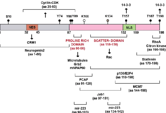
Figure 19: Human p27Kip1 protein: post-translational modifications and direct interactions . 46 Figure 20: Regulation of p27Kip1 ... 47
Figure 21: Crystal structure of the p27Kip1/Cdk2/CyclinA complex ... 49
Figure 22: Control of cell-cycle progression by p27Kip1 ... 50
Figure 23: Regulation of G1/S transition ... 51
Figure 24: p27Kip1 phosphorylation regulates subcellular localization and degradation ... 56
Figure 25: Ras pathway activation ... 60
Figure 26: Rac pathway ... 61
Figure 27: RhoA pathway ... 62
Figure 28: p27Kip1 regulates stathmin activity ... 64
8
Figure 30: Brap regulates neurogenesis ... 67
Figure 31: CyclinE functions and regulation ... 70
Figure 32: Discrimination of the different cell-cycle phases ... 78
Figure 33: Example of immunolabeled brain slice at E16 ... 78
Figure 34: Analysis of non-electroporated APs in the VZ at E12, E14 and E16 ... 80
Figure 35: S and G2 phase repartition of wild type APs nuclei within the VZ depth at E16 as revealed by immunolabeling. ... 82
Figure 36: Clonal electroporation strategy ... 83
Figure 37: Apical movement of an AP nucleus ... 84
Figure 38: Analysis of non-electroporated APs nuclei in the VZ at E12, E14 and E16 as revealed by immunolabeling ... 85
Figure 39: Exemples of APs BrdU labelling ... 90
Figure 40: Schematic representation of our current data on INM ... 91
Figure 41: Schematic representation INM during corticogenesis ... 93
Figure 42: Different forms of the p27kip1 C-terminal domain protein and their interactions.. 99
Figure 43: Strategy of experimentation ... 100
Figure 44: APs behavior following p27-wt-Cter overexpression... 101
Figure 45: Example of immunolabeled APs within the different cell-cycle phases ... 103
Figure 46: Overexpression of p27-wt-Cter modifies APs nuclei repartition in the depth of the VZ as revealed by immunolabelling ... 104
Figure 47: Overexpression of p27-wt-Cter modifies APs cell-cycle progression as revealed by immunolabelling ... 105
Figure 48: Clonal electroporation strategy ... 106
Figure 49: Confocal quantification of p27kip1 immunolabelling in the VZ and SVZ 24 hours after electroporation ... 108
Figure 50: Impact of p27Kip1 KO on the apical movement and cell-cycle progression ... 109
Figure 51: The C-terminal domain of p27Kip1 modifies APs nuclei repartition ... 111
Figure 52: APs distribution within the cell-cycle at E16 as revealed by immunolabelling .... 112
Figure 53: Important interactions with p27Kip1 for INM ... 113
Figure 54: APs distribution within the cell-cycle at E16 as revealed by immunolabelling. .. 116
Figure 55: Coupling between cell-cycle progression and INM is modified following p27-wt-Cter overexpression in APs ... 120
9
Figure 56: Summary cartoon showing that S and G2 phases nuclei are found lower in the
depth of the VZ in absence of endogeneous p27Kip1 ... 121
Figure 57: Schematic representation illustrating that cell-cycle progression is modified following p27-wt-Cter overexpression ... 122
Figure 58: Human C-terminal domain of p27Kip1 protein: post-translational modifications and direct interactions ... 124
Figure 59: p27-wt-Cter overexpression in APs influences the coupling between cell-cycle progression and INM through interaction with MTs and RhoA ... 127
Figure 60: Strategy of experimentation ... 131
Figure 61: Progenitors cell-cycle duration as revealed by TLV analysis ... 132
Figure 62: INM phases duration is not modified in CyclinE overexpressing APs ... 133
Figure 63: INM velocity is modified in APs overexpressing CyclinE ... 134
Figure 64: The apical movement of APs nuclei is more linear after CyclinE overexpression 135 Figure 65: Example of immunolabeled APs in the different cell-cycle phases ... 137
Figure 66: Example of immunolabeled brain sections for each condition ... 138
Figure 67: Progenitors distribution in the cell-cycle at E16 as revealed by immunolabeling. ... 139
Figure 68: CyclinE modifies the INM behavior of APs nuclei ... 140
Figure 69: Example of immunolabeled brains 48 hours after electroporation ... 142
Figure 70: APs position during mitosis ... 143
Figure 71: Angles of dividing APs ... 144
Figure 72: Example of immunolabeled brain slices of each condition 24 hours after electroporation ... 146
Figure 73: Pax6, Tbr2 and Ki67 markers expression in the VZ and in the SVZ ... 147
Figure 74: Schematic representation of CyclinE actions on progenitors during neurogenesis ... 148
Figure 75: Schematic representations of cell-cycle progression in control and CyclinE GOF APs ... 149
Figure 76: Schematic INM progression during the cell-cycle in control and CyclinE GOF progenitors ... 154
Figure 77: Schematic representation of the regulation of the BPs pool by CyclinE ... 158
10
Tables
Table 1: Summary of S, late S and G2 APs nuclei variations in the repartition withinthe depth of the VZ in the different conditions compared to control (p27Kip1 lox/-) APs ... 114 Table 2: Summary of S, late S and G2 APs nuclei variations in the repartition withinthe depth of the VZ in the different conditions compared to the p27-wt-Cter expressing APs ... 115 Table 3: Summary of of APs distribution in the cell-cycle in the different conditions compared to the (p27Kip1 lox/-) APs ... 117 Table 4: Summary of APs distribution in the cell-cycle in the different conditions compared to the p27-wt-Cter expressing condition ... 118 Table 5: Number of progenitors tracked in each condition (N) ... 136 Table 6: Table representing the cell-cycle parameters in hours ... 150
Supplementary figures
Supplementary figure 1: There are no major modifications of nuclei repartition 24h after overexpression of p27-Cter different forms ... 210 Supplementary figure 2: S and G2 phases nuclei are found lower in the VZ in absence of endogeneous p27Kip1 ... 211 Supplementary figure 3: Loss of interaction with microtubules affects S and G2 phase as well as the apical movement ... 213 Supplementary figure 4: Loss of interaction with microtubules and RhoA restores cell-cycle progression and modifies S phase nuclei repartition ... 214 Supplementary figure 5: Plasmidic vectors used for electroporation ... 215
11
Abbreviation list
AA: Aminino-Acid
Aβ42: Amyloid-beta 42 protein AJ: Adherens Junction
Akt: RAC-alpha serine/threonine-protein kinase
AMP: Adenosine Monophosphate AMPK: AMP activated protein Kinase AP: Apical Progenitor
APC: Anaphase-Promoting Complex aPKC: Protein Kinase C
Asp: Aspartic acid
ASP: Abnormal Spindle Protein
ASPM: Abnormal Spindle Microtubule
Assembly
ATP: Adenosine Triphosphate
BicD2: Protein bicaudal D homolog 2 BP: Basal Progenitor
BCR: B-Cell Receptor
BRCA1: Breast cancer type 1 susceptibility
protein homolog
Brap: BRCA1-associated protein BrdU: Bromodeoxyuridine bRG: basal Radial Glia
BTB: Bric-à-brac, Tramtrack, Broad complex
C-ter: C-terminal domain
CAK: Cyclin Activating Kinase
CBP/p300: CREB-binding protein/p300 CDC: Cell-Division Control protein
Cdh1: Cadherin-1
CDK: Cyclin-Dependant Kinase CENPF: Centromere protein F Cep120: Centrosomal protein 120
Cip/Kip: CDK interacting protein/Kinase
inhibitory protein
CK2: Casein Kinase 2
CKI: Cyclin-dependant Kinase Inhibitor CKS1: Cyclin-dependent Kinases regulatory
Subunit 1
CLS: Centrosomal Localization Signal CNS: Central Nerveous System CP: Cortical Plate
Cre: Causes recombination
CRM1: Chromosomal Maintenance 1
Cul: Cullin
DAPI: 4',6-diamidino-2-phénylindole
Dirk: Dual specificity
tyrosine-phosphorylation-regulated kinase 1B
DNA: Deoxyribonucleic Acid
DOCK7: Dedicator of cytokinesis protein 7 Drp1: Dynamin related protein 1
12
E2F: E2 transcription Factor
ELAV: Embryonic Lethal Abnormal Vision EMT: Epithelial to Mesenchymal Transition ER: Endoplasmic Reticulum
ERK: Extracellular signal-Regulated Kinase Far1: Factor arrest resistant 1
Fbw7: F-box/WD repeat-containing protein 7
FOXO: Forkhead box protein O G1 and G2: Gap 1 and Gap 2 γ: µg/µL
GEF: Guanine Exchange Factor GFP: Green Fluorescent Protein GMO: Genetically Modified Organism GOF: Gain Of Function
Grb2: Growth factor receptor-bound
protein 2
GSCF: Granulocyte colony-stimulating factor
GTPase: Guanine Triphosphatase GZ: Germinal Zones
h: hours
HBSS: Hank’s buffered salt solution Hes: hairy and enhancer of split-1
HOOK3: Protein Hook homolog 3 HMTase: Histone Methyl Transferase KD: Knock Down
Ki67: Kiel 67
KIF1A: Kinesin Family member 1A KO: Knock Out
Id3: Inhibitor of DNA binding 3
INK4: Inhibitor of cyclin-dependant Kinase
4
INM: Interkinetic Nuclear Migration IRES: Internal Ribosome Entry Site IZ: Intermediate Zone
Jab1: Jun activation domain-binding
protein 1
K: Lysin
KASH: Klarsicht/ANC-1/Syne-1 homology
Ki67: Kiel 67
KIS: Kinase Interacting Stathmin Klp54D: Kinesin-like protein 54D LH: Linker Helix
LIMK: LIM-motif containing protein kinases
Lis1: Lissencephaly 1 Lys : Lysin
M: Mitotic
MAP: Microtubule-Associated Protein MAPK: Mitogen-Activated Protein Kinase MAT: Mesenchymal Ameboid Transition MBS: Myosin Binding Subunit
MCM: Mini Chromosome Maintenance mDia: mammalian Diaphanous formin
13
MLC: Myosin Light Chain min: minutes
miR: microRNA
Mirk: Minibrain-related kinase MT: microtubule
Myc: Myelocytomatosis oncogene cellular
homolog
NDS: Normal Donkey Serum NE: Nuclear Envelope NES: Nuclear Export Signal Ngn: Neurogenin
NGS: Normal Goat Serum NLS: Nuclear Localization Signal NPM/B23: Nucleophosmin/B23 N-ter: N-terminal domain
Nud: Nuclear distribution protein
Nup133: Nuclear pore complex protein
133
OSVZ: Outer Subventricular Zone p220NPAT: p220Nuclear Protein Ataxia Telangiectasia PAK: p21-activated kinase
Par: Partitioning defective protein Pax6: Paired box protein 6
PBS: Phosphate Buffer Saline PCAF: p300/CBP-Associated Factor PCM1: Pericentriolar Material 1
PCNA: Proliferating Cell Nuclear Antigen PD166285:
6-(2,6-dichlorophenyl)-2-[4-[2- (diethylamino)ethoxy]anilino]-8-methylpyrido[2,3-d]pyrimidin-7-one
Pi3K: Phosphoinositide 3 kinase
PIM: proviral integration site for moloney
murine leukemia virus
PIRH2: p53-induced RING-H2-type ubiquitine ligase
PMLC2: Phospho-Myosin Light Chain 2 polyA: polyadenylic acid tail
PPM1G: Protein Phosphatase 1G PPM1H: Protein Phosphatase 1H PSE: Pseudodtratified Epithelia PTB: Polypyrimidine Tract-Binding R: Arginin
Rac: ras-related C3 botulinum toxin
substrate
Ran BP2: RAN binding protein 2
Ras: Rat sarcoma protein Rb: retinoblastoma protein RBX1: RING-box protein 1 RGC: Radial Glial Cell
Rho: Ras homolog gene family RNA: Ribonucleic Acid
RO3306: (5Z)-5-(quinolin-6-ylmethylidene)- 2-[(thiophen-2-ylmethyl)amino]-1,3-thiazol-4(5H)-one
14
ROCK: Rho-associated protein Kinase Rp58: Repressor protein 58
RSK: Ribosomal S6 Kinase RT: Room Temperature S: Synthesis
SCF: Skp-cdc53-F-box ubiquitin ligase
complex
SEM: Standard Error Ser: Serin
Sic1: Stoichiometric inhibitor of Clb-Cdk Skp2: S-phase kinase-associated protein 2 Smad: mothers against decapentaplegic
homolog
Sos: Son of sevenless Sp1: Specificity protein 1
Src: Rous sarcoma oncogene cellular
homolog
SUMO1: Small Ubiquitin-like Modifier 1 SUN: SUN domain-containing protein SVZ: Subventricular Zone
Syne: Nesprin
TACC3: Transforming Acidic Coiled-Coil
protein 3
TAG-1: Transient Axonal Glycoprotein-1 Tbr2: T-box brain 2
tbRG: transient basal Radial Glia TBS: Tris-Buffer Saline
Tc: cell-cycle duration
TF: Transcription Factor
TGFβ: Transforming Growth Factor β Thr: Threonin
TLV: Time Lapse Videomicroscopy Tpx2: Targeting protein for Xklp2 Tyr: Tyrosin
UTR: Untranslated Region VZ: Ventricular Zone
WAVE: Wiskott-Aldrich syndrome
protein-family Verprolin homologous protein
WT: Wild Type
15
Abstract (in english)
The balance between proliferation and differentiation of Apical Progenitors (APs) is a crucial mechanism for proper cerebral cortex histogenesis, and its dysregulation causes important cortical malformations. In the Ventricular Zone (VZ) of the cortex, the nucleus of APs performs a stereotyped movement of Interkinetic Nuclear Migration (INM) during cell-cycle progression. This study aims to characterize the coupling between INM and cell-cell-cycle progression. First, I present a refined description of the INM dynamics with respect to cell-cycle progression. I show that both INM and cell-cell-cycle dynamics vary from early to late stages of corticogenesis. I have examined the impact of p27Kip1 C-terminal domain (p27-Cter) on the proliferative behavior of APs. We observed that p27-Cter expression modifies the INM as well as the distribution of APs within the cell-cycle. We evidenced that the interaction between p27Kip1 and microtubules plays a key role in the apical movement of APs. Finally, I studied the effect of CyclinE protein overexpression on the coupling between INM and cell-cycle progression. We observed that CyclinE overexpression, which decreases G1 phase duration, also impacts INM, via a modification of the velocity and directionality of the INM movements.
16
Résumé (en français)
L'équilibre entre prolifération et différenciation des Progéniteurs Apicaux (PA) est un mécanisme crucial de l'histogénèse du cortex cérébral dont la dérégulation est à l'origine de malformations corticales importantes. Dans la zone ventriculaire (ZV) du cortex, le noyau des PA effectue un mouvement stéréotypé de Migration Nucléaire Intercinétique (MNI) au cours du cycle cellulaire. Cette étude a pour but de caractériser le couplage entre le cycle-cellulaire et la MNI. Je présente tout d’abord une description révisée de la dynamique de la MNI en fonction de la progression dans le cycle-cellulaire. Je montre également que la relation entre la MNI et le cycle-cellulaire évolue au cours du développement cortical. J'ai examiné l'impact du domaine C-terminal de la protéine p27Kip1 (p27-Cter) sur le comportement prolifératif des PA. Nous avons observé que l’expression de p27-Cter modifie à la fois la distribution des PA dans le cycle cellulaire et leur répartition dans la ZV. Nos résultats montrent que la liaison directe entre p27kip1 et les microtubules joue un rôle important dans le mouvement apical des PA. Enfin, j’ai étudié l’effet de la surexpression de la protéine CyclinE sur le couplage entre le cycle-cellulaire et la MNI. La surexpression de la CyclinE, qui diminue la durée de la phase G1, module également la MNI via des modifications de la vitesse et de la directionalité des mouvements apicaux et basaux.
17
Introduction
I. Cortical development
The mammalian cerebral cortex plays a major role in higher cognitive functions and its development is the focus of sustained investigations.
The neocortex is composed of 6 layers which harbor morphologically and functionally distinct neurons (Ramón y Cajal, 1899; figure 1A). These layers are formed through a process called corticogenesis which starts at Embryonic day (E)11 in the mouse and implies the coordination of proliferation, differentiation and migration of neurons in radial and tangential directions (reviewed in Price and Willshaw, 2000; Dehay and Kennedy, 2007). Pyramidal neurons are positioned in the cortex via an inside-out process (figure 1B) with oldest neurons located closer to the ventricle and youngest neurons close to the pial surface. Layer I corresponds to Cajal-Retzius (CR) cells, which have migrated tangentially toward the Cortical Plate (CP) early in neurogenesis. Layer II to VI are positioned below, above the subplate (SP) (Ayala et al., 2007; Cooper, 2008).
Figure 1: The mouse cortex is composed of 6 cortical layers. A: Cortical layers in the adult mouse brain. The adult CP is divided into 6 layers: from layer I at the pial surface to layer VI close to the
ventricle. B: Formation of the neocortex via the inside-out migration process. As corticogenesis proceeds, newborn neurons migrate to their final location though the CP. The neocortex is formed through an inside-out migration process with later born neurons settling in the upper layers. The Cajal-Retzius (CR) cells are shown in yellow and the Subplate (SP) cells in purple. Extracted from Cooper, 2008.
18
During mammalian development, the cerebral cortex emerges from the neuroectoderm. At the end of neurulation and neural tube closure, between E7 and E9 in the mouse model, the early neuroepithelium is composed of neuroepithelial stem cells (NESCs). Before the onset of neurogenesis, proliferating cells form a pseudostratified neuroepithelium that lines the lateral ventricle. At embryonic day 11 (E11) in the mouse, the neuroepithelial progenitors give rise to the progenitor cells of the cortex, the Apical Progenitors (APs) (see part II of the introduction p.15) which are located in the Ventricular Zone (VZ) where they proliferate (Malatesta et al., 2000; Noctor et al., 2001). APs extend radially through the telencephalic wall; they bear processes attached to the ventricular surface and to the pial surface. The APs are capable of self-renewal and have extensive proliferative abilities. The nuclei of the APs undergo the Inter Kinetic Migration during their cell-cycle progression in the VZ. They also give rise to neurons and to the Basal Progenitors (BPs) which form the secondary Germinal Zone (GZ): the Subventricular Zone (SVZ) (Haubensak et al., 2004). Following exit from the cell-cycle and differentiation, newborn neurons then migrate radially through the Intermediate Zone (IZ) to reach their final location in the CP (Ayala et al., 2007).
19 Figure 2: Neurogenesis in the mouse developing cortex. Cortical neurogenesis lasts 8 days in the
mouse, from E11 to E19. APs are located in the VZ, they are able to self renew and to give rise to BPs or neurons. BPs delaminate to settle in the SVZ, they divide to give rise to two neurons. Neurons migrate through the IZ to reach their final location in the CP. Adapted from Lodato et al., 2015.
Mature neuron Immature neuron BP AP
20
II.
Apical Progenitors
1 Cell biology properties of Apical Progenitors
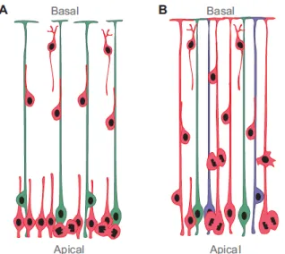
At the onset of neurogenesis, neuroepithelial cells become a cell type called Neural Progenitor Cells (NPCs) or Apical Progenitors (APs) (Taverna and Huttner, 2010) or Radial Glial Cells (RGCs) (Malatesta et al., 2011; Noctor et al., 2001) that divide at the apical surface. Götz and colleagues first identified RGCs as cortical progenitors in the mouse developing cortex (Malatesta et al., 2011). Looking for cells expressing Pax6 (Götz et al., 1998), they found that all progenitors and RGCs were Pax6 positive cells. These cells not only act as a scaffold for neuron migration but also generate neurons and astrocytes (Malatesta et al., 2000; see figure 3). Kriegstein and colleagues used retroviral infections in embryonic rat brains and showed that RGCs proliferate and give rise to neurons (Noctor et al., 2001). In this thesis, these cells will be referred as APs.
Figure 3: RGCs are also neuronal progenitors. A: Former corticogenesis model. RGCs (green) were
believed to have a cable role, guiding neuron (red) migration. These cells were supposed to become glial cells later during development. B: Actual corticogenesis model. RGCs are progenitors able to give rise to neurons (red), glial cells (green) or both (blue). Extracted from Malatesta and Götz, 2013.
APs are still highly related to the neuroepithelial cells. They have an apico-basal polarity and span the entire cortical wall. However, their nuclei reside in the VZ (Figure 4A). APs have a bipolar morphology and they extend two processes: one contacting the ventricular surface, the apical process, and one contacting the basal membrane at the pial surface, the basal process. This structural polarization underlies key features of the cell biology mechanisms controlling self-renewal and cell fate of APs, as will be reviewed below.
21 Figure 4: APs nuclei in the VZ form a pseudostratified epithelium. A: APs nuclei in the VZ. APs span
the entire cortical wall but their nuclei are restricted to the VZ. Adapted from Lee and Norden, 2013.
B: Pseudostratified epithelia. Progenitors are packed and their nuclei move along the apico-basal
axis. Extracted from Lee and Norden, 2013. C: Comparison between a simple cuboidal epithelium
and two columnar epithelia. Increasing the degree of pseudostratification of the epithelium allows
more productivity at the apical surface, where mitosis takes place. Here, the apical area remains fixed (5a) but epithelial thickness is augmented, and nuclei span the entire thickness (from a to 4a). Extracted from Miyata et al., 2015.
The VZ is a pseudostratified epithelium
Pseudostratification is a characteristic of a highly proliferative tissue (Lee and Norden, 2013). It is a widely conserved cellular architecture. Indeed, it is observed in the Drosophila wing disc, the Nematostella ectoderm, the Zebrafish retina and in vertebrate tissues such as the liver or the intestine (Meyer et al., 2011; Norden et al., 2009; Bort et al., 2006; Grosse et al., 2011), and, as studied here, in the developing central nervous system (Malatesta et al., 2011; Noctor et al., 2001). In these epithelia, progenitors are densely packed and their nuclei are found in various positions along the apico-basal axis (Figure 4B).
Indeed, a characteristic phenomenon in all pseudostratified epithelia is Interkinetic Nuclear Migration (INM). It is a stereotyped migration movement during cell-cycle progression where the nucleus periodically moves up and down within the cytoplasm. It moves away from the ventricle during G1 phase to undergo the S phase basally, near the SVZ. During G2 phase, the nucleus moves in the apical direction, toward the ventricle so as to divide at the ventricular border (Sauer and Walker, 1959; Hayes and Nowakowski, 2000;
B
22
Figure 4A). As explained by Miyata et al., epithelia exhibiting a high degree of pseudostratification allow more mitosis at the ventricular border. Therefore, these epithelia are more productive (Figure 4C; Miyata et al., 2015).
2 The apical process The cilium
The apical plasma membrane, which forms the ventricular surface, makes a protrusion, the primary cilium (Dubreuil et al., 2007; Figure 5A). The cilium is an organelle involved in intercellular signaling. Since it protrudes from the apical plasma membrane, it is able to detect signals in the ventricular fluid (reviewed in Singla and Reiter, 2006). The primary cilium is made of the axoneme, a microtubular structure with a 9 + 0 configuration generated during interphase from the basal body. The basal body is also a microtubular structure forming the base of the cilium that originates from the centrosome (Figure 5A and 5B), which is comprised of the mother and daughter centrioles (Figure 5C). It is a microtubule-organizing center or a spindle pole during active cell division but it also templates the assembly of the primary cilium. As a consequence, the primary cilium will provide the centrosomes for the mitotic spindle, so it persists during the cell-cycle until the onset of M phase (reviewed in Santos and Reiter, 2008).
23 Figure 5: The primary cilium and the centrosomes. A: Cilium localization. The primary cilium
protrudes from the apical plasma membrane. The basal body originates from the centrosome and forms the base of the cilium. Extracted from Taverna and Huttner, 2010. B: Electron micrograph of
the primary cilium of a canary brain and detailed structure of the basal body and the primary cilium. The basal body is constituted of 9 triplets of microtubules and the cilium of 9 doublets of
microtubules. Scale bar = 1µm. Extracted from Singla and Reiter, 2006. C: Centrosome-cilium
complex. The mother centriole of the centrosome forms the basal body and the cilium protrudes from
the apical plasma membrane (in blue). The cilium is surrounded by the ciliary membrane (in red). Extracted from Mahjoub et al., 2013.
The cilium apical location in APs was seen for a long time as the main reason why the nucleus undergo mitosis at the ventricular surface (Miyata, 2008). However, recent findings contradict this assomption, as detailed below (see introduction section III, p29; Spear and Erickson, 2012; Hu et al., 2013; Strzyz et al., 2015).
This organelle is associated with cell-cycle progression. Cilia are generated during interphase and ciliary resorption triggers cell-cycle re-entry. In fact, the gradual loss of cilia may promote the formation of the Jouberin-mediated β-catenin complex to facilitate G1/S transition through the transcription of the CyclinD gene (Kim and Tsiokas, 2011).
The primary cilium of neuroepithelial cells concentrates prominin-1 particles which are released as extracellular membrane particles. This release into the neural tube fluid allows the reduction of APs apical surface and modifies the cell composition by depleting stem-cell characteristic membrane microdomains (Dubreuil et al., 2007). In addition, the
B A
24
decreased apical surface of APs occurs along with the switch from proliferative to neurogenic divisions. It is associated with a change in the distribution of the apical membrane from a symmetric to an asymmetric inheritance by the daughter cells (Kosodo et al., 2004).
The Golgi apparatus
The Golgi is an organelle that sorts secretory proteins and membrane components. It is also involved in the establishment and the maintenance of epithelial cell polarity. Taverna et al studied the Golgi in APs. They found that it is located in the apical process between the apical domain and the nucleus (Figure 6). In mammalian cell culture, it is associated with the centrosome physically and functionally since they are both linked to cell-cycle progression, polarized cell migration and ciliogenesis. However, the Golgi was not pericentrosomal in APs but was always found at a minimum distance of 8 µm from the centrosome. This feature is an evolutionarily conserved feature in columnar epithelia (Taverna et al., 2016).
Figure 6: Golgi localization. The Golgi is located in the apical process between the apical domain and
the nucleus. Extracted from Taverna et al., 2016.
The apical domain
The apical plasma membrane of APs constitutes the ventricular surface. It is separated from the basolateral membrane domain by adherens junction (AJ). They maintain adhesion between APs and are made of cadherins and catenins which connect the intracellular actin network (Götz and Huttner, 2005). When cycling, the APs remain in contact with the basal lamina via their basal plasma membrane while they are connected to neighbouring APs by AJ belt at the apical end of the lateral plasma membrane.
25
This apical domain was first shown to be a fate determinant. Indeed, the switch from symmetric inheritance to asymmetric inheritance of the apical membrane is linked to the switch from proliferative to neurogenic divisions (Kosodo et al., 2004). However, Shitamukai et al showed that the daughter cells’ fate was independent of the size of the inherited apical membrane. Moreover, inheritance of apical junctions does not impact the decision to self-renew or differentiate (Shitamukai et al., 2011).
The apical domain is enriched in Par-complex proteins (Par3, Par6, aPKC) and Cdc42 (Kosodo et al., 2004; Cappello et al., 2006; Imai et al., 2006). Apically located Par-complex proteins promote self-renewing divisions in APs (Costa et al., 2008). Imai et al showed that disruption of aPKC causes the loss of AJ, which in turn affects the neuroepithelium architecture. Apical-basal polarity is lost and there is a retraction of the apical process along with impaired INM. However, neurogenesis initiates normally (Imai et al., 2006).
Rho family small GTPases (RhoA, Rac1 and Cdc42) control cytoskeletal organization and cell adhesion. RhoA is essential in the maintenance of the apical attachment by AJ (Katayama et al., 2011; Herzog et al., 2011; Cappello et al., 2012). Cappello et al showed the importance of AJ in the maintenance of apico-basal polarity and self-renewing of APs and, in extension in mammalian neurogenesis. They observed that Cdc42 activates the Par complex to maintain AJ. In absence of Cdc42, APs progressively become BPs (Cappello et al., 2012).
3 The basal process
The basal process was originally identified as a cable guiding the migration of newly formed neurons toward the cortical plate. “The migrating cells are apposed to elongated, radially oriented, immature glial processes which span the full thickness of the cerebral wall.” (Rakic, 1972).
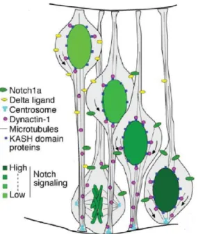
Shitamukai et al showed that inheritance of APs basal process was associated with self-renewal whereas its sister differentiates. Following spindle orientation modification, they observed that the daughter cell located basally to its sister inherits the basal process. These cells retain the basal process but not the apical one and delaminate to self-renew outside the VZ. This behavior is very much like what is observed in the primate with OSVZ precursors. They were also the first ones to identify OSVZ progenitors in normal conditions. Shitamukai et al observed that Notch is activated in the daughter cell that inherits the basal
26
process. Conversely it tends to be inactivated in the cell that does not inherit the basal process. Forcing Notch activation does not affect the basal process segregation but instead leads to the re-establishment of a basal process by the daughter cell that did not inherit it (Shitamukai et al., 2011).
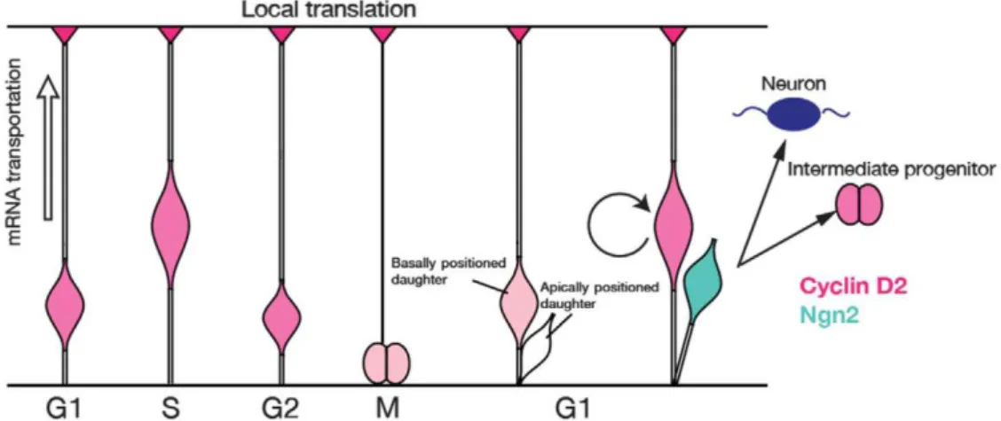
The basal process has been shown to be a site of active transportation and local translation of cell-cycle regulators, for instance CyclinD2 mRNA (figure 7). Tsunekawa et al looked for cellular factors influencing mitotic progenies to become APs or neurons. They observed a polarized CyclinD2 expression in the basal process which appears at the onset of neurogenesis and is maintained subsequently. There is actually an active transport of CyclinD2 mRNA to the basal endfoot that is further locally translated. The basal process is preferentially inherited by the daughter cell that becomes an AP. CyclinD2 overexpression increases the population of APs, while the loss of CyclinD2 increases the population of neurons. CyclinD2 is described as a “basal fate determinant” (Tsunekawa et al., 2012).
Figure 7: CyclinD2 mRNA and protein localization during the cell-cycle. CyclinD2 mRNA is
transported to the basal endfoot in S-G2 transition and translated into protein. During mitosis, CyclinD2 mRNA or protein is inherited by the basal daughter cell that becomes an AP (in pink). The daughter cell that does not inherit the basal process expresses Neurogenin2 (Ngn2 in blue) and becomes an intermediate progenitor (in pink) or a neuron (in dark blue). Extracted from Tsunekawa et al., 2012.
CyclinD controls the G1 cell-cycle restriction point (Sherr and Roberts, 1999). G1 reduction through forced expression of CyclinD was described to promote cell-cycle reentry at the expense of differentiation (Pilaz et al., 2009). Tsunekawa and Osumi investigated if basal process inheritance influences cell fate toward progenitor production. They observed that it does not always modify the total cell-cycle duration compared with the other daughter cell. CyclinD2 promotes p27Kip1 exportation to the cytoplasm leading to its
27
degradation (Susaki et al, 2007). Since p27Kip1 promotes neurogenesis and radial migration of post-mitotic neurons (Nguyen et al, 2006), inherited CyclinD2 may inhibit neurogenesis and promote cell proliferation via a p27Kip1-dependent mechanism (Tsunekawa and Osumi, 2012).
Okamoto et al showed that the basal process is essential for APs to undergo INM. They knockdowned TAG-1, thus leading to basal process shortening. These shortened APs did not perform INM and were observed accumulated at the ventricular surface (Okamoto et al., 2013).
The endoplasmic reticulum
The Endoplasmic Reticulum (ER) is found in the apical and the basal processes of APs. However, the basal process lacks Golgi-derived membrane constituents. These findings suggest there must be an unconventional vesicular secretory pathway that bypasses the Golgi apparatus to deliver the newly synthetized membrane components (Taverna et al., 2016).
28
III. Interkinetic Nuclear Migration
1 What is Interkinetic Nuclear Migration?INM was discovered by Sauer in 1935 (Sauer, 1935) and further characterized by Sauer and Walker in the early neural tube of the chick embryo. Using thymidine-H3 they showed that S phase cells nuclei are located basally within the epithelium and mitotic cells nuclei of the same population are located apically, thus suggesting nuclei migrate apically to divide at the ventricular border (Sauer and Walker, 1959).
Later, Hayes and Nowakowski studied the mouse E14 cortex using a cumulative labeling strategy (thymidine-H3 and BrdU) to understand the relationship between cell position in the VZ and the cell-cycle phase. They show that S phase nuclei do not change position and start the apical movement when entering G2 phase (Hayes and Nowakowski, 2000). Hence, during G1 phase the nucleus moves basally, undergoes S-phase basally at the same level and moves apically during G2 phase to divide at the ventricular border (see figure 8).
Figure 8: Schematic INM in the developing neocortex. The nuclei of the cycling progenitors move in
synchrony with cell-cycle progression. Following mitosis, the nucleus migrates basally during G1 phase, spends S phase at the same position basally and migrates apically during G2 phase to divide apically. Extracted from Hayes and Nowakowski, 2000.
INM is a conserved feature and can be found in a variety of tissues. It exists in Drosophila imaginal discs (Meyer et al., 2011), Nematostella larvae (Meyer et al., 2011), somites in the chick embryo (Langman and Nelson, 1968), in the vertebrate neuroepithelia -such as the Zebrafish retina (Norden et al., 2009) or the rodent cortex (Malatesta et al.,
29
2011; Noctor et al., 2001; see figure 9)-, in embryonic intestinal epithelium (Grosse et al., 2011) and in embryonic midgut (Yamada et al., 2013), liver, lung and pancreas (Bort et al., 2006).
Figure 9: INM in the VZ of the developing rodent neocortex. Coronal section and schematic
represention of INM in the rodent forebrain. APs are located in the VZ where they undergo INM. They extend radially through the telencephalic wall; they have processes attached to the ventricular surface and to the pial surface. Extracted from Kulikova et al., 2011.
2 Forces behind migration Basal nuclear migration
Schenk et al demonstrated that myosin II inhibition following blebbistatin treatment specifically disturbed the basal movement of INM in the mouse developing cortex. They described that myosin II-mediated constriction of the apical process pushes the nucleus in the basal direction as shown in figure 10A (Schenk et al., 2009). In the regenerating Zebrafish retina, the basal movement also relies on actomyosin (Lahne et al., 2015). Tsai et al obtained contradicting results using shRNA against myosin II and blebbistatin. Indeed, they did not find any significant effect on INM following myosin II inhibition. Moreover, they showed that the basal movement of INM relies on microtubules (MTs) and requires the kinesin KIF1A to move the nucleus toward the microtubule plus extremity (figure 10B) because forced expression of a RNA directed against KIF1A leads to a defect in the basal migration (Tsai et al., 2010).
30
Figure 10: Putative motors of basal nuclear migration in the mouse cortex. A: Myosin II drives the basal migration of INM. Actomyosin (in green) pushes the nucleus during the basal movement.
Extracted from Schenk et al., 2009. B: Involvement of microtubules in basal migration. Microtubules (in blue) extend basally (plus end) from the centrosome (pink circle). The kinesin KIF1 carries the nucleus along the microtubules toward the plus end. Adapted from Dantas et al., 2016.
Kosodo et al reported that the basal movement relies on passive forces generated by the displacement of nuclei moving apically. They added fluorescent microbeads on cultured mouse brain slices, the beads were integrated apically into the tissue using a magnet at the basal side. They observed the beads were transported basally but never exhibited an apical movement once reaching the basal side of the VZ. These results indicate that the basal movement is a non-autonomous process conditioned by the apical movement (Kosodo et al., 2011). Leung et al support these findings as they describe a stochastic movement arising passively from APs nuclei migrating apically in the Zebrafish retina. Indeed, following S phase arrest, APs nuclei do not perform the apical movement toward the ventricle thus impairing the basal movement of APs nuclei that underwent mitosis (Leung et al., 2011).
These findings are rather controversial and have not been clarified yet. Actin (Schenk et al., 2009) and microtubules are observed, with the latter encaging the nucleus (Norden et al., 2009, Tsai et al., 2010). However, their participation in the basal movement is not clear. Microtubules may serve to prevent the nucleus from “back-sliding” or may be required for a more active and persistent movement (Spear and Erikson, 2012).
31
Apical nuclear migration
Messier and Auclair reported that the absence of microtubules impedes the apical movement of INM in the chick neural tube (Messier and Auclair, 1973). Another study from Webster and Langman using cytochalasin B, which inhibits actin filament polymerization, shows that apical mitosis and cytokinesis are inhibited in the developing mouse cortex (Webster and Langman, 1978). Later, actin inhibition via cytochalasin B was shown to interfere with apical migration in the chick embryo retina (Murciano et al., 2002).
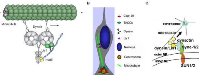
Microtubules are described to be involved in the apical movement during mouse cortical development (Gambello et al., 2003). The protein Lis1 interacts with the microtubule motor cytoplasmic dynein (Faulkner et al., 2000; Smith et al., 2000) and increases the affinity of dynein for microtubules (Vallee et al., 2012; figure 11A). It was observed, using in situ analysis, that Lis1 mutant embryonic mouse brains display an impaired apical movement (Gambello et al., 2003). Later, Tsai et al analyzed Lis1 RNA interference using live imaging experiments in rat brains. Interfering with Lis1 abolished apical migration, supporting previous findings from Gambello et al (Tsai et al., 2005).
Figure 11: Dynein mediates the apical movement of INM. A: Interaction between Lis1, NudE and dynein. Lis1 (light green and black) interacts with NudE (yellow and blue) and dynein (grey). Lis1
increases the dynein affinity for microtubules (dark green). Adapted from Vallee et al., 2012. B:
Microtubules are involved in apical migration. Cep120 recruits TACCs to the centrosome localized in
the cilia protruding from the apical process. They promote the assembly of microtubules from the centrosome. The nucleus is further carried by dynein along these microtubules toward the minus end (centrosomes). Adapted from Xie et al., 2007. C: SUN1/2 and Syne-1/2 mediate the connexion
between microtubules and nucleus. Inner NE proteins SUN1/2 interact with outer NE proteins Syne-1
and Syne-2. Syne-1 and Syne-2 bind dynein/Lis1 complex to carry the nucleus toward the minus end of microtubules (centrosome). Adapted from Zhang et al., 2009.
32
Xie et al showed that the centrosomal protein Cep120 recruits the Transforming Acidic Coiled-Coil protein 3 (TACC3) to the centrosome to promote microtubule elongation from the centrosome. The knockdown (KD) of one of these proteins impairs the apical movement of INM, thus suggesting microtubules are an essential motor of the apical movement in the mouse developing cortex (Xie et al., 2007; figure 11B). DOCK7 protein binds the centrosome as well as the protein TACC3 and prevents TACC3 from promoting microtubule extension in the mouse cortex. Indeed, following DOCK7 overexpression, APs nuclei display an impaired apical movement and divide away from the ventricular surface. Conversely, DOCK7 KD accelerates the apical movement (Yang et al., 2012). Pericentriolar Material 1 (PCM1) is a scaffold protein that recruits centrosomal proteins (Hames et al., 2005) such as Hook3 to pericentriolar satellites. When interaction between PCM1 and Hook3 is lost in the mouse developing cortex, the centrosome ability to organize microtubules is impaired. Thereby, INM is defective, as testified by abventricular mitosis (Ge et al., 2010). Cappello et al demonstrated that the Nuclear distribution protein C (NudC), which interacts with Lis1 (Morris et al., 1998), is required for apical migration in embryonic rat brain (Cappello et al., 2011). This result is consistent with the requirement of dynein-associated microtubules for the apical movement.
Del Bene et al studied the mokZebrafishmutant in which the dynactin, an adaptor between the dynein and its cargo (reviewed in Schroer, 2004) is disrupted. They showed that INM is modified in the retina: the nucleus moves faster and further during the basal movement and slower during the apical movement. The division was often observed to take place away from the apical surface. Furthermore, disruption of Syne2 (or Nesprin2), which contains a KASH domain, linking the nucleus to the cytoskeleton (Starr and Fischer, 2005) also impairs INM (Del Bene et al., 2008). Altogether, these findings suggest a role for microtubule associated dynein in the apical movement to carry the nucleus to the apical surface. However, no failure in INM following dynactin disruption was observed in the Zebrafish retina by Norden et al. They impaired the dynein-dynactin complex using a dominant negative of dynein and found that dynein plays a role in the apical movement but is not necessary for it. Furthermore, the absence of stable microtubules or the absence of microtubules does not impair the apical movement (Norden et al., 2009). These results are in contradiction with previous findings (Del Bene et al., 2008). Indeed, in the Zebrafish retina the apical movement appears to rely largely on actomyosin contractions as myosin II
33
inhibition blocks INM (Norden et al., 2009). Later, Leung et al showed that at the onset of G2, myosin concentrates at the basal side of the nucleus and pushes it toward the ventricular surface (Leung et al., 2011), thereby confirming previous findings (Norden et al., 2009). Actin and ROCK signaling were found to be necessary for the apical movement in the Drosophila wing imaginal disc and the Nematostella larvae since either addition of Cytochalasin D or inhibition of the RhoA-ROCK pathway abolished the movement in both species (Meyer et al., 2011). Rujano et al found that INM in the Drosophila optic lobe requires myosin II. The Abnormal Spindle Microtubules Assembly (ASPM) orthologue Abnormal Spindle Protein (ASP) interacts with myosin II at the basal side of the neuroepithelium. ASP controls myosin II polarization and, following disruption, the apical movement is impaired (Rujano et al., 2013). Actin involvement in INM of retinal progenitors was recently confirmed by the Norden group in the Zebrafish retina. They altered aPKC distribution, thereby inducing the loss of actin filament organization. The apical movement was subsequently impaired and abventricular divisions were observed (Stzryz et al., 2015). Lahne et al studied the Zebrafish regenerating retina and observed that actin accumulates at the rear of the nucleus and contraction of actomyosin mediates INM (Lahne et al., 2015). Although ROCK is required to facilitate the apical movement, they showed that following inhibition of ROCK, the basal movement is impaired, thereby supporting findings from Schenk et al (Schenk et al., 2009).
The CK2 protein (formerly known as casein kinase 2) is a MAP involved in microtubule stabilization (Lim et al., 2004). Inhibition of CK2 blocks the apical movement in the rat retina. Indeed, stable microtubule levels are decreased with concomitant increase of free tubulin dimers compared with control retina (Carneiro et al., 2008). This work is in contradiction with the findings obtained is the Zebrafish retina (Norden et al., 2009). The microtubule-associated protein Tpx2 was shown to redistribute from the nucleus to the apical process during S-G2 transition, thus regulating the apical movement in the mouse cortex. Tpx2 modulates microtubule organization to promote apical nuclear migration (Kosodo et al., 2011). Microtubules are involved in the apical movement in the Nematostella larvae since abventricular mitosis are observed following colchicine mediated disruption of microtubules (Meyer et al., 2011).
A study in the chick neural tube and the mouse cortex described that the apical movement relies on microtubules and the mitotic rounding involves actin (Spear and
34
Erickson, 2012). Zhang et al analyzed the apical movement of APs from Sun1/2 double KO or Syne-1/2 (Nesprin 1/2) double KD brains and observed that the apical movement was slower. They demonstrated that SUN1 and SUN2 recruit Syne-2 to the Nuclear Envelope (NE). Furthermore, Syne-2 interacts with the dynein-dynactin complexes but also kinesin on the NE. Hence, SUN1/2 and Syne-2 connect microtubules to the NE and these proteins are needed for an efficient INM movement (Zhang et al., 2010; figure 11C). The importance of these proteins during INM in the mouse retina was also demonstrated (Yu et al., 2011). Dynein recruitment to the nuclear pore was further explored in the rat developing brain by Hu et al. They interfered with two pathways that recruit dynein, RanBP2-BicD2 and Nup133-CENP-F (see figure 12A), and observed an inhibition of the apical movement along with abventricular divisions. Inhibition of the RanBP2-BicD2 pathway arrested nuclei more basally than inhibition of the Nup133-CENP-F pathway (figure 12B), suggesting a sequential activation of those pathways (Hu et al., 2013). The team later showed that these two pathways of dynein recruitment are independently regulated. Indeed, the Cyclin Dependant-Kinase CDK1 targets BicD2 to the NE, which drives the apical movement and allows CENP-F nuclear exit thereby triggering dynein recruitment (figure 12C) (Baffet et al., 2015).
35 Figure 12: Recruitment of dynein to the NE regulates the microtubule based apical movement of INM. A: Different dynein recruitment to the NE. The nucleoporin RanBP2 recruits BicD2, which binds
dynactin and dynein (Splinter et al., 2012). The nucleoporin Nup133 recruits CENP-F (Bolhy et al., 2011), which binds NudE and NudEL. NudE and NudEL then directly bind Lis1 and dynein (Niethammer et al., 2000; Sasaki et al., 2000). Adapted from Hu et al., 2013. B: Impaired dynein recruitment to the
NE leads to apical movement arrest. Inhibition of the RanBP2-BicD2 pathway arrested the nuclei
more basally than the inhibition of the Nup133-CENPF-F pathway did. Extracted from Hu et al., 2013.
C: CDK1 regulates dynein recruitment to the NE. CDK1 targets BicD2 to the NE and drives CENP-F
nuclear exit. Extracted from Baffet et al., 2015.
Differences between pseudostratified epithelia
This brief overview of the analysis of the forces driving basal and apical movements reveals that there is no consensus regarding the mechanisms behind INM. The perspectives proposed by the Norden group to better understand INM shed some light on this prospect.
The thickness of the Pseudostratified Epithelium (PSE) seems to have a great influence on the machinery used by the progenitors to drive the apical movement. Strzyz et al proposed a ranking where they differentiate (i) short PSE, measuring between 20 and 30 µm, where nuclei are arranged into two or three nuclear layers (ii) intermediate PSE, measuring between 30 and 60 µm, where nuclei are arranged into four or five nuclear layers
A B
36
and (iii) long PSE that can measure up to a few millimeters, where nuclei are arranged into more than eight nuclear layers (Strzyz et al., 2015, see figure 13).
In intermediate PSE, such as the Drosophila imaginal disc or the Zebrafish retina, measuring around 50 µm (Lee and Norden, 2013), the apical movement is driven by actomyosin (Meyer et al., 2011; Norden et al., 2009). However, Del Bene et al showed that the apical movement in the Zebrafish retina is slower in absence of dynactin, which is an argument in favor of microtubule involvement during the apical movement (Del Bene et al., 2008). Norden et al explained this contradiction by the fact that the nucleus movement had been measured for 1 hour, which is not a sufficient time window to observe the rapid apical movement occurring before mitosis (Norden et al., 2009). In the Nematostella, both microtubules and actin are required during apical INM, which is in contradiction with the discussed theory since the length of the epithelium is about 50 µm. Actomyosin may be the necessary motor in intermediate PSE because the progenitors process is thicker and thus contains more cytoplasm compared to long PSE (Lee and Norden, 2013; Strzyz et al., 2016).
In long PSE such as the chick neural tube, measuring about 80 µm in later stages, or the rodent cortex measuring about 100 µm (Okamoto et al., 2014) the apical movement appears to rely mostly on microtubules (Gambello et al., 2003; Tsai et al., 2005; etc) but some studies reported that actomyosin is important in the process (Webster and Langman, 1978; Murciano et al., 2002). A more recent study suggests that actomyosin has a role in allowing the cell to adopt a round shape to enter mitosis (Spear and Erickson, 2012). As epithelia were growing thicker, microtubules might have been an evolutionary response for the nucleus to be carried apically more efficiently. However, there is no consensus to distinguish if microtubules and actomyosin or microtubules alone are required in the apical movement. The mechanisms behind it may depend on other factors than the thickness, such as the tissue characteristics or the developmental period.
37
Figure 13: The apical movement is driven by different molecular motors depending on the PSE thickness. In short PSE, actomyosin triggering mitotic cell rounding appears to be sufficient to push
the nucleus apically. In intermediate PSE, actomyosin is the motor of the apical movement. In long PSE cells, such as radial glial cells, dynein associated microtubules drive the apical movement. Arrows represent the time. Adapted from Norden et al., 2017.
Directionality of INM
The apical movement of INM was described to be faster and more directional than the basal movement, which is reported to be rather stochastic in the mouse and the Zebrafish models (Xie et al., 2007; Norden et al., 2009; Leung et al., 2011; Kosodo et al., 2011; Tsai et al., 2010; Okamoto et al., 2014).
However, in the ferret, the apical movement is less directional and the basal movement is more directional compared to the mouse developing cortex. Accordingly, a reduced nuclear density in the periventricular space is observed in mice (Okamoto et al., 2014). This is likely explained by the greater density of apical endfeet in ferrets. Indeed, the stiffness of the VZ at the apical surface is more important in ferret compared to mouse (Nagasaka et al., 2016; see figure 14).
38
Figure 14: Differences between mouse and ferret VZ, APs and associated INM. The thickness of the
VZ is more important and APs are slender in the ferret. The apical movement is less directional and the basal movement is more directional in the ferret. The density of apical endfeet is more important in the ferret explaining the greater stiffness of the apical surface. Extracted from Nagasaka et al., 2016.
Coupling between cell-cycle progression and INM
Nuclei are described to migrate basally during G1, spend S phase basally and migrate apically so as to divide at the ventricular border. So what happens if INM is impeded or if the cell-cycle is delayed, accelerated or simply stopped?
Murciano et al blocked INM and observed that the cell-cycle continues and cells further divide basally (Murciano et al., 2002), thereby suggesting that the cell-cycle can be uncoupled from INM. Tsai et al observed contradicting results; they reported that blocking the apical movement using Lis1 RNAi prevents mitosis (Tsai et al., 2005). This could be explained by the fact that in absence of Lis1, division is blocked. However, in this study no mitotic event was observed and other studies reported that division ceases at the prometaphase state (Tsai et al., 2005). Okamoto et al showed that APs with a shortened basal process, which are unable to undergo INM, continue cell-cycle progression and divide to give rise to progenies (Okamoto et al., 2013). More recently, Carabalona et al observed that impaired basal migration does not affect cell-cycle progression. Indeed, the cycling fraction and cells proportion within each phase of the cell-cycle were not modified (Carabalona et al., 2016).
39
Ueno et al showed that inhibition of mitosis prevents the basal movement and that S phase arrest prevents apical migration (Ueno et al., 2006). Arrested cells nuclei keep the position they were occupying before exiting the cell-cycle. Ueno et al demonstrate here that cell-cycle is required for INM progression. It should be noted that the reverse is not true and cells can progress in the cell-cycle when INM is blocked (Murciano et al., 2002; Cappello et al., 2006; Tamai et al., 2007). This was confirmed by Leung et al, who arrested APs nuclei in S phase and observed the absence of apical movement (Leung et al., 2011).
Sargeant et al increased G2-M duration using a morphine treatment and observed slower apical movement, thereby showing a coupling between cell-cycle progression and INM (Sargeant et al., 2008). Kosodo et al arrested cells in G1 and observed nuclear accumulation at the basal side of the VZ. Furthermore, following arrest in S phase, the apical movement was slower compared to non-arrested nuclei (Kosodo et al., 2011). APs nuclei thus appear to stay blocked at the position in which they were during INM following cell-cycle arrest.
Recently, Hu et al showed that apical nuclear migration is required for mitosis to take place. They suggest the nucleus must reach the ventricular surface to enter mitosis (Hu et al., 2013). These findings were contradicted by Strzyz et al, who observed that mitosis was not restricted to the ventricular surface as it has been described in previous studies (Murciano et al., 2002; Cappello et al., 2006; Tamai et al., 2007). They discovered that the apical movement was triggered by CDK1 at the end of S phase (Strzyz et al., 2015). This result is an argument in favor of a coupling between cell-cycle and INM since CDK1 regulates entry in G2 phase (Sherr and Roberts, 1995). Baffet et al explained how CDK1 triggers the apical movement in the rat cortex. They showed that CDK1 regulates dynein recruitment to the NE (Baffet et al., 2015). Since dynein does not appear to be involved in the apical movement in the Zebrafish retina (Norden et al., 2009), CDK1 may have kept the role of coupling G2 phase entry with apical movement initiation during evolution and adapted this role by regulating dynein interaction to the NE.
3 Centrosome behavior and mitosis in APs
It is interesting to study the mitotic behavior of APs performing INM and its consequences. In this phenomenon, there is a central role for centrosomes.