HAL Id: inserm-00398108
https://www.hal.inserm.fr/inserm-00398108
Submitted on 17 May 2010HAL is a multi-disciplinary open access archive for the deposit and dissemination of sci-entific research documents, whether they are pub-lished or not. The documents may come from teaching and research institutions in France or abroad, or from public or private research centers.
L’archive ouverte pluridisciplinaire HAL, est destinée au dépôt et à la diffusion de documents scientifiques de niveau recherche, publiés ou non, émanant des établissements d’enseignement et de recherche français ou étrangers, des laboratoires publics ou privés.
Revisiting the stimulus-secretion coupling in the adrenal
medulla: role of gap junction-mediated intercellular
communication.
Claude Colomer, Michel G Desarménien, Nathalie C. Guérineau
To cite this version:
Claude Colomer, Michel G Desarménien, Nathalie C. Guérineau. Revisiting the stimulus-secretion coupling in the adrenal medulla: role of gap junction-mediated intercellular communication.. Molec-ular Neurobiology, Humana Press, 2009, 40 (1), pp.87-100. �10.1007/s12035-009-8073-0�. �inserm-00398108�
#MOLN-D-09-00009
Revisiting the stimulus-secretion coupling in the adrenal medulla: role of gap
junction-mediated intercellular communication
Claude Colomer1,2,3,4
, Michel G. Desarménien1,2,3,4
and Nathalie C. Guérineau1,2,3,4 1
Institute of Functional Genomics, Montpellier, France; 2
CNRS UMR5203, Montpellier, France; 3INSERM U661, Montpellier, France; 4University of Montpellier, Montpellier, France.
Abstract
The current view of stimulation-secretion coupling in adrenal neuroendocrine chromaffin cells holds that catecholamines are released upon trans-synaptic sympathetic stimulation mediated by acetylcholine released from the splanchnic nerve terminals. However, this traditional vertical scheme would merit to be revisited in the light of recent data. Although electrical discharges invading the splanchnic nerve endings are the major physiological stimulus to trigger catecholamine release in vivo, growing evidence indicate that intercellular chromaffin cell communication mediated by gap junctions represents an additional route by which biological signals (electrical activity, changes in intracellular Ca2+ concentration,...) propagate between adjacent cells and trigger subsequent catecholamine exocytosis. Accordingly, it has been proposed that gap junctional communication efficiently helps synapses to lead chromaffin cell function, and in particular hormone secretion. The experimental clues supporting this hypothesis are presented and discussed with regards to both interaction with the excitatory cholinergic synaptic transmission and physiopathology of the adrenal medulla.
Index Entries: Gap junctions, adrenal medulla, innervation, chromaffin cells, cholinergic
synaptic transmission, catecholamine release, stimulus-secretion coupling, development, stress, physiopathology.
Overview of the neurogenic control of catecholamine release in the adrenal medulla
The adrenal medulla is the latest link of the sympathoadrenal axis and is involved in the ability of the organism to generate appropriate reaction to stressful situations. This adaptive response to stressor agents involves the secretion of adrenal catecholamines (adrenaline and noradrenaline) by the neuroendocrine chromaffin cells. Once released into the blood circulation, catecholamines contribute to the regulation of multiple functions including cardiovascular function as well as energy metabolism, thus helping the organism to cope with a changing environment. With regards to catecholamine secretion by chromaffin cells, the common view is that it is chiefly controlled by synaptically released acetylcholine at the splanchnic nerve terminal-chromaffin cell synapses (1,2; Fig. 1). The first demonstration of a neuronal control of the release of catecholamines from the adrenal gland comes from the pioneer work of Dreyer (3), showing that the stimulation of the splanchnic nerve caused an outflow of the “pressor activity” from the adrenal. This “pressor activity” has been further identified as being mediated by the release of adrenaline (4). In the early twentieth century, numerous studies have confirmed that catecholamine release from the adrenal medulla is controlled by the splanchnic nerve activity (5,6). Within the medulla, chromaffin cells are organized into discrete cell complexes (7) and each chromaffin cell receives its own synaptic inputs, up to five synapses per cell (8-10). Because it is currently proposed that each chromaffin cell cluster functions as an independent unit to release catecholamines after activation of the splanchnic nerve (11), it was of physiological interest to characterize
stimulus-secretion coupling at the cell cluster level. In particular, recent studies have focused on the functional role of direct intercellular communication between chromaffin cells.
It has long been thought that, since the triggering of hormone release is chiefly achieved by synaptic neurotransmission at the splanchnic nerve-chromaffin cell contacts, there was no need for an additional stimulatory signal exchanged between chromaffin cells. The present article reviews the evidence supporting the presence of cell-to-cell coupling mediated by gap junctions between adrenal chromaffin cells and the consequences of this coupling on hormone secretion, an emerging area of potential physiological/physiopathological interest that has received too little attention so far.
Gap junctions in the adrenal medullary tissue
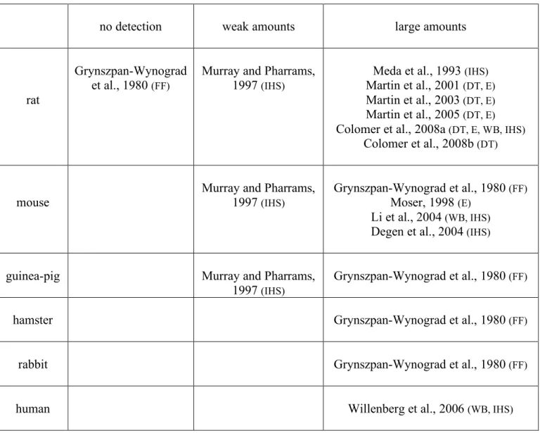
Gap junctions are specialized regions of the plasma membrane in which protein oligomers establish a contact between adjacent cells, thereby forming conduits for intercellular communication that allows coupled cells to exchange nutrients, metabolites, ions, second messengers and small molecules under ~1000 Da. With regards to molecular identity, gap junctions in chordate are chiefly assemblies of connexins (12-14). Indeed, regarding the more recently discovered pannexin family (15-18), their contribution to functional gap junctions in vertebrates is still unclear. Their functional roles are only beginning to emerge. To date, expression of pannexins in the adrenal gland has not been reported, neither in the cortex nor in the medulla. By contrast, connexins are widely present in the adrenal gland, with a preferential expression in the zona fasciculata and zona reticularis of the cortex (19). In the medulla, connexin-built gap junctional plaques have been originally described in the eighties from observations of freeze-fractured specimens (20). By using this experimental procedure, gap junction clusters have been found in several species including mouse, guinea-pig, hamster and rabbit but, surprisingly not in rat. Later on, the use of immunohistofluorescence and
immunoblot techniques revealed the presence of connexins in the rat adrenal medulla (21-23). More recently, gap junctions have also been found in human medulla (24). Table 1 summarizes the available data concerning the presence of connexin-related gap junctions in the adrenal medulla of different species.
Connexin subtypes expressed in the adrenal medulla
Since the cloning of the first members at the end of the 1980s, the connexin family has considerably expanded, and there are about 20 identified genes (14). To date, six connexins (Cx26, Cx29, Cx32, Cx36, Cx43 and Cx50) have been found in the adrenal medulla (Table 2). Their expression not only varies between species but also differs between normal and tumoral tissue. Cx43 appears to have the highest expression, in rat (19,21-23), in mouse
(19,25-27), in guinea-pig (19) and in human (24). By contrast, expression of Cx50 seems to
be restricted to human, both in normal medulla and in pheochromocytomas (24). Not suprinsingly, Cx36, a connexin present in neuronal tissues (28) is also expressed in the adrenal medulla, which originates from the neural crest, and in the rat tumoral cell line PC-12
(29). Interestingly, acquisition of a tumoral phenotype appears to be accompanied by a change
in connexin expression. Indeed, Cx26 and Cx32, mainly identified in exocrine but not in endocrine glands (21), are present in human pheochromocytomas while they are absent from normal medulla (24).
Brief focus on gap junction hemichannels
The formation of a gap junction channel results from the apposition of two gap junction hemichannels, which are delivered to the plasma membrane (30,31) where they diffuse laterally into cell-contact regions to dock head-to-head with partner connexins present on the neighboring cell. Hemichannels have long been considered as mere structural precursors of
gap junctions. This concept of hemichannels being only the halves of gap junction channels began to be questioned after the finding that they can generate ionic currents and modulate cell activity (32). Functional hemichannels have been described in various tissues such as glial cells, retina or heart (33-35) and more recently in PC-12 cells (36). They have been reported to contribute to paracrine communication or to the integration of the incoming information (37-39). When open, hemichannels that are traveling on the plasma membrane prior to incorporation into gap junction plaques allow diffusion of ions and small molecules across the plasma membrane (37,40,41). It is noteworthy that some results are still discussed
(42). Various factors can be released from a cell in the extracellular medium through
hemichannels, such as ATP from Cx36- and Cx43-built hemichannels (43,44,45), or glutamate from Cx32-built hemichannels (46). As mentioned above, adrenal chromaffin cells express both Cx36 and Cx43 and although the expression of connexin-built hemichannels has not yet been reported in adrenal medullary tissue, one could reasonably propose that gap junction hemichannels might be potent modulators of the adrenal medulla physiology. If this hypothesis is confirmed, connexins would contribute to two modes of communication in the adrenal medulla : i) direct cell-cell communication between chromaffin cells coupled by gap junctional channels and ii) diffuse paracrine communication, targeting several cell types, through hemichannels. Because gap junction channels and hemichannels can be differently regulated by a same molecule (47,48), both communication modes likely contribute to specific and distinct functions in a tissue.
Adrenal medullary cell types expressing connexin-built gap junctions
Since the adrenal medullary tissue is mainly composed of neuroendocrine chromaffin cells, gap junctional communication was first investigated at the chromaffin cell level. Suspicion that chromaffin cells may be electrically coupled in situ first came from
electrophysiological recordings of membrane potentials in hemi-sectioned gland (49,50). Later on, comparison of the membrane properties of chromaffin cells measured in thin acute tissues slices, to those of isolated cultured cells, suggested that chromaffin cells are electrically coupled (22,51). Confirming this hypothesis, dual patch-clamp recordings from chromaffin cell pairs showed that electrical signals can propagate from the stimulated to the non-stimulated cell (22,23,52,53). Regarding the molecular determinants supporting gap junctional communication between chromaffin cells, transcripts encoding Cx36 and Cx43 were detected in single chromaffin cells by using RT-PCR (22) and related proteins were next detected by immunoblot and immunohistofluorescence techniques (23). An unsolved issue deals with the specificity of gap junctional coupling between chromaffin cells, in particular with regards to the hormonal nature of coupled cells. Chromaffin cells can be divided into two separate populations, depending on the hormone secreted (54). To date, we do not know whether gap junctions connect only adrenaline-containing cells or only noradrenaline-containing cells or both. Because i) these two populations of chromaffin cells are innervated and regulated by morphologically different nerve terminals originating from distinct spinal cord and brain regions (55-58) and ii) gap junctional coupling can be modulated by the cholinergic synaptic activity (52), elucidating this question would help understanding the contribution of gap junctions to hormone secretion.
Although chromaffin cells are the predominantly expressed cellular constituents of the adrenal medulla, it also contains non neuroendocrine cells including sustentacular cells, homologous in nature to Schwann cells (59,60). As extensively documented in other tissues including both neuronal and endocrine tissues, glial cells or glial-like cells are highly coupled by gap junctions (61-63). Sustentacular cells in the adrenal medulla are likely gap junction-coupled, as evidenced by the expression of Cx29 (27). It is noteworthy that the presence of gap junctions in the other medulla tissue components, i.e. ganglion cells, nerve fibers
originated from pre-ganglionic sympathetic axons, connective-vascular tissue, small intensely fluorescent (SIF) cells and pluripotent stem cells (60,64,65) has not been reported yet.
Functional roles of gap junction-mediated cell-to-cell communication in the adrenal medulla: the stimulus-secretion coupling revisited
While gap junctions in the adrenal cortex have been extensively studied, very few works have been devoted to the functional characterization of gap junctional coupling in the medulla. Until the beginning of the two thousand-years, the current view with regards to stimulus-secretion coupling in the adrenal medulla was that there is no need for a direct coupling between chromaffin cells to ensure hormone release since each chromaffin cell receives its own synaptic input. However, a series of recently obtained data allows confronting this view.
Gap junctional communication between chromaffin cells: a tool for synchronizing their secretory activity
The hypothesis of a direct cell-to-cell communication between adrenal chromaffin cells amplifying the secretory signal in the intact gland has been initially proposed by Ceña and collaborators (66). At the same time, the publication of a series of works illustrating the influence of chromaffin cell-cell contacts on levels of tyrosine hydroxylase, an enzyme involved in catecholamine biosynthesis, strongly supported this hypothesis (67-69), although the nature of the intrinsic membrane protein involved was not characterized. A decade later, a junctional pathway coupling mouse chromaffin cells in situ has been reported to potentially underlie the simultaneous firing of a cell cluster (51). This contrasts with a study performed in rat and published one year before reporting that the spreading of electrical activity between rat chromaffin cells induced by transmural stimulation of the splanchnic nerve would be mostly
dependent on nerve activity rather than on cell-to-cell coupling (70). More recently, the presence of functional gap junction-mediated electrical coupling between rat chromaffin cells has been reported in acute adrenal slices (22). It is noteworthy that the percentage of coupled chromaffin cells is species- and gender-dependent with a higher percentage in female versus male, and also differs as a function of the physiological state of the animals (Fig. 2A). In addition, simultaneous recordings from chromaffin cell pairs allowing to monitor the junctional current flowing between two cells revealed two populations of coupled chromaffin cells: a weakly coupled cell population and a robustly coupled cell population (22,23). Again, the proportion of the two populations differs between male and female rats and also differs as a function of the physiological state of the rats (normal versus stressed, 23; adult versus neonate 53; Fig. 2B). As remarkable differences, one can point out i) the dramatic up-regulation of electrical coupling in stressed rats that is associated with the appearance of robust coupling in half of cell pairs and ii) the increased electrical coupling in neonate rats when compared to adults. These results will be discussed later.
Strengthening the role of gap junctions in adrenal catecholamine release, it is noteworthy that the higher level of gap junctional communication in rat female parallels i) higher basal circulating adrenaline and noradrenaline concentrations, as compared to male
(71), and ii) an increased catecholaminergic activity of the adrenal medulla in female (72,73).
The underlying mechanisms still remain to be elucidated. One attractive hypothesis is that the expression level of connexins may be higher in female. This would be especially efficient for Cx43, which exhibits a higher unitary conductance than Cx36 (74,75). This hypothesis is strengthened both by steroid hormone-induced regulation of Cx36 and Cx43 expression levels and by a sexually dimorphic regulation of Cx43 (76-79). Although hormonal and gender-specific regulation of Cx36 and Cx43 has not been yet reported in the adrenal medulla, this
could likely be part of the explanation for different gap junctional coupling strength and extent between male and female chromaffin cells.
The contribution of Cx36- and/or Cx43-built channels to weak versus robust coupling is unknown but, since Cx36 and Cx43 channels exhibit different voltage sensitivities (80), their roles in propagating electrical signals between adjacent cells likely differ. The weak voltage dependence of Cx36 channels suggests that electrical coupling may not be disrupted during secretagogue-mediated action potential discharges. By contrast, Cx43 channels that are highly voltage-sensitive may predominantly be regulated during firing. Through the coupling of electrical activities, gap junctional communication provides the basis for coordinating spontaneous and agonist-induced synchronous multicellular [Ca2+]i increases (22,23,81). The synchrony of [Ca2+]i changes amplifies catecholamine release in response to excitation of a single chromaffin cell (22), and could therefore constitute an efficient complement to synaptic neurotransmission. This complementary action of gap junctional coupling and neuronal command is particularly obvious in situations in which the neuronal control of catecholamine release is not fully competent, that is at birth, in splanchnectomized adrenals or in response to pharmacological blockade of postsynaptic nicotinic receptors (52,53,82). In summary, it is now clear that gap junction-mediated communication between chromaffin cells complements the nerve command, or even relays it in some cases. Both signals contribute to the synchronization of electrical activity, ensuing [Ca2+]i transients and hormone secretion.
Although the function of gap junctional communication to help synapses is of major interest for the adrenal secretory tissue, one could reasonably think that gap junctions between chromaffin cells might be involved in other functions, based in particular on the fact that chromaffin cells do not represent a homogeneous cell population. Chromaffin cells can be distinguished by the peptidergic content of their secretory granules (83,84), their localization within the lobule (at the periphery versus at the center), their nearness with the cortex or their
vicinity with blood vessels. A specific coupling of cells belonging to one of these populations may be relevant in physiological conditions (such as during embryogenesis, postnatal development, ...) or in pathological conditions (such as during inflammation, infectious diseases, ...). The exhaustive study of gap junctional communication (in terms of connexin subtypes expressed, coupling strength, coupling extent, ...) between these different chromaffin cell populations would likely unmasks not yet anticipated roles of gap junctions and connexins in the medullary tissue function. Indeed, gap junction channels are permeant for several second messengers including Ca2+, cAMP and cGMP which regulate numerous cell activities. Additionally and independently of their gap junctional function, connexins also directly interact with a wide variety of signaling intracellular and membrane-associated proteins and can regulate gene expression and cell function (85,86). Gap junctions are now perceived, not only as channels between neighboring cells, but also as signaling complexes that regulate cell function and transformation. Subsequently, it should be considered in further studies that, beyond a tool for synchronizing catecholamine release, connexins may also be involved in many chromaffin cell functions such as differentiation, growth, division, repair, adhesion.
Gap junctional communication between sustentacular cells
To date, a unique study describes the presence of connexin-built gap junctions between adrenal sustentacular cells, mediated by Cx29 channels (27), which were recently reported as a novel marker for cells of the defined Schwann cell lineage (87). Nevertheless, based on other reports on sustentacular cells (88) and other glial-like cell types (63,89), we propose that gap junction-coupled sustentacular cells may form a long-distance communication route, as described for the anterior pituitary folliculostellate cell network (90). This hypothesis is consistent with a recent study suggesting that adrenal medulla sustentacular
cells take an active part in Ca2+ metabolism, regulating indirectly the synthesis and release of catecholamines from chromaffin cells (91).
Contribution of gap junctions to interactions between chromaffin cells and other elements of the adrenal tissue
In the medulla, hormone secretion from chromaffin cells results from the precise coordination and integration of cellular and intercellular signals arising from distinct inputs. Not only can catecholamine secretion of a single chromaffin cell be modulated by neighboring chromaffin cells if gap junction-coupled (22), but secretion can also be regulated by other tissue components. First, splanchnic nerve endings synapsing onto chromaffin cells are major inputs controlling intracellular signaling and ensuing catecholamine release
(2,92,93). Is gap junctional communication between chromaffin cells able to interact with
synaptic cholinergic neurotransmission? Although the underlying mechanisms are not totally elucidated, it is clear that gap junction-mediated cell-to-cell communication is under a tonic inhibitory control exerted by synaptic activity, as also reported in other neuronal tissues
(94,95). Indeed, studies performed under experimental conditions leading to impairment of
synaptic transmission (pharmacological blockade of post-synaptic nicotinic receptors, unilateral splanchnectomy, immature synaptic transmission in newborn rats) showed an up-regulation of gap junctional coupling between chromaffin cells (52,53). Second, sustentacular cells that are commonly observed in close proximity to chromaffin cells and that elongate their thin processes between secretory cells (60) are also good candidates to modulate signaling pathways in adjacent chromaffin cells and therefore catecholamine release. Assuming that sustentacular cells can form a functional circuitry intermingled between chromaffin cells, they may also have a privileged role in coordinating the biological activities
of distant chromaffin cells in both physiological and pathological conditions, and thus may modulate hormone release.
At the level of the whole adrenal gland, that is cortex and medulla, it is likely that gap junctional communication between medullary cells may influence activities of cortical cells and further release of steroids and sex hormones. Although no data ascertain this hypothesis yet, several evidences support it: i) morphological data show close appositions of chromaffin cells with cortical cells (96,97) in particular with dehydroepiandrosterone (DHEA)-producing cells (98), and ii) functional interaction mediated by paracrine factors between chromaffin and cortical cells have been extensively reported (99). In particular, the release of catecholamines locally supports influence of chromaffin on neighboring cortical cells (100). Thus, we propose that, by modulating catecholamine secretion, gap junctional coupling may indirectly affect cortical cell function. Reciprocally, as previously reported for induction of catecholamine enzymes in chromaffin cells by glucocorticoids (101), steroids and sex hormones released from the cortex may also influence junctional coupling in the medulla. Because adrenocortical factors are involved in many cell functions (differentiation, growth, apoptosis, hormone release, ...), their action on chromaffin gap junctional communication may be particularly relevant in physiological/pathological situations such as development or tumorigenesis.
Gap junctional communication in the developing adrenal medulla: towards the identification of the mechanism responsible for adrenal catecholamine secretion in response to birth-induced stress
In developing neuronal tissues, a finely tuned coordination between gap junction-mediated electrical coupling and synaptic neurotransmission (102,103) is crucial for synapse formation. At birth, the synaptic transmission is immature in the rat adrenal medulla (53) and it becomes fully competent during the first post-natal week (104,105). Maturation of
cholinergic synaptic transmission results from a 2-fold increase in synaptic current amplitude without significant change in synaptic current frequency (53). In parallel, the gap junction-mediated coupling is prominent at birth (52; Fig. 3A). With the help of the extracellular matrix protein agrin (53,82), electrical coupling then decreases, in coincidence with the establishment of functional and mature chemical synapses. What can be the role of gap junctions between chromaffin cells in newborn rats and can they be involved in catecholamine secretion? During birth, neonate rats are subjected to various stresses, including hypoxia, hypoglycemia or glycopenia. During this period, the adrenal medulla exerts a very specific protective function, in particular against hypoxia, involving a variety of respiratory, cardiovascular and metabolic adjustments (105). In the absence of functional synapses originating from the splanchnic nerve, this function is handled directly by chromaffin cells via their ability to respond to hypoxia by a non-neurogenic catecholamine release (106). It is likely that cell-to-cell communication mediated by extracellular factors is not appropriate for the massive catecholamine mobilization which is needed at that time. Gap junction-mediated fast signal propagation would therefore become physiologically relevant. The contribution of gap junctional communication to the non-neurogenic response is also indicated by the fact that both the high occurrence of electrical coupling and the non-neurogenic response observed in neonates decrease during the first postnatal week and are inhibited by establishment of functional splanchnic synapses (52,53,104). In addition, the non-neurogenic response to hypoxia involves calcium ions and reactive oxygen species (107,108), two messengers that are diffusible through gap junctions (13,109-111). Since only a proportion of chromaffin cells are sensitive to hypoxia in late embryos and neonates (106), we propose that gap junctional communication facilitates the spreading of the response from hypoxia-sensitive cells to the whole population of chromaffin cells and thus plays a major role in the extent of the response of the adrenal medulla to the birth-related hypoxic stress. In view of the impact of this
non-neurogenic response on the adaptation of respiratory and cardiac function to the extrauterine life (104), this would provide gap junctions with a major role in early neonatal life.
Beyond the role of junctional communication as support to catecholamine secretion in neonates, it is likely that gap junctions also play important functions during adrenal embryogenesis, in particular with regards to acquisition of a chromaffin cell fate and subsequent acquisition of an adrenergic or a noradrenergic phenotype. In addition, the acquisition of a specific neuropeptidergic phenotype during development (112) may also be helped by gap junctional communication. By promoting factor exchange between cells, gap junctions may contribute to a harmonious exposure of sympathoadrenal progenitors to morphogenetic factor-induced instructive signals (65,113).
Gap junctional communication: involvement in physiopathology of the adrenal gland The “stressed” adrenal medulla
A rise in catecholamine plasma levels is a key event in response to stressors. Consistent with a crucial role of the adrenal gland in stress-induced catecholamine secretion, the "stressed" medulla is significantly remodeled when compared to an "unstressed" gland (Fig. 3B). In particular, the gap junction-mediated metabolic and electrical coupling between chromaffin cells is dramatically up-regulated (23,114). Regarding electrical coupling, the most striking change is the appearance of robustly coupled chromaffin cells, leading to the transmission of action potentials and ensuing [Ca2+]i transients between coupled cells. We
propose that the up-regulated gap junctional communication between chromaffin cells, similarly to what has been observed in pancreatic beta-cells (115,116), might serve as a general model to better understand how secretory tissues dynamically adapt to an increased hormonal demand. In addition to an up-regulation of gap junctional communication, synaptic transmission between splanchnic nerve terminals and chromaffin cells is also enhanced in
stressed rats (114). We propose that all these changes contribute to optimize the stimulus-secretion coupling efficiency in the adrenal gland. From a mechanistic view-point, the “stressed” medulla represents an interesting model in which interactions between gap junction-mediated coupling and synaptic transmission are differently regulated from the “unstressed” medulla. In particular, the tonic inhibitory control exerted by the synaptic neurotransmission on gap junctional communication is masked in stressed animals at variance with what is observed in unstressed animals (52). The underlying mechanisms have not been investigated yet. One plausible possibility is the synaptic release of noncholinergic factors (ATP, nitric oxide, ...) or neurotransmitters (VIP, PACAP, ...) by presynaptic terminals
(117-121). Supporting our hypothesis, the relative amplitude of their release depends on the pattern
of electrical activity in the incoming nerve (118,122). Some of these transmitters are known to regulate gap junctional cell-cell communication (123-125). Although not yet investigated in the adrenal medulla, one of their functions would be to counteract acetylcholine-mediated regulation of gap junctional communication between chromaffin cells.
The tumoral adrenal medulla
Tumor development is a process resulting from the disturbance of various cellular functions including cell proliferation, adhesion and motility, in which gap junctional communication is strongly implicated. Thus, there is growing evidence supporting the etiologic implication of gap junctional communication disorders in tumorigenesis (126,127). In addition, some genes encoding proteins involved in cell-to-cell communication, including connexin genes, appear to form a family of tumor-suppressor genes (128,129). More recently, the finding of aberrant connexin phosphorylation in tumoral cells (130) has strengthened the emergence of gap junctions as attractive targets for anti-tumoral therapy (131,132). Regarding adrenal medulla, pheochromocytomas are catecholamine-secreting tumors that arise from
chromaffin tissue within adrenal and extra-adrenal sites (133). With regards to gap junctions, pheochromocytomas express Cx26 and Cx32, contrasting with the normal human medulla
(24). Whether this change in connexin expression pattern originates or results from
tumorigenesis still remains to be elucidated. In addition, connexin50-built gap junctions have been found in human intra-adrenal pheochromocytomas with a predominant expression in benign versus malign tumors (24). Mice deficient for Cx32 are more sensitive to radiation-induced tumorigenesis and develop pheochromocytomas more frequently, suggesting that connexins and gap junctional intercellular communication may function as tumor suppressors/modulators (129,134). Although gap junction-mediated cell-to-cell communication has not been extensively studied in the tumoral adrenal medulla, it is likely that gap junction expression is altered, as observed in numerous tumoral tissues, including endocrine tissues (135-137).
Gap junctional communication between chromaffin cells: which added value for the adrenal medulla?
Keeping in mind that each chromaffin cell receives several synaptic inputs (8), the finding of a functional gap junction-mediated communication between chromaffin cells raises the question of the added value for the physiology and/or pathology of the gland. This issue is of great interest more especially as we have previously demonstrated that gap junctional communication and synaptic transmission interact with each other. In particular, when the cholinergic synaptic transmission is not competent, the junctional coupling is significantly enhanced and thus relays neurotransmission in catecholamine secretion (52,53,82). In stressful conditions, these two pathways undergo profound remodeling (23,114) and seem to act in synergy, allowing the adrenal medulla to respond to an increased catecholamine demand. However, this finding calls into question the relevance of the up-regulated gap
junctional coupling since synaptic neurotransmission, the major stimulus triggering catecholamine release, is also enhanced and could on its own trigger increased hormone secretion. Attempting to address this issue, we hypothesize that gap junctional communication between chromaffin cells may have either a synergistic or an opposite action to synaptic transmission on catecholamine secretion, depending on both splanchnic nerve discharge frequency and gap junctional coupling strength (Fig. 4). In basal conditions when the hormonal need is weak, the firing nerve discharge is low (≤1 Hz) and chromaffin cells exhibit an action potential firing matching the sympathetic tone and release catecholamines at a modest rate (138). Under these conditions, gap junction-mediated coupling may help chromaffin cells to coordinate their activities (electrical events and ensuing calcium changes) and may therefore represent an efficient complement to synaptic transmission to amplify catecholamine release, as previously proposed (22; Fig. 4A). Gap junctional intercellular communication would also facilitate catecholamine release in case of a sudden increase in splanchnic nerve discharge frequency, as observed in response to an acute stress (Fig. 4B). When the firing discharge of the splanchnic nerve is high (>10 Hz) and the hormonal need is large and sustained as observed in response to chronic stress, increased strength of gap junctional coupling (23,114) could also have a synergistic effect with synaptic transmission (Fig. 4C). The occurrence of such a synergy would need the inhibitory control of synaptic transmission on junctional communication to be impaired. To date, the cellular mechanisms shutting down this inhibitory control remain to be elucidated. Several hypothesis, not mutually exclusive, would be that in stressed animals, i) the junctional communication is facilitated by synaptic factors (139) co-released with acetylcholine during intense discharges of sympathetic preganglionic nerve fibers (140) or by factors released from peptidergic sensory afferents that innervate the adrenal medulla (141) and/or ii) the gap junctional coupling is positively regulated by other factors, originating from the medulla itself or from
the cortex, such as the adrenocorticotropin hormone ACTH, which is a potent modulator of gap junction channels (142).
In case of very robust coupling combined with a high nerve firing frequency, gap junctional communication would not act in synergy with synaptic activity, but conversely would counteract synaptic transmission and have a buffering and filtering action on signal propagation (Fig. 4D). This would avoid a huge catecholamine release potentially harmful for the organism or even lethal. Indeed, as stated by Aunis and Langley (58), catecholamine exocytosis from chromaffin cells must be strictly regulated to avoid excessive release of potentially toxic molecules. We based our hypothesis on a concept arising from mathematical modeling of gap junctional coupling in pancreatic beta-cells, called the "channel sharing" theory (143). The "channel sharing" process acts as a protective effect by quantitatively filtering signal propagation between coupled cells, in so that the coupled cells become less excitable, thus preventing any deleterious signal propagation between cells.
Concluding remarks and future perspectives
Since the pioneering work of W. W. Douglas in the sixties (88,144-147), the study of stimulus-secretion coupling in the adrenal medulla has been continuously pursued. This stimulus-secretion coupling now appears far more intricate than was previously envisioned and its deciphering still represents a major domain of interest for neurobiologists engaged in the study of the regulation of body homeostasis.
The finding that chromaffin cells are electrically coupled in situ and that this coupling is remodeled under physiological/pathological conditions is, in our view, one of the most significant results obtained over the last decade in the field of adrenal stimulus-secretion coupling. Nevertheless, whether this gap junction-mediated communication between chromaffin cells is relevant for the regulation of catecholamine secretion in the entire animal
still remains to be determined. Although in vivo measurements of catecholamine secretion in response to splanchnic nerve stimulation have been performed (148), the contribution of gap junctions was not investigated. One experimental approach allowing to solve this latter issue would be to conduct in vivo pharmacological manipulation of gap junctions during electrophysiological recordings of the adrenal medulla combined with the blood measurement of catecholamine secretion. Through the constant development of new innovative strategies, this issue will be likely addressed in the next future.
Acknowledgments
The authors thank Drs. Dominique Aunis and François Molino for critical reading of the manuscript and Mireille Passama for help with the artwork. This work has been supported by the Centre National de la Recherche Scientifique, Institut National de la Santé et de la Recherche Médicale, Ministère de l’Enseignement Supérieur et de la Recherche, Fondation pour la Recherche Médicale, ARC Régionale and Région Languedoc-Roussillon.
References
1. Axelrod J. (1971) Noradrenaline: fate and control of its synthesis. Science 173, 598-606.
2. de Diego A. M., Gandía L., and García A. G. (2008) A physiological view of the central and peripheral mechanisms that regulate the release of catecholamines at the adrenal medulla. Acta Physiol. 192, 287-301.
3. Dreyer G. P. (1898) On secretory nerves to the suprarenal capsules. Am. J. Physiol. 2, 203-219.
4. Feldberg W., Minz B., and Tsudzimura H. (1934) The mechanism of the nervous discharge of adrenaline. J. Physiol. 81, 286-304.
5. Elliott T. R. (1912) The control of the suprarenal glands by the splanchnic nerves. J.
Physiol. 44, 374-409.
6. Elliott T. R. (1913) The innervation of the adrenal glands. J. Physiol. 46, 285-290. 7. Hillarp N.A. (1946) Functional organization of the peripheral autonomic innervation.
Acta Anat. 4[Suppl], 1-153.
8. Coupland R. E. (1965) Electron microscopic observations on the structure of the rat adrenal medulla. II. Normal innervation. J. Anat. 99, 255-272.
9. Carmichael S. W. (1986) Morphology and innervation of the adrenal medulla. In
Stimulus-secretion Coupling, vol. 1, ed. Rosenheek K and Lelkes P, pp 1-29. CRC
Press, Boca Raton, FL, USA.
10. Coupland R. E. (1989) The natural history of the chromaffin cell—twenty-five years on the beginning. Arch. Histol. Cytol. 52, 331-341.
11. Iijima T., Matsumoto G., and Kidokoro Y. (1992) Synaptic activation of rat adrenal medulla examined with a large photodiode array in combination with a voltage-sensitive dye. Neuroscience 51, 211-219.
12. Unwin P. N. (1987) Gap junction structure and the control of cell-to-cell communication. Ciba Found. Symp. 125:78-91.
13. Sáez J. C., Berthoud V. M., Branes M. C., Martinez A. D., and Beyer E. C. (2003) Plasma membrane channels formed by connexins: their regulation and functions.
Physiol. Rev. 83, 1359-1400.
14. Mehta P. P. (2007) Introduction: a tribute to cell-to-cell channels. J. Membr. Biol. 217, 5-12.
15. Bruzzone R., Hormuzdi S. G., Barbe M. T., Herb A., and Monyer H. (2003) Pannexins, a family of gap junction proteins expressed in brain. Proc. Natl. Acad. Sci.
U. S. A. 100, 13644-13649.
16. Panchin Y. V. (2005) Evolution of gap junction proteins--the pannexin alternative. J.
Exp. Biol. 208, 1415-1419.
17. Barbe M. T., Monyer H., and Bruzzone R. (2006) Cell-cell communication beyond connexins: the pannexin channels. Physiology (Bethesda) 21:103-114.
18. Shestopalov V. I., and Panchin Y. (2008) Pannexins and gap junction protein diversity. Cell. Mol. Life Sci. 65, 376-394.
19. Murray S.A., and Pharrams S. Y. (1997) Comparison of gap junction expression in the adrenal gland. Microsc. Res. Tech. 36, 510-519.
20. Grynszpan-Wynograd O., and Nicolas G. (1980) Intercellular junctions in the adrenal medulla: a comparative freeze-fracture study. Tissue Cell 12, 661-672.
21. Meda P., Pepper M.S., Traub O., Willecke K., Gros D., Beyer E., Nicholson B., Paul D., and Orci L. (1993) Differential expression of gap junction connexins in endocrine and exocrine glands. Endocrinology 133, 2371-1378.
22. Martin A. O., Mathieu M-N., Chevillard C., and Guérineau N. C. (2001) Gap junctions mediate electrical signaling and ensuing cytosolic Ca2+
increases between chromaffin cells in adrenal slices: a role in catecholamine release. J. Neurosci. 21, 5397-5405. 23. Colomer C., Olivos Ore L. A., Coutry N., Mathieu M-N., Arthaud S., Fontanaud P.,
Iankova I., Macari F., Thouënnon E., Yon L., Anouar Y., and Guérineau N. C. (2008) Functional remodeling of gap junction-mediated electrical communication between adrenal chromaffin cells in stressed rats. J. Neurosci. 28, 6616-6626.
24. Willenberg H. S., Schott M., Saeger W., Tries A., Scherbaum W. A., and Bornstein S. R. (2006) Expression of connexins in chromaffin cells of normal human adrenals and in benign and malignant pheochromocytomas. Ann. N. Y. Acad. Sci. 1073, 578-583. 25. Li X., Olson C., Lu S., and Nagy J. I. (2004) Association of connexin36 with zonula
occludens-1 in HeLa cells, betaTC-3 cells, pancreas, and adrenal gland. Histochem.
Cell. Biol. 122, 485-498.
26. Degen J., Meier C., Van Der Giessen R. S., Söhl G., Petrasch-Parwez E., Urschel S., Dermietzel R., Schilling K., De Zeeuw C. I., and Willecke K. (2004) Expression pattern of lacZ reporter gene representing connexin36 in transgenic mice. J. Comp.
Neurol. 473, 511-525.
27. Eiberger J., Kibschull M., Strenzke N., Schober A., Büssow H., Wessig C., Djahed S., Reucher H., Koch D. A., Lautermann J., Moser T., Winterhager E., and Willecke K. (2006) Expression pattern and functional characterization of connexin29 in transgenic mice. Glia 53, 601-611.
28. Condorelli D. F., Belluardo N., Trovato-Salinaro A., and Mudò G. (2000) Expression of Cx36 in mammalian neurons. Brain Res. Brain Res. Rev. 32, 72-85.
29. Lu S. J., Li H., Zhou F. H., Zhang J. J., and Wang L. X. (2007) Connexin 36 is expressed and associated with zonula occludens-1 protein in PC-12 cells. Gen.
Physiol. Biophys. 26, 33-39.
30. Falk M. M. (2000) Biosynthesis and structural composition of gap junction intercellular membrane channels. Eur. J. Cell Biol. 79, 564-574.
31. Dermietzel R., Meier C., Bukauskas F., and Spray D. C. (2003) Following tracks of hemichannels. Cell. Commun. Adhes. 10, 335-340.
32. Malchow R. P., Qian H., and Ripps H. (1993) Evidence for hemi-gap junctional channels in isolated horizontal cells of the skate retina. J. Neurosci. Res. 35, 237–245.
33. Hofer A., and Dermietzel R. (1998) Visualization and functional blocking of gap junction hemichannels (connexons) with antibodies against external loop domains in astrocytes. Glia 24, 141-154.
34. Ebihara L., Xu X., Oberti C., Beyer E. C., and Berthoud V. M. (1999) Co-expression of lens fiber connexins modifies hemi-gap-junctional channel behavior. Biophys. J. 76, 198-206.
35. Kondo R. P., Wang S. Y., John S. A., Weiss J. N., and Goldhaber J. I. (2000) Metabolic inhibition activates a non-selective current through connexin hemichannels in isolated ventricular myocytes. J. Mol. Cell. Cardiol. 32, 1859-1872.
36. Belliveau D. J., Bani-Yaghoub M., McGirr B., Naus C. C., and Rushlow W. J. (2006) Enhanced neurite outgrowth in PC-12 cells mediated by connexin hemichannels and ATP. J. Biol. Chem. 281, 20920-20931.
37. Bennett M. V., Contreras J. E., Bukauskas F. F., and Sáez J. C. (2003) New roles for astrocytes: gap junction hemichannels have something to communicate. Trends
Neurosci. 26, 610-617.
38. Sáez J. C., Contreras J. E., Bukauskas F. F., Retamal M. A., and Bennett M. V. (2003) Gap junction hemichannels in astrocytes of the CNS. Acta Physiol. Scand. 179, 9-22. 39. Kamermans M., and Fahrenfort I. (2004) Ephaptic interactions within a chemical
synapse: hemichannel-mediated ephaptic inhibition in the retina. Curr. Opin.
Neurobiol. 14, 531-541.
40. Trexler E. B., Bennett M. V., Bargiello T. A., and Verselis V. K. (1996) Voltage gating and permeation in a gap junction hemichannel. Proc. Natl. Acad. Sci. U. S. A.
93, 5836-5841.
41. Evans W. H., De Vuyst E., and Leybaert L. (2006) The gap junction cellular internet: connexin hemichannels enter the signalling limelight. Biochem. J. 397, 1-14.
42. Spray D. C., Ye Z. C., and Ransom B. R. (2006) Functional connexin "hemichannels": a critical appraisal. Glia 54, 758-773.
43. Schock S. C., Leblanc D., Hakim A. M., and Thompson C. S. (2008) ATP release by way of connexin 36 hemichannels mediates ischemic tolerance in vitro. Biochem.
Biophys. Res. Commun. 368, 138-144.
44. Belliveau D. J., Bani-Yaghoub M., McGirr B., Naus C. C., and Rushlow W. J. (2006) Enhanced neurite outgrowth in PC12 cells mediated by connexin hemichannels and ATP. J. Biol. Chem. 281, 20920-20931.
45. Kang J., Kang N., Lovatt D., Torres A., Zhao Z., Lin J., and Nedergaard M. (2008) Connexin 43 hemichannels are permeable to ATP. J. Neurosci. 28, 4702-4711.
46. Takeuchi H., Jin S., Wang J., Zhang G., Kawanokuchi J., Kuno R., Sonobe Y., Mizuno T., and Suzumura A. (2006) Tumor necrosis factor-alpha induces neurotoxicity via glutamate release from hemichannels of activated microglia in an autocrine manner. J. Biol. Chem. 281, 21362-21368.
47. Retamal M. A., Froger N., Palacios-Prado N., Ezan P., Sáez P. J., Sáez J. C., and Giaume C. (2007) Cx43 hemichannels and gap junction channels in astrocytes are regulated oppositely by proinflammatory cytokines released from activated microglia.
J. Neurosci. 27, 13781-13792.
48. De Vuyst E., Decrock E., De Bock M., Yamasaki H., Naus C. C., Evans W. H., and Leybaert L. (2007) Connexin hemichannels and gap junction channels are differentially influenced by lipopolysaccharide and basic fibroblast growth factor. Mol.
Biol. Cell 18, 34-46.
49. Nassar-Gentina V., Pollard H. B., and Rojas E. (1988) Electrical activity in chromaffin cells of intact mouse adrenal gland. Am. J. Physiol. 254, C675-683.
50. Holman M. E., Coleman H. A., Tonta M. A., and Parkington H. C. (1994) Synaptic transmission from splanchnic nerves to the adrenal medulla of guinea-pigs. J Physiol.
478, 115-124.
51. Moser T. (1998) Low-conductance intercellular coupling between mouse chromaffin cells in situ. J. Physiol. 506, 195-205.
52. Martin A. O., Mathieu M-N., and Guérineau N. C. (2003) Evidence for long-lasting cholinergic control of gap juctional communication between adrenal chromaffin cells.
J. Neurosci. 23, 3669-3678.
53. Martin A. O., Alonso G., and Guérineau N. C. (2005) Agrin mediates a rapid switch from electrical coupling to chemical neurotransmission during synaptogenesis. J. Cell
Biol. 169, 503-514.
54. Hillarp N. A., and Hokfelt B. (1953) Evidence of adrenaline and noradrenaline in separate adrenal medullary cells. Acta Physiol. Scand. 30, 55-68.
55. Grynszpan-Winograd O. (1974) Adrenaline and noradrenaline cells in the adrenal medulla of the hamster: a morphological study of their innervation. J. Neurocytol. 3, 341-361.
56. Bereiter D. A., Engeland W. C., and Gann D. S. (1987) Adrenal secretion of epinephrine after stimulation of trigeminal nucleus caudalis depends on stimulus pattern. Neuroendocrinology 45, 54-61.
57. Edwards S. L., Anderson C. R., Southwell B. R., and McAllen R. M. (1996) Distinct preganglionic neurons innervate noradrenaline and adrenaline cells in the cat adrenal medulla. Neuroscience 70, 825-832.
58. Aunis D. and Langley K. (1999) Physiological aspects of exocytosis in chromaffin cells of the adrenal medulla. Acta Physiol. Scand. 167, 89–97.
59. Kameda Y. (1996) Immunoelectron microscopic localization of vimentin in sustentacular cells of the carotid body and the adrenal medulla of guinea pigs. J.
Histochem. Cytochem. 44, 1439-1449.
60. Díaz-Flores L., Gutiérrez R., Varela H., Valladares F., Alvarez-Argüelles H., and Borges R. (2008) Histogenesis and morphofunctional characteristics of chromaffin cells. Acta Physiol. 192, 145-163.
61. Orthmann-Murphy J. L., Abrams C. K., and Scherer S. S. (2008) Gap junctions couple astrocytes and oligodendrocytes. J. Mol. Neurosci. 35, 101-116
62. Morand I., Fonlupt P., Guerrier A., Trouillas J., Calle A., Remy C., Rousset B., and Munari-Silem Y. (1996) Cell-to-cell communication in the anterior pituitary: evidence for gap junction-mediated exchanges between endocrine cells and folliculostellate cells. Endocrinology 137, 3356-3367.
63. Fauquier T., Guérineau N. C., McKinney R. A., Bauer K., and Mollard P. (2001) Folliculostellate cell network: a route for long-distance communication in the anterior pituitary Proc. Natl. Acad. Sci. U. S. A. 98, 8891-8896.
64. Anderson D. J. (1993) Molecular control of cell fate in the neural crest: the sympathoadrenal lineage. Annu. Rev. Neurosci. 16, 129-158.
65. Huber K. (2006) The sympathoadrenal cell lineage: specification, diversification, and new perspectives. Dev. Biol. 298, 335-343.
66. Ceña V., Nicolas G. P., Sanchez-Garcia P., Kirpekar S. M., and Garcia A. G. (1983) Pharmacological dissection of receptor-associated and voltage-sensitive ionic channels involved in catecholamine release. Neuroscience 10, 1455-1462.
67. Acheson A. L., and Thoenen H. (1983) Cell contact-mediated regulation of tyrosine hydroxylase synthesis in cultured bovine adrenal chromaffin cells. J. Cell Biol. 97, 925-928
68. Saadat S., and Thoenen H. (1986) Selective induction of tyrosine hydroxylase by cell-cell contact in bovine adrenal chromaffin cell-cells is mimicked by plasma membranes. J.
Cell Biol. 103, 1991-1997.
69. Saadat S., Stehle A. D., Lamouroux A., Mallet J., and Thoenen H. (1987) Influence of cell-cell contact on levels of tyrosine hydroxylase in cultured bovine adrenal chromaffin cells. J. Biol. Chem. 262, 13007-13014.
70. Kajiwara R., Sand O., Kidokoro Y., Barish M. E., and Iijima T. (1997) Functional organization of chromaffin cells and cholinergic synaptic transmission in rat adrenal medulla. Jpn J. Physiol. 47, 449-464.
71. Weinstock M., Razin M., Schorer-Apelbaum D., Men D., and McCarty R. (1998) Gender differences in sympathoadrenal activity in rats at rest and in response to footshock stress. Int. J. Dev. Neurosci. 16, 289-295.
72. Fernández-Ruiz J. J., Bukhari A. R., Martínez-Arrieta R., Tresguerres J. A., and Ramos J. A. (1988) Effects of estrogens and progesterone on the catecholaminergic activity of the adrenal medulla in female rats. Life Sci. 42, 1019-1028.
73. Fernandez-Ruiz J. J., Bukhari A. R., Hernandez M. L., Alemany J., and Ramos J. A. (1989) Sex- and age-related changes in catecholamine metabolism and release of rat adrenal gland. Neurobiol. Aging 10, 331-335.
74. Valiunas V., Bukauskas F. F., and Weingart R. (1997) Conductances and selective permeability of connexin43 gap junction channels examined in neonatal rat heart cells.
Circ. Res. 80, 708-719.
75. Srinivas M., Rozental R., Kojima T., Dermietzel R., Mehler M., Condorelli D. F., Kessler J. A., and Spray D. C. (1999) Functional properties of channels formed by the neuronal gap junction protein connexin36. J. Neurosci. 19, 9848-9855.
76. Petrocelli T., and Lye S. J. (1993) Regulation of transcripts encoding the myometrial gap junction protein, connexin-43, by estrogen and progesterone. Endocrinology 133, 284-290.
77. Yu W., Dahl G., and Werner R. (1994) The connexin43 gene is responsive to oestrogen. Proc. Biol. Sci. 255, 125-132.
78. Shinohara K., Funabashi T., Nakamura T. J., and Kimura F. (2001) Effects of estrogen and progesterone on the expression of connexin-36 mRNA in the suprachiasmatic nucleus of female rats. Neurosci. Lett. 309, 37-40.
79. Gulinello M., and Etgen A. M. (2005) Sexually dimorphic hormonal regulation of the gap junction protein, CX43, in rats and altered female reproductive function in CX43+/- mice. Brain Res. 1045, 107-115.
80. Gonzalez D., Gomez-Hernandez J. M., and Barrio L. C. (2007) Molecular basis of voltage dependence of connexin channels: an integrative appraisal. Prog. Biophys.
Mol. Biol. 94, 66-106.
81. Yamagami K., Moritoyo T., Wakamori M., and Sorimachi M. (2002) Limited intercellular spread of spontaneous Ca2+
signals via gap junctions between mouse chromaffin cells in situ. Neurosci. Lett. 323, 97-100.
82. Martin A. O., Alonso G., and Guérineau N. C. (2005) An unexpected role for agrin on cell-to-cell coupling during synaptogenesis. Med. Sci. (Paris) 21, 913-915.
83. Metz-Boutigue M. H., Goumon Y., Lugardon K., Strub J. M., and Aunis D. (1998) Antibacterial peptides are present in chromaffin cell secretory granules. Cell. Mol.
Neurobiol. 18, 249-266.
84. Crivellato E., Nico B., and Ribatti D. (2008) The chromaffin vesicle: advances in understanding the composition of a versatile, multifunctional secretory organelle.
85. Jiang J. X., and Gu S. (2005) Gap junction- and hemichannel-independent actions of connexins. Biochim. Biophys. Acta 1711, 208-214.
86. Dbouk H. A., Mroue R. M., El-Sabban M. E., and Talhouk R. S. (2009) Connexins: a myriad of functions extending beyond assembly of gap junction channels. Cell.
Commun. Signal. 7,4.
87. Li J., Habbes H. W., Eiberger J., Willecke K., Dermietzel R., and Meier C. (2007) Analysis of connexin expression during mouse Schwann cell development identifies connexin29 as a novel marker for the transition of neural crest to precursor cells. Glia
55, 93-103.
88. Vogalis F., Hegg C. C., and Lucero M. T. (2005) Electrical coupling in sustentacular cells of the mouse olfactory epithelium. J. Neurophysiol. 94, 1001-1012.
89. Soji T., and Herbert D. C. (1989) Intercellular communication between rat anterior pituitary cells. Anat. Rec. 224, 523-533.
90. Fauquier T., Lacampagne A., Travo P., Bauer K., and Mollard P. (2002) Hidden face of the anterior pituitary. Trends Endocrinol. Metab. 13, 304-309.
91. Rodriguez H., Filippa V., Mohamed F., Dominguez S., and Scardapane L. (2007) Interaction between chromaffin and sustentacular cells in adrenal medulla of viscacha (Lagostomus maximus maximus). Anat. Histol. Embryol. 36, 182-185.
92. Douglas W. W. (1968) Stimulus-secretion coupling: the concept and clues from chromaffin and other cells. Br. J. Pharmacol. 34, 451-474.
93. Wakade A. R. (1981) Studies on secretion of catecholamines evoked by acetylcholine or transmural stimulation of the rat adrenal gland. J. Physiol. 313, 463-480.
94. Chang Q., Pereda A., Pinter M. J., and Balice-Gordon R. J. (2000) Nerve injury induces gap junctional coupling among axotomized adult motor neurons. J. Neurosci.
95. Mentis G. Z., Díaz E., Moran L. B., and Navarrete R. (2002) Increased incidence of gap junctional coupling between spinal motoneurones following transient blockade of NMDA receptors in neonatal rats. J. Physiol. 544, 757-764.
96. Bornstein S. R., Ehrhart-Bornstein M., Usadel H., Böckmann M., and Scherbaum W. A. (1991) Morphological evidence for a close interaction of chromaffin cells with cortical cells within the adrenal gland. Cell Tissue Res. 265, 1-9.
97. Schinner S., and Bornstein S. R. (2005) Cortical-chromaffin cell interactions in the adrenal gland. Endocr. Pathol. 16, 91-98.
98. Sicard F., Ehrhart-Bornstein M., Corbeil D., Sperber S., Krug A. W., Ziegler C. G., Rettori V., McCann S. M., and Bornstein S. R. (2007) Age-dependent regulation of chromaffin cell proliferation by growth factors, dehydroepiandrosterone (DHEA), and DHEA sulfate. Proc. Natl. Acad. Sci. U. S. A. 104, 2007-2012.
99. Bornstein S. R., Ehrhart-Bornstein M., and Scherbaum W. A. (1997) Morphological and functional studies of the paracrine interaction between cortex and medulla in the adrenal gland. Microsc. Res. Tech. 36, 520-533.
100. Bornstein S.R., and Ehrhart-Bornstein M. (1992) Ultrastructural evidence for a paracrine regulation of the rat adrenal cortex mediated by the local release of catecholamines from chromaffin cells. Endocrinology 131, 3126-3128.
101. Pohorecky L. A., Piezzi R. S., and Wurtman R. J. (1970) Steroid induction of phenylethanolamine-N-methyl transferase in adrenomedullary explants: independence of adrenal innervation. Endocrinology 86, 1466-1468.
102. Kandler K. (1997) Coordination of neuronal activity by gap junctions in the developing neocortex. Semin. Cell Dev. Biol. 8, 43-51.
103. Bruzzone R., and Dermietzel R. (2006) Structure and function of gap junctions in the developing brain. Cell Tissue Res. 326, 239-248.
104. Seidler F. J., and Slotkin T. A. (1985) Adrenomedullary function in the neonatal rat: responses to acute hypoxia. J. Physiol. 358, 1-16.
105. Slotkin T. A., and Seidler F. J. (1988) Adrenomedullary catecholamine release in the fetus and newborn: secretory mechanisms and their role in stress and survival. J. Dev.
Physiol. 10, 1-16.
106. García-Fernández M., Mejías R., and López-Barneo J. (2007) Developmental changes of chromaffin cell secretory response to hypoxia studied in thin adrenal slices.
Pflügers Arch. 454, 93-100.
107. Rico A. J., Prieto-Lloret J., Gonzalez C., and Rigual R. (2005) Hypoxia and acidosis increase the secretion of catecholamines in the neonatal rat adrenal medulla: an in vitro study. Am. J. Physiol. Cell. Physiol. 289, C1417-1425.
108. Thompson R. J., Buttigieg J., Zhang M., and Nurse C. A. (2007) A rotenone-sensitive site and H2O2 are key components of hypoxia-sensing in neonatal rat adrenomedullary chromaffin cells. Neuroscience 145, 130-141.
109. Spray D. C., and Bennett M. V. (1985) Physiology and pharmacology of gap junctions. Annu. Rev. Physiol. 47, 281-303.
110. Alexander D. B., and Goldberg G. S. (2003) Transfer of biologically important molecules between cells through gap junction channels. Curr. Med. Chem. 10, 2045-2058.
111. Upham B. L., and Trosko J. E. (2009) Oxidative-dependent integration of signal transduction with intercellular gap junctional communication in the control of gene expression. Antioxid. Redox Signal. 11, 297-307.
112. Henion P. D., and Landis S. C. (1990) Asynchronous appearance and topographic segregation of neuropeptide-containing cells in the developing rat adrenal medulla. J
113. Huber K., Combs S., Ernsberger U., Kalcheim C., and Unsicker K. (2002) Generation of neuroendocrine chromaffin cells from sympathoadrenal progenitors: beyond the glucocorticoid hypothesis. Ann. N. Y. Acad. Sci. 971, 554-559.
114. Colomer C., Lafont C., and Guérineau N. C. (2008) Stress-induced intercellular communication remodeling in the rat adrenal medulla. Ann. N. Y. Acad. Sci. 1148, 106-111.
115. Collares-Buzato C. B., Leite A. R., and Boschero A. C. (2001) Modulation of gap and adherent junctional proteins in cultured neonatal pancreatic islets. Pancreas 23,177-185.
116. Leite A. R., Carvalho C. P., Furtado A. G., Barbosa H. C., Boschero A. C., and Collares-Buzato C. B. (2005) Co-expression and regulation of connexins 36 and 43 in cultured neonatal rat pancreatic islets. Can. J. Physiol. Pharmacol. 83,142-151.
117. Maubert E., Tramu G., Croix D., Beauvillain J.-C., and Dupouy J.-P. (1990) Co-localization of vasoactive intestinal polypeptide and neuropeptide Y immunoreactivities in the nerve fibers of the rat adrenal gland. Neurosci. Lett. 113, 121-126.
118. Wakade T. D., Blank M. A., Malhotra R. K., Pourcho R., and Wakade A.R. (1991) The peptide VIP is a neurotransmitter in rat adrenal medulla: physiological role in controlling catecholamine secretion. J. Physiol. 444, 349-362.
119. Guo X., and Wakade A. R. (1994) Differential secretion of catecholamines in response to peptidergic and cholinergic transmitters in rat adrenals. J. Physiol. 475, 539-545. 120. Zimmermann H. (1994) Signalling via ATP in the nervous system. Trends Neurosci.
121. Marley P. D., McLeod J., Anderson C., and Thomson K. A. (1995) Nerves containing nitric oxide synthase and their possible function in the control of catecholamine secretion in the bovine adrenal medulla. Auton. Nerv. Syst. 54, 184-194.
122. Malhotra R. K., and Wakade A. R. (1987) Non-cholinergic component of rat splanchnic nerves predominates at low neuronal activity and is eliminated by naloxone. J. Physiol. 383, 639-652.
123. Ngezahayo A., and Kolb H. A. (1994) Regulation of gap junctional coupling in isolated pancreatic acinar cell pairs by cholecystokinin-octapeptide, vasoactive intestinal peptide (VIP) and a VIP-antagonist. J. Membr. Biol. 139, 127-136.
124. Rörig B., and Sutor B. (1996) Regulation of gap junction coupling in the developing neocortex. Mol. Neurobiol. 12, 225-249.
125. Baldridge W. H., Vaney D. I., and Weiler R. (1998) The modulation of intercellular coupling in the retina. Semin. Cell. Dev. Biol. 9, 311-318.
126. Hotz-Wagenblatt A., and Shalloway D. (1993) Gap junctional communication and neoplastic transformation. Crit. Rev. Oncog. 4, 541-558.
127. Czyz J. (2008) The stage-specific function of gap junctions during tumourigenesis.
Cell. Mol. Biol. Lett. 13, 92-102.
128. Yamasaki H., Mesnil M., Omori Y., Mironov N., and Krutovskikh V. (1995) Intercellular communication and carcinogenesis. Mutat. Res. 333, 181-188.
129. King T. J., and Lampe P. D. (2004) Mice deficient for the gap junction protein Connexin32 exhibit increased radiation-induced tumorigenesis associated with elevated mitogen-activated protein kinase (p44/Erk1, p42/Erk2) activation.
Carcinogenesis 25, 669-680.
130. Lampe P. D., and Lau A. F. (2004) The effects of connexin phosphorylation on gap junctional communication. Int. J. Biochem. Cell Biol. 36, 1171-1186.
131. Trosko J. E., and Ruch R. J. (2002) Gap junctions as targets for cancer chemoprevention and chemotherapy. Curr. Drug Targets 3, 465-482.
132. Pointis G., Fiorini C., Gilleron J., Carette D., and Segretain D. (2007) Connexins as precocious markers and molecular targets for chemical and pharmacological agents in carcinogenesis. Curr. Med. Chem. 14, 2288-2303.
133. Tischler A. S. (2008) Pheochromocytoma and extra-adrenal paraganglioma: updates.
Arch. Pathol. Lab. Med. 132, 1272-1284
134. King T. J., Gurley K. E., Prunty J., Shin J. L., Kemp C. J., and Lampe P. D. (2005) Deficiency in the gap junction protein connexin32 alters p27Kip1 tumor suppression and MAPK activation in a tissue-specific manner. Oncogene 24, 1718-1726.
135. Murray S. A., Davis K., Fishman L. M., and Bornstein S. R. (2000) Alpha1 connexin 43 gap junctions are decreased in human adrenocortical tumors. J. Clin. Endocrinol.
Metab. 85, 890-895.
136. Umhauer S., Ruch R. J., and Fanning J. (2000) Gap junctional intercellular communication and connexin 43 expression in ovarian carcinoma. Am. J. Obstet.
Gynecol. 182, 999-1000.
137. Naoi Y., Miyoshi Y., Taguchi T., Kim S. J., Arai T., Maruyama N., Tamaki Y., and Noguchi S. (2008) Connexin26 expression is associated with aggressive phenotype in human papillary and follicular thyroid cancers. Cancer Lett. 262, 248-256.
138. Fulop T., and Smith C. (2007) Matching native electrical stimulation by graded chemical stimulation in isolated mouse adrenal chromaffin cells. J. Neurosci. Methods
166, 195-202.
139. Hatton G. I. (1998) Synaptic modulation of neuronal coupling. Cell Biol. Int. 22, 765-780.