Anteroposterior axis patterning by early canonical Wnt signaling during hemichordate development
37
0
0
Texte intégral
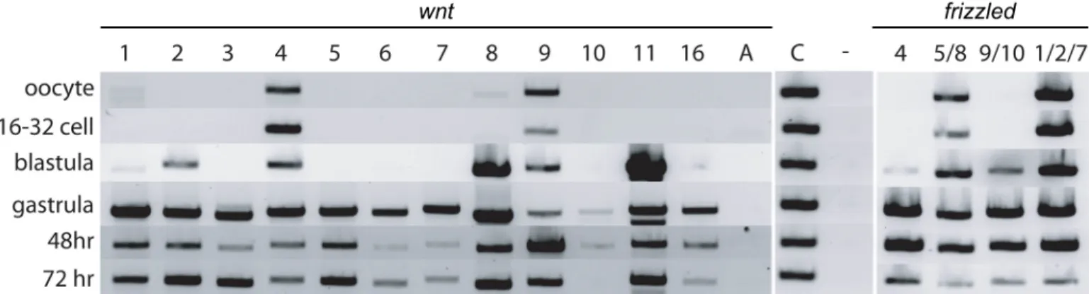
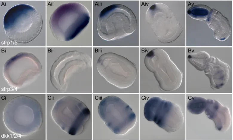
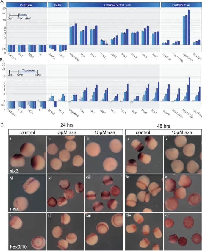
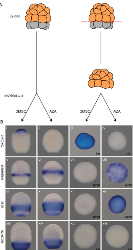
Figure


+7


Documents relatifs