HAL Id: inserm-02179369
https://www.hal.inserm.fr/inserm-02179369
Submitted on 10 Jul 2019
HAL is a multi-disciplinary open access
archive for the deposit and dissemination of
sci-entific research documents, whether they are
pub-lished or not. The documents may come from
teaching and research institutions in France or
abroad, or from public or private research centers.
L’archive ouverte pluridisciplinaire HAL, est
destinée au dépôt et à la diffusion de documents
scientifiques de niveau recherche, publiés ou non,
émanant des établissements d’enseignement et de
recherche français ou étrangers, des laboratoires
publics ou privés.
Molecular cloning and characterization of endogenous
SV40 dna from human HBL-100 cells
Claude Saint-Ruf, P. Nardeux, J. Cebrian, M. Lacasa, C. Lavialle, Roland
Cassingena
To cite this version:
Claude Saint-Ruf, P. Nardeux, J. Cebrian, M. Lacasa, C. Lavialle, et al.. Molecular cloning and
characterization of endogenous SV40 dna from human HBL-100 cells. International Journal of Cancer,
Wiley, 1989, 44 (2), pp.367-372. �10.1002/ijc.2910440230�. �inserm-02179369�
Int. J . Cancer: 367-372 (1989)
Publication de I'Union Internationale Contra 1e Cancer
0 1989 Alan R. Liss, Inc.
MOLECULAR CLONING AND CHARACTERIZATION OF ENDOGENOUS SV,, DNA
FROM HUMAN HBL-100 CELLS
c.
SAINT-RuF', P. NARDEUX', J. CEBRIAN~, M. LACASA~,c.
LAVIALLE' and R. CASSINGENA',3'UPR6, W P R 2 (CNRS), Institut de Recherches Scient$ques sur le Cancer, 94802 Villejuif Ckdex, France.
The human HBL-100 cell line harbours SV, D N A inte- grated in tandem at a unique site. The SV,, T-antigen ex- pressed in these cells is defective in a function essential to the replication of the viral genome. The integrated SV,, se- quences were molecularly cloned in a bacteriophage, and a subclone (plasmid pSVHBI) containing a complete SV, D N A was isolated. As compared to SV, wild-type strain 776, se- quence analysis of pSVHBl early region revealed the pres- ence of several D N A alterations. Among these, a point mu- tation at position 3199, predicting a change at amino-acid 540 of arginine to isoleucine, was shown by marker rescue to be responsible for the deficiency of T-antigen. This novel muta- tion further delimits one of the T-antigen domains involved in SV, D N A replication. Transfection experiments demon- strated that the transforming activity of the SV, genome from HBL-I00 cells is still preserved. Moreover, several trans- formed human cell clones thus obtained could be perma- nently established in culture.
The epithelial HBL-100 cell line, established in vitro from the milk of an apparently healthy woman, exhibits character- istics of transformation from the very beginning and evolves during in vitro maintenance until it becomes tumorigenic in nude mice.
In a previous publication (Caron de Fromentel et a l . , 1985) we reported the unexpected finding that HBL-100 cells harbour SV,, DNA integrated in tandem at a unique site in the cellular genome. By in situ hybridization, the integration site of the viral DNA was shown to be located on human chromosome 15, at band 15q24 (Marlhens et a l . , 1988). It seems likely that virus infection occurred in vivo, possibly by SV40 administered inadvertently with a polio virus vaccine (Caron de Fromentel et
a l . , 1985).
With regard to the SV40 T-antigen expressed in HBL-100 cells, we have previously shown (Caron de Fromentel et a l . , 1985) that it can bind to the SV40 origin of replication but that this property is not sufficient to permit the rescue of the inte- grated viral genome after fusion with permissive CV-1 cells, although rescue takes place upon fusion with permissive COS- 7 cells which harbour a replication-competent SV,, T-antigen gene. This indicates that this protein has a defect which does not interfere with its DNA-binding properties but affects at least one function essential to the replication of the viral ge- nome.
In order to address this issue, as part of a project concerning the biological and molecular characterization of the HBL- 100 cell line, we have molecularly cloned the endogenous SV,, DNA, sequenced its early region and identified the alteration responsible for the lack of viral DNA replication. In addition, we have determined the capacity of the cloned viral genome to transform human and rodent cells in culture.
MATERIAL AND METHODS
Nomenclature
The nucleotide numbering system used for the SV, genome is that of Buchman et al. (1982) and is based on the 5.243 base-pair (bp) genome, with the unique BgZI palindrome cen- tred on position 015243. Specific nucleotide positions in the cloned inserts of plasmids pSVHBl and pSVHB2 are referred
to by their analogous positions in the genome of the wild-type SV,, strain 776.
Cell cultures
HBL-100 is an epithelial cell line established in vitro from the milk of an apparently healthy woman (Polanowski et al., 1976; Gaffney, 1982); CV-1 is an established line of epithelial monkey kidney cells, fully permissive for lytic growth of SV, (Manteuil et a l . , 1973); ICIG-7 is a diploid fibroblast cell line with a limited in vitro life span, derived from normal human embryonic lung (Macieira-Coelho and Azzarone, 1982) and semi-permissive for SV,, infection; 3T3-Vill is a contact- inhibited Swiss mouse embryonic cell line established in our laboratory and non-permissive for SV40 infection.
All these lines were grown routinely in Eagle's minimal essential medium supplemented with 10% newborn calf serum (MEM 10).
DNA preparation and hybridization
Preparation of high-molecular-weight cellular DNA and of SV,, DNA, electrophoresis in agarose gels, nick-translation of DNA probes and blot hybridization were performed as previ- ously described (Caron de Fromentel et a l . , 1985).
Bacterial strains, bacteriophages and plasmids
All the procedures involving bacteriophages and plasmids were carried out according to Maniatis et a l . (1982). Esche-
richia coli strains Q359 and Q358 (Karn et a l . , 1980), used as
recipients for bacteriophages, were a gift from Dr. 0. Brison (IGR, Villejuif, France). Bacterial strain AG1, derived from E .
coli DH1 (Hanahan, 1983) and used for plasmid preparations,
as well as bacteriophage AEMBL3 (Frischauf et a l . , 1983) were purchased from Stratagene, San Diego, CA. Plasmids pUC13 (Messing and Vieira, 1982) and pSVTneoZ (Sarasin et a l . , 1987), which contains a functional early region from wild-
type SV,, DNA, were provided by Dr. M. James (IRSC, Villejuif, France). Plasmid pLASwt (Daya-Grosjean et a l . , 1987), containing an origin-minus SV40 DNA early region and expressing a fully functional T-antigen, was a gift from Dr. L. Daya-Grosjean (IRSC, Villejuif, France). Packaging and se- quencing kits were purchased from Stratagene. Bacterial strain DH5aF' and plasmid pTZ18R (plus helper phage M13K07) used for marker rescue experiments were purchased from BRL, Gaithersburg, MD, and Pharmacia, Uppsala, Sweden, respec- tively.
Molecular cloning of integrated SV,, DNA
High-molecular-weight genomic DNA from HBL- 100 cells was digested to completion with BglII. The DNA fragments were ligated to the arms of AEMBL3 bacteriophage vector, generated by cleavage with BamHI. Packaging and propaga- tion of the DNA recombinants in E . coli strain Q359 were carried out by standard procedures (Maniatis et a l . , 1982). This
3To whom reprint requests should be addressed.
368 SAINT-RUF ET AL.
partial library was screened by plaque hybridization using in
vitro nick-translated SV40 DNA as a probe. DNA sequencing
Sequencing was performed by the dideoxy-termination method (Sanger et al., 1977) with alkali-denatured plasmid DNA templates (Hattori and Sakaki, 1986) and deoxyadeno- sine 5'-a-(35S)thiotriphosphate (Amersham, Little Chalfont, UK). In order to sequence the SV40 early region contained in plasmid pSVHBl (Fig. l), 2 strategies were followed. (1)
pSVHB 1 was doubly digested with Sac1 and NcoI generating a
Sad-NcoI fragment containing the pUC13 sequences and the
complete SV, early region. Unidirectional deletions were in- troduced into the latter, from the NcoI site, by the method of Henikoff (1984), the only difference being that mung bean nuclease was used instead of S1 nuclease. The deleted frag- ments were circularized by ligation so that the deletion break points were positioned next to the M13 reverse primer site from pUC13. Plasmids thus obtained, with deletions of variable length into the SV,, sequences, were cloned and amplified. The sequence of overlapping DNA stretches of 200-300 nu- cleotides was then determined using the M13 reverse primer (Stratagene). (2) Confirmation of the results was obtained by direct sequencing of pSVHBl using a selected set of 17-mer oligonucleotides synthesized with an Applied 380 B apparatus (BRL) and purified on an HPLC column by Dr. J. Armier (IRSC, Villejuif, France).
Marker rescue
Four synthetic oligonucleotides were prepared by Dr.
J. Armier as described above: h4wt (5'-TTTACAAA- T C T G G C C T G C A ) ; h 4 m u t ( 5 ' - T T T A C A A A - CCCATAGC): h5mut (5'-AAAACTCCAGTTCCCATAGC). A plasmid designated pTSVHB was constructed in order to use a procedure based on the method of Zoller and Smith (1983) for oligonucleotide-directed mutagenesis. This plasmid was obtained by inserting the SV,, early region isolated from pSVHBl by digestion with K m I and EcoRI into the D T Z ~ ~ R vector. This vector 1
T A T G G C C T G C A ) , h 5 w t ( 5 ' - A A A A C T C C A A T T -
HBLlOO
XEHBBg
pSVHB2
pSVHBl
bears an' f l phage origin of replication
E c o R l
I OR/
which is required for the synthesis of single-stranded DNA in the presence of M13K07 helper phage. Single-stranded DNA was purified following M13K07 helper phage infection of E .
coli DH5aF' carrying the pTSVHB plasmid. One pmole of
single-stranded template was annealed with appropriate pairs of phosphorylated oligonucleotide primers (h4mut and h5mut: h4wt and h5mut; h4mut and h5wt; h4wt and h5wt; 5 pmole of each oligonucleotide). The resulting hybrid DNAs were then filled in with the Klenow DNA polymerase and ligated. A small aliquot of each reaction mixture was used to transform AG 1 cells. The supercoiled plasmid DNAs thus obtained were purified and transfected into subconfluent CV- 1 cells using the DEAE-dextran method of Kimura and Dulbecco (1972). Three days later, low-molecular-weight DNA was selectively ex- tracted from each culture by the procedure of Hirt (1967). The purified DNA was digested with KpnI and DnpI and analyzed by Southern blot hybridization using in vitro nick-translated pSVHBl as a probe.
Growth in soft agar
(1964) was used.
The method described by MacPherson and Montagnier
Transforming activity of SV,, DNA
DNA transfection was carried out using the calcium phos- phate co-precipitation method (Wigler et al., 1978). Human ICIG-7 cells (pass. 20-28) or mouse 3T3-Vill cells (pass. 33) were seeded into 100-mm Falcon dishes at a concentration of 1.5 x lo6 cells/lO ml MEM lO/dish. The next day, an equiv- alent number of cultures was transfected with 1 ml/culture of DNA-calcium phosphate co-precipitate containing either 40 pg of calf thymus DNA, as a control, or 1 p,g of SV, DNA- containing recombinant plus 40 p,g of camer calf thymus DNA. After 6 hr at 37"C, the medium was removed and 10 ml of fresh MEM lO/dish were added. Two days later, the cells were trypsinized and seeded either in liquid medium or in soft agar at a concentration of 2 X lo5 cells/60-mm Falcon dish. Transformed foci or colonies were scored after 3 4 weeks' incubation. EcoRl Bgl I Bgl I1 I I I I I 1 I I I I I 1 I I I 1 1 I I l K b p I 1
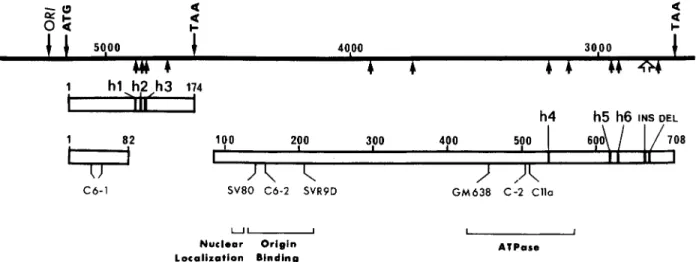
FIGURE 1 - Physical maps of SV, DNA integrated sequences and cloned inserts. The map at the top shows the structure of the SV,, DNA
sequences (white areas) integrated within the genome of HBL-100 cells (black areas), as determined by Southern blotting. AEHBBg is a phage recombinant containing the whole integrated viral DNA with flanking cellular sequences inserted in AEMBL3 DNA (dotted areas). pSVHB2 and pSVHBl are recombinant plasmids harbouring fragments from the AEHBBg insert subcloned in pUC13 (shaded areas). The line drawn below the pSVHBl map delimits the early SV, region subjected to sequence analysis.
1 1po 200 300 400 500 600
Imrnunojluorescence
Expression of SV4,-specific T-antigen was detected by in- direct immunofluorescence (Wicker and Avrameas, 1969), with Syrian hamster anti-SV, tumour serum and fluorescein isothiocyanate-conjugated rabbit antiserum to hamster y-glob- ulin.
708
RESULTS
Molecular cloning of integrated SV,, DNA
Previous results had shown that the SV,, T-antigen ex- pressed in HBL-100 cells is affected in a function essential to the replication of the viral genome (Caron de Fromentel et al., 1985). In order to identify the structural alteration responsible for the inactivation of this function, we have molecularly cloned the SV, DNA integrated in tandem (Fig. 1). Previous data had shown that the viral DNA sequences were contained in a unique 12-kbp BglII fragment of HBL-100 genomic DNA (Caron de Fromentel et al., 1985). Thus, 0.6 p.g of HBL-100 DNA were digested with BglII and ligated to 1.5 pg of BamHI- cleaved hEMBL3 phage DNA. Approximately 250,000 re- combinants were obtained, 100,000 of which were screened with an SV, DNA probe leading to the isolation of one pos- itive recombinant (AEHBBg). Upon digestion with various re- striction enzymes and electrophoresis on agarose gels, the XEHBBg DNA displayed the expected band patterns. Two overlapping fragments of about 5.2 kbp, obtained by cleavage with either BamHI or EcoRI, were isolated and subcloned in the pUC13 plasmid vector (Fig. 1). The 2 recombinant plas- mids pSVHBl and pSVHB2 were extensively studied by re- striction enzyme mapping. It was established that pSVHB 1 harbours a complete SV40 genome whereas pSVHB2 contains an incomplete viral genome with a truncated early region flanked by a cellular sequence of about 2.1 kbp. The junction between the viral and cellular sequences has been located at nucleotide 4037 interrupting the coding sequence of T-antigen at the 260th codon, which would lead to a truncated protein of
about 34 kDa. When compared to the wild-type SV,, strain
I
4
U
L
I I I 1
776 by restriction enzyme mapping, a noticeable difference resides in the size of the Hind 111 C DNA fragment from both pSVHB 1 and pSVHB2 inserts. Refined analysis of both plas- mids doubly digested with Hind 111 and Kpn I showed that the fragment containing the SV,, origin of replication has a size of approximately 290 bp whereas the same wild-type fragment is 366 bp long. This size difference (approx. 70 bp) would be consistent with the presence, in the strain of SV, which orig- inally infected the HBL-100 cells, of a single copy of the 72-bp enhancer which is found duplicated in strain 776 DNA. This was actually confirmed by sequence analysis of this region in pSVHBl (data not shown).
Sequencing of SV,, early region contained in pSVHB1
The SV40 genomic portion contained in pSVHB1, corre- sponding to the complete early region, encompassing nucle- otides 150 to 0 and 5243 to 2600 in the sense of the early RNA was sequenced and compared to that of the wild-type SV40 strain 776. As shown in Figures 2 and 3a, 11 nucleotide sub- stitutions were found, 3 of them (hl, h2 and h3) altering the amino-acid sequence of small t-antigen and 3 of them (h4, h5 and h6) modifying the C-terminal part of large T-antigen. In
addition, 2 changes which involve several nucleotides but which do not shift the reading frame were observed in the T-antigen coding sequence, proximal to the stop codon: (1) an insertion of 9 bp in the region around nucleotide 2795, which had already been reported for SV,, strains 777 (Manos and Gluzman, 1985) and LP (Feunten et al., 1981); (2) a deletion
of 6 bp in the region around nucleotide 2770, not previously described (Figs. 2 and 3b). Two of the conservative nucleotide changes mentioned above have been observed at position 3755 for strain 777 and at position 2757 for other SV,, strains (Manos and Gluzman, 1985). To our knowledge, none of the other modifications listed in Figure 3a have been described as yet. The 3 point mutations h l
,
h2 and h3 affecting the amino- acid sequence of small t-antigen would be without consequence for SV, replication since mutants with deletions covering this region display a normal lytic cycle (Bouck et al., 1978;a U I- U -Nuclear Origin Localization Binding I 1 AlPare
FIGURE 2 - Location of the mutations identified by sequence analysis in the SV, early region of pSVHB 1. The black line at the top represents the nucleotide sequence of the SV, early region. The positions of the initiation and termination codons for small t- and large T-antigens are
indicated. OR1 corresponds to the origin of viral DNA replication. Below the black line, the small arrows show the positions of the single nucleotide substitutions, and the large arrow that of both the insertion of 9 bp and the deletion of 6 bp (see “Results”). The uppermost box represents the coding sequence for small t-antigen, where the 3 predicted amino-acid changes h l , h2 and h3 have been located. The 2 lower boxes delimit the coding region for large T-antigen, interrupted by the sequence spliced out in the corresponding transcript. h4, h5 and h6 show the position of 3 amino-acid substitutions, INS, that of a 3 amino-acid insertion and DEL, that of a 2 amino-acid deletion. Below the 2 lower boxes are indicated the positions of known large T-antigen mutations (see References cited in Gish and Botchan, 1987) that inactivate SV, DNA
replication and which are clustered in 3 separate domains of the protein. Portions of large T-antigen that encode various functions of this protein are bracketed. Nucleotide and amino-acid numbering is based on that of SV, wild-type strain 776.
370 SAINT-RUF ET AL.
A POINT MUTATIONS
nucleotide amino acid name
N" change N" change 4 8 7 9 4 8 5 3 4 8 3 9 4 7 5 0 3 9 3 0 3 7 5 5 3 1 9 9 3 1 1 7 2 9 5 0 2 9 1 8 2 8 1 7 2 7 5 7 ATG to ATA GGA to GIA S T to _ACT TGG to TGL CAG to C M AAI to AAG A D to AIA AGA lo AGG A n to ACT D T to AAT TCC to T C I TAG to T A I 95 Met to Ile h l 1 0 4 Ala to Val h2 1 0 9 Ala lo Thr h3 conservative conservative conservative 5 4 0 Arg lo Ile h4 conservative 6 2 3 Ile lo Thr h5 6 3 4 Asp to Asn h6 conservative conservative B INSERTION-DELETION 2801 2 7 6 0 wt 776 GCC.CCT.CAG. . . .TCC.TCA.CAG.TCT.GTT.CAT.GAT.CAT.AAT.CAG.CCA.
Ala Pro Gln Ser Ser Gln Ser Val His Asp His Asn Gln Pro
673 686
pSVHBI GCC.CCT.CAG.CCC.TCA.CAG.TCC.TCA.CAG.TCT.GTT.CAT.~T. , .CAG.CCA. Ala Pro Gln Pro Ser Gln Ser Ser Gln Ser Val His Asp Gln Pro
FIGURE 3 - (a) Single nucleotide and amino-acid changes found in the early region o f SV,, cloned in pSVHB1. (b) Partial sequence o f pSVHB1, compared to the homologous region of SV, strain 776, showing the insertion of 9 nucleotides coding for 3 amino-acids, and the deletion of 2 nucleotides coding for 2 amino-acids.
Feunten et al., 1978; Shenk et al., 1976; Sleigh et al., 1978). One deletion mutant (dl 2199) encompassing the h6 modifica- tion does not show any significant difference in virus yield on CV-1 cells, as compared to wild-type SV40 (Pecceu et al.,
1987). The deletion of 6 bp around nucleotide 2770 would not be expected to affect viral DNA replication since 2 deletion mutants (dl 1066 and dl 1140) overlapping this region are positive for this function (Pipas et al., 1983). Consequently, we were left with the h4 and h5 mutations as being possibly responsible for the defectiveness of the T-antigen expressed in HBL-100 cells. In order to solve this question we performed marker rescue experiments.
Marker rescue with synthetic oligonucleotides
Marker rescue experiments were carried out with synthetic oligonucleotides covering the sites of the h4 and h5 mutations.
As described in "Material and Methods", for each of these sites 2 oligonucleotides were synthesized, one with the wild- type sequence (h4wt, h5wt) and the other with the mutated
sequence (h4mut, h5mut). Single-stranded pTSVHB plasmid
DNA, containing the SV40 early region from pSVHB1, was simultaneously annealed with each of the following pairs of oligonucleotides: h4wt and h5mut, h4mut and h5wt, h4wt and h5wt, h4mut and h5mut. This strategy was chosen to take into account the possibility that both the h4 and h5 mutations might be responsible for the inactivation of T-antigen. After primer extension and amplification in bacteria, the resulting DNA molecules were transfected into permissive CV- 1 cells. The
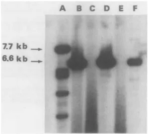
DNA from Hirt extracts was then linearized with KpnI and digested with DpnI in order to degrade the input DNA which had not replicated in CV-1 cells. The results of Southern blot analysis are shown in Figure 4. The 7.7-kb pSVTneoZ plas- mid, used as a positive control, did replicate (lane A). As expected, use of the h4mut and h5mut oligonucleotide pair combination did not lead to replication of the 6.6-kb pTSVHB plasmid DNA (lane E). In contrast, rescue occurred with h4wt plus h5wt (lane D) indicating that, indeed, at least one of the 2 mutations was responsible for the T-antigen defect. That it is
solely the h4 mutation which is involved is demonstrated by the efficient replication of pTSVHB observed following comple- mentation with the h4wt plus h5mut combination (lane B). This is confirmed by the fact that the h5wt oligonucleotide, in conjunction with h4mut, was inefficient (lane C).
Transfonning activity of SV,, DNA from HBL-I00 cells
Since we had identified multiple mutations throughout the early region of the SV,, DNA cloned from HBL-100 cells, it was of interest to determine whether any of them might have affected its transforming activity in rodent and human cells. A preliminary experiment was carried out by transfecting recip- ient human ICIG-7 cells with high-molecular-weight genomic DNA from HBL-100 cells. Three foci were obtained, of which one led to the establishment of a permanent cell line harbouring integrated SV40 DNA, expressing SV,, T-antigen in 100% of the cells and capable of growth in soft agar (data not shown). In order to extend this result and obtain quantitative data, trans- fection experiments were performed using AEHBBg, pSVHB 1 and pSVHB2 DNAs, in comparison with the origin-minus pLASwt DNA. The latter was chosen as a control in order to take into account the inability of the former DNAs to replicate in the recipient semi-permissive human cells. As shown in Table I, XEHBBg DNA was able to induce the formation of foci in both mouse 3T3 and human ICIG-7 cells. It was also able to induce the ICIG-7 cells to form colonies in agar. In 2 independent experiments (3 and 4), transforming efficiency was not significantly different from that obtained with control pLASwt DNA. While pSVHBl
,
which contains the complete SV40 early region, is also transforming, pSVHB2, which har- bours a truncated early region, is without effect. Several clones picked at random from mouse 3T3 or human ICIG-7 cells transformed either in liquid medium or in soft agar were all 100% SV, T-antigen-positive as determined by immunofluo- rescence. Moreover, several of the transformed ICIG-7 clones could be established as permanent cell lines after a short crisis period (3-4 weeks). Taken together, these results clearly show that the complete early region which has been subcloned in pSVHBl (Fig. l), despite multiple mutations, is fully func- tional for cell transformation.FIGURE 4 - Marker rescue with synthetic oligonucleotides. South- em blot analysis of KpnI- and DpnI-digested DNA extracted from CV-1 cells after transfection with plasmid pTSVHB complemented with the following pairs of oligonucleotides: h4wt
+
h5mut (lane B), h4mut+
h5wt (lane C), h4wt+
h5wt (lane D), h4mut+
h5mut (lane E). Lane A KpnI- and DpnI-digested DNA extracted from CV-1cells after transfection with pSVT neo Z, used as a positive control.
Lane F: EcoR1-cleaved pTSVHB DNA, used as a marker.
TABLE I - TRANSFORMATION OF MOUSE 3T3-VI1.I. AND HUMAN ICIG-7 CELLS BY CLONED SV, DNA
Experiment Recipient Transfecting Number
:Fyi:
of foci2 colonies2number cells DNA'
1 3T3-Vill XEHBBg 42 ND' 2 ICIG-7 XEHBBg 13 15 pLASwt 28 18 1C1G-7 XEHBBg 41 23 3 1C1G-7 XEHBBg 6 4 12 pLASwt ND 10 5 ICIG-7
$ $ ~ ~ ~
ND 0'In each experiment, control cultures transfected with calf thymus DNA alone never gave rise to foci or agar colonies.-2Per 4 X 10⁶ c e l l ~ . - ~ N D = not done.
DISCUSSION
HBL-100 cells obtained from 4 different sources express SV4,-specific T-antigen and harbour SV, DNA integrated in tandem at a unique site (Caron de Fromentel et al., 1985). In the present work we have molecularly cloned the complete viral sequences flanked on both sides by cellular sequences. Subcloning of this DNA allowed us to isolate 2 recombinant plasmids harbouring either a complete SV,, genome (pSVHB1) or an incomplete viral genome with a truncated early region (pSVHB2). Structural analysis of the latter showed that it could code only for a truncated protein of about 34 kDa, but no evidence of expression of such a protein was obtained by immunoprecipitation (data not shown). We there- fore concentrated on a detailed study of the pSVHBl early region capable of expressing the 94-kDa protein previously demonstrated in HBL-100 cells and shown to be defective for viral DNA replication (Caron de Fromentel et al., 1985). In order to locate the mutation(s) involved in this defect, we determined the sequence of pSVHB 1 early region. Taking as a reference SV40 wild-type strain 776, the analysis of this se- quence revealed the presence of 13 alterations (1 1 nucleotide substitutions, one 9-bp insertion, one 6-bp deletion). Among these, the point mutation designated h4, at position 3199 and predicting a change at amino-acid 54C of arginine to isoleu- cine, was the only one responsible for the deficiency of T- antigen, as demonstrated by marker rescue experiments.
By comparison with other SV,, mutants previously mapped in the early region (Fig. 2), the h4 mutation is in the proximity of a cluster of DNA lesions shown to be defective in a function essential to the replication of viral DNA. Moreover, mutant C l l b (Manos and Gluzman, 1984) which has an amino-acid change 9 codons downstream from h4, at position 549, is only partially affected in its replication. It appears, therefore, that the novel h4 mutation further delimits one of the domains involved in SV,, DNA replication. No mutations in the coding sequence of large T-antigen have been detected upstream of h4, which is consistent with our previous observations that T-antigen from HBL- 100 cells has an intranuclear localization and is capable of binding to the SV,, origin of replication (Caron de Fromentel et al., 1985).
A second issue addressed in the present work concerned the transforming activity of the SV40 genome harboured by HBL- 100 cells. That such an activity is preserved has been clearly shown by transfection experiments using either HBL- 100 ge- nomic DNA or cloned SV,, DNA sequences. Furthermore, we have shown that this activity is due to the complete early region contained in pSVHB1, despite the various mutations identi- fied. The fact that several of the transformed cell clones that we have isolated were capable of indefinite growth in culture is consistent with our previous hypothesis (Caron de Fromentel et
372 SAINT-RUE ET AL. al., 1985) that the HBL-100 endogenous SV,, DNA may have
been responsible for immortalization of these cells.
Genetic evidence is presently available (Gish and Botchan, 1987) indicating that the lytic replication and the transforming capacity of SV, are separable by mutation of the viral T- antigen gene. In addition, since human cells are semi- permissive for SV, replication, it has been suggested that the continued growth of SV,-transformed human cells imposes selective pressure against the replicative function and in favour of the transforming activity of the viral T-antigen gene. These considerations may explain the emergence of the HBL- 100 cell
REFERE BOUCK, N., BEALES, N., SHENK, T., BERG, P. and DI MAYORCA, G., New region of the simian virus 40 genome required for efficient viral transformation. Proc. nat. Acad. Sci. (Wash.), 75, 1274-1278 (1978). BUCHMAN, A.R., BURNETT, L. and BERG, P . , The SV, nucleotide se- quence. In: J. Tooze (ed.), DNA tumor viruses, 2nd ed., revised, pp. 799-841, Cold Spring Harbor Laboratory, New York (1982).
CARON DE FROMENTEL, C . , NARDEUX, P.C., SOUSSI, T., LAVIALLE, C., ESTRADE, S., CARLONI, G., CHANDRASEKARAN, K. and CASSINGENA, R., Epithelial HBL-100 cell line derived from milk of an apparently healthy woman harbours SV, genetic information. Exp. Cell Res., 160, 83-94 (1985).
DAYA-GROSJEAN, L., JAMES, M.R., DROUGARD, C. and SARASIN, A., An immortalized Xeroderma pigmentosum, group C, cell line which replicates SV, shuttle vectors. Mutation Res., 183, 185-196 (1987).
FEUNTEN, J., CARMICHAEL, G . , NICOLAS, J.C. and KRESS, M., Mutant carrying deletions in the two simian virus 40 early genes. J . Virol., 40,
FEUNTEN, J., KRESS, M., GARDES, M. and MONIER, R., Viable deletion mutants in the SV, early region. Proc. nat. Acad. Sci. (Wash.), 75, 44554459 (1978).
FRISCHAUF, A.M., LEHRACH, H., POUSTKA, A. and MURRAY, N . , Lambda replacement vectors carrying polylinker sequences. J . mol. Biol., 170, 827-842 (1983).
GAFFNEY, E.V., A cell line (HBL-100) established from human breast milk. Cell Tissue Res., 227, 563-568 (1982).
GISH, W.R. and BOTCHAN, M.R., Simian virus 40-transformed human cells that express large T-antigens defective for viral DNA replication. J .
Virol., 61, 2864-2876 (1987).
HANAHAN, D., Studies on transformation of Escherichia coli with plas- mids. J. mol. Biol., 166, 557-580 (1983).
HATTORI, M. and SAKAKI, Y., Dideoxy sequencing method using dena- tured plasmid templates. Anal. Biochem., 152, 232-238 (1986). HENIKOFF, S., Unidirectional digestion with exonuclease 111 creates tar-
geted breakpoints for DNA sequencing. Gene, 28, 351-359 (1984). HIRT, B., Selective extraction of polyoma DNA from infected mouse cell cultures. J mol. Biol., 26, 365-369 (1967).
KARN, J., BRENNER, S., BARNETT, L. and CESARENI, G., Novel bacte- riophage cloning vector. Proc. nar. Acad. Sci (Wash.), 77, 5172-5176 (1980).
KIMURA, G. and DULBECCO, R., Isolation and characterization of temper- ature-sensitive mutants of Simian virus 40. Virology, 49, 3 9 4 4 0 3 (1972). MACIEIRA-COELHO, A. and AZZARONE, B., Aging of human fibroblasts is a succession of subtle changes in the cell cycle and has a final short stage with abrupt events. Exp. Cell Res., 141, 325-332 (1982).
MACPHERSON, I. and MONTAGNIER, L., Agar suspension culture for the 625-634 (1981).
line, the progenitor cells of which-as already suggested (Ca- ron de Fromentel et ul., 1 9 8 5 t m i g h t have been infected in the mammary gland of the milk donor by an SV,, virus orig- inally contaminating a polio virus vaccine.
ACKNOWLEDGEMENTS
The authors are indebted to Miss S. Estrade and to Miss J . StCvenet for excellent technical assistance and to Mrs. P. Blanchin for typing the manuscript. This work was supported by the CNRS, the Association pour la Recherche sur le Cancer and the Foundation pour la Recherche MCdicale.
LNCES
selective assay of cells transformed by polyoma virus. Virology, 23, 291- 294 (1964).
MANIATIS, T., FRITSCH, E.F. and SAMBROOK, J., Molecular cloning: a
laboratory manual. Cold Spring Harbor Laboratory, New York (1982). MANOS, M.M. and GLUZMAN, Y., Simian virus 40 large T-antigen point mutants that are defective in viral DNA replication but competent in on- cogenic transformation. Mol. cell. Biol., 4, 1125-1 133 (1984). MANOS, M.M. and GLUZMAN, Y., Genetic and biochemical analysis of transformation-competent, replication-defective simian virus 40 large T- antigen mutants. J. Virol., 53, 120-127 (1985).
MANTEUIL, S., PAGBS, J., STBHELIN, D. and GIRARD, M., Replication of simian virus 40 deoxyribonucleic acid: analysis of the one step growth cycle. J. Virol., 11, 98-106 (1973).
MARLHENS, F., SAINT-RUF, C., NARDEUX, P., LAVIALLE, C., BROUTY- BouB, D., DUTRILLAUX, B. and CASSINGENA, R., Karyotype evolution of the human HBL-100 cell line and mapping of the integration site of SV, DNA. Ann. GCnCr., Paris, 31, 81-86 (1988).
MESSING, J. and VIEIRA, J., A new pair of M I 3 vectors for selecting either DNA strand of double digest restriction fragments. Gene, 19, 269-276 (1982).
PECCEU, F., KOMLY, A,, GARDES, M. and FEUNTEN, J., Properties of simian virus 40 mutants lacking the Asp,-Glu-Asp stretch at the carboxyl- terminus of large T-antigen. Virology, 160, 4 8 5 4 8 8 (1987).
PIPAS, J.M., PEDEN, K.W.C. and NATHANS, D., Mutational analysis of simian virus 40 T-antigen: isolation and characterization of mutants with deletions in the T-antigen gene. Mol. cell. Biol., 3, 203-214 (1983).
POLANOWSKI, F.P., GAFFNEY, E.V. and BURKE, R.E., HBL-100, a cell line established from human breast milk. In Vitro, 12, 328 (1976). SANGER, F., NICKLEN, S. and COULSON, A.R., DNA sequencing with chain-terminating inhibitors. Proc. nat. Acad. Sci. (Wash.), 74, 5463- 5467 (1977).
SARASIN, A , , MENCK, C.F.M. and JAMES, M.R., Shuttle vector-host sys- tems for analysis of mutagenesis in mammalian cells. Phorobiochem. Pho-
tobiophys., Suppl., 343-351 (1987).
SHENK, T.E., CARBON, J. and BERG, P., Construction and analysis of
viable deletion mutants of simian virus 40. J. Virol., 18, 664-671 (1976). SLEIGH, M.L., TOPP, W.C., HANICH, R. and SAMBROOK, J.F., Mutants of SV, with an altered small t protein are reduced in their ability to transform cells. Cell, 14, 79-88 (1978).
WICKER, R. and AVRAMEAS, S., Localization of virus antigens by enzyme- labelled antibodies. J. gen. Virol., 4, 4 6 5 4 7 1 (1969).
WIGLER, M., PELLICER, A , , SILVERSTEIN, S. and AXEL, R., Biochemical transfer of single-copy eucaryotic genes using total cellular DNA as donor. Cell, 14, 725-731 (1978).
ZOLLER, M. J. and SMITH, M., Oligonucleotide-directed mutagenesis of
DNA fragments cloned into M13 vectors. In: S.P. Colowick and N.O. Kaplan (eds.), Methods in enzymology, Vol. 100, pp. 468-500, Academic Press, New York (1983).


![Comparison of the Z/γ[superscript ∗] + jets to γ + jets cross sections in pp collisions at √s = 8 TeV](data:image/gif;base64,R0lGODlhAQABAIAAAP///wAAACH5BAEAAAAALAAAAAABAAEAAAICRAEAOw==)