HAL Id: hal-03254023
https://hal.sorbonne-universite.fr/hal-03254023
Submitted on 8 Jun 2021
HAL is a multi-disciplinary open access
archive for the deposit and dissemination of
sci-entific research documents, whether they are
pub-lished or not. The documents may come from
teaching and research institutions in France or
abroad, or from public or private research centers.
L’archive ouverte pluridisciplinaire HAL, est
destinée au dépôt et à la diffusion de documents
scientifiques de niveau recherche, publiés ou non,
émanant des établissements d’enseignement et de
recherche français ou étrangers, des laboratoires
publics ou privés.
Bacterial coinfection in critically ill COVID-19 patients
with severe pneumonia
Alexandre Elabbadi, Matthieu Turpin, Grigoris Gerotziafas, Marion Teulier,
Guillaume Voiriot, Muriel Fartoukh
To cite this version:
Alexandre Elabbadi, Matthieu Turpin, Grigoris Gerotziafas, Marion Teulier, Guillaume Voiriot, et al..
Bacterial coinfection in critically ill COVID-19 patients with severe pneumonia. Infection, Springer
Verlag, 2021, 49 (3), pp.559 - 562. �10.1007/s15010-020-01553-x�. �hal-03254023�
https://doi.org/10.1007/s15010-020-01553-x
CORRESPONDENCE
Bacterial coinfection in critically ill COVID‑19 patients with severe
pneumonia
Alexandre Elabbadi
1· Matthieu Turpin
1· Grigoris T. Gerotziafas
2,3· Marion Teulier
1· Guillaume Voiriot
1,4·
Muriel Fartoukh
1,4Received: 24 August 2020 / Accepted: 3 November 2020 / Published online: 3 January 2021 © Springer-Verlag GmbH Germany, part of Springer Nature 2021
Abstract
Severe 2019 novel coronavirus infectious disease (COVID-19) with pneumonia is associated with high rates of admission to
the intensive care unit (ICU). Bacterial coinfection has been reported to be rare. We aimed at describing the rate of bacterial
coinfection in critically ill adult patients with severe COVID-19 pneumonia. All the patients with laboratory-confirmed severe
COVID-19 pneumonia admitted to the ICU of Tenon University-teaching hospital, from February 22 to May 7th, 2020 were
included. Respiratory tract specimens were obtained within the first 48 h of ICU admission. During the study period, 101
patients were referred to the ICU for COVID-19 with severe pneumonia. Most patients (n = 83; 82.2%) were intubated and
mechanically ventilated on ICU admission. Overall, 20 (19.8%) respiratory tract specimens obtained within the first 48 h.
Staphylococcus aureus was the main pathogen identified, accounting for almost half of the early-onset bacterial etiologies.
We found a high prevalence of early-onset bacterial coinfection during severe COVID-19 pneumonia, with a high
propor-tion of S. aureus. Our data support the current WHO guidelines for the management of severe COVID-19 patients, in whom
antibiotic therapy directed to respiratory pathogens is recommended.
Keywords
Coronavirus disease 2019 · Bacterial coinfection · Staphylococcus aureus · Intensive care unit · Pneumonia
Abbreviations
COVID 19
Coronavirus infectious disease
ICU
Intensive care unit
SAPSII
Simplified acute physiology score
SARS-CoV-2 Severe acute respiratory syndrome
corona-virus 2
SOFA
Sequential Organ Failure Assessment
Introduction
Severe 2019 novel coronavirus infectious disease
(COVID-19) with pneumonia is associated with high rates of
admis-sion to the intensive care unit (ICU) and in-hospital
mor-tality [
1
]. Information about the rates of coinfection with
SARS-CoV-2 and one or more additional microorganisms
Electronic supplementary material The online version of thisarticle (https ://doi.org/10.1007/s1501 0-020-01553 -x) contains supplementary material, which is available to authorized users. * Alexandre Elabbadi
560 A. Elabbadi et al.
1 3
Methods
All the patients with laboratory-confirmed severe COVID-19
pneumonia admitted to the 42-bed ICU of Tenon
University-teaching hospital, from February 22 to May 7th, 2020 were
included. Respiratory tract specimens were obtained within
the first 48 h of ICU admission. Direct examination and
quan-titative cultures were performed on usual media for sputum,
tracheal aspirate, plugged telescoping catheter, or
bronchoal-veolar lavage, considering the respective positivity thresholds:
10
6cfu/ml, 10
5cfu/ml, 10
3cfu/ml and 10
4cfu/ml. Empirical
antimicrobial therapy combined broad-spectrum
antibiot-ics (a third-generation cephalosporin plus a macrolide), and
oseltamivir along the Flu season.
Results
During the study period, 101 patients were referred to the ICU
for COVID-19 with severe pneumonia after 8 (5.5–11) days
of symptoms onset, and 1 (0–2) day of hospitalization in the
wards. They were 79 men (78.2%), aged 61 (53–69) years,
with moderate overweight [body mass index 27.6 (24.5–31)]
and frequent comorbid conditions, mainly arterial
hyper-tension (n = 66; 65.3%) and diabetes (n = 33; 32.7%). Most
patients (n = 83; 82.2%) were intubated and mechanically
ven-tilated on ICU admission. The SAPSII score and SOFA score
were 27 (22–37) and 3 (2–5), respectively. By the end of the
study period, 21 patients (21%) had died, 12 (11.9%) were still
hospitalized in the ICU, while 51 (50.4%) and 17 (16.8%) had
been discharged to conventional wards or long-term
rehabilita-tion care units, respectively.
Overall, 20 (19.8%) respiratory tract specimens obtained
within the first 48 h of ICU admission yielded positive culture
at or above the thresholds for at least one pathogen (n = 12),
and below the thresholds (n = 8, including 6 with prior
anti-biotic therapy before ICU admission, and 1 associated with
pneumococcal bacteremia) (Table
1
and Table S1).
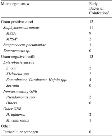
Staphy-lococcus aureus was the main microorganism identified,
accounting for almost half of the early-onset bacterial
etiolo-gies (Table
2
). Late-onset bacterial superinfections were
diag-nosed after 7.5 days (4–11) in 48 patients, and were mainly
related to Pseudomonas aeruginosa. There was no difference
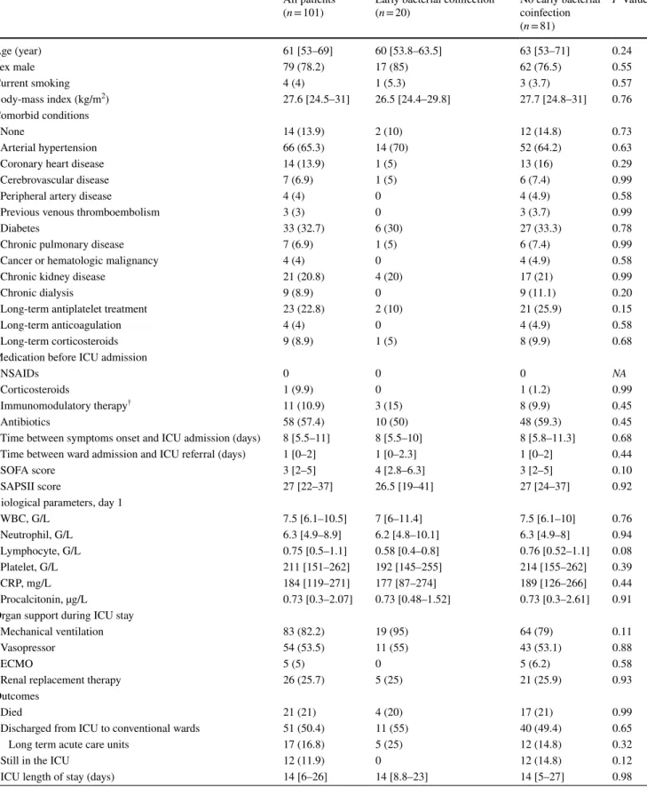
in comorbidities or admission clinical and laboratory
char-acteristics between patients with or without early bacterial
coinfection, except a trend towards a more pronounced
lym-phopenia (Table
1
).
Discussion
We found a high prevalence of early bacterial coinfection
during severe COVID-19 pneumonia, with a high
propor-tion of S. aureus. Data from China and South-east Asia
pointed to a low prevalence of bacterial coinfection in
patients with COVID-19 pneumonia [
3
]. In one cohort in
which this information was reported in detail, including
201 patients hospitalized for COVID-19 pneumonia [of
whom 53 (26%) were admitted to the ICU], none had
doc-umented bacterial co-infection
2. If the high rate of
coin-fection with S. aureus has been well described in Flu [
4
],
first reported cohorts do not mention bacterial co-infection
as a common feature of COVID-19 with pneumonia [
5
].
Our findings are consistent with those of two recent series
which focused on the early bacterial coinfection
associ-ated with SARS-CoV-2 pneumonia, and highlighted that
S. aureus was one of the main identified microorganism,
using molecular diagnostic tests alone or in association
with conventional tests [
6
,
7
]. Interestingly, procalcitonin
level did not differ between the patients with and without
associated bacterial coinfection, as already reported by
Kreitmann et al. [
7
] raising the question of the usefulness
of this biomarker to help for identifying early bacterial
coinfection during COVID-19 pneumonia.
Our findings support the current WHO guidelines for the
management of severe COVID-19 patients, in whom
anti-biotic therapy directed to respiratory pathogens is
recom-mended [
8
].
This is a single-center study, so our findings should be
extrapolated with caution. However, clinicians should be
alert of the high proportion of S. aureus co-infection during
COVID-19 pneumonia.
Table 1 Baseline characteristics, management and outcomes of the COVID19 cohort All patients
(n = 101) Early bacterial coinfection(n = 20) No early bacterial coinfection (n = 81) P Value Age (year) 61 [53–69] 60 [53.8–63.5] 63 [53–71] 0.24 Sex male 79 (78.2) 17 (85) 62 (76.5) 0.55 Current smoking 4 (4) 1 (5.3) 3 (3.7) 0.57 Body-mass index (kg/m2) 27.6 [24.5–31] 26.5 [24.4–29.8] 27.7 [24.8–31] 0.76 Comorbid conditions None 14 (13.9) 2 (10) 12 (14.8) 0.73 Arterial hypertension 66 (65.3) 14 (70) 52 (64.2) 0.63
Coronary heart disease 14 (13.9) 1 (5) 13 (16) 0.29
Cerebrovascular disease 7 (6.9) 1 (5) 6 (7.4) 0.99
Peripheral artery disease 4 (4) 0 4 (4.9) 0.58
Previous venous thromboembolism 3 (3) 0 3 (3.7) 0.99
Diabetes 33 (32.7) 6 (30) 27 (33.3) 0.78
Chronic pulmonary disease 7 (6.9) 1 (5) 6 (7.4) 0.99
Cancer or hematologic malignancy 4 (4) 0 4 (4.9) 0.58
Chronic kidney disease 21 (20.8) 4 (20) 17 (21) 0.99
Chronic dialysis 9 (8.9) 0 9 (11.1) 0.20
Long-term antiplatelet treatment 23 (22.8) 2 (10) 21 (25.9) 0.15
Long-term anticoagulation 4 (4) 0 4 (4.9) 0.58
Long-term corticosteroids 9 (8.9) 1 (5) 8 (9.9) 0.68
Medication before ICU admission
NSAIDs 0 0 0 NA
Corticosteroids 1 (9.9) 0 1 (1.2) 0.99
Immunomodulatory therapy† 11 (10.9) 3 (15) 8 (9.9) 0.45
Antibiotics 58 (57.4) 10 (50) 48 (59.3) 0.45
Time between symptoms onset and ICU admission (days) 8 [5.5–11] 8 [5.5–10] 8 [5.8–11.3] 0.68 Time between ward admission and ICU referral (days) 1 [0–2] 1 [0–2.3] 1 [0–2] 0.44
SOFA score 3 [2–5] 4 [2.8–6.3] 3 [2–5] 0.10
SAPSII score 27 [22–37] 26.5 [19–41] 27 [24–37] 0.92
Biological parameters, day 1
WBC, G/L 7.5 [6.1–10.5] 7 [6–11.4] 7.5 [6.1–10] 0.76 Neutrophil, G/L 6.3 [4.9–8.9] 6.2 [4.8–10.1] 6.3 [4.9–8] 0.94 Lymphocyte, G/L 0.75 [0.5–1.1] 0.58 [0.4–0.8] 0.76 [0.52–1.1] 0.08 Platelet, G/L 211 [151–262] 192 [145–255] 214 [155–262] 0.39 CRP, mg/L 184 [119–271] 177 [87–274] 189 [126–266] 0.44 Procalcitonin, µg/L 0.73 [0.3–2.07] 0.73 [0.48–1.52] 0.73 [0.3–2.61] 0.91 Organ support during ICU stay
Mechanical ventilation 83 (82.2) 19 (95) 64 (79) 0.11
562 A. Elabbadi et al.
1 3
Author contributions MF, AE, MT collected, analyzed, and interpreted the data. AE and MF drafted the manuscript. The authors read and approved the final manuscript.
Funding The authors declare that they have no funding source. Availability of data and materials The datasets and materials used and/ or analyzed during the current study are available from the correspond-ing author on reasonable request.
Compliance with ethical standards
Competing interest The authors have no conflict of interest to declare. Ethics approval and consent to participate This is a non-interventional data-based research, using the care data collected during patients stay, involving all the consecutive critically ill COVID-19 patients with severe pneumonia admitted to the ICU in Tenon Hospital during the pandemic. There is no processing of indirectly identifiable data, or chaining with data from other sources, or long-term patient follow-up for this research. Patients and proxies were informed, written consent was waived.
References
1. Wu C, et al. Risk factors associated with acute respiratory distress syndrome and death in patients with coronavirus disease 2019 pneumonia in Wuhan, China. JAMA Intern Med. 2019. https :// doi.org/10.1001/jamai ntern med.2020.0994.
2. Kim D, Quinn J, Pinsky B, Shah NH, Brown I. Rates of co-infection between SARS-CoV-2 and other respiratory pathogens. JAMA. 2020. https ://doi.org/10.1001/jama.2020.6266.
3. Wang D, et al. Clinical characteristics of 138 hospitalized patients with 2019 novel coronavirus-infected pneumonia in Wuhan, China. JAMA. 2019. https ://doi.org/10.1001/jama.2020.1585. 4. Chertow DS, Memoli MJ. Bacterial coinfection in influenza: a
grand rounds review. JAMA. 2013;309(3):275–82. https ://doi. org/10.1001/jama.2012.19413 9.
5. Rawson TM, et al. Bacterial and fungal co-infection in individuals with coronavirus: a rapid review to support COVID-19 antimicro-bial prescribing. Clin Infect Dis. 2020. https ://doi.org/10.1093/cid/ ciaa5 30.
6. Contou D, et al. Bacterial and viral co-infections in patients with severe SARS-CoV-2 pneumonia admitted to a French ICU. Ann Intensive Care. 2020;10(1):119. https ://doi.org/10.1186/s1361 3-020-00736 -x.
7. Kreitmann L, Monard C, Dauwalder O, Simon M, Argaud L. Early bacterial co-infection in ARDS related to COVID-19. Intensive Care Med. 2020;46(9):1787–9. https ://doi.org/10.1007/s0013 4-020-06165 -5.
8. “Clinical management of severe acute respiratory infection when COVID-19 is suspected https ://www.who.int/publi catio ns-detai l/ clini cal-manag ement -of-sever e-acute -respi rator y-infec tion-when-novel -coron aviru s-(ncov)-infec tion-is-suspe cted.” .
Table 2 Bacterial microorganism(s) identified in severe COVID-19 pneumonia
a MRSA methicillin-resistant S. aureus (MRSA), in one renal
trans-plant recipient, and one patient without identified risk factor
† defined as microorganism(s) identified within the first 48 h of ICU
admission. More than one bacterium was identified in 5 patients
Microorganism, n Early Bacterial Coinfection† Gram-positive cocci 12 Staphylococcus aureus 11 MSSA 9 MRSAa 2 Streptococcus pneumoniae 1 Enterococcus sp. 0 Gram-negative bacilli 13 Enterobacteriaceae E. coli 2 Klebsiella spp. 2
Enterobacter, Citrobacter, Hafnia spp. 4
Serratia 0 Non-fermenting GNB Pseudomonas spp. 2 Others 0 Other GNB H. influenza 2 M. catarrhalis 1 Other Intracellular pathogen 0