HAL Id: hal-03251712
https://hal.sorbonne-universite.fr/hal-03251712
Submitted on 7 Jun 2021
HAL is a multi-disciplinary open access
archive for the deposit and dissemination of
sci-entific research documents, whether they are
pub-lished or not. The documents may come from
teaching and research institutions in France or
abroad, or from public or private research centers.
L’archive ouverte pluridisciplinaire HAL, est
destinée au dépôt et à la diffusion de documents
scientifiques de niveau recherche, publiés ou non,
émanant des établissements d’enseignement et de
recherche français ou étrangers, des laboratoires
publics ou privés.
Kinetics of Anti–SARS-CoV-2 IgG Antibodies in
Hemodialysis Patients Six Months after Infection
Hamza Sakhi, Djamal Dahmane, Philippe Attias, Thomas Kofman, Magali
Bouvier, Nathanael Lapidus, Slim Fourati, Khalil El Karoui
To cite this version:
Hamza Sakhi, Djamal Dahmane, Philippe Attias, Thomas Kofman, Magali Bouvier, et al.. Kinetics
of Anti–SARS-CoV-2 IgG Antibodies in Hemodialysis Patients Six Months after Infection. Journal
of the American Society of Nephrology, American Society of Nephrology, 2021, 32 (5), pp.1033-1036.
�10.1681/ASN.2020111618�. �hal-03251712�
RAPID COMMUNICATIONS www.jasn.org
Kinetics of Anti–SARS-CoV-2 IgG Antibodies in
Hemodialysis Patients Six Months after Infection
Hamza Sakhi,1,2Djamal Dahmane,1 Philippe Attias,3Thomas Kofman,4Magali Bouvier,5,6Nathanael Lapidus,7,8 Slim Fourati,5,6 and Khalil El Karoui,1,2 *
Due to the number of contributing authors, the affiliations are listed at the end of this article. ABSTRACT
BackgroundThe humoral response against severe acute respiratory syndrome coronavirus 2 (SARS-CoV-2) in the hemodialysis population, including its dynamics over time, remains poorly understood.
MethodsTo analyze initial and long-term humoral responses against SARS-CoV-2 in a hemodialysis population, we retrospectively evaluated findings from SARS-CoV-2 IgG serologic assays targeting the nucleocapsid antigen or spike antigen up to 6 months of follow-up in patients on hemodialysis in the Paris, France, region who had recovered from coronavirus disease 2019 (COVID-19).
ResultsOur analysis included 83 patients (median age 65 years); 59 (71%) were male and 28 (34%) had presented with severe COVID-19. We observed positive initial SARS-CoV-2 IgG antinucleocapsid serology in 74 patients (89%) at a median of 67 days postdiagnosis. By multivariable analysis, immunocompromised status was the only factor significantly associated with lack of an IgG antinucleocapsid antibody response. Follow-up data were available at 6 months postdiagnosis for 60 of 74 patients (81%) with positive initial antinucleocapsid serology, and 15 (25%) of them had negative antinucleocapsid serology at month 6. In total, 14 of 15 sera were tested for antispike antibodies, 3 of 14 (21%) of which were also negative. Overall, 97% of antinucleocapsid-antibody–positive specimens were also antispike-antibody positive. Female sex, age .70 years, and nonsevere clinical presentation were independently associated with faster IgG antinucleocapsid titer decay in multivariable analysis. After adjustment for sex and age .70 years, non-severe clinical presentation was the only factor associated with faster decay of IgG antispike antibodies.
ConclusionsThis study characterizes evolution of the SARS-CoV-2 antibody re-sponse in patients on hemodialysis and identifies factors that are associated with lack of seroconversion and with IgG titer decay.
doi: https://doi.org/10.1681/ASN.2020111618
Coronavirus disease 2019 (COVID-19), caused by severe acute respiratory syn-drome coronavirus 2 (SARS-CoV-2) in-fection, is associated with a severe risk of mortality in patients on hemodialysis.1,2
The humoral response against SARS-CoV-2 in the hemodialysis population, including its dynamics over time, remains
poorly understood.3,4 Given the
pan-demic’s ongoing waves of new infections and the need for future vaccination strat-egies, the characterization of this response appears to be an important unmet need. In this study, we analyzed the initial and long-term humoral response against SARS-CoV-2 in a hemodialysis population.
METHODS
In this multicenter study, the study pop-ulation included 83 patients undergoing hemodialysis in the Paris, France, area who had recovered from COVID-19; 76 patients were previously described.2
Clinical data were retrospectively re-corded. A severe form of COVID-19 was defined by the need for oxygen therapy. Immunocompromised status was char-acterized by one of the following factors: former organ transplant, HIV infection, recent (within ,6 months) immuno-suppressive therapy, or chemotherapy.
We collected 241 sequential serum samples, which were analyzed with the
Received November 20, 2020. Accepted January 24, 2021.
*The Mondor NephroCov Study Group members are Vincent Audard, Bouthenia Bentaarit, Anna Boueilh, Sébastien Gallien, Philippe Grimbert, So-phie Hue, Nizar Joher, Narindra Jouan, Larbi Lam-riben, Jean-Daniel Lelievre, Raphael Lepeule, Matthieu Mahevas, Marie Matignon, Giovanna Melica, Julie Oniszczuk, Jean-Michel Pawlotsky, Thomas Stehle, William Vindrios, and Charlotte Wemmert.
H.S., D.D., P.A., and T.K. contributed equally to this work.
Published online ahead of print. Publication date available at www.jasn.org.
Correspondence: Dr. Khalil El Karoui, Department of Nephrology and Transplantation, AP-HP, Hôpital Henri Mondor, and INSERM U955, Institut Mondor de Recherche Biomédicale, 51 avenue du Maréchal de Lattre de Tassigny 94000 Créteil, France. Email: khalil.el-karoui@inserm.fr
Copyright © 2021 by the American Society of Nephrology
JASN 32: 1033–1036, 2021.
JASN 32: 1033–1036, 2021 ISSN : 1046-6673/3205-1033 1033
on behalf of Mondor NephroCov Study Group
R A PI D C O M M U N IC A TI O N S
Abbot SARS-CoV-2 IgG Architect sys-tem (targeting the nucleocapsid anti-gen). Of the 83 participants, 25 had two serial measurements of IgG levels, and 52 had at least three serial measure-ments. We used the Ortho Clinical Di-agnostics Vitros IgG assay (targeting the spike antigen) to analyze 113 samples.
Data collection was declared to the French Commission Nationale de l’In-formatique et des Libertés, registration 2218583. This protocol was submitted to the approbation of Paris Centre Insti-tutional Review Board.
Statistical Analyses
Categorical and continuous variables were expressed as count (percentage) and median (interquartile range, IQR), respectively. When appropriate, chi-squared or Fisher’s exact tests were used for categorical comparison, and t test or Mann–Whitney for continuous variables.
Variables associated with seroconver-sion were analyzed by logistic regresseroconver-sion. All variables with a P value,0.2 in uni-variable analysis were included in the multivariable analysis. Stepwise back-ward selection on the basis of the Akaike information criterion was then used for the final multivariable model. In the subset of patients who experienced sero-conversion, we modeled the SARS-CoV-2 antibody decay with random-intercept linear models to account for intrasubject correlations. Antibody titers were log10
transformed to estimate the evolution of geometric mean titers after their peaks were reached, and factors associated with their decrease were identified by testing the interaction between time and patients’ characteristics.
Results were analyzed with GraphPad Prism software version 9.0.0 and R soft-ware version 4.0.3.
RESULTS
We retrospectively studied SARS-CoV-2 serologic assay findings in 83 patients who received in-center hemodialysis at five different centers in the Paris area, and who were still alive after their
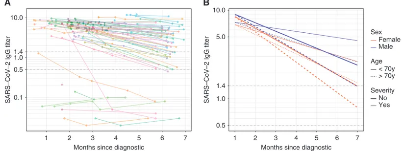
diagnosis with COVID-19 in March 2020. Baseline patient characteristics are described in Supplemental Tables 1 and 2. Thefirst serologic evaluation was per-formed a median of 67 (IQR 39) days after COVID-19 diagnosis. Among the 83 patients, 74 (89%) had positive SARS-CoV-2 IgG antinucleocapsid or antispike serology (Figure 1A). Nine pa-tients had negative initial IgG serology on the basis of the antinucleocapsid as-say at a median of 51 (IQR 32.5) days postdiagnosis; sera from six of these nine patients were also tested for anti-spike antibodies, which were similarly negative. Detailed characteristics of these patients are provided in Supplemental Table 3.
We found no association between the absence of IgG response and initial dis-ease severity, but glomerular disdis-eases and immunosuppression were more fre-quent among patients who did not ex-hibit a SARS-CoV-2 antibody response (Supplemental Table 2). We observed similar results after excluding patients without PCR-confirmed diagnosis (Supplemental Table 4).
By multivariable analysis, an immu-nocompromised status was the only fac-tor significantly associated with the ab-sence of IgG antinucleocapsid antibody response (odds ratio, 13.8; 95% con fi-dence interval [95% CI], 2.76 to 69.13; P50.001) (Supplemental Table 5).
Follow-up until month 6 was avail-able for 65 patients (78%). In 60 of these patients with positive initial antinucleo-capsid serology, 15 (25%) had an anti-nucleocapsid antibody level below the limit of positivity (index ratio ,1.4). Among these 15 patients, 13 (87%) had an index ratio between 0.5 and 1.4, and two (13%) had an index ratio ,0.49 (Figure 1A). Only a low initial SARS-CoV-2 antibody value was associated with long-term negative antinucleocapsid antibody level (7.1; IQR, 2.3 versus 3.44; IQR, 1.6; P,0.0001) (Supplemental Table 6). Of the 15 sera samples with anti-nucleocapsid antibody level below the limit of positivity, 14 were tested for anti-spike antibodies, and three of the 14 (21%) were also negative with this assay (Figure 2).
The estimated slope of IgG antinu-cleocapsid titer weekly decrease was 20.022 log10(95% CI,20.03 to 20.02),
meaning the geometric mean titer de-creased weekly by 4.9% (95% CI, 4.1 to 5.7). Female sex (20.0081 log10; 95% CI,
20.00026 to 20.016; P50.05), age .70 years (20.0091 log10; 95% CI,
20.017 to 20.0018; P50.02), and nonse-vere clinical presentation (20.014 log10;
95% CI,20.0069 to 20.021; P5,0.001) were independently associated with a faster IgG antinucleocapsid titer decay in mul-tivariable analysis (Figure 1B). Regarding antispike assay, all 74 antinucleocapsid-positive specimens were also antispike positive, except for two (3%) patients, who repeatedly tested negative for anti-spike antibodies despite being positive for antinucleocapsid antibodies. The es-timated slope of the antispike IgG titer weekly decrease was20.0080 log10(95%
CI,20.011 to 20.0045), meaning the geo-metric mean titer decreased weekly by 1.8% (95% CI, 1.0 to 2.6). After adjustment for sex and age.70 years, non-severe clin-ical presentation was the only factor asso-ciated with a steeper decay (20.0082 log10;
95% CI,20.015 to 20.0015; P50.02).
Significance Statement
The humoral response over time against severe acute respiratory syndrome coro-navirus 2 (SARS-CoV-2) is poorly un-derstood. The authors investigated the long-term kinetics of the antibody re-sponse to SARS-CoV-2 (specifically, IgG against nucleocapsid and spike antigens), in 83 patients on in-center hemodialysis who recovered from coronavirus disease 2019 (COVID-19). They found that 10% of patients had no initial seroconversion, which was associated with immunocom-promised status; in patients with serocon-version, IgG antibodies decayed over time. Factors associated with this de-cline included older age, female sex, and nonsevere clinical presentation. About 25% of patients had negative IgG anti-nucleocapsid serology after 6 months, whereas most patients maintained anti-spike antibodies. By characterizing the evolution of the SARS-CoV-2 antibody response, these findings might help better define future therapeutic and preventive approaches against COVID-19 in patients on hemodialysis.
RAPID COMMUNICATIONS www.jasn.org
DISCUSSION
In this study, we describe the 6-month kinetics of IgG antibody response against SARS-CoV-2 in patients on he-modialysis. On the basis of assayfindings for antinucleocapsid and antispike anti-bodies, we observed a lack of seroconver-sion in 10% of patients, as previously suggested in the general population and in rare patients on hemodialysis.4
Interestingly, we show that lack of sero-conversion was associated with immu-nocompromised status, as previously suggested,3 which may explain the
fre-quency of patients with glomerular disease in this group. Whether immunosuppres-sion also affects seroconverimmunosuppres-sion in patients not on dialysis remains to be studied. In-terestingly, a lack of seroconversion was observed in 7.9%–33% of patients (de-pending on the assay) of a recent cohort
of patients who had undergone kidney transplant.5
Long-term evolution in anti–SARS-CoV-2 antibodies is still a matter of de-bate, although virus-specific IgG decline seems to occur in most individuals.6,7In
our study, the antibody response of pa-tients on hemodialysis seems similar to that of previously reported healthy indi-viduals. Our models suggest IgG decline continues over time and is indepen-dently associated with female sex, non-severe disease, and older age. Whether this decline is also associated with loss of neutralizing antibodies or cellular re-sponse to SARS-CoV-2 will require fur-ther study.7 Of note, this IgG decline
leads to antinucleocapsid antibody titers below the cutoff value in 25% of patients 6 months after diagnosis, although most patients had levels in the gray zone (sig-nal to cutoff ratio between 0.5 and 1.4, Figure 1A) and maintain antispike antibodies.
This retrospective study has some limitations. Lack of seroconversion in patients who are RT-PCR negative should be interpreted with caution, al-though reported RT-PCR sensitivity is approximately 80% in the hemodialysis population.3 In addition, our findings
1 2 3 4 5 6 7
Months since diagnostic 0.1
0.5 1.0 1.4 10.0
SARS–CoV–2 IgG titer
A
B
0.5 1.0 1.4 5.0 10.0 1 2 3 4 5 6 7Months since diagnostic
SARS–CoV–2 IgG titer
Sex Age Severity Female Male < 70y > 70y No Yes
Figure 1. Evolution of SARS-CoV-2 IgG antinucleocapsid (NC) antibody titer until 6 months after diagnosis. (A) Evolution of SARS-CoV-2 IgG titer for each patient over time (spaghetti plot). Cutoff for negative serology was defined according to the manufacturer (Index sample/
control,1.4: dashed line). The zone between the dashed lines (1.4 and 0.5) represents the equivocal zone. The y-axis is plotted in
logarithmic scale. (B) Predicted SARS-CoV-2 antibody decay according to age, sex, and disease severity (multivariable model). Cutoff
for negative serology: index sample/control,1.4; dashed line. The zone between the dashed lines (1.4 and 0.5) represents the
equivocal zone. 0 5 10 15 20 1 1 2 3 4 5 6 7
Months since diagnostic
SARS–CoV–2 anti–spike IgG titer
Figure 2. Evolution of SARS-CoV-2 IgG antispike antibody titers until 6 months after di-agnosis. Evolution of SARS-CoV-2 IgG titer for each patient over time (spaghetti plot).
Cutoff for negative serology was defined according to the manufacturer (Index S/C ,1:
dashed line).
www.jasn.org RAPID COMMUNICATIONS
would have been enriched by the use of different serologic assays, earlier in dis-ease evolution (notably in patients who did not survive), by evaluating different antibody subclasses and characterizing cellular immunity, which is not neces-sarily parallel to the humoral response.
However, this characterization of the evolution of the SARS-CoV-2 antibody response in patients on hemodialysis, and the identification of factors associ-ated with lack of seroconversion and with IgG titer decay, might help to better define future therapeutic and preventive approaches, including vaccination strategies.
DISCLOSURES
K. El Karoui reports receiving research funding from Amgen, Otsuka, and Sanofi, and honoraria from Alexion and Otsuka. S. Fourati reports receiving honoraria from Abbott Diagnostics. All remaining authors have nothing to disclose.
FUNDING None.
ACKNOWLEDGMENTS
Hamza Sakhi, Philippe Attias, Thomas Kofman, and Khalil El Karoui designed the study; Hamza
Sakhi, Philippe Attias, Thomas Kofman, Djamal Dahmane, Larbi Lamriben, Slim Fourati, and Khalil El Karoui collected data; Hamza Sakhi, Philippe Attias, Thomas Kofman, Djamal Dah-mane, Larbi Lamriben, Thomas Stehlé, Vincent Audard, Julie Oniszczuk, and Nizar Joher cared for the study patients; Hamza Sakhi, Philippe Attias, Thomas Kofman, Nathanael Lapidus, Slim Fourati, and Khalil El Karoui analyzed the data; Hamza Sakhi, Nathanael Lapidus, Slim Fourati, and Khalil El Karoui wrote the paper; and all authors provided feedback and critical review.
SUPPLEMENTAL MATERIAL
This article contains the following supplemental material online at http://jasn.asnjournals.org/ lookup/suppl/doi:10.1681/ASN.2020111618/-/ DCSupplemental.
Supplemental Table 1. Clinical and biological presentation of patients at diagnosis.
Supplemental Table 2. Clinical characteristics of patients according to SARS-CoV-2 IgG antinucleo-capsid response.
Supplemental Table 3. Detailed characteristics of patients with absence of SARS-CoV-2 IgG antinu-cleocapsid response.
Supplemental Table 4. Clinical characteristics of patients with RT-PCR confirmed diagnosis, ac-cording to their initial SARS-CoV-2 IgG antinu-cleocapsid antibody response.
Supplemental Table 5. Multivariable analysis of factors associated with the absence of initial SARS-CoV-2 IgG antinucleocapsid antibody response.
Supplemental Table 6. Patient characteristics ac-cording to their long-term SARS-CoV-2 IgG anti-nucleocapsid antibody response.
REFERENCES
1. Couchoud C, Bayer F, Ayav C, Béchade C, Brunet P, Chantrel F, et al.: Low incidence of SARS-CoV-2, risk factors of mortality and the course of illness in the French national cohort of dialysis patients. Kidney Int 98: 1519–1529, 2020
2. Chawki S, Buchard A, Sakhi H, Dardim K, El Sakhawi K, Chawki M, et al.; HD-CovIDF Study Group: Treatment impact on COVID-19 evo-lution in hemodialysis patients. Kidney Int 98: 1053–1054, 2020
3. Clarke C, Prendecki M, Dhutia A, Ali MA, Sajjad H, Shivakumar O, et al.: High preva-lence of asymptomatic COVID-19 infection in hemodialysis patients detected using sero-logic screening. J Am Soc Nephrol 31: 1969–1975, 2020
4. Tang H, Tian J-B, Dong J-W, Tang X-T, Yan Z-Y, Zhao Y-Y, et al.: Serologic detection of SARS-CoV-2 infections in hemodialysis cen-ters: A multicenter retrospective study in Wuhan, China. Am J Kidney Dis 76: 490–499.e1, 2020
5. Prendecki M, Clarke C, Gleeson S, Greathead L, Santos E, McLean A, et al.: Detection of SARS-CoV-2 antibodies in kidney transplant recipients. J Am Soc Nephrol 31: 2753–2756, 2020
6. Ripperger TJ, Uhrlaub JL, Watanabe M, Wong R, Castaneda Y, Pizzato HA, et al.: Orthogonal SARS-CoV-2 serological assays enable sur-veillance of low-prevalence communities and reveal durable humoral immunity. Immunity 53: 925–933.e4, 2020
7. Wajnberg A, Amanat F, Firpo A, Altman DR, Bailey MJ, Mansour M, et al.: Robust neutraliz-ing antibodies to SARS-CoV-2 infection persist for months. Science 370: 1227–1230, 2020
AFFILIATIONS
1Assistance Publique des Hôpitaux de Paris, Hôpitaux Universitaires Henri Mondor, Department of Nephrology, Centre de Référence Maladie Rare « Syndrome Néphrotique Idiopathique », Fédération Hospitalo-Universitaire « Innovative therapy for immune disorders », Créteil, France 2University Paris Est Créteil, Institut National de la Santé et de la Recherche Médical, (INSERM) U955, Institut Mondor de Recherche Biomédicale (IMRB), Equipe 21, Créteil, France
3Department of Nephrology and Dialysis, Hôpital Privé Nord Parisien, Sarcelles, France
4Association Néphrologique Développement Rein Artificiel, Unité de Dialyse des Buttes Chaumont, Paris, France 5
Assistance Publique des Hôpitaux de Paris, Hôpitaux Universitaires Henri Mondor, Service de Virologie, Centre National de Référence Hépatites B, C, et delta, Créteil, France.
6
Department of Virology, University Paris Est Créteil, Institut National de la Santé et de la Recherche Médicale, Créteil, France 7Public Health Department, Saint-Antoine Hospital, Paris, France
8Sorbonne Université, Institut Pierre Louis d’Epidémiologie et de Santé Publique, Paris, France
1036 JASN JASN 32: 1033–1036, 2021
Supplemental data
Table of content
Supplemental Table 1. Clinical characteristics of patients according to SARS-CoV-2 IgG
anti-NC response
Supplemental Table 2. Detailed characteristics of patients with absence of SARS-CoV-2 IgG
anti-NC response
Supplemental Table 3. Clinical characteristics of patients with RT-PCR confirmed diagnosis,
according to their initial SARS-CoV-2 IgG anti-NC antibody response
Supplemental Table 4. Multivariable analysis of factors associated with the absence of initial
SARS-CoV-2 IgG anti-NC antibody response
Supplemental Table 5. Patient characteristics according to their long-term SARS-CoV-2 IgG
anti-NC antibody response
Supplemental Table 6. Clinical and biological presentation of patients at diagnosis
Supplemental Figure 1: Evolution of SARS-CoV-2 IgG anti-spike antibody titers until 6
months after diagnosis
Supplemental Table 1. Clinical characteristics of patients according to SARS-CoV-2 IgG
anti-NC response
VARIABLE ALL PATIENTS N = 83 SEROPOS. N = 74 SERONEG. N = 9 P VALUEAge (years), median (IQR) 65 (19.5) 64 (19.5) 66 (22) 0.274
Male, n (%) 59 (71) 54 (73) 5 (56) 0.427
Cause of nephropathy 0.002
Diabetes and/or vascular nephropathy, n (%) 55 (66) 52 (70) 3 (33) -
Glomerular disease, n (%) 16 (19) 10 (14) 6 (67) -
Other, n (%) 12 (14) 12 (16) 0 (0) -
Time from dialysis initiation (years), median (IQR) 3 (3.8) 3 (3.9) 2 (4.1) 0.584 Comorbidities
Diabetes mellitus, n (%) 40 (48) 37 (50) 3 (33) 0.485
Hypertension, n (%) 79 (95) 71 (96) 8 (89) 0.374
BMI >25 kg/m2, n (%) 40/76 (52) 37/68 (54) 3/8 (38) 0.465
Coronary disease, n (%) 21 (25) 17 (23) 4 (44) 0.221
Peripheral artery disease n (%) 28 (34) 24 (32) 4 (44) 0.483
Chronic heart failure 7 (8) 5 (6) 2 (22) 0.165
COPD/asthma 5 (6) 5 (6) 0 (0) >0.999
Cirrhosis, n (%) 2 (2) 2 (2) 0 (0) 0.569
Cancer, n (%) 5 (6) 5 (7) 0 (0) 0.873
Auto-immune disease, n (%) 6 (7) 2 (3) 4 (44) 0.001
Immunocompromised, n (%) 16 (19) 10 (17) 6 (67) 0.001
Former kidney transplantation, n (%) 6 (7) 5 (6) 1 (11) 0.509
Other organ transplantation, n (%) 1 (11) 1 (13) 0 (0) >0.999
Immunosuppressive treatment, n (%) 11 (13) 7 (9) 4 (44) 0.016 HIV, n (%) 4 (4) 3 (4) 1 (11) 0.374 RAS inhibitor, n (%) 41 (49) 38 (51) 3 (33) 0.483 ACE inhibitor, n (%) 16 (19) 14 (18) 2 (22) <0.999 ARB, n (%) 25 (30) 24 (33) 1 (11) 0.264 Characteristics Clinical presentation Asymptomatic, n (%) 12 (14) 10 (14) 2 (22) 0.612 Mild presentation, n (%) 43 (52) 39 (53) 4 (44) 0.732 Severe presentation, n (%) 28 (34) 25 (34) 3 (33) >0.999 Hospitalized, n (%) 41 (49) 36 (49) 5 (56) 0,696 Ct-scan (>50% lesions), n (%) 10/37 (26) 9/33 (27) 1/4 (25) >0.999 RT-PCR diagnosis, n (%) 64/80 (80) 59/71 (83) 5/9 (56) 0.073
First SARS-CoV-2 IgG: value (Index S/C), median (IQR) 5.8 (4.56) 6.5 (3.84) 0.06 (0.14) <0.0001 Time from diagnosis (days), median (IQR) 67 (39) 68 (41.5) 51 (32.5) 0.11
Specific therapy
Hydrochloroquine, n (%) 2 (2.5) 2 (2.82) 0 (0) >0.999
Macrolides, n (%) 14 (16.8) 13 (17.6) 1 (11.1) >0.999
Convalescent plasma, n (%) 0 (0) 0 (0) 0 (0) -
Severe or mild presentation was defined according to the need of oxygen therapy. ACE: angiotensin converting enzyme, ARB: angiotensin receptor blocker, COPD: chronic obstructive pulmonary disease, HIV: human immunodeficiency virus, RAS: renin angiotensin system
Supplemental table 2. Detailed characteristics of patients with absence of SARS-CoV-2 IgG anti-NC response
PAT. : patient; FSGS : Focal and segmental glomerulosclerosis; HT : Hypertension; DM : Diabetes mellitus ; OW :overweight ; CHF : chronic heart failure ; Vasc: vascular ; Dysf. : dysfunction ;HLH: hemophagocytic lymphohistiocytosis; IS :Immunosuppressive ; O2 : Oxygen therapy; Y : yes ; N = : No COPD: chronic obstructive pulmonary disease
PAT. AG E S EX CAUSE OF NEPHROPATHY TIME FROM DIALYSIS INITIATION (YEARS) CV RISK FACTOR VASCU. DISEASE ORGA N DYSF. OTHER IS THERAP Y DIAG NOSI S SYMPTOMS CT-scan H O S PIT. 2 O TH ERA PY P1 63 M ANCA vasculitis nephropathy <1 HT ; DM ;
smoker ; OW Y - ANCA vasculitis
Steroid ; Cylcophosp hamide CT-scan Cough, anosmia, agueusia severe Y Y
P2 66 F AA amyloidosis 1 HT ; OW N - Rheumatoid arthritis
with amyoidosis Steroid
RT-qPCR Cough, Fever
moder
ate Y Y
P3 91 M Vascular 2 HT ; smoker Y CHF - -
CT-scan Fever, Dyspnea mild Y Y
P4 74 F FSGS 1 HT ; DM ;
OW N - HIV -
RT-qPCR cough - N N
P5 69 M Diabetic & vascular
nephropathy 9 HT ; DM ; smoker Y - - - RT-qPCR UK - Y N P6 72 F ANCA vasculitis
nephropathy 5 HT N - ANCA vasculitis MMF
CT-scan Cough, , Fever - Y Y
P7 28 M FSGS <1 - N CHF ; COPD HLH Steroid ; Etoposide RT-qPCR UK N N P8 48 M Glomerular disease 4 HT N - - - RT-qPCR No symptoms - N N P9 50 F Glomerular disease 9 HT Y - - -
Supplemental table 3. Clinical characteristics of patients with RT-PCR confirmed diagnosis,
according to their initial SARS-CoV-2 IgG anti-NC antibody response
Severe or mild presentation was defined according to the need of oxygen therapy. ACE: angiotensin converting enzyme, ARB: angiotensin receptor blocker, COPD: chronic obstructive pulmonary disease, HIV: human immunodeficiency virus, RAS: renin angiotensin system
VARIABLE ALL PATIENTS N = 64 SEROPOS. N = 59 SERONEG. N = 5 P VALUE
Age (years), median (IQR) 65 (21) 65 (22) 66 (33.5) 0.563
Male, n (%)
49 (78)
46 (78)
3 (60)
0.583Cause of nephropathy 0.093
Diabetes and/or vascular nephropathy, n (%) 41 (64) 39 (66) 2 (40) -
Glomerular disease, n (%) 12 (19) 9 (15) 3 (60) -
Other, n (%) 11 (17) 11 (19) 0 (0) -
Time from dialysis initiation (years), median (IQR) 3 (4) 3 (4.1) 1.2 (2.2) 0.07
Comorbidities
Diabetes mellitus, n (%)
32 (50)
30 (51)
2 (40)
>0.999Hypertension, n (%)
61 (95)
57 (97)
4 (80)
0.220BMI >25 kg/m2, n (%)
31/58 (53)
29/53 (55)
2/5 (40)
0.656Coronary disease, n (%)
15 (23)
14 (24)
1 (20)
>0.999Peripheral artery disease, n (%)
22 (34)
21 (36)
1 (20)
0.652Chronic heart failure
6 (9)
5 (8)
1 (20)
0.399COPD/asthma
5 (8)
5 (8)
0 (0)
>0.999Cirrhosis, n (%)
2 (3)
2 (3)
0 (0)
>0.999Cancer, n (%)
4 (6)
4 (7)
0 (0)
>0.999Auto-immune disease, n (%)
4 (6)
2 (3)
2 (40)
0.028Immunocompromised, n (%)
12 (19)
9 (15)
3 (60)
0.042Former kidney transplantation, n (%)
4 (6)
4 (7)
0 (0)
>0.999Other organ transplantation, n (%)
1 (2)
1 (2)
0 (0)
>0.999Immunosuppressive treatment, n (%)
9 (14)
7 (12)
2 (40)
0.141 HIV, n (%)4 (6,)
3 (5)
1 (20)
0.284 RAS inhibitor, n (%)31 (48)
29 (49)
2 (40)
>0.999 ACE inhibitor, n (%)11 (17)
10 (17)
1 (20)
>0.999 ARBS, n (%)20 (32)
19 (33)
1 (20)
>0.999 Characteristics, n (%) Clinical presentation Asymptomatic, n (%)8 (13)
7 (12)
1 (20)
0.499 Mild, n (%)33 (52)
30 (51)
3 (60)
>0.999 Severe, n (%)23 (36)
22 (37)
1 (20)
0.646 Hospitalized, n (%)34 (53)
32 (54)
2 (40)
0.659Supplemental Table 4. Multivariable analysis of factors associated with the absence of initial
SARS-CoV-2 IgG anti-NC antibody response
VARIABLE
OR (95%CI)
P VALUE
Chronic heart failure
No
Ref.
Yes
7.551 (0.920-61.966)
0.060
Immunocompromised status
No
Ref.
Supplemental Table 5. Patient characteristics according to their long-term SARS-CoV-2 IgG
anti-NC antibody response
VARIABLE ALL PATIENTS N = 60 SEROPOS. N = 45 SERONEG. N = 15 P VALUE
Age (years), median (IQR)
63 (18)
63 (15)
63 (20)
0.925
Male, n (%)
43 (72)
34 (76)
9 (60)
0.324
Cause of nephropathy
Diabetes and/or vascular nephropathy, n (%)
43 (72)
31 (68)
12 (80)
0.806
Glomerular disease, n (%)
9 (15)
7 (16)
2 (13)
-
Other, n (%)
8 (13)
7 (16)
1 (7)
-
Time from dialysis initiation (years), median (IQR)
3 (3.3)
3.3 (3.2)
2.1 ( 3.4)
0.522
ComorbiditiesDiabetes mellitus, n (%)
30 (50)
21 (47)
9 (60)
0.371
Hypertension, n (%)
59 (98)
44 (97)
15 (100)
>0.999
BMI >25 kg/m2, n(%)
32 (59)
24 (57)
8 (60)
>0.999
Coronary disease, n (%)
12 (20)
9 (20)
3 (20)
>0.999
Peripheral artery disease n (%)
18 (31)
11 (24)
7 (47)
0.192
Chronic heart failure
3 (5)
3 (7)
0 (0)
0.566
COPD/asthma
3 (5)
2 (4)
1 (7)
>0.999
Cirrhosis, n (%)
2 (3)
1 (2)
1 (7)
>0.999
Cancer, n (%)
4 (6)
3 (7)
1 (7)
>0.999
Auto-immune disease, n (%)
1 (1)
1 (2)
0 (0)
>0.999
Immunocompromised, n (%)
7 (12)
4 (9)
3 (20)
0.351
Former kidney transplantation, n (%)
3 (5)
1 (2)
2 (13)
0.151
Other organ transplantation, n (%)
1 (11)
1 (13)
0 (0)
>0.999
Immunosuppressive treatment, n (%)
4 (7)
3 (7)
1 (7)
>0.999
HIV, n (%)3 (5)
2 (4)
1 (7)
>0.999
RAS inhibitor, n (%)34 (57)
26 (58)
8 (53)
0.764
ACE inhibitor, n (%)12 (20)
8 (17)
4 (27)
0.479
ARB, n (%)22 (37)
18 (41)
4 (27)
0.325
Characteristics Clinical presentation Asymptomatic, n (%)9 (15)
6 (13)
3 (20)
0.678
Mild, n (%)33 (55)
24 (53)
9 (53)
0.768
Severe, n (%)18 (30)
15 (33)
3 (20)
0.517
Hospitalized, n (%)26 (43)
21 (47)
5 (33)
0.367
IgG pic value (Index S/C), median (IQR)
6.7 (3.8)
7.1 (2.3)
3.44 (1.6)
<0.0001
Time from diagnosis(days), median (IQR)70.5 (54)
71 (68)
70 (35)
0.323
Last follow upIgG value (Index S/C), at last Fup, median (IQR)
3.240 (3.79)
4.02 (3.05)
0.97 (0.68)
<0.0001
Time from diagnosis (days), median (IQR)189.5 (14.8)
189 (15)
190 (9)
0.764
Severe or mild presentation was defined according to the need of oxygen therapy. ACE: angiotensin converting enzyme, ARB: angiotensin receptor blocker, COPD: chronic obstructive pulmonary disease, HIV: human immunodeficiency virus, RAS: renin angiotensin system, Fup : Follow up
Supplemental Table 6. Clinical and biological presentation of patients at diagnosis
ALL PATIENTS N = 83 Clinical presentation Asymptomatic, n (%) 12 (14.4) Fever, n (%) 39 (46.9) Dyspnea, n (%) 16 (19.2) Fatigue, n (%) 31 (37.3) Flu-like symptoms, n (%) 36 (43.3) Diarrhea, n (%) 12 (14.4) Anosmia, n (%) 2 (2.40)Time from symptoms onset (days), median (IQR) 2 (4.5)
Biological presentation
C-reactive protein (g/l), median (IQR) 57.5 (106.9)
Procalcitonin (ng/ml), median (IQR) 1.3 (2.2)
Hemoglobin (g/dl), median (IQR) 10.25 (2.3)
Leucocytes (G/l), median (IQR) 4.7 (3)
Lymphocytes (G/l), median (IQR) 0.796 (0.71)
Severity assessment
Oxygen therapy, n (%) 28 (34)