HAL Id: inserm-02490585
https://www.hal.inserm.fr/inserm-02490585
Submitted on 25 Feb 2020HAL is a multi-disciplinary open access
archive for the deposit and dissemination of sci-entific research documents, whether they are pub-lished or not. The documents may come from teaching and research institutions in France or abroad, or from public or private research centers.
L’archive ouverte pluridisciplinaire HAL, est destinée au dépôt et à la diffusion de documents scientifiques de niveau recherche, publiés ou non, émanant des établissements d’enseignement et de recherche français ou étrangers, des laboratoires publics ou privés.
regulation by PDE3 and PDE4 is lost in heart failure
Sarah Idres, Germain Perrin, Valérie Domergue, Florence Lefebvre, Susana
Gomez, Audrey Varin, Rodolphe Fischmeister, Véronique Leblais, Boris
Manoury
To cite this version:
Sarah Idres, Germain Perrin, Valérie Domergue, Florence Lefebvre, Susana Gomez, et al.. Contribu-tion of BKCa channels to vascular tone regulaContribu-tion by PDE3 and PDE4 is lost in heart failure. Cardio-vascular Research, Oxford University Press (OUP), 2019, 115 (1), pp.130-144. �10.1093/cvr/cvy161�. �inserm-02490585�
1
Contribution of BKCa channels to vascular tone regulation by PDE3 and PDE4 is lost inheart failure
Sarah Idres1, Germain Perrin1, Valérie Domergue2, Florence Lefebvre1, Susana Gomez1,
Audrey Varin1, Rodolphe Fischmeister1, Véronique Leblais1 and Boris Manoury1.
1 Signalling and cardiovascular pathophysiology - UMR-S 1180, Univ. Paris-Sud, INSERM,
Université Paris-Saclay, 92296, Châtenay-Malabry, France
2 UMS IPSIT, Univ. Paris-Sud, Université Paris-Saclay, 92296, Châtenay-Malabry, France
Short title: BKCa-mediated PDE regulation of artery tone
Correspondence to: B Manoury, UMR-S 1180, Faculté de Pharmacie, Université Paris-Sud, 5 rue J-B Clément, 92296 Châtenay-Malabry, France.
E-mail: boris.manoury@u-psud.fr; Tel: +33-1.46.83.59.06; Fax: +33-1.46.83.54.75.
Keywords: BKCa channel, phosphodiesterase, cAMP, coronary artery, heart failure
Word count: 8382
Total number of figures and tables: 8 main Figures, 4 supplementary Tables and 3 supplementary Figures.
2
AbstractAims
Regulation -cyclic adenosine monophosphate (cAMP) involves many effectors, including the large conductance, Ca2+-activated, K+ (BK
Ca) channels. In arteries,
cAMP is mainly hydrolyzed by type 3 and 4 phosphodiesterases (PDE3, PDE4). Here, we examined the specific contribution of BKCa channels to tone regulation by these PDEs in rat
coronary arteries, and how this is altered in heart failure.
Methods and Results
Concomitant application of PDE3 (cilostamide) and PDE4 (Ro-20-1724) inhibitors increased BKCa unitary channel activity in isolated myocytes from rat coronary arteries. Myography was
conducted in isolated, U46619-contracted coronary arteries. Cilostamide or Ro-20-1724 induced a vasorelaxation that was greatly reduced by iberiotoxin, a BKCa channel blocker.
Ro-20- -adrenergic agonist
isoprenaline or the adenylyl cyclase activator L-858051. Iberiotoxin abolished the effect of PDE inhibitors on isoprenaline but did not on L-858051. In coronary arteries from rats with heart failure induced by aortic stenosis, contractility and response to acetylcholine were dramatically reduced compared to arteries from sham rats, but relaxation to PDE inhibitors was retained. Interestingly, however, iberiotoxin had no effect on Ro-20-1724- and cilostamide-induced vasorelaxations in heart failure. Expression of the BKCa -subunit, of a 98 kDa PDE3A and of a 80 kDa PDE4D were lower in heart failure compared to sham coronary arteries while that of a 70 kDa PDE4B was increased. Proximity ligation assays demonstrated that PDE3 and PDE4 were localized in the vicinity of the channel.
Conclusion
BKCa channels mediate the relaxation of coronary artery induced by PDE3 and PDE4 inhibition.
This is achieved by co-localization of both PDEs with BKCa channels, enabling tight control of
cAMP available for channel opening. Contribution of the channel is prominent at rest and on -adrenergic stimulation. This coupling is lost in heart failure.
3
AbbreviationsAC: Adenylyl cyclase ACh: Acetylcholine
- -(pore forming) subunit of the BKCa channel
-AR: Beta-adrenergic receptor
BKCa: Large conductance, Ca2+-activated K+ channel
CA: Coronary artery
-cyclic adenosine monophosphate -cyclic guanosine monophosphate Cil: Cilostamide
CRC: Concentration-response curve
DPO-1: diphenyl phosphine oxide-1 EF: left ventricle ejection fraction IBMX: 3-isobutyl-1-methylxanthine
HF: Heart failure IBTX: Iberiotoxin ISO: Isoprenaline
Kv: Voltage-dependent K+ channel
KATP: ATP-sensitive K+ channel
K80: 80 mmol/L KCl -modified Krebs solution
L85: L-858051
LADCA: Left anterior descending coronary artery LV: Left ventricle
NPo: Average number of open channels
NFo: Opening frequency of N channels
PDE: Cyclic nucleotide phosphodiesterase PLA: Proximity ligation assay
PP: Patch potential Ro: Ro-20-1724
RyR: Ryanodine receptor
STX: stromatoxin-1 TL: tibia length
4
1. IntroductionAdequate vasoreactivity of the coronary circulation is key for cardiovascular homeostasis. However coronary reserve is altered in cardiac hypertrophy and heart failure (HF)1-5. Abnormal
coronary flow in HF may originate from arterial wall remodelling, atherothrombosis and vascular rarefaction. In addition, impaired endothelial-dependent vasodilatation and relaxant responses i -cyclic adenosine monophosphate (cAMP)-elevating agents6-8, have
been reported in patients with chronic HF1, 6 and animal models of HF7-9.
Production of cAMP is achieved by adenylyl cyclase (AC), while its hydrolysis is catalyzed by cyclic nucleotide phosphodiesterases (PDEs). There are 11 described families of PDEs that encompass 21 genes and a myriad of variants10. PDE3 and PDE4 are classically reported to
account for the major part of the Ca2+-independent, cAMP-PDE activity in vascular smooth
muscle cells (VSMCs) from various species11, 12. Selective pharmacological inhibition of PDE3
or PDE4 is well known to elevate cAMP concentration in tissue12, 13 and to promote relaxation
of VSMCs in various vascular beds, including coronary artery (CA)13-15. This response is
generally considered to be mediated by protein kinase A16, although cross-activation of protein
kinase G17 and stimulation of exchange protein directly activated by cAMP (EPAC)18, 19 are
also documented. The potential downstream mechanisms that can be targeted by these pathways are numerous, encompassing effectors of Ca2+ handling and regulators of the Ca2+
sensitivity of the contractile apparatus16.
Nevertheless, it remains unclear whether PDE3 and PDE4 control evenly cAMP concentrations in all compartments of VSMCs, or rather work by restricting cAMP in the vicinity of some particular effectors that would be predominant for the relaxing effect evoked by PDE inhibition. Importantly, evidence gathered by our laboratory and others in various models suggest that specific PDE subfamilies are non-uniformly localized near discrete cellular effectors and thus delineate restricted compartments that hinder the spreading of cAMP10, 20.
According to this paradigm, such a compartmentalization would allow fine tuning of cAMP signals, and disorganization of these signalling platforms in disease might jeopardize cellular homeostasis20. In blood vessels, the actual existence of tone regulating signalling domains
that would include PDE3 and PDE4 remains elusive. Although already examined in endothelial cells10, the potential tethering of PDE with molecular effectors of the cAMP pathway has never
been investigated in contractile vascular smooth muscle layer. Large conductance, Ca2+-activated K+ (BK
Ca) channels are key regulators of vascular and
non-vascular smooth muscle tone21-23. Activation of BK
Ca channels by intracellular Ca2+ influx or
local and transient Ca2+ release from the ryanodine receptor (i.e. Ca2+ sparks)23, 24 repolarizes
5
of the VSMCs. In addition, BKCa channels are well documented cellular effectors of cAMPsignalling in VSMCs: i) pharmacological inhibition of BKCa channels reduces the relaxing effect
of cAMP-elevating agents 25, 26; ii) the BK
Ca current is enhanced by such agents, an effect that
may either be promoted directly by phosphorylation21, or indirectly via potentiation of Ca2+
sparks26, 27. Of note, BK
Ca channel activity was reported to be depressed in several animal
models of cardiovascular disorders22 including HF28.
Because the BKCa channel is a target of the cAMP pathway with pivotal role in regulation of
vasomotricity, we tested the hypothesis that BKCa channels may be key effectors by which
PDE3 and PDE4 control vascular tone in rat CA. We explored the modalities of such a coupling, and how this is impacted by the establishment of HF.
2. Materials and Methods
An expanded detailed version of Materials and Methods is available online.
2.1 Animals and surgical procedures
All animal care and experimental procedures conformed to the European Community guiding principles in the care and use of animals (2010/63/EU) and complied with French institution's application decrees for animal care and handling. Aortic stenosis was mimicked in rats under anaesthesia and analgesia, by placing a stainless steel hemoclip (0.6 mm-internal diameter) on the ascending aorta via thoracic incision, as previously described29.The surgical procedure
was carried out on 3-week-old rats, under anaesthesia with a ketamine-xylazine mix (75 mg/kg 10 mg/kg, respectively, 0.3 mL/100g, i.p.). Buprenorphine chlorhydrate (0.03 mg/kg,
0.2mL/100g, s.c.) was administered twice daily for 3 days beginning at the end of the surgery. Age-matched sham animals underwent the same procedure without placement of the clip. Operated animals were sacrificed 22 weeks after surgery, using sodium pentobarbital (150 mg/kg, i.p.).
2.2 Coronary artery isolation and smooth muscle cells preparation
Male, 8-10-week-old Wistar rats or above-mentioned operated rats were anesthetized by injection of pentobarbital (150 mg/kg, i.p.). The left anterior descending coronary artery (LADCA, inner diameter 100 300 µm) was carefully isolated and either freshly used for vascular reactivity, submitted to digestion steps in order to obtain isolated smooth muscle cells (SMCs) used for patch-clamp or in situ immunolabelling, or immediately frozen for biochemical analysis.
6
2.3 cAMP-PDE activity assay and PDE inhibitorscAMP-PDE activity was measured by a radioenzymatic assay according to the method reported by Thompson and Appleman30 and adapted to vascular tissue9. Reactions were
carried out with or without PDE inhibitors, namely 100 µmol/L 3-isobutyl-1-methylxanthine (IBMX), as a non-selective PDE inhibitor, 1 µmol/L cilostamide (Cil.), as a selective PDE3 inhibitor and 10 µmol/L Ro-20-1724 (Ro), as a selective PDE4 inhibitor (Table S1). IBMX-sensitive PDE activity, PDE3 and PDE4 activities were defined as the fraction of total activity that was inhibited by corresponding inhibitor.
2.4 Vascular reactivity measurement
Assessment of vascular reactivity was conducted in segments of LADCA mounted on a wire myograph (DMT, Aarhus, Denmark), as previously described31. Vessels were isolated and
2(H2O)2 2.5, MgSO4(H2O)7
1.2, KH2PO4 1.2, glucose 11, NaHCO3 25, bubbled with 95% O2 and 5% CO2. All vessels were
transiently challenged with 80 mmol/L KCl -modified Krebs solution (K80) to evaluate contractile capacity. Endothelial function was evaluated in all vessels by measuring the relaxation induced by 1 µmol/L acetylcholine (ACh) following contraction with the thromboxane A2 mimetic U46619 (0.3-3 µmol/L) so as to obtain a response as close as possible to the K80
response. In experiments set out to study the vasorelaxant effect of PDE inhibition, the rings were contracted with U46619 (0.3-3 µmol/L). Once steady contraction was obtained, Cil was added. To study the additional effect of PDE4 inhibition, PDE4 inhibitor Ro (10 µmol/L) was added on top of Cil. In other vessels, this order was reversed. In other experiments, vasorelaxant agonists were added on U46619-contracted vessels in a stepwise, cumulative fashion to establish concentration-response curve (CRC). When addressing the role of ion channels or PDEs in relaxant responses, inhibitors or relevant vehicle were applied during 10 min before U46619. Contractile responses were expressed in mN/mm and relaxant responses were expressed in %, relative to the contraction amplitude obtained with U46619.
2.5 Patch-clamp experiments
Unitary channel activity recording was performed in freshly isolated LADCA SMCs, using either cell-attached or inside-out configurations of the patch-clamp technique17, 32, 33. Composition of
extracellular bath solution was33 (mmol/L): KCl 140, MgCl
2 10, CaCl2 0.1, Hepes 10, D-glucose
30, pH=7.2 adjusted with KOH. High K+ concentration in the bath was used to clamp cell
membrane potential close to 0 mV. Experiments were conducted at room temperature (20-23°C). For cell-attached recordings, pipette (2-5 MOhm) solution33 contained (mmol/L): KCl 5,
NaCl 110, MgCl2 1, CaCl2 2, Hepes 10, pH=7.4 adjusted with NaOH. In some experiments, 0.1
7
potential (PP) of 40 mV. Perfusion with PDE inhibitors was started after the vehicle (DMSO 0.03%) had been perfused for 2-5 min and channel activity was stable. Channel activity was analyzed using PClamp 10 (Molecular Devices Inc., Sunnyvale, CA, USA) and average number of open channels (NPo), mean open time, opening frequency (NFo) and unitary currentamplitude were calculated. For inside-out recordings, pipette contained (mmol/L): KCl 140, MgCl2 1, CaCl2 0.1, Hepes 10, pH=7.4 adjusted with KOH. Several PP were tested to build up
the current-PP relationship of the conductance detected.
2.6 Western Blot analysis
Western blotting was performed as previously described using other vascular tissues9. Protein
samples were prepared in standard loading buffer under reducing conditions and heated at 95°C for 5 min except for BKCa detection. Following primary antibodies were used: rabbit
anti-PDE3A (1/10000; kind gift from Dr. Chen Yan, University of Rochester Medical Center, NY, USA), rabbit anti-PDE4A (1/5000); rabbit anti-PDE4B (1/1000), mouse anti-PDE4D (1/10000), kind gifts from Dr. Marco Conti (University of California, San Francisco, CA, USA); and a mouse anti-BKCa - -s.u. 1/500; #75-022, purchased from University of California
Davis/NIH Neuromab facility). GAPDH was used for normalization. Results were expressed relatively to the mean expression level in the sham group.
2.7 Proximity ligation assay (PLA)
PLA protocol was carried out according to the recommendation of manufacturer (Duolink® PLA, Sigma-Aldrich, St Quentin-Fallavier, France) using fixed LADCA SMCs. The following primary antibodies were used at the indicated dilutions: above-mentioned anti-BKCa -s.u.
(1/300), anti-PDE3A (1/400), anti-PDE4B (1/100), or a pan-PDE4 (rabbit, 1/100; #PD4-101AP, FabGennix, Frisco, TX, USA). Preparations were incubated overnight at 4°C with the anti-BKCa
-s.u. antibody and one type of anti-PDE antibody. Preparations incubated with only one antibody were used as negative control. Subsequent steps and image acquisition were performed using similar parameters for all slides testing a given antibody association. PLA images were acquired with a laser scanning confocal microscope (excitation: 554 nm; emission: 579 nm). Using the ImageJ 1.50b software all single cell images corresponding to one given couple of antibodies were converted into 8-bit and binarized using a common threshold value. Results were expressed as the percentage of cell area covered by PLA signal. Considering the average diameter of an antibody being 10 nm, this technique allows to detect co-localization of proteins in a 40 nm range.
8
2.8 Histological evaluation of coronary arteries-thick) were stained by the trichrome method and digitally scanned. Wall thickness over vessel diameter ratio was analyzed using the ImageJ software.
2.9 Data and statistical analysis
CRCs obtained for each vessel were fitted with the Hill equation using Prism 7 software (Graphpad Software, La Jolla, CA, USA) and pharmacological parameters, namely pD2 and
maximal effect (Emax), were obtained. Data were expressed as mean ± SEM. N generally
represents the number of rats unless otherwise specified; while n represented the number of cells (electrophysiology and PLA experiments) or arteries (histological evaluation). Two-group comparisons were performed using either t- -test, or non-parametric Mann-Whitney test when relevant, except for paired comparisons that were performed using the Wilcoxon signed-rank paired test. In comparisons involving more than 2 groups, 1-way ANOVA followed by Holm-Sidak multiple comparison post-test was used. When comparing the effect of IBTX in sham and HF animals, 2-way ANOVA was used. Comparisons of CRCs, and assessment of PDE inhibitors effects on tension in sham and HF were performed using 2-way ANOVA for repeated measures. Where relevant, a nested ANOVA was performed to fit a mixed effect model. Values of P<0.05 were considered for statistical significance.
3. Results
3.1 Characterization of PDE3 and PDE4 activity in rat coronary artery
Because previous studies9, 12 demonstrated that PDE3 and PDE4 were the most abundant
cAMP-PDEs in vasculature, we first verified their respective contributions in rat LADCA. Total cAMP-hydrolyzing activity amounted to 76±11 pmol/min/mg (N=7 independent assays with tissue collected from 23 rats). Selective PDE3 inhibitor, Cil, (1 µmol/L), and selective PDE4 inhibitor, Ro (10 µmol/L), inhibited 55±5% and 33±2% of the total PDE activity, respectively (N=5-6). In comparison, the non-selective PDE inhibitor, IBMX (100 µmol/L) decreased the total PDE activity by 91±3% (N=4). These results confirm that PDE3 and PDE4 are the main contributors to cAMP hydrolysis in rat LADCA.
3.2 BKCa channels largely contribute to vasorelaxation induced by PDE3 and PDE4
inhibition
To address whether vasoactive effects of PDE3 and PDE4 inhibition are mediated by BKCa
9
with U46619 (0.3-3 µmol/L, Table S2A), in the presence or absence of IBTX (0.1 µmol/L), a selective BKCa channel inhibitor (Table S1). Figure 1A-B shows that Cil and Ro relaxedcontracted LADCA on average by 40±11% and 26±5%, respectively. Both Ro and Cil effects were greatly blunted by IBTX. Furthermore, joint addition of Cil and Ro induced a synergistic relaxation which was still significantly decreased by 50% upon IBTX pretreatment. These results provided evidence that, in rat LADCA, the vasorelaxant effect of PDE3 or PDE4 inhibitors involves activation of BKCa channels. IBTX was still as effective in inhibiting relaxant
response to combined Ro and Cil in LADCA rings where the endothelium had been voluntarily deteriorated (Figure 1C), showing that the coupling between PDE3/4 and BKCa channel takes
place in the smooth muscle layer and is not influenced by endothelium-derived components. The contributions of alternative vascular K+ channels were also investigated in precontrated
LADCA (Table S2A) using selective inhibitors (Table S1). Interestingly the Kv7 blocker XE991
(30 µmol/L) also inhibited the action of combined Ro and Cil, while DPO-1 (1 µmol/L), stromatoxin-1 (0.1 µmol/L), selective blockers of Kv1 and Kv2 channels, respectively, or the
KATP channel blocker glibenclamide (10 µmol/L) had no effect (Figure 1D). These data indicate
that the relaxing effects of Ro and Cil in rat LDCA require specific activity of BKCa and Kv7
channels.
Ca2+ sparks can activate BK
Ca channels and regulate smooth muscle tone24. Thus, to address
the relevance of this pathway in the relaxant effect of PDE3/4 inhibition in precontracted rat LADCA, we repeated the above protocol using high concentration of ryanodine (30 µmol/L, Table S1-S2A) to prevent Ca2+ release from internal stores via the ryanodine receptor (RyR).
We found that, in contrast to IBTX, ryanodine did not significantly alter the relaxant responses induced by Cil and Ro. This indicates that ryanodine-sensitive BKCa channel regulation is not
a prominent mechanism in the relaxation induced by PDE3/4 inhibitors in rat LADCA.
3.3 Simultaneous inhibition of PDE3 and PDE4 increased BKCa channel activity
In order to verify whether PDE3 and PDE4 control the activity of BKCa channels, we examined
the effect of inhibition of PDE3 or PDE4 on BKCa channel activity in freshly isolated LADCA
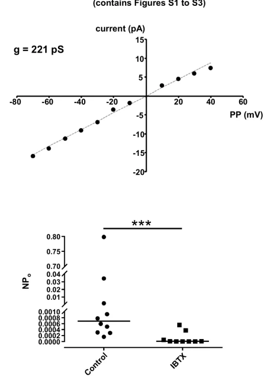
SMCs. Using inside-out patches in symmetrical [K+] and bath with high [Ca2+] (0.1 mmol/L), a
unitary conductance of 221 pS was measured, consistent with properties of BKCa channel in
VSMCs (Figure S1A). Using the cell-attached configuration of the patch-clamp technique and physiological K+ gradient, channel activity was observed in most membrane patches and was
dramatically silenced when pipette solution contained IBTX (Figure S1B), strongly supporting that this conductance was carried by BKCa channels. Average unitary current in the presence
of 0.03% DMSO amounted to 5.4±0.4 pA, at a 40 mV PP, and the channel open state probability NPo averaged to 0.029±0.006 (n=26), consistent with other reports in similar
10
conditions17. Figure 2 and Table S3 show the effects of PDE3 and PDE4 inhibition on channelactivity in cell-attached patches. When perfused alone, Ro or Cil had no obvious effect on channel activity. However, simultaneous perfusion with Ro and Cil significantly increased NPo,
mean open time and NFo. Thus BKCa channel activity can be regulated by PDE3 and PDE4.
3.4 BKCa channels differentially participate in the potentiating effect of PDE3 and PDE4
-adrenergic or AC stimulation
So far, we have shown that BKCa channels are key players in the relaxing effect of PDE
inhibitors on rat LADCA rings under resting cAMP level. In order to investigate the relevance of this phenomenon under stimulation of the cAMP pathway, the response of LADCA to isoprenaline (ISO), a - -AR) agonist, was examined (Figure 3A-D, Table S2B). ISO induced a concentration-dependent relaxation of LADCA rings (pD2=7.7±0.1,
Emax=90±4%, Figure 3A). Both PDE4 (Ro, 10 µmol/L) and PDE3 (Cil, 1 µmol/L) inhibition
shifted the CRC of ISO to the left and increased its pD2 value to 8.3±0.1 and 8.2±0.1,
respectively (P<0.01 for both), indicating a potentiating effect (Figure 3A and C). Pretreatment with IBTX reduced the Emax of ISO response to 54±4% (P<0.001) but not its pD2 (7.6±0.1;
Figure 3B and D). Interestingly, neither Ro nor Cil had any effect on ISO response in the presence of IBTX (Figures 3B and D). These results indicate that BKCa channels are important
-adrenergic vasorelaxation. They further highlight a key role of BKCa channels
in mediating the potentiating effect of PDE4 inhibition -adrenergic response.
We also studied the effects of PDE3 and PDE4 inhibition on the response to a direct AC activator, L-858051 (L85, a hydrophilic forskolin analog, Figure 3E-H, Table S2C). L85 induced a concentration-dependent relaxation (pD2=6.5±0.1) which was potentiated by Ro and Cil
(pD2=7.1±0.1, P<0.001, and 6.8±0.1, P<0.05, respectively; Figure 3E and G). IBTX hampered
the response to L85, by decreasing its pD2 value to 5.9±0.1 (P<0.001; Figure 3F and H).
Interestingly, Cil and Ro still potentiated L85 response in the presence of IBTX (Figure 3F and H). These data indicate that the potentiating effect of PDE3/4 inhibitors on the vasorelaxant response to direct AC stimulation does not require functional BKCa channels.
3.5 The coupling of BKCa channels with PDE3 and PDE4 is functionally absent in HF
LADCA
We then explored whether the above-described coupling between BKCa channels and PDE3/4
is altered in HF situation, by using a rat model of chronic cardiac pressure overload9, 29.
Echocardiographic analysis in independent series of animals are presented in Table S4. Rats which had aortic stenosis surgery displayed significant increases in left ventricle (LV) mass, end diastolic and end systolic volumes, and a significant decrease in LV ejection fraction (EF)
11
compared to sham-operated animals. Thus this model is featured with hypertrophic remodelling of the myocardium, LV dilation and decreased systolic function. The weight of the lung normalized to tibia length (LW/TL) collected after sacrifice in rats with aortic stenosis correlated significantly with the decrease in systolic function (e.g. EF, P<0.05, Pearson correlation coefficients), and with the increase in LV dimensions (e.g. diastolic and end-systolic volumes, P<0.05 and P<0.01, respectively) and parameters related to LV hypertrophy (e.g. LV mass, P<0.01). Heart weight correlated only with LV mass and posterior wall thickness, diastolic (P<0.01) and systolic (P<0.001). Because the outcome of the model showed great individual variability29, only rats with LW/TL ratio on sacrifice above 650 mg/cm,(as an evidence of lung congestion, a sign of HF) were included in the series used for experiments on LADCA (Figure 4A). Furthermore, histological examination revealed that CA from HF animals displayed thickening of their wall and perivascular fibrosis (Figure 4B). LADCA isolated from HF rats (HF-LADCA) showed decreased contractile capacity to both K80 and U46619 (1 µmol/L), compared to LADCA isolated from sham rats (Figure 4C and Table S2D). Moreover, HF-LADCA displayed a significant reduction of the relaxant effect of ACh, an archetypical endothelium-mediated response, in comparison with sham (Figure 4D). Thus HF-LADCA are characterized by an alteration of their contractile capacity, accompanied by signs of endothelial dysfunction.
As already shown above, Ro relaxed sham-LADCA (P<0.05, before versus after Ro addition, 2-way ANOVA for repeated measures) and this response was smaller in the presence of IBTX (Figure 5A). In HF-LADCA, relaxation produced by Ro was generally poor, yet globally significant (P<0.05) and this response was not affected by IBTX (Figure 5A). Cil did not evoke any robust response in this series in sham (P=0.07). Still, Cil relaxed HF-LADCA (P<0.05). Response to Cil was globally higher in HF than in sham (P<0.01, 2-way ANOVA), with no effect of IBTX. (Figure 5B). Furthermore, upon simultaneous application of Ro and Cil, synergistic
relaxation (P<0.001) were observed in both HF- and sham-LADCA but the response was depressed by IBTX in sham-LADCA only (Figure 5C). Therefore, contribution of BKCa channel
to the relaxing effect of PDE3/4 inhibition is lost in HF-LADCA.
3.6 Expression level of BKCa -subunit, PDE3 and PDE4 in HF-LADCA
In order to provide a molecular basis that would explain this last result, we characterized the expressions of BKCa channel and PDE3/4 in sham- and HF-LADCA (Figures 6 and S2). The
amount of the pore- -s.u. of the BKCa channel was about half-decreased in HF-
compared to sham-LADCA (Figure 6A). Quantification of PDE3A and PDE4D revealed a 2-fold decrease in short isoforms of 98 kDa and 80 kDa isoform, respectively (Figure 6B). By contrast, a robust 4-fold increase in the amount of a 70 kDa PDE4B isoform was found in HF-
12
compared to sham-LADCA. PDE4A did not reveal any significant difference between HF and sham.3.7 Proximity between PDE3/4 isoforms and BKCa channels
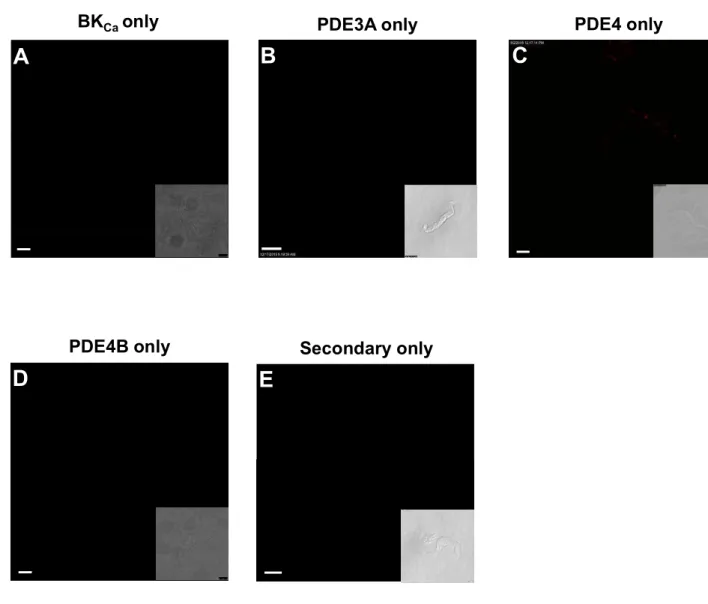
In order to reveal whether PDE isoforms localize in the vicinity of BKCa channels, experiments
using PLA were performed in SMCs freshly isolated from either HF- or LADCA. In sham--s.u. of the BKCa
channel associated with either a PDE3A antibody, a pan-specific PDE4 antibody (documented to detect PDE4A and PDE4D isoforms) or a PDE4B antibody (Figure 7). Controls incubated with only one of these antibodies showed no signal except for the pan-specific PDE4 antibody (Figure S3). However, this signal was considered to be negligible compared to the signal obtained by coupling with anti-BKCa antibody. Quantification and mixed model analysis
revealed no significant changes of the PLA signals in HF-LADCA cells.
4. Discussion
Here, we addressed the extent and modalities of the contribution of BKCa channels in mediating
the vasodilating properties of PDE3 and PDE4 inhibitors. This was performed using rat LADCA isolated from either healthy rats or animals with severe HF. We report novel findings: (i) The existence of a signalosome involving PDE3/4 and BKCa channels is supported by PLA data
that revealed in situ spatial proximity between PDE isoforms and the channel within a 40 nm range. (ii) PDE3 and PDE4 control BKCa channel activity. (iii) The relative contribution of BKCa
channel to the relaxing effects of selective PDE inhibitors depends on the status of cAMP synthesis (either unstimulated, stimulated via -AR, or direct AC stimulation) and is globally equivalent for PDE3 and PDE4. (iv) Inhibition of the RyR did not affect the relaxant responses to PDE3 or PDE4 inhibition. (v) In a model exhibiting signs of severe HF associated with hypertrophic remodelling of the myocardium, LV dilation and decreased systolic function, the contribution of BKCa channel to the regulation of coronary tone by PDE3 and PDE4
disappeared, although PDE3 and PDE4 inhibitors were still able to relax the vessels. This was associated with decreased expression of BKCa channel -s.u. and of short forms of PDE3A and PDE4D. Altogether, these data provide new insights on how regulation of a specific cAMP effector by PDEs translates into fine-tuning of the vascular tone. Moreover, our study provides an unprecedented observation of an altered coupling between vascular PDEs and a cAMP effector, namely the BKCa channel, in a cardiovascular disorder.
13
Effects obtained with PDE3 and PDE4 inhibitors at selective concentrations (Table S1) were overall consistent with previous data obtained in various vascular beds9, 11-13, 15, 34. Usingselective block with IBTX, we demonstrated that vasorelaxation by PDE3 or PDE4 inhibition in rat LDCA depended substantially on BKCa. Few studies formerly explored such participation of
BKCa channels in the relaxation evoked by PDE3/4 inhibition in other vascular beds. Regarding
PDE3 inhibition, BKCa inhibitors showed either no or little effect in human or guinea-pig
pulmonary artery (PA), respectively35, 36. In line with our study, Li et al.37reported that relaxation
of rabbit aorta by cilostazol, a PDE3 inhibitor, was almost suppressed by the BKCa channel
blocker paxilline. Considering PDE4 inhibition, a couple of reports mentioned that the relaxant effect of rolipram was only partially inhibited by BKCa inhibitors in human PA35 and porcine CA13.
Although heterogeneity of conclusions among reports may originate from diversity of protocols used (e.g. type and concentration of contractile agonist or PDE inhibitor used), the relative contribution of BKCa channels may also vary among vascular beds and species.
According to our data, Kv7 channels are interesting supplementary candidates for mediating
the effects of PDE3 and PDE4 inhibitors. Kv7.1, Kv7.4 and Kv7.5 subunits were reported to be
robustly expressed in rat CA38 and inhibited by 30 µmol/L XE991 (Table S1). Accumulating
evidence suggest that Kv7 act in parallel with BKCa channels to mediate cAMP vasorelaxant
pathways in various vascular beds19, 38. Since PDE4 inhibition with rolipram was recently
shown to enhance native Kv7.5 while Kv7.4 was insensitive39, channels including the Kv7.5
subunit may contribute to the XE991-sensitive action of PDE inhibitors in our study.
We found that in rat LADCA SMCs, simultaneous PDE3 and PDE4 inhibition induced a clear stimulation of BKCa unitary channel activity. However, neither PDE3 nor PDE4 inhibitor used
alone was sufficient to increase NPo. Cilostazol was recently reported to stimulate channel
activity in rabbit aortic SMCs37. However, a high concentration was used (10 µmol/L) so that
the effect may also be attributed to other PDEs, such as PDE540. Inhibition of a single PDE
may not be sufficient for detectable increase of channel activity possibly because of overlapping activity of PDE3 and PDE4. Absence of endothelial-derived NO-cGMP pathway in isolated myocytes may enhance PDE3 activity and mask the effect of PDE4 inhibition9, 11, 12.
Why then PDE3 inhibition had no apparent effect is unclear. At a low basal rate of cAMP production, inhibition of a single PDE family may only yield small or sporadic rise in cAMP concentration resulting in small increase in BKCa channel activity. It is accepted that, because
input resistance of vascular smooth muscle cells is high (1010 ohm), very few ion channels
need to be open to have substantial effect on membrane potential24. Therefore, it is possible
that even hardly detectable activation of BKCa channel may evoke relaxation of SMC.
In line with earlier work focusing on other vascular beds12, 34, our data in CA show that inhibitors
cAMP-14
elevating agent, and L85, a direct AC activator. Besides, responses to these stimulators were inhibited by IBTX, an observation consistent with previous studies25. Interestingly, IBTX-AR stimulation, but not under broad AC -AR-stimulated cAMP is preferentially controlled by PDEs acting on BKCa channels, while L85 response is impeded by PDEs acting on other effectors.
This would be consistent with compartmentalization of cAMP in these cells, created by signalling domains including PDE3/4, BKCa -AR. A signalosome involving BKCa
2-AR, L-type Ca2+ channels and the scaffolding protein AKAP79/150, was
previously characterized in VSMCs41. In other cell types, PDE4D was detected in a
macromolecular complex 2-AR whereas PDE4B was associated with the L-type
Ca2+ channel (reviewed in Mika et al., 201220). To our knowledge, there is no available
evidence for localization of PDE3 isoforms in such sarcolemma complex involving the -AR. Here, we took advantage of the novel PLA technique to show that PDE3 and PDE4 isoforms are localized in the vicinity of BKCa channels in LADCA SMCs. Nevertheless, resolution of this
technique is limited since two antigens as far as 40 nm from each other can generate a PLA signal. Further data supporting the existence of a macromolecular complex might be given by biochemical methods although these are challenging to use in native small arteries yielding small amount of material. Yet, our data offer new insights toward the characterization of a new signalling complex with important functional relevance in regulating arterial tone -AR stimulation. Since recent studies in bladder SMCs showed that PDE4 inhibition elevated BKCa
activity by stimulating Ca2+ release42, 43, it was of interest to address whether this coupling was
relevant in vascular SMCs. Our data did not support a major role of such a mechanism in the vasorelaxant effects of Cil and Ro in unstimulated conditions. This implies that Ca2+
participating to BKCa activation may rather originate from Ca2+ influx23.
4.2 Contribution of BKCa channel to the relaxant action of PDE3/4 inhibition is lost in HF Importantly, in clear contrast with age-matched sham animals, BKCa channels did not mediate
the relaxant effect of PDE3 and PDE4 inhibitors in arteries from HF rat. This indicates that the relaxation was mediated by other mechanisms than BKCa channels activation, which may
include action on other ion channels, increased Ca2+ pumping, decreased Ca2+-sensitivity of
the myosin light chain phosphorylation, or uncoupling of contractile machinery16. A
straightforward explanation would be that collapse of the IBTX-sensitive contribution was a direct consequence of the decrease of the amount of BKCa -s.u. observed in
HF-LADCA. Such a down-regulation of BKCa channel expression was previously described in
mesenteric arteries from mice which developed HF following myocardial infarction28 and in
other models of cardiovascular disorders22, and was generally associated with a reduced
15
alteration of channel expression in a rat model with cardiac hypertrophy following injection of ISO44. In the latter work, however, animals presented only mild remodelling, whereas the ratsstudied here were submitted to chronically elevated afterload and displayed dramatic cardiac hypertrophy and lung congestion.
In parallel, we report a decrease of short PDE3A and PDE4D isoforms, together with a marked increase in a short PDE4B isoform in the HF-LADCA. Decrease in PDE3A expression was reported to be a feature of SMC switching to a proliferative phenotype45, consistent with the
intense vascular wall remodelling that was seen in the CA from HF rats. By contrast, expression of short forms of PDE4D are reported to be more robustly expressed in cultured VSMC upon prolonged stimulation with cAMP-elevating agents46.Finally, robust increase in
the short PDE4B was also observed in aorta using the same HF model in rats9. Thus this
alteration may constitute a hallmark of remodelling vascular tissue, at least in HF.
A limitation to our study was that contractility of LADCA from rats with HF was strikingly reduced compared to sham (Figure 4C) and this was associated with wall thickening and remodelling. By contrast, contractility was slightly increased in aortic rings from rat submitted to the same model9. Exposure of both vascular beds to contrasted hemodynamic stress
(LADCA being upstream the stenosis whereas aorta being downstream) may explain differential alteration of contractility. Still, a milder aortic banding model in guinea pig yielded CA with thickened wall but normal active media stress8, while septal arteries from rats with
congestive HF five weeks following myocardial infarction displayed quasi-normal structure with higher response to contractile stimuli47. Thus loss of reactivity of CA in our study may be related
to specific hemodynamic stress caused by severe stenosis rather than deterioration of myocardial function. The exact mechanisms remain to be determined and may include deleterious wall remodelling with a loss of muscular capacity. The conclusion of our study will have to be taken cautiously when extended to other forms of HF such as post-myocardial infarction and HF with preserved ejection fraction.
Coronary reserve is well known to be altered in severe cardiac hypertrophy, including in patients or animal models submitted to chronic afterload4, 5. The observation that combined
PDE3 and PDE4 inhibition yielded almost maximal relaxation of LADCA suggests that dilatory mechanisms are still functional, although the BKCa component is apparently abolished.
Because our study focused on large arteries mounted on an isometric myograph, caution must be taken when extending our conclusions to in vivo changes in coronary reserve, which does not depend solely on vasodilatory mechanisms, but also on basal blood flow and minimal coronary resistance in the whole myocardium circulation.
16
4.3 ConclusionIn conclusion, this study identifies cAMP-PDE BKCa channel coupling as a key signalling
pathway for fine tuning of vascular tone (Figure 8). Our results demonstrate that the contribution of this mechanism to global tone regulation by PDEs varies depending on the mode of cAMP stimulation and in pathophysiological context of HF. The last result may be explained by decreased expression of BKCa channel in HF. Further studies are needed to
delineate the structural determinants of cyclic nucleotide compartmentation among various effectors in the vascular smooth muscle and how they are modified in disease.
5. Funding
This work was supported by the University Paris-Sud (Attractivité 2013 grant to BM); the grant [ANR-10-LABX-33] as members of the Laboratory of Excellence LERMIT; and the French
for a PhD fellowship awarded to SI.
6. Acknowledgments
We are thankful to Dr Chen Yan (University of Rochester Medical Center, NY, USA), and Dr Marco Conti (University of California, San Francisco, CA, USA) for kindly providing PDE3A and PDE4A/B/D antibodies, respectively. Dr Delphine Mika, Dr Guillaume Pidoux, Dr Milia Belacel-Ouari and Dr Bertrand Crozatier are acknowledged for kindly providing expertise on specific protocols. Françoise Gaudin performed tissue sections and staining at the IPSIT PHIC facility at Univ. Paris-Sud.
7. Conflict of Interest
None declared.
8. References
1. Treasure CB, Vita JA, Cox DA, Fish RD, Gordon JB, Mudge GH, Colucci WS, Sutton MG, Selwyn AP, Alexander RW, Ganz P. Endothelium-dependent dilation of the coronary microvasculature is impaired in dilated cardiomyopathy. Circulation 1990;81:772-779.
17
2. Vatner SF, Hittinger L. Coronary vascular mechanisms involved in decompensationfrom hypertrophy to heart failure. J Am Coll Cardiol 1993;22:34A-40A.
3. Krams R, Kofflard MJ, Duncker DJ, Von Birgelen C, Carlier S, Kliffen M, ten Cate FJ, Serruys PW. Decreased coronary flow reserve in hypertrophic cardiomyopathy is related to remodeling of the coronary microcirculation. Circulation 1998;97:230-233. 4. Marcus ML, Doty DB, Hiratzka LF, Wright CB, Eastham CL. Decreased coronary
reserve: A mechanism for angina pectoris in patients with aortic stenosis and normal coronary arteries. N Engl J Med 1982;307:1362-1366.
5. Tsai SH, Lu G, Xu X, Ren Y, Hein TW, Kuo L. Enhanced endothelin-1/Rho-kinase signalling and coronary microvascular dysfunction in hypertensive myocardial hypertrophy. Cardiovasc Res 2017;113:1329-1337.
6. Inoue T, Sakai Y, Morooka S, Hayashi T, Takayanagi K, Yamaguchi H, Kakoi H, Takabatake Y. Vasodilatory capacity of coronary resistance vessels in dilated cardiomyopathy. Am Heart J 1994;127:376-381.
7. Kiuchi K, Sato N, Shannon RP, Vatner DE, Morgan K, Vatner SF. Depressed beta-adrenergic receptor- and endothelium-mediated vasodilation in conscious dogs with heart failure. Circ Res 1993;73:1013-1023.
8. McGoldrick RB, Kingsbury M, Turner MA, Sheridan DJ, Hughes AD. Left ventricular hypertrophy induced by aortic banding impairs relaxation of isolated coronary arteries. Clin Sci (Lond) 2007;113:473-478.
9. Hubert F, Belacel-Ouari M, Manoury B, Zhai K, Domergue-Dupont V, Mateo P, Joubert F, Fischmeister R, Leblais V. Alteration of vascular reactivity in heart failure: Role of phosphodiesterases 3 and 4. Br J Pharmacol 2014;171:5361-5375.
10. Maurice DH, Ke H, Ahmad F, Wang Y, Chung J, Manganiello VC. Advances in targeting cyclic nucleotide phosphodiesterases. Nat Rev Drug Discov 2014;13:290-314.
11. Komas N, Lugnier C, Stoclet JC. Endothelium-dependent and independent relaxation of the rat aorta by cyclic nucleotide phosphodiesterase inhibitors. Br J Pharmacol 1991;104:495-503.
12. Polson JB, Strada SJ. Cyclic nucleotide phosphodiesterases and vascular smooth muscle. Annu Rev Pharmacol Toxicol 1996;36:403-427.
13. Kaneda T, Kubota T, Fujimoto K, Urakawa N, Nakajyo S, Shimizu K. Effects of rolipram on U46619-induced contraction and cyclic nucleotide content in the porcine coronary artery. J Smooth Muscle Res 2010;46:17-29.
14. Rump AF, Acar D, Klaus W. A quantitative comparison of functional and anti-ischaemic effects of the phosphodiesterase-inhibitors, amrinone, milrinone and levosimendan in rabbit isolated hearts. Br J Pharmacol 1994;112:757-762.
15. Lindgren S, Andersson KE. Effects of selective phosphodiesterase inhibitors on isolated coronary, lung and renal arteries from man and rat. Acta Physiol Scand 1991;142:77-82.
16. Morgado M, Cairrao E, Santos-Silva AJ, Verde I. Cyclic nucleotide-dependent relaxation pathways in vascular smooth muscle. Cell Mol Life Sci 2012;69:247-266. 17. White RE, Kryman JP, El-Mowafy AM, Han G, Carrier GO. cAMP-dependent
vasodilators cross-activate the cgmp-dependent protein kinase to stimulate BKCa
channel activity in coronary artery smooth muscle cells. Circ Res 2000;86:897-905. 18. Roberts OL, Kamishima T, Barrett-Jolley R, Quayle JM, Dart C. Exchange protein
activated by cAMP (EPAC) induces vascular relaxation by activating Ca2+-sensitive K+
channels in rat mesenteric artery. J Physiol 2013;591(20):5107-23.
19. Stott JB, Barrese V, Greenwood IA. Kv7 channel activation underpins EPAC-dependent
relaxations of rat arteries. Arterioscler Thromb Vasc Biol 2016;36:2404-2411.
20. Mika D, Leroy J, Vandecasteele G, Fischmeister R. PDEs create local domains of cAMP signaling. J Mol Cell Cardiol 2012;52:323-329.
18
21. Wu RS, Marx SO. The BK potassium channel in the vascular smooth muscle andkidney: alpha- and beta-subunits. Kidney Int 2010;78:963-974.
22. Hu XQ, Zhang L. Function and regulation of large conductance Ca2+-activated K+
channel in vascular smooth muscle cells. Drug Discov Today 2012;17:974-987. 23. Latorre R, Castillo K, Carrasquel-Ursulaez W, Sepulveda RV, Gonzalez-Nilo F,
Gonzalez C, Alvarez O. Molecular determinants of BK channel functional diversity and functioning. Physiol Rev 2017;97:39-87.
24. Nelson MT, Cheng H, Rubart M, Santana LF, Bonev AD, Knot HJ, Lederer WJ. Relaxation of arterial smooth muscle by calcium sparks. Science 1995;270:633-637. 25. Price JM, Cabell JF, Hellermann A. Inhibition of cAMP mediated relaxation in rat
coronary vessels by block of Ca2+ activated K+ channels. Life Sci 1996;58:2225-2232.
26. Porter VA, Bonev AD, Knot HJ, Heppner TJ, Stevenson AS, Kleppisch T, Lederer WJ, Nelson MT. Frequency modulation of Ca2+ sparks is involved in regulation of arterial
diameter by cyclic nucleotides. Am J Physiol 1998;274:C1346-1355.
27. Wellman GC, Nathan DJ, Saundry CM, Perez G, Bonev AD, Penar PL, Tranmer BI, Nelson MT. Ca2+ sparks and their function in human cerebral arteries. Stroke
2002;33:802-808.
28. Wan E, Kushner JS, Zakharov S, Nui XW, Chudasama N, Kelly C, Waase M, Doshi D, Liu G, Iwata S, Shiomi T, Katchman A, D'Armiento J, Homma S, Marx SO. Reduced vascular smooth muscle BK channel current underlies heart failure-induced vasoconstriction in mice. FASEB J 2013;27:1859-1867.
29. Feldman AM, Weinberg EO, Ray PE, Lorell BH. Selective changes in cardiac gene expression during compensated hypertrophy and the transition to cardiac decompensation in rats with chronic aortic banding. Circ Res 1993;73:184-192. 30. Thompson WJ, Appleman MM. Multiple cyclic nucleotide phosphodiesterase activities
from rat brain. Biochemistry 1971;10:311-316.
31. Nyborg NC, Baandrup U, Mikkelsen EO, Mulvany MJ. Active, passive and myogenic characteristics of isolated rat intramural coronary resistance arteries. Pflugers Arch 1987;410:664-670.
32. Singer JJ, Walsh JVJ. Characterization of calcium-activated potassium channels in single smooth muscle cells using the patch-clamp technique. Pflugers Arch 1987;408:98-111.
33. White RE, Darkow DJ, Lang JL. Estrogen relaxes coronary arteries by opening BKCa
channels through a cgmp-dependent mechanism. Circ Res 1995;77:936-942.
34. Maurice DH, Crankshaw D, Haslam RJ. Synergistic actions of nitrovasodilators and isoprenaline on rat aortic smooth muscle. Eur J Pharmacol 1991;192:235-242.
35. Bardou M, Goirand F, Bernard A, Guerard P, Gatinet M, Devillier P, Dumas JP, Morcillo EJ, Rochette L, Dumas M. Relaxant effects of selective phosphodiesterase inhibitors on U46619 precontracted human intralobar pulmonary arteries and role of potassium channels. J Cardiovasc Pharmacol 2002;40:153-161.
36. Rieg AD, Rossaint R, Verjans E, Maihofer NA, Uhlig S, Martin C. Levosimendan relaxes pulmonary arteries and veins in precision-cut lung slices - the role of K-channels, cAMP and cGMP. PLoS One 2013;8:e66195.
37. Li H, Hong da H, Son YK, Na SH, Jung WK, Bae YM, Seo EY, Kim SJ, Choi IW, Park WS. Cilostazol induces vasodilation through the activation of Ca2+-activated K+
channels in aortic smooth muscle. Vascul Pharmacol 2015;70:15-22.
38. Khanamiri S, Soltysinska E, Jepps TA, Bentzen BH, Chadha PS, Schmitt N, Greenwood IA, Olesen SP. Contribution of Kv7 channels to basal coronary flow and
active response to ischemia. Hypertension 2013;62:1090-1097.
39. Mani BK, Robakowski C, Brueggemann LI, Cribbs LL, Tripathi A, Majetschak M, Byron KL. Kv7.5 potassium channel subunits are the primary targets for PKA-dependent
19
enhancement of vascular smooth muscle Kv7 currents. Mol Pharmacol2016;89:323-334.
40. Sudo T, Tachibana K, Toga K, Tochizawa S, Inoue Y, Kimura Y, Hidaka H. Potent effects of novel anti-platelet aggregatory cilostamide analogues on recombinant cyclic nucleotide phosphodiesterase isozyme activity. Biochem Pharmacol 2000;59:347-356. 41. Liu G, Shi J, Yang L, Cao L, Park SM, Cui J, Marx SO. Assembly of a Ca2+-dependent
BK channel signaling complex by binding to beta2 adrenergic receptor. EMBO J 2004;23:2196-2205.
42. Xin W, Li N, Cheng Q, Petkov GV. BK channel-mediated relaxation of urinary bladder smooth muscle: A novel paradigm for phosphodiesterase type 4 regulation of bladder function. J Pharmacol Exp Ther 2014;349(1):56-65.
43. Zhai K, Chang Y, Wei B, Liu Q, Leblais V, Fischmeister R, Ji G. Phosphodiesterase types 3 and 4 regulate the phasic contraction of neonatal rat bladder smooth myocytes via distinct mechanisms. Cell Signal 2014;26:1001-1010.
44. Kim N, Chung J, Kim E, Han J. Changes in the Ca2+-activated K+ channels of the
coronary artery during left ventricular hypertrophy. Circ Res 2003;93:541-547.
45. Dunkerley HA, Tilley DG, Palmer D, Liu H, Jimmo SL, Maurice DH. Reduced phosphodiesterase 3 activity and phosphodiesterase 3A level in synthetic vascular smooth muscle cells: Implications for use of phosphodiesterase 3 inhibitors in cardiovascular tissues. Mol Pharmacol 2002;61:1033-1040.
46. Tilley DG, Maurice DH. Vascular smooth muscle cell phosphodiesterase (PDE) 3 and PDE4 activities and levels are regulated by cyclic AMP in vivo. Mol Pharmacol 2002;62:497-506.
47. Stassen FR, Fazzi GE, Leenders PJ, Smits JF, De Mey JG. Coronary arterial hyperreactivity and mesenteric arterial hyporeactivity after myocardial infarction in the rat. J Cardiovasc Pharmacol 1997;29:780-788.
20
9. Figure LegendsFigure 1. BKCa channel inhibition inhibited the relaxant effect of cAMP-PDE inhibitors in rat LADCA. Selective PDE4 inhibitor (Ro, 10 µmol/L), or PDE3 inhibitor (Cil, 1 µmol/L), or both
inhibitors were applied on LADCA rings contracted with U46619 (U46, 0.3-3 µmol/L) in the presence of BKCa channel inhibitor IBTX (A, B, C, 0.1 µmol/L), various ion channel inhibitors (D) or relevant vehicle (veh., A-D). A: Examples of traces obtained in the absence (veh., left-hand side) or in the presence of IBTX (right-left-hand side). B: Scatter plot showing individual data points and mean relaxation ± SEM, in percentage of initial contraction. (+): IBTX; (-): veh. C: Relaxant effect by Ro + Cil was still inhibited by IBTX in rings after the endothelium was voluntarily damaged (endo(-)). D: Relaxant effect of combined application of Ro and Cil was not altered by either ryanodine (30 µmol/L), glibenclamide (glib.,10 µmol/L), DPO-1 (1 µmol/L), or stromatoxin (STX, 0.1 µmol/L), but was reduced by XE991 (30 µmol/L). Shown are individual data and means ± SEM. N=5-13. **: P<0.01, ***: P<0.001 (Mann-Whitney test). ###
(ANOVA followed by Holm- test).
Figure 2. Ro (10 µmol/L) together with Cil (1 µmol/L) increased NPo, mean open time and
NFo of BKCa channels in LADCA SMCs. Both inhibitors had no significant effect when used alone. A: Data from 3 independent cell-attached patches are shown, during DMSO perfusion (a, b and c) and during Ro+Cil perfusion of the same cells ( and , respectively). Patch
potential was 40 mV. NPo associated to the displayed traces are indicated. Amplitude
histograms generated from the whole analysed period in each condition are also shown. B-D: Summary individual (grey dots) and mean±SEM (black lines) data of NPo (B), mean open time
(C) and opening frequency (NFo, D) are presented. n=5-11 cells from N=5-7 rats. *: P<0.05 vs.
DMSO (Wilcoxon signed-rank test performed on means per rat).
Figure 3. Effect of PDE4 or PDE3 inhibition on concentration-response curves (CRCs) to isoprenaline or L-858051 in LADCA rings. PDE4 (Ro, 10 µmol/L: A, B, E, F) or PDE3 (Cil
1 µmol/L: C, D, G, H) inhibition was tested on CRCs to isoprenaline (ISO, A-D) or L-858051 (L85, E-H) in LADCA rings incubated with IBTX, in the right panels, or relevant vehicle, in the left panels. In the right panels (B, D, F, H), dashed line represents the CRC fit from control data. N=6-14. ***: P<0.001 (2-way ANOVA for repeated measures).
Figure 4. Rats with aortic stenosis exhibit signs of heart failure, remodelling of the wall of large coronary arteries and altered vasoreactivity of LADCA. A: Summary values of
organ weight over tibia length (TL) ratios for heart, lung, kidney and liver, as well as body weight from sham rats and rats with aortic stenosis included for LADCA isolation and histology (HF; lung weight/TL >650 mg/cm). N=23-26; $$$ -test). B: Representative
21
left ventricle cross-sections from sham and HF rats stained with Masson trichrome, displaying large arteries. The scatter plot represents quantification of [wall/vessel diameter] ratio in large arteries (n=22-23 from N=3 rats per group; #: P<0.05, nested ANOVA). C-D: Vasoreactivity in LADCA from sham and HF rats: contractile response to K80 and 1 µmol/L U46619 (C; N=7 rats, ***: P<0,001, Mann-Whitney test) and relaxant response to ACh (D; N=7 rats, : P<0,001, 2-way ANOVA for repeated measures). Scatter plots also show mean values ± SEM.Figure 5. BKCa channel inhibition has no effect on the relaxant effect of PDE3/4 inhibitors in HF-LADCA. PDE4 inhibitor (A, Ro, 10 µmol/L), PDE3 inhibitor (B, Cil, 1 µmol/L) or both
inhibitors (C) were applied on U46619-contracted (1-3 µmol/L) LADCA rings from sham and HF rats in the presence of IBTX (+) or vehicle (-). Scatter plot showing individual data points and mean relaxation ± SEM, in percentage of initial contraction. N=4-7. *: P<0.05, **: P<0.01 (2-way ANOVA followed by Holm-Sidak test). In B, effect of Cil was overall significantly higher
-way ANOVA).
Figure 6. Expression of BKCa and PDE proteins in sham and HF rat LADCA. Expression
of BKCa -s.u. (A), PDE3A, PDE4A, PDE4B and PDE4D (B, from left to right, respectively) proteins in LADCA isolated from sham or HF rats. Representative immunoblots obtained for these proteins and matching GAPDH signal are shown. Arrows indicate the bands that were quantified. Scatter plots show individual values and mean ± SEM from relative densitometric quantification normalized to GAPDH and averaged signal in sham. N=5 for BKCa -s.u. and PDE4B, N=9 for PDE3A, N=6 for PDE4A and N=6-7 for PDE4D. *: P<0.05, **: P<0.01 (Mann -Whitney test).
Figure 7. Evidence for PDE3/4-BKCa -subunit signals. PLA using an antibody against BKCa
-subunit associated with anti-PDE3A (A), anti-PDE4 (pan specific, B) or anti-PDE4B (C) antibody in sham- (images in upper panel, empty circles in scatter plot) or HF- (images in middle panel, filled circles) LADCA SMCs. Representative images are shown. Scale bar: 5 µm. Inset shows transmitted light image. Plots in lower panels show respective quantifications of PLA signal (individual data and mean ± SEM) of n=12-84 cells from 2-3 rats.
Figure 8. Schematic representation of the differential contribution of BKCa channels to
the relaxant effects of PDE3 and PDE4 inhibitors in rat LADCA. A. Under basal cAMP
production rate, Ro and Cil produce a relaxation in which the BKCa channels play essential role.
K+ channels limit depolarization of cell membrane potential, resulting in closing L-type Ca2+
channels (LTCC), reduction of intracellular calcium concentration ([Ca2+]
i) and relaxation. This
22
also participate in the action of PDE inhibitors. B. Under stimulation of cAMP production via the -AR (ISO), Ro and Cil action depends mostly on coupling with BKCa channels. Underbroad stimulation of the AC with L85, participation of the channel is not apparent, and other effectors may mediate the relaxation. C. In LADCA from rats with heart failure, participation of the channel to the relaxant action of Ro and Cil is absent, in contrast with sham. Variation of expression of BKCa ( ), short form of PDE3A and PDE4D ( ) and PDE4B ( ) proteins are
Figure 1
A
Ro Ro U46 Cil U46 5 min 1 mN/mm Ro IBTX Cil U46 U46 Ro+ veh.
+ IBTX
IBTXB
IBTX
-
+
-
+
-
+
Ro
Cil
Ro + Cil
R
e
la
x
a
ti
o
n
(
%
)
0 50 100**
***
**
endo.
(-)C
**
Ro + Cil
0 50 100 ###D
0 50 100R
e
la
x
a
ti
o
n
(
%
)
R
e
la
x
a
ti
o
n
(
%
)
B
C
D
Figure 2
A
DMSO
5 pARo + Cil
NPo= 0.0072 NPo= 0.2520 NPo= 0.0362 NPo= 0.2634 Ampl. (pA) 0 100 200 20 10 0 0 20 10 0 100 200 0 20 40 20 10 0 0 20 10 0 20 40 NPo= 0.0053 NPo= 0.1669 40 80 0 0 10 20 40 80 0 0 10 20 Ampl. (pA) Ampl. (pA) Ampl. (pA) Ampl. (pA) Ampl. (pA) 0.0 0.2 0.4 0.6 N Po 0 50 100 150 m e an o p en t im e ( m s ) 0 2 4 6 N Fo (s -1) NPomean open time
control Ro -10 -8 -6 -4 100 50 0
Log ([ISO], mol/L)
A
R el ax at io n ( % ) 100 50 0Log ([ISO], mol/L) IBTX Ro + IBTX
B
-10 -8 -6 -4 R el ax at io n ( % )Log ([ISO], mol/L)
100 50 0 -10 -8 -6 -4 control Cil
C
R el ax at io n ( % )Log ([ISO], mol/L) IBTX Cil + IBTX 100 50 0
D
-10 -8 -6 -4 R el ax at io n ( % )E
Log ([L85]), mol/L) -10 -8 -6 -4 control Ro 100 50 0 R el ax at io n ( % ) -10 -8 -6 -4 100 50 0 IBTX Ro + IBTX Log ([L85]), mol/L)F
R el ax at io n ( % )G
100 50 0 Log ([L85], mol/L) -10 -8 -6 control Cil R el ax at io n ( % ) -4H
-10 -8 -6 -4 100 50 0 Log ([L85], mol/L) IBTX Cil + IBTX R el ax at io n ( % )Figure 3
K80 U46619 0 2 4 6 C o n tr a c ti o n ( m N /m m )
***
***
sham HFD
sham HF sham pD2 = 7.4±0.1 Emax = 85.2±6.1% pD2 = 6.9±0.2 Emax = 50.1±10.9% ** -10 -8 -6 100 50 0 R e la x a ti o n ( % ) Log ([ACh], M) §§ §C
b o d y w e ig h t (g ) sham HF w e ig h t / T L ( g /c m ) $$$ 0.0 0.5 1.0 1.5 2.0 $$$A
heart lung 0 2 4 6 liver 0 200 400 600 800 1000 body 0.20 0.25 0.30 0.35 0.40 0.45 kidney $$$ w e ig h t / T L ( g /c m ) w e ig h t / T L ( g /c m ) sham HFB
[w a ll / v e s s e ld ia m e te r] r a ti o sham#
0.0 0.1 0.2 0.3Figure 4
R
e
la
x
a
ti
o
n
(
%
)
Ro + Cil
C
0 50 100-IBTX
+
sham
HF
+
-**
*
R
e
la
x
a
ti
o
n
(
%
)
Ro
A
+
-+
-sham
HF
IBTX
0 50 100 0 50 100R
e
la
x
a
ti
o
n
(
%
)
Cil
B
-IBTX
+
sham
HF
+
-Figure 5
Figure 6
130 100 sham HFB
PDE4A 130 100 sham HF 100 70 PDE4B sham HF 70 100 PDE4D sham HF 35 35 35 35GAPDH GAPDH GAPDH GAPDH
PDE3A
kDa kDa kDa kDa
A
130 100 BKCa -s.u. sham HF 35 GAPDH kDa 0.0 0.4 0.8 1.2 BKCa -s.u. sham HF R e la ti v e e x p re s s io n 0 1 2 3 4 5 HF shamPDE3A PDE4A PDE4B
R e la ti v e e x p re s s io n 6 PDE4D
% o f c e ll a re a 0 5 10 15
BK
Ca– PDE3A
% o f c e ll a re a 0 5 10 15BK
Ca– PDE4
HF shamBK
Ca– PDE4
HF
A
BK
Ca– PDE3A
B
sham
% o f c e ll a re a 0 5 10 15 20 25BK
Ca– PDE4B
C
BK
Ca– PDE4B
Figure 7
A
B
C
SUPPLEMENTARY MATERIAL
CONTENTS
1. Detailed Material and Methods
p. 2
2. Supplemental Tables
p. 11
1. Detailed Material and Methods
1.1 Animals and surgical procedures
All animal care and experimental procedures conformed to the European Community guiding principles in the care and use of animals (Directive 2010/63/EU of the European Parliament) and authorizations to perform animal experiments according to application decrees were obtained from the French Ministry of Agriculture, Fisheries and Food (No. C92-019-01, 03 August 2016). A total of 164 rats were used in the experiments described here. This included 8-12-week-old, male Wistar rats that were used for basic physiological exploration. In addition, 37 rats with HF sacrificed 22 weeks after surgical stenosis of the ascending aorta were used. The surgical procedure was carried out on 3-week-old rats, under anaesthesia with a ketamine-xylazine mix (75 mg/kg 10 mg/kg, respectively, 0.3 mL/100g, i.p.). Aortic stenosis was mimicked by placing a stainless steel hemoclip (0.6 mm-internal diameter) on the ascending aorta via thoracic incision, as previously described1, 2. Also, 37 age-matched control animals
underwent the same procedure without placement of the clip. Buprenorphine chlorhydrate (0.03 mg/kg, 0.2 mL/100g, s.c.) was administered twice daily for 3 days beginning at the end of the surgery.
Echocardiography was performed at 22 weeks after surgery on 11 SHAM-operated and 14 stenosed rats using a 12 MHz transducer (Vivid 7, General Electric Healthcare). Anaesthesia was induced and maintained using 3% and 1.5% isoflurane, respectively. Two-dimensional-guided (2D) M-mode echocardiography was used to determine wall thickness and left ventricular chamber diameter at systole and diastole, and contractile parameters such as fractional shortening and ejection fraction. Left ventricular mass (LVM, mg) was calculated according to the Penn formula home-adapted for the rat heart:
with IVSd: end-diastolic interventricular septum thickness; LVIDd: end-diastolic LV internal diameter; LVPWd: end-diastolic LV posterior wall thickness3. Volume (V) of the left ventricular
chamber was evaluated using the Teicholz formula: , where D is the diameter of the chamber4.