Short Communication
Vitamin C deficiency in weanling guinea pigs: differential expression
of oxidative stress and DNA repair in liver and brain
Jens Lykkesfeldt
1*, Gilberto Perez Trueba
2, Henrik E. Poulsen
2and Stephan Christen
31Section of Biomedicine, Department of Veterinary Pathobiology, Faculty of Life Sciences, University of Copenhagen, 9 Ridebanevej, DK-1870 Frederiksberg C, Denmark
2Department of Clinical Pharmacology Q, Copenhagen University Hospital, 20 Blegdamsvej, DK-2200, Kbh K, Copenhagen, Denmark 3Institute of Infectious Diseases, University of Berne, Friedbu¨hlstrasse 51, CH-3010 Berne, Switzerland
(Received 1 February 2007 – Revised 29 March 2007 – Accepted 30 April 2007)
Neonates are particularly susceptible to malnutrition due to their limited reserves of micronutrients and their rapid growth. In the present study, we examined the effect of vitamin C deficiency on markers of oxidative stress in plasma, liver and brain of weanling guinea pigs. Vitamin C deficiency caused rapid and significant depletion of ascorbate (P, 0·001), tocopherols (P, 0·001) and glutathione (P, 0·001), and a decrease in superoxide dismutase activity (P¼ 0·005) in the liver, while protein oxidation was significantly increased (P¼ 0·011). No changes in lipid oxi-dation or oxidatively damaged DNA were observed in this tissue. In the brain, the pattern was markedly different. Of the measured antioxidants, only ascorbate was significantly depleted (P, 0·001), but in contrast to the liver, ascorbate oxidation (P¼ 0·034), lipid oxidation (P, 0·001), DNA oxidation (P¼ 0·13) and DNA incision repair (P¼ 0·014) were all increased, while protein oxidation decreased (P¼ 0·003). The results show that the selective preservation of brain ascorbate and induction of DNA repair in vitamin C-deficient weanling guinea pigs is not sufficient to prevent oxidative damage. Vitamin C deficiency may therefore be particularly adverse during the neonatal period.
Vitamin C deficiency: Weanling guinea pigs: Oxidative stress markers: DNA repair
Pre- and postnatal malnutrition may have serious conse-quences for neuronal development and growth1,2. The impact of vitamin C deficiency, beyond that of scurvy, has not been studied in detail. Vitamin C, for example, is known to play an important role in maintaining brain function3. The brain is particu-larly susceptible to oxidative damage and therefore highly depen-dent on proper maintenance of redox homeostasis, especially during development, when brain metabolism and growth are maximal4. An imbalance in redox homoestasis has also been implicated in neurodegenerative diseases of ageing and other neurological disorders such as schizophrenia5 – 8.
It has been known for a while that vitamin C is preferen-tially retained in the brain during deficiency9. As established recently, this is due to the high-affinity sodium ascorbate cotransporter SVCT210. Experiments with homozygous SVCT2-knockout mice have shown that this transporter is essential for perinatal survival10, indicating that vitamin C is crucial for early brain development, although the precise mechanism of death is unknown. Moreover, studies of com-bined vitamin C and E deficiency in guinea pigs have revealed extensive neuronal damage despite a relatively modest increase in oxidative stress in the brain11,12.
Unfortunately, neonatal vitamin C deficiency is fairly common. In a study with 127 pregnant Brazilian women, it was found that 40 % of the smokers and 27 % of the non-smo-kers had hypovitaminosis C (i.e. a plasma concentration , 23 mmol/l) and that this condition was passed on to their fetuses13. Major epidemiological studies have shown that this prevalence of hypovitaminosis C can largely be extended to the Western world14. Vitamin C deficiency due to inadequate perinatal feeding (e.g. with pasteurized milk) is also fairly common15.
Like man, guinea pigs completely depend on dietary vitamin C16,17. In the present study, we used newly weaned guinea pigs to test if neonates are susceptible to vitamin C deficiency and if even small changes in antioxidant status in the brain result in deleterious events as measured by oxidative damage.
Methods
Animals and study design
The study was approved by the Danish Animal Experimen-tation Inspectorate. Dunkin Hartley guinea pigs were born in our animal facility by breeder guinea pigs obtained from
* Corresponding author: Dr Jens Lykkesfeldt, fax þ 45 35 35 35 14, email [email protected] Abbreviations:Asc, ascorbate; MDA, malondialdehyde; TBA, thiobarbituric acid.
British Journal of Nutrition (2007), 98, 1116–1119 doi: 10.1017/S0007114507787457
qThe Authors 2007
British
Journal
of
Nutrition
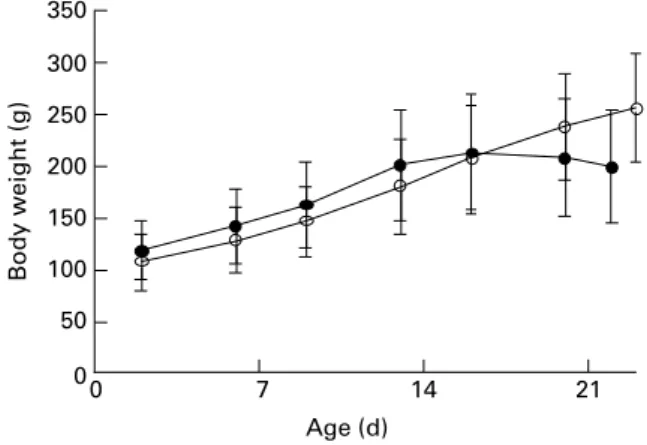
https:/www.cambridge.org/core/terms. https://doi.org/10.1017/S0007114507787457Statens Serum Institut (Allerød, Denmark), that had been maintained at the animal facility for at least 2 weeks. After 2 d, the neonates were taken from their mothers and random-ized into two weight-matched groups (control group, n 8; deficient group, n 12). All animals were housed in plastic cages (four per cage) and fed pathogen-free standard diets (Altromin International, Lage, Germany). The diets were essentially identical except for vitamin C content. Control ani-mals were placed on a normal guinea pig diet (Diet #3010, 1036 mg vitamin C/kg)18, while deficient animals were sup-plied with a diet low in vitamin C (Diet #2010, 36 mg vitamin C/kg)19. The dietary regimen was continued for about 3 weeks. Animals were checked daily by educated staff and weighed twice a week (Fig. 1). No clinical signs of scurvy were observed during the experiment.
After 3 weeks, the animals were anaesthetized using pento-barbital (55 mg/kg containing 2 % lidocain, by intraperitoneal injection). Heparin (500 units; 100 ml) was injected intracar-dially using a 27G needle and after 1 min a 2 ml blood sample was obtained by cardiac puncture using a 18G needle while carefully avoiding haemolysis. The animals were perfused with 100 ml ice-cold PBS, after which liver and brain were removed and immediately frozen at 2 808C until further analysis. Blood samples were immediately centri-fuged (2000g, 5 min, 48C). One aliquot (200 ml) of plasma was acidified with an equal volume of 10 % meta-phosphoric acid containing 2 mM-EDTA, briefly vortex mixed and frozen at 2 808C for ascorbate (Asc) and dehydroascorbic acid analysis. The remaining plasma was stored neat in aliquots at 2 808C.
Biochemical analyses
Asc and dehydroascorbic acid in meta-phosphoric acid-stabil-ized plasma and tissues were analysed by HPLC with coulo-metric detection as described previously20,21. Because of the very low vitamin C levels in plasma of animals fed with the deficient diet, dehydroascorbic acid was below the detection limit of this assay. a-Tocopherol and g-tocopherol were ana-lysed by HPLC with amperometric detection as described22. Glutathione was measured as described by Hissin & Hilf23. Superoxide dismutase activity was quantified by the pyrogallol method24. Plasma oxidizability was quantified essentially as
described by Kontush & Beisiegel25 except that lag time was objectively based on a sigmoidal curve-fitting model with subsequent calculation of the inflexion. The intercept between the baseline and inflexion was used as lag time.
Malondialdehyde (MDA) was used as an index of lipid oxidation and was assessed by thiobarbituric acid (TBA) derivatization, followed by specific quantification of the genu-ine MDA(TBA)2adduct by HPLC with fluorescence detection as described previously26. Protein carbonyls were measured using the ZenTech ELISA kit (Alexis Corporation) based on the method by Buss & Winterbourn27. Oxidatively modified DNA as measured by 8-oxo-deoxyguanosine content was quantified by HPLC with electrochemical detection as described28and expressed per 106unmodified guanosine resi-dues. Base excision repair was estimated by the nicking tech-nique as described earlier29. Protein concentrations were measured by using the Bradford method30.
Statistical analysis
Effects were analysed by one-way ANOVA followed by post hoc t-tests. A two-tailed P value less than 0·05 was considered statistically significant. All data are reported as means and standard deviations.
Results and discussion
In the present study, we examined the effect of vitamin C deficiency on liver and brain redox homeostasis in weanling guinea pigs. We were particularly interested in the brain, since it is highly susceptible to oxidative damage and known to accumulate high concentrations of Asc, the reduced and anti-oxidative form of vitamin C. Although no significant differ-ences in growth were observed over the course of the study (Fig. 1), the deficient guinea pigs showed clear signs of growth arrest after 3 weeks and would have developed scurvy and died had the experiment been allowed to continue. Apart from its antiscorbutic effect, vitamin C plays a pivotal role in redox homeostasis3,31,32.
Weanling guinea pigs fed for 3 weeks on a vitamin C-deficient diet displayed severe depletion of plasma and liver Asc, showing less than 1 % Asc compared to control ani-mals (P, 0·001; Table 1). In contrast, the depletion of Asc in the brain was only about 70 %. This preferential preservation of vitamin C in the brain is in agreement with other studies9,33 and is most likely the result of active transport of vitamin C across the blood – brain barrier.
Asc depletion in plasma and liver was accompanied by a general change in the antioxidant status. Thus, vitamin C deficiency caused a decrease of a- and g-tocopherol in plasma and liver, and resulted in lower glutathione levels and superoxide dismutase activity in the liver, suggesting a collapse of the redox homeostasis in this tissue (Table 1). In contrast, Asc was the only antioxidant in the brain that was significantly decreased. The significant 2·5-fold increase in dehydroascorbic acid observed in vitamin C-deficient animals (P¼ 0·034) is indicative of elevated oxidative stress in the brain. Thus, increased antioxidant action of Asc combined with inadequate recycling capacity for vitamin C results in increased turnover as recognized by the increased presence of dehydroascorbic acid.
Fig. 1. Weights of 2-d-old neonatal guinea pigs maintained on either a con-trol diet (W) or a vitamin C-deficient diet (†) for 3 weeks. No significant differ-ence was observed over the course of the study period. Values are means with standard deviations depicted by vertical bars.
Vitamin C deficiency in weanling guinea pigs 1117
British
Journal
of
Nutrition
https:/www.cambridge.org/core/terms. https://doi.org/10.1017/S0007114507787457
Vitamin C deficiency was also associated with increased oxidative damage. In plasma, lipid oxidation was significantly increased in vitamin C-deficient animals (P¼ 0·013) and the resistance to lipid oxidation (lag time) was significantly dimin-ished to less than one-quarter of that of animals fed with the normal diet (P, 0·001). In the liver, protein oxidation was increased (P¼ 0·011) while lipid and DNA oxidation were unchanged.
As with the antioxidants, the pattern of oxidative damage was different in the brain. Here, MDA was increased by 130 % in deficient animals compared to that of controls (P, 0·001). In contrast, protein oxidation was significantly lower (P¼ 0·003). Whether this is related to vitamin C’s role in protein synthesis is not known. With regard to DNA damage, both oxidatively damaged DNA and DNA repair capacity was assessed, the latter as 8-oxo-deoxyguanosine gly-cosylase activity. Since de novo synthesis of DNA is only possible during cell proliferation, the capacity to repair damaged DNA is of major importance for the ability of the cells to survive an oxidative insult. In the brain, DNA incision repair was 38 % higher in deficient animals compared to con-trols (P¼ 0·014), suggesting an up-regulation of DNA repair in response to the increased oxidative stress. This response was apparently adequate, since 8-oxo-deoxyguanosine did not significantly increase in the vitamin C-deficient animals (P¼ 0·13).
Recently, it was reported that in vitro-cultured cells do not respond to oxidative DNA damage by inducing repair34. The authors concluded that DNA damage per se does not induce DNA repair34. However, it is possible that DNA repair was already maximally induced in their in vitro system, or that induction was no longer possible due to cell transformation. Our in vivo data, however, suggest that either the reduced levels of antioxidants per se or a secondary effect of such depletion indeed do induce DNA repair in vivo.
Neonatal guinea pigs appear to be more susceptible to malnu-trition compared to their older counterparts, possibly due to their limited reserve of micronutrients and their rapid growth. This became apparent in the present study as the neonates began suffering growth arrest and even weight loss, usually among the first signs of emerging lameness in the hind limbs and clinical scurvy, already after 3 weeks (Fig. 1), while in older animals (e.g. 3 months old) the same condition is normally reached only after more than 5 weeks on a deficient diet35. Thus, neonates appear to be particularly susceptible to vitamin C deficiency. Conclusion
The present results show that in weanling guinea pigs, vitamin C deficiency results in altered brain redox homeostasis and increased lipid oxidation. Preferential preservation of vitamin C in the brain over other tissues, antioxidant function of Asc Table 1. Biomarkers of oxidative stress and damage in plasma, liver and brain of weanling guinea pigs after
3 weeks on a vitamin C-deficient diet compared to animals on a control diet
(Mean values and standard deviations)
Controls (n 8) Deficient (n 12)
Mean SD Mean SD P value*
Plasma
Ascorbate (mm) 168·3 49·1 1·0 0·5 ,0·001
a-Tocopherol (mm) 5·8 1·8 3·8 1·4 0·017
g-Tocopherol (mm) 0·10 0·04 0·02 0·01 ,0·001
Malondialdehyde (mmol/l) 0·34 0·10 0·61 0·15 0·013
Plasma oxidizability lag time (min) 268 25 62 30 ,0·001
Liver
Ascorbate (nmol/g tissue) 2835 623 26·2 4·6 ,0·001
Dehydroascorbic acid (% of total vitamin C) 3·3 1·8 5·3 9·7 NS
a-Tocopherol (nmol/g tissue) 33·8 9·9 15·0 3·2 ,0·001
g-Tocopherol (nmol/g tissue) 1·0 0·4 0·2 0·1 ,0·001
Glutathione (nmol/g tissue) 3511 490 1822 755 ,0·001
Superoxide dismutase (mg/mg protein) 7·8 0·6 6·5 1·1 0·005
Malondialdehyde (nmol/g tissue) 128 15 127 23 NS
Protein carbonyls (nmol/mg protein) 151 5 171 18 0·011
8-Oxo-deoxyguanosine (1/106dG) 12·9 4·6 11·4 3·8 NS
Base excision repair (%, arbitrary units) 1·22 0·14 1·04 0·22 NS Brain
Ascorbate (nmol/g tissue) 1450 138 453 80 ,0·001
Dehydroascorbic acid (% of total vitamin C) 6·5 8·2 14·8 4·0 0·034
a-Tocopherol (nmol/g tissue) 17·0 1·7 15·3 2·4 NS
g-Tocopherol (nmol/g tissue) 0·4 0·1 0·4 0·1 NS
Glutathione (nmol/g tissue) 1189 80 1146 92 NS
Superoxide dismutase (mg/mg protein) 2·6 0·3 2·5 0·5 NS
Malondialdehyde (nmol/g tissue) 186 47 429 79 ,0·001
Protein carbonyls (pmol/mg protein) 77·6 5·5 64·3 9·0 0·003
8-Oxo-deoxyguanosine (1/106dG)† 3·18 0·52 3·71 0·51 NS
Base excision repair (%, arbitrary units) 0·24 0·05 0·33 0·09 0·014 dG, unmodified guanosine residues.
* Compared to control group. † Control, n 4; deficient, n 8. J. Lykkesfeldt et al. 1118
British
Journal
of
Nutrition
https:/www.cambridge.org/core/terms. https://doi.org/10.1017/S0007114507787457and induction of DNA incision repair provide some protection against oxidative damage to DNA and proteins. To strengthen the link between oxidative stress and disease, future studies should include the evaluation of brain injury. Also, the expression of SVCT2 in the brain during vitamin C deficiency may be of importance in understanding the preferential preser-vation of vitamin C in the brain.
Acknowledgements
Annie B. Kristensen, Jytte Nielsen, Corinne Siegenthaler, Jane Povlsen and the KVL animal facility are thanked for excellent technical assistance. This study was supported by National Research Council grant 21-01-0525 to J. L. The laboratory of S. C. is supported by grants from the Swiss National Science Foundation (3100-108236) and National Institute of Neurological Disorders and Stroke (R01 NS33997).
References
1. Guesry P (1998) The role of nutrition in brain development. Prev Med 27, 189 – 194.
2. Morgane PJ, Austin-LaFrance R, Bronzino J, Tonkiss J, az-Cintra S, az-Cintra L, Kemper T & Galler JR (1993) Prenatal mal-nutrition and development of the brain. Neurosci Biobehav Rev 17, 91 – 128.
3. Rice ME (2000) Ascorbate regulation and its neuroprotective role in the brain. Trends Neurosci 23, 209 – 216.
4. Rougemont M, Do KQ & Castagne V (2002) New model of glutathione deficit during development: effect on lipid peroxi-dation in the rat brain. J Neurosci Res 70, 774 – 783.
5. Bains JS & Shaw CA (1997) Neurodegenerative disorders in humans: the role of glutathione in oxidative stress-mediated neuronal death. Brain Res Brain Res Rev 25, 335 – 358. 6. Mahadik SP & Mukherjee S (1996) Free radical pathology and
antioxidant defense in schizophrenia: a review. Schizophr Res 19, 1 – 17.
7. Mukerjee S, Mahadik SP, Scheffer R, Correnti EE & Kelkar H (1996) Impaired antioxidant defense at the onset of psychosis. Schizophr Res 19, 19 – 26.
8. Schulz JB, Lindenau J, Seyfried J & Dichgans J (2000) Gluta-thione, oxidative stress and neurodegeneration. Eur J Biochem 267, 4904 – 4911.
9. Kuo CH, Yonehara N & Yoshida H (1979) Subcellular ascorbic acid in scorbutic guinea pig brain. J Nutr Sci Vitaminol (Tokyo) 25, 9 – 13.
10. Sotiriou S, Gispert S, Cheng J, et al. (2002) Ascorbic-acid trans-porter Slc23a1 is essential for vitamin C transport into the brain and for perinatal survival. Nat Med 8, 514 – 517.
11. Burk RF, Christensen JM, Maguire MJ, Austin LM, Whetsell WO Jr, May JM, Hill KE & Ebner FF (2006) A combined deficiency of vitamins E and C causes severe central nervous system damage in guinea pigs. J Nutr 136, 1576 – 1581. 12. Hill KE, Montine TJ, Motley AK, Li X, May JM & Burk RF
(2003) Combined deficiency of vitamins E and C causes paraly-sis and death in guinea pigs. Am J Clin Nutr 77, 1484 – 1488. 13. Madruga de Oliveira A, de Carvalho Rondo PH & Barros SB
(2004) Concentrations of ascorbic acid in the plasma of preg-nant smokers and nonsmokers and their newborns. Int J Vitam Nutr Res 74, 193 – 198.
14. Lykkesfeldt J (2006) Smoking depletes vitamin C: should smo-kers be recommended to take supplements? In Cigarette Smoke and Oxidative Stress pp. 237 – 260 [B Halliwell and HE Poulsen, editors]. Berlin: Springer Verlag.
15. Ratanachu-Ek S, Sukswai P, Jeerathanyasakun Y & Wongtapra-dit L (2003) Scurvy in pediatric patients: a review of 28 cases. J Med Assoc Thai 86, Suppl. 3, S734 – S740.
16. Chatterjee IB (1973) Evolution and the biosynthesis of ascorbic acid. Science 182, 1271 – 1272.
17. Nandi A, Mukhopadhyay CK, Ghosh MK, Chattopadhyay DJ & Chatterjee IB (1997) Evolutionary significance of vitamin C biosynthesis in terrestrial vertebrates. Free Radic Biol Med 22, 1047 – 1054.
18. Altromin (2007) Altromin Diet 3010 Composition. http:// www.altromin.de/content/db.php?nummer¼ 3010&sprache¼ de. 19. Altromin (2007) Altromin Diet 2010 Composition. http:// www.altromin.de/content/db.php?nummer¼ 2010&sprache¼ de. 20. Lykkesfeldt J (2000) Determination of ascorbic acid and dehy-droascorbic acid in biological samples by high-performance liquid chromatography using subtraction methods: reliable reduction with Tris[2-carboxyethyl]phosphine hydrochloride. Anal Biochem 282, 89 – 93. [Published erratum appears in Anal Biochem (2001) 292, 173.].
21. Lykkesfeldt J (2002) Measurement of ascorbic acid and dehy-droascorbic acid in biological samples. In Current Protocols in Toxicology, pp. 761 – 7615 [M Maines, LG Costa, E Hodson and JC Reed, editors]. New York: John Wiley & Sons. 22. Sattler W, Mohr D & Stocker R (1994) Rapid isolation of lipo-proteins and assessment of their peroxidation by high-perform-ance liquid chromatography postcolumn chemiluminescence. Meth Enzymol 233, 469 – 489.
23. Hissin PJ & Hilf R (1976) A fluorometric method for determi-nation of oxidized and reduced glutathione in tissues. Anal Bio-chem 74, 214 – 226.
24. Marklund S & Marklund G (1974) Involvement of the superoxide anion radical in the autoxidation of pyrogallol and a convenient assay for superoxide dismutase. Eur J Biochem 47, 469–474. 25. Kontush A & Beisiegel U (1999) Measurement of oxidizability
of blood plasma. Methods Enzymol 299, 35 – 49.
26. Lykkesfeldt J (2001) Determination of malondialdehyde as dithiobarbituric acid adduct in biological samples by HPLC with fluorescence detection: comparison with ultraviolet-visible spectrophotometry. Clin Chem 47, 1725 – 1727.
27. Buss IH & Winterbourn CC (2002) Protein carbonyl measure-ment by ELISA. Methods Mol Biol 186, 123 – 128.
28. Gedik CM & Collins A (2005) Establishing the background level of base oxidation in human lymphocyte DNA: results of an interlaboratory validation study. FASEB J 19, 82 – 84. 29. Riis B, Risom L, Loft S & Poulsen HE (2002) OGG1 mRNA
expression and incision activity in rats are higher in foetal tissue than in adult liver tissue while 8-oxo-20-deoxyguanosine levels are unchanged. DNA Repair (Amst) 1, 709 – 717. 30. Bradford MM (1976) A rapid and sensitive method for the
quantitation of microgram quantities of protein utilizing the principle of protein-dye binding. Anal Biochem 72, 248 – 254. 31. Buettner GR & Schafer FQ (2004) Ascorbate as an antioxidant.
In Vitamin C: Its Functions and Biochemistry in Animals and Plants, pp. 173 – 188 [H Asard, JM May and N Smirnoff, editors]. Oxford: BIOS Scientific Publishers.
32. Rice ME & Russo-Menna I (1998) Differential compartmentali-zation of brain ascorbate and glutathione between neurons and glia. Neuroscience 82, 1213 – 1223.
33. Hughes RE, Hurley RJ & Jones PR (1971) The retention of ascorbic acid by guinea-pig tissues. Br J Nutr 26, 433 – 438. 34. Bercht M, Flohr-Beckhaus C, Osterod M, Runger TM, Radicella
JP & Epe B (2007) Is the repair of oxidative DNA base modi-fications inducible by a preceding DNA damage induction? DNA Repair (Amst) 6, 367 – 373.
35. Lykkesfeldt J (2002) Increased oxidative damage in vitamin C deficiency is accompanied by induction of ascorbic acid recy-cling capacity in young but not mature guinea pigs. Free Radic Res 36, 567 – 574.
Vitamin C deficiency in weanling guinea pigs 1119
British
Journal
of
Nutrition
https:/www.cambridge.org/core/terms. https://doi.org/10.1017/S0007114507787457