Percutaneous Cervical Vertebroplasty in a Multifunctional
Image-Guided Therapy Suite: Hybrid Lateral Approach to C1 and
C4 Under CT and Fluoroscopic Guidance
R.W. Huegli,
1S. Schaeren,
2A.L. Jacob,
1J.B. Martin,
3S.G. Wetzel
11Department of Radiology, University Hospital of Basel, Basel, Switzerland 2Department of Orthopaedic Surgery, University of Hospital, Basel, Switzerland 3
Department of Radiology, University Hospital of Geneva, Geneva, Switzerland
Abstract
A 76-year-old patient suffering from two painful osteolytic
metastases in C1 and C4 underwent percutaneous vertebroplasty by
a hybrid technique in a multi-functional image-guided therapy
suite (MIGTS). Two trocars were first placed into the respective
bodies of C1 and C4 under fluoroscopic computed tomography
guidance using a lateral approach. Thereafter, the patient was
transferred on a moving table to the digital subtraction angiography
unit in the same room for implant injection. Good pain relief was
achieved by this minimally invasive procedure without
complica-tions. A hybrid approach for vertebroplasty in a MIGTS appears to
be safe and feasible and might be indicated in selected cases for
difficult accessible lesions.
Key words:
Percutaneous vertebroplasty—Computed
tomogra-phy—Fluoroscopy—Cervical
spine—Osteolytic
metastases—
Bone implant—Pain treatment
Percutaneous vertebroplasty (PVP) is increasingly gaining
accep-tance as standard of care for patients suffering painful osteolytic
metastases of the spine [1]. Percutaneous injection of acrylic
ce-ment allows a pain relief by stabilization of the vertebral body [2,
3].
Our study describes the treatment of two cervical osteolytic
lesions located in the right lateral atlas and in the body of C4 by a
single-stage hybrid intervention avoiding movement of the patient
during the procedure. After a computed tomgraphy (CT)-guided
trocar placement into the osteolytic metastases, digital subtraction
angiography (DSA)-guided implant injection was conducted in the
same room after a translation of the patient in the exact same
position on a rail-guided table.
Case Report
A 76-year-old woman with known metastatic disease from an adenocarci-noma of unknown origin was admitted to the authorsÕ hospital for treatment of refractory pain in the upper cervical spine. Conventional images and CT of the
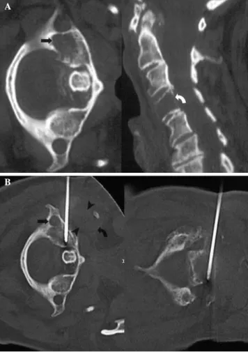
cervical spine showed an osteolytic lesion of the atlas in the right lateral portion and a second osteolysis in the body of C4 (Fig. 1A). On magnetic resonance imaging (MRI), there was no epidural tumor involvement.
In an interdisciplinary meeting among orthopedic surgeons, neuroradi-ologists, and interventional radineuroradi-ologists, the decision was made to treat the two osteolytic metastases of C1 and C4 with percutaneous vertebroplasty, aiming for stability and pain control. In order to employ the best possible technique for access path, injection control, and, at the same time, avoiding patient repositioning during the intervention, a single-stage hybrid lateral approach under CT and fluoroscopic guidance was proposed.
The procedure was performed under general anesthesia in a multi-functional image-guided therapy suite (MIGTS), which integrates the technology and infrastructure for sterile image-guided therapy with CT, fluoroscopy, DSA, and a new OR table [4, 5]. The patient was fixed in a left lateral position on the intervention table and immobilized on a vacuum mattress. Prophylactic antibiotic therapy with a third-generation cepha-losporine (Rocephine 2 g intravenously; Roche Pharma, Reinach, Swit-zerland) was applied.
Intra-interventional imaging was performed on a spiral CT scanner (GE Hispeed Advantage; GE Medical Systems, Milwaukee, WI, USA; imaging parameter 140 kV, 380 mA, slice thickness 3 mm, pitch 1.5, slice increment 2.5 mm). First, a contrast-enhanced high-resolution volumetric scan from the skull base to the level of C5 was performed to define the access path and to demonstrate the course of the cervical arteries. Under CT fluoroscopic guidance using a collimation of 3mm, a 14G 11-cm trephine-type needle with screw threads (OstycutÒ; Bard Products, Angiomed, Berlin, Germany) was inserted in a strict axial direction just anterior to the vertebral artery and posterior to the internal carotid artery to reach the osteolytic destroyed lateral mass of C1. A similar procedure was conducted for the osteolytic lesion in the vertebral body of C4 (Fig. 1B). Penetration of the respective lesions could easily be achieved. After verification of the correct needle placement, the intervention table was driven via a remote control on a rail system into the fluoroscopy unit (Philips Integris V 5000; Philips Medical Systems, The Netherlands). Phlebography, performed by injection of a few milliliters of contrast material (Iopamiro 300; Iopamidolum, Bracco S.P.A. Milano, Italy) in a strict lateral position under fluoroscopic control, revealed a leakage into the intervertebral space at the level of C1. In C4, a strong hypervascularization of the osteolysis was demonstrated, but no leakage toward the vertebral artery was observed.
A cement composition of 18 ml polymethyl methacrylate (PMMA) (Simplex P, methylmethacrylate; Howemedica, Rutherford, NJ) powder, 2 ml barium powder, and 5 ml liquid polymer (Simplex P, Polymer; Howe-medica)] providing up to 8 min of extended polymerization time under room temperature [6, 7] was loaded into a 10-cc injector device (10 cc Leveen; BSC, Watertown, MA, USA). The injector was connected to the Correspondence to: R.W. Huegli, M.D.; email: [email protected]
ª Springer Science+Business Media, Inc. 2005 Published Online: 4 July 2005
CardioVascular
and
Interventional
Radiology
Cardiovasc Intervent Radiol (2005) 28:649–652 DOI: 10.1007/s00270-004-0159-5
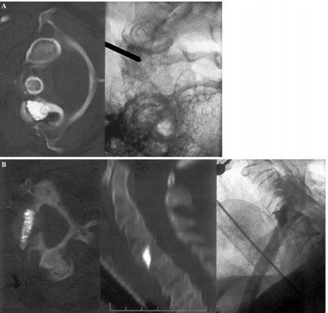
needles and the cement preparation was injected under continuous fluoro-scopic control mainly in the lateral projections with intermittent checks in the anteroposterior projection in order to gain optimal filling while avoiding significant PMMA leakage toward the spinal canal and the vertebral arteries. Approximately 1 ml of PMMA was administered at C1 and 2 ml at C4 level. On both levels, a good filling of the osteolytic lesions could be obtained (Fig. 2A,B).
Vertebroplasty was concluded uneventfully within a total time of less than 2 h and the patient exhibited no neurologic symptoms when she woke up from general anesthesia. Substantial pain relief resulted and she no longer required pain medication. The follow-up was unremarkable, with stable pain relief 6 month after the intervention.
Discussion
Various approaches for vertebroplasty of the cervical spine were
mentioned in the literature depending on the location. The single
treatment of C1 described in the literature was performed via a
posterior approach after coil occlusion of the vertebral artery [8].
For the C2 level, a transoral procedure was proposed [9, 10],
whereas the mid and lower cervical spines are usually assessed by
an anterolateral approach [6]. In the reported case with a patient
suffering from two painful cervical osteolyses at the C1 and C4
level, an intervention using an established access route without
patient repositioning was not possible. Therefore, we decided to
apply a lateral approach with similar access paths to reach both
lesions in the same patient position. In order to perform this access
safely, a visualization of vital structures adjacent to the
metasta-ses—primarily the course of the vertebral and carotid arteries—
was mandatory. We therefore chose a hybrid approach by CT
guidance for the needle placement and fluoroscopic guidance for
cementing.
A combination of CT-guided needle placement and
fluoro-scopically controlled implant injection has been already described
originally by Gangi et al. [11]. Our technique differed from the
procedure outlined in their article based on the technologies
available to us: First, the procedure was performed in a MIGTS
that integrates the technology and infrastructure for sterile
image-guided interventions [4]. The state-of-the-art imaging modalities,
including spiral CT and a high-resolution DSA unit, provided an
optimal visualization for both needle placement and implant
injection. Repositioning of the patient is avoided by a rail system
that enables the transfer of the intervention table with a remote
control from the CT to the DSA unit. Second, the stable patient
fixation on a vacuum mattress in a lateral position offers a good
operator control. Third, we used a 14G trephine-type needle with
screw threads; as in the presented case, a precise entering of a
small area for safe cement delivery was mandatory. This needle
type allowed a very exact advancement [12], as the penetration of
the cortex is achieved with a drilling action (see http://
www.emedicine.com/radio/topic 844.htm). In order to find the
correct entry point at the cortical surface, especially in cases with a
tangential entry point, hammering might be helpful to accomplish
bony contact.
Although there exist new cements on the market in which
barium or zirkondioxid is already added [13, 14], we still utilized a
conventional PMMA product because of familiarity with the
mechanical properties and the dynamic of polymerization.
Venography with an iodinated contrast agent before injecting
the PMMA is carried out to characterize collateralization to the
basivertebral venous complex [15]. In patients with severe allergy
to iodinated contrast media or chronic renal insufficiency,
gado-linium-based agents have been used [16].
The good analgetic effect in this patient was achieved with
only small amounts of approximately 1 ml PMMA at the C1 level
and 2 ml at the C4 level. This matches the observation in
bio-mechanical studies that a restoration of vertebral body integrity
resulting in patients´ pain relief can be achieved with only small
volumes of cement [17]. Moreover, the application of relatively
small cement portions increases the safety of the interventional
procedure by diminishing the risk of leakage to sensitive
structures.
In conclusion, the case presented demonstrates that a
single-step hybrid lateral access under CT and DSA guidance is safe and
feasible without patient repositioning. This approach seems to be
fast, reliable, and easy to perform in selected patients with multiple
lesions and difficult access paths such as in the upper cervical
spine.
Fig. 1. A. Axial computed tomography showing an osteolyic lesion of the right lateral atlas mass (arrow) and another osteolysis in a sagittal reconstruction of C4 (curved arrow). The right-sided osteo-lytic lesion of the atlas abuts the vertebral artery. The cortex along the vertebral artery is intact, whereas at the medial cortex, a fracture zone with mild displacement can be delineated. Note the cortical breakthrough of the posterior vertebral surface at the C4 level.B. Lateral access path to the osteolytic lesion in C1 (left) and the entirely destroyed C4 (right) between the internal carotid artery (curved ar-row)/internal jugular vein (arrowhead) anteriorly and the vertebral artery (arrow) posteriorly under CT fluoroscopic guidance.
Acknowledgement. S.G. Wetzel was supported, in part, by the Swiss Na-tional Science Foundation (grant 3200-066634/1).
References
1. Cotten A, Boutry N, Cortet B, et al. (1998) Percutaneous vertebropl-asty: state of the art. Radiographics 18:311–320; discussion 320–313 2. Gangi A, Guth S, Imbert JP, et al. (2003) Percutaneous vertebroplasty:
indications, technique, and results. Radiographics 23:e10
3. Tong FC, Cloft HJ, Joseph GJ, et al. (2000) Transoral approach to cervical vertebroplasty for multiple myeloma. Am J Roentgenol 175:1322–1324
4. Jacob AL, Regazzoni P, Steinbrich W, et al. (2000) The multifunctional therapy room of the future: image guidance, interdisciplinarity, inte-gration and impact on patient pathways. Eur Radiol 10:1763–1769 5. Messmer P, Jacob AL, Fries E, et al. (2001) [Technology integration
and process management. Concept and implementation of a new platform for simultaneous diagnosis and therapy of acutely ill and in-jured patients and for elective computer assisted surgery (CAS)]. Un-fallchirurg 104:1025–1030
6. Deramond H, Depriester C, Galibert P, et al. (1998) Percutaneous vertebroplasty with polymethylmethacrylate. Technique, indications, and results. Radiol Clin North Am 36:533–546
7. Martin JB, Jean B, Sugiu K, et al. (1999) Vertebroplasty: clinical experience and follow-up results. Bone 25:11S–15S
8. Wetzel SG, Martin JB, Somon T, et al. (2002) Painful osteolytic metastasis of the atlas: treatment with percutaneous vertebroplasty. Spine 27:E493–E495
9. Gailloud P, Martin JB, Olivi A, et al. (2002) Transoral vertebroplasty for a fractured C2 aneurysmal bone cyst. J Vasc Intervent Radiol 13:340–341
10. Martin JB, Gailloud P, Dietrich PY, et al. (2002) Direct transoral ap-proach to C2 for percutaneous vertebroplasty. Cardiovasc Intervent Radiol 25:517–519
11. Gangi A, Kastler BA, Dietemann JL (1994) Percutaneous vertebropl-asty guided by a combination of CT and fluoroscopy. Am J Neuroradiol 15:83–86
12. Brugieres P, Gaston A, Voisin MC, et al. (1992) CT-guided percuta-neous biopsy of the cervical spine: a series of 12 cases. Neuroradiology 34:358–360
Fig. 2. A. Fluoroscopy image during the filling process of the right C1 body lesion and axial computed tomography after vertebroplasty. The distritbution of cement can be better appreciated on CT images than plain films. No leakage toward the vertebral artery can be
ob-served.B. Post-procedural axial CT with sagittal reconstruction and lateral radiograph of the cervical spine after vertebroplasty revealing a sufficient cement filling at the C4 level. No cement leakage toward the spinal canal and the vertebral artery can be observed.
13. Breusch SJ, Kuhn KD (2003) [Bone cements based on polymethyl-methacrylate]. Orthopade 32:41–50
14. Schnurer SM, Gopp U, Kuhn KD, et al. (2003) [Bone substitutes]. Orthopade 32:2–10
15. McGraw JK, Heatwole EV, Strnad BT, et al. (2002) Predictive value of intraosseous venography before percutaneous vertebroplasty. J Vasc Intervent Radiol 13:149–153
16. McGraw JK, Strnad BT, Patzik SB, et al. (2000) Carbon dioxide and gadopentetate dimeglumine venography to guide percutaneous ver-tebroplasty. Cardiovasc Intervent Radiol 23:485–487
17. Belkoff SM, Mathis JM, Jasper LE, et al. (2001) The biomechanics of vertebroplasty. The effect of cement volume on mechanical behavior. Spine 26:1537–1541