Adrian Spreng Peter Netzer Joerg Mattich Hans-Peter Dinkel Peter Vock Hanno Hoppe Received: 11 February 2005 Revised: 24 April 2005 Accepted: 29 April 2005 Published online: 18 June 2005
# Springer-Verlag 2005
Importance of extracolonic findings at IV
contrast medium-enhanced CT colonography
versus those at non-enhanced CT colonography
Abstract To compare the clinical importance of extracolonic findings at intravenous (IV) contrast-enhanced CT colonography versus those at non-enhanced CT colonography. IV con-trast medium-enhanced (n=72) and non-enhanced (n=30) multidetector CT colonography was performed in 102 symptomatic patients followed by conventional colonoscopy on the same day. The impact of extracolonic findings on further work up and treatment was assessed by a review of patient records. Extracolonic findings were divided into two groups: either leading to further work up respec-tively having an impact on therapy or not. A total of 303 extracolonic findings were detected. Of those, 71% (215/303) were found on IV contrast-enhanced CT, and 29% (88/303) were found on non-enhanced CT colono-graphy. The extracolonic findings in
25% (26/102) of all patients led to further work up or had an impact on therapy. Twenty-two of these patients underwent CT colonography with IV contrast enhancement, and four with-out. The percentage of extracolonic findings leading to further work up or having an impact on therapy was higher for IV contrast-enhanced (31%; 22/72) than for non-enhanced (13%; 4/30) CT scans (P=0.12). IV contrast-enhanced CT colonography produced more extracolonic findings than non-enhanced CT colonography. A substantially greater proportion of findings on IV contrast-enhanced CT colonography led to further work up and treatment than did non-enhanced CT colonography. Keywords Extracolonic findings . Virtual colonoscopy . CT
colonography . Abdominal CT
Introduction
First described by Vining et al. in 1994, CT colonography, or so-called “virtual colonoscopy,” is a rapidly evolving minimally invasive technique for examination of the entire colon and detection of colorectal neoplasms [1]. Previous studies have shown that CT colonography has the potential to become a valuable clinical screening method for colo-rectal neoplasms [2–6].
In contrast to other screening tools for colorectal cancer, such as conventional colonoscopy and barium enema, CT colonography allows for simultaneous visualization of ex-tracolonic pathologies within the abdominal organs,
ves-sels, bone, and soft tissue [7]. Such findings are reported to be useful in staging cancer, explaining a patient’s present-ing symptoms, and detectpresent-ing other potentially serious dis-orders [8]. On the other hand, extracolonic findings may involve phenomena already known or may lead to un-necessary further work up, which can be problematic for both ethical and economical reasons [9,10].
Only a few studies have examined the clinical relevance of extracolonic findings at CT colonography [7,9–12]. These studies examining non-enhanced CT scans of the abdomen report large differences in the prevalence of extracolonic findings and the need for additional work up. To our knowledge, the present study is the first to compare the A. Spreng . J. Mattich . H.-P. Dinkel .
P. Vock . H. Hoppe (*) Institute of Diagnostic Radiology, Inselspital, University of Bern, Freiburgstrasse 10, 3010 Bern, Switzerland e-mail: hanno.hoppe@insel.ch Tel.: +41-31-6322435 Fax: +41-31-6324874 P. Netzer Department of Gastroenterology, Inselspital, University of Bern, Bern, Switzerland
clinical relevance of extracolonic findings at IV contrast-enhanced CT colonography with those at non-contrast-enhanced CT colonography.
Patients and methods Patients
One hundred and two adult patients referred to our gastro-enterology clinic for conventional colonoscopic evaluation of symptoms (including hematochezia, positive hemoccult test, iron deficiency anemia, or personal or family history of colonic neoplasms) were enrolled in the study. The study’s trial protocol was approved by the institute’s ethics committee and performed in accordance with the revised Declaration of Helsinki of 1989. Each patient provided written consent after being fully informed regarding the study protocol. In all patients, CT colonography was fol-lowed by conventional colonoscopy the same morning.
CT colonography
On the day before the examinations, each patient was given a wet bowel preparation consisting of 4 l of methylcellulose as prescribed by the participating gastroenterologist. No oral contrast medium was administered. CT colonography was performed with an Asteion four-channel multidetector CT scanner (Toshiba, Tokyo, Japan). A flexible rubber cath-eter with a rectal balloon was inserted into the rectum by the investigating radiologist. The patient’s colon was then insufflated with room air according to the patient’s toler-ance. The catheter was clamped and left in the rectum, and a single supine scout CT image was obtained to verify adequate bowel distension. If bowel distension was in-adequate, additional air was insufflated into the rectum. Once bowel distension was adequate, CT colonography was performed. In 72 of the 102 patients, 120 ml (flow-rate: 3 ml/s; scan delay: 60 s) of iopromide IV contrast medium containing 300 mg/ml iodine (Ultravist 300, Berlex Labo-ratories, Montville, USA) was power injected, followed first by CT colonography in the supine position in a cranio-caudal direction to image the entire region of the colon and rectum, then by examination in the prone position without additional injection of contrast medium. In 30 of the 102 patients, administration of IV contrast medium was con-traindicated due to an elevated creatinine level, known renal insufficiency, or contrast media allergy.
CT parameters included 4×2-mm detector collimation, 120 kV, 0.75-s gantry rotation, 200 mAs and a pitch of 1.375. The entire abdomen and pelvis were scanned during a breath hold of approximately 30 s. Axial CT images were
reconstructed as 2-mm slices with a 1-mm reconstruction interval.
Image analysis
Methods for polyp detection have been described else-where [6]. The reconstructed supine and prone datasets were transferred to an Advantage Windows workstation (Version 4.0, General Electric Medical Systems, Milwau-kee, WI) running on Sun Ultra Sparc 60 hardware (Sun Microsystems, Mountain View, CA) featuring two Sun Ultra Sparc II 450 MHz central processing units and 2 gigabytes of random access memory. Images for extraco-lonic findings were read by a board-certified radiologist and a radiology resident. Since the images were read during clinical routine they were not always evaluated by the same readers throughout the study. The supine acquisitions were IV contrast enhanced in 72 patients and non-enhanced in 30 patients; no further IV contrast medium was adminis-tered for prone scans. Images were viewed as continuous 2-mm axial sections with regular abdominal contrast window display settings (level 50 H, width 450 H). The window/ level settings could be adjusted manually at the worksta-tion. The basal portions of the lungs were also viewed using standard lung window settings (level−450 H, width 1,850 H). If necessary, additional multiplanar reformatting was performed using the workstation. Both readers were unaware of the patients’ medical history, but knew that the examined patient cohort consisted of symptomatic pa-tients. One radiologist per study was responsible for dic-tating an official CT colonography report, which included both intra- and extracolonic findings plus suggestions for possible work-up procedures. The report was sent to the referring clinician.
Definitions
Extracolonic findings were divided into two groups: either leading to further work-up respectively having an impact on therapy or not. Extracolonic findings were classified by organ system as hepatic (lesion, fatty liver), vascular (aneurysm, thrombosis, sclerosis, varices), gallbladder (cho-lecystitis, polyps) or cholangio (cholestasis), pulmonary (nod-ule, infiltrate, emphysema, fibrosis) or pleural (pleuritis, effusion, calcification, thickening), musculoskeletal(mass including hematoma or lipoma, osteolysis, arthrosis), urogen-ital (renal mass, renal cyst, renal duplication, nephrolithia-sis, adrenal mass, prostatic enlargement, testicular hydrocele, uterine myoma, any calcification), cardiac (pericardial effu-sion, coronary artery calcification, cardiomegaly), splenic (lesion, splenomegaly, accessory spleen), pancreatic (mass,
pancreatitis, calcification), hernia (inguinalhernia, hiatal her-nia), or other (enlarged lymph nodes, ascites, esophageal or gastric mass). The definition of a fatty liver on non-con-trast-enhanced CT was mean CT Hounsfield units lower in the liver than in the spleen or on contrast-enhanced CT if the liver is less attenuating than muscle, since comparison with the spleen is not accurate on contrast-enhanced CT scans [13]. Prostate enlargement was defined as a prostate volume larger than 25 ml. Enlarged abdominal lymph nodes were defined as short axis larger than 10 mm.
Follow-up
The minimum follow-up time after CT colonography was 6 months, the maximum 30 months. The files for each patient with extracolonic findings were reviewed to determine the number and results of any examination [e.g., CT, ultra-sound (US), angiography, biopsy, ERCP, MRI, gastros-copy) performed because of extracolonic findings at CT colonography. The numbers and results of such examina-tions or surgical procedures performed on the basis of CT colonography were tabulated. An effort was made to deter-mine whether any extracolonic findings were known before or if they were initially discovered at CT colonography and whether the extracolonic finding was made at IV contrast-enhanced or non-contrast-enhanced CT colonography.
Statistics
The chi-squared test with Yates’ continuity correction was performed to compare the distribution of extracolonic find-ings made with and without IV contrast enhancement. The age and sex distribution of patients with unenhanced and IV contrast-enhanced CT-colonography was calculated and tested for degrees of difference using the Student’s t-test
or chi-squared test, respectively. A P-value of less than 0.05 was considered significant.
Results
One hundred and two subjects were enrolled in the study (63 men, 39 women; age range: 20–91 years; mean age: 66 years). Complete conventional colonoscopy to the cecum was achieved in 94 patients. No patient had to be excluded from evaluation of extracolonic findings. In 72 patients CT colonography was performed after application of IV trast medium; in 30 patients IV contrast medium was con-traindicated. Of the 72 patients receiving IV contrast, 46 were male and 26 were female, and of the 30 patients not receiving IV contrast 17 were male and 13 were female
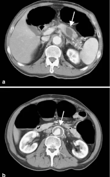
Fig. 1 IV contrast-enhanced CT scan of a 66-year-old female pa-tient with known metastasis of an ovarian carcinoma to the right iliac muscle shows previously unknown thrombosis of the right femoral vein (arrow), probably due to compression by the muscle tumor. Successful treatment with heparin
Table 1 The number of extra-colonic findings at CT colonog-raphy in 102 patients, number of previously known findings, and corresponding numbers of find-ings on CT scans with and without IV contrast enhancement
A patient could have more than one finding
Finding No. of
findings
No. of previously known findings Findings on IV contrast-enhanced CT scans Findings on non-enhanced CT scans IV contrast Non-enhanced Hepatic 40 3 2 30 10 Vascular 48 6 0 33 15 Gallbladder/cholangio 18 0 0 13 5 Pulmonary/pleural 32 3 0 24 8 Musculoskeletal 13 1 0 9 4 Urogenital 65 2 0 48 17 Cardiac 15 0 0 7 8 Splenic 8 0 0 6 2 Pancreatic 3 1 0 2 1 Hernia 20 1 0 14 6 Other 41 2 0 29 12
(P=0.6). For the 72 patients receiving IV contrast, the mean age was 64.7 for males and 69.8 for females (P=0.1), and for the patients not receiving IV contrast 65.8 for males and 65.1 for females (P=0.9).
Conventional colonoscopy found a high number of co-lorectal neoplasms, which was 122 lesions (carcinoma,n= 8; adenoma, n=67; hyperplastic bowel mucosa, n=47) in 49 patients. The results of polyp detection at CT colo-nography were reported earlier [6].
A total of 303 extracolonic findings were recorded, of which 71% (215/303) were seen at IV-enhanced CT
colo-nography and 29% (88/303) at CT colocolo-nography without enhancement (Table 1). Eighty nine percent (91/102) of patients had at least one extracolonic finding (Fig.1). Of these patients, 75% (68/91) underwent CT scan with IV contrast enhancement, and 25% (23/91) without. Eleven percent of patients (11/102) had no extracolonic findings. Of these, 4% (4/102) underwent CT scan with IV contrast enhancement and 7% (7/102) without. Ninety-four percent (68/72) of patients with IV contrast-enhanced CT colonog-raphy had extracolonic findings, compared to 77% (23/30) without IV contrast enhancement (P=0.02).
Table 2 Extracolonic findings at IV contrast enhanced CT colonography that resulted in additional work-up IV contrast enhanced extracolonic findings resulting in additional work up
Finding No. resulting in work up/no. of findings
Type of work up Results of work up No.
resulting in surgery Abdominal aortic aneurysm (>4 cm) 4 CT (n=2), CT follow-up (n=1), angiography (n=1)
Abdominal aortic aneurysm (n=4) with stenting (n=1) or aortoduodenal fistula (n=1)
2
Pancreatic mass 2 Surgery (n=2) Pancreatitis (n=1), anastomosis insufficiency (n=1)
2
Hepatic lesion 1 CT guided biopsy Cholangio–Ca 1
Pelvic mass 2 Laparoscopy (n=2) Ovarian abscess (n=1) Abscess caused by sigma diverticulitis (n=1)
2
Adrenal mass 1 CT Adenoma 0
Cholecystitis 2 US (n=2) Cholecystitis with antibiotic
treatment (n=2)
1
Esophageal mass 1 Gastroscopy Tumor 1
Lung nodule 1 CT and PET Lung cancer 1
Musculoskeletal mass 1 MRI Carcinoma 1
Renal mass 1 Staging CT Renal cell cancer 1
Iliacal artery aneurysm 1 Angiography Aneurysm with stenting 1
Cholestasis 1 ERCP, liver biopsy,
1 aboratory tests
Liver cirrhosis, abnormal liver function tests
0
Thrombosis 3 Heparinization based on
initial findings (n=3)
Regression of thrombosis (n=3) 0
Pleuritis 1 CT Pleural effusion 0
Table 3 Extracolonic findings at non-enhanced CT colonogra-phy that resulted in additional work up
Non-enhanced CT colonography findings resulting in additional work up Finding No. resulting
in work up/no. of findings Type of work up Results of work up No. resulting in surgery
Hepatic lesion 1 CT Metastasis 1
Ovarian mass 1 Staging CT Ovarian cancer, no metastasis
1
Adrenal mass 1 MRI Adenoma (1 cm, 5 HU) 0
Pulmonary fibrosis 1 High resolution chest CT
Seven percent (21/303) of extracolonic findings were previously known, including hepatic lesions (n=5), vascu-lar aneurysms (n=6), lung nodules (n=3), an adrenal mass, ovarian cyst, inguinal hernia with bowel, metastasis of the iliac muscle, esophageal mass, pancreatic metastasis, and gastric carcinoma.
The extracolonic findings in 25% (26/102) of all patients led to further work up or had an impact on therapy. Twenty-two of these patients had a CT colonography with IV con-trast enhancement (Table 2), and four without (Table 3). Calculated per patient, 31% (22/72) of all patients with IV contrast enhancement and 13% (4/30) of all patients with-out underwent further work up (P=0.12). Of these, 32% (22/68) of patients with IV contrast and 17% (4/23) of patients without IV contrast had further work up (P=0.26). One patient died before work up could be performed (Fig. 2). Nineteen patients underwent additional imaging examinations following CT colonography, including CT (n= 10), CT-guided biopsy (n=1), US (n=2), angiography
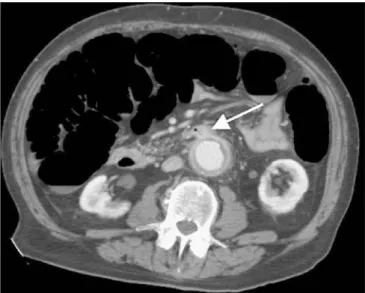
Fig. 2 Non-enhanced CT colonography of an 80-year-old male patient with partially calcified, mainly solid renal mass of the left kidney (arrow). Further work up was precluded by death of patient soon afterwards of unrelated cause
Fig. 3 IV contrast-enhanced CT colonography of a 59-year-old male patient with a surgically treated esophageal carcinoma. Dila-tation of the bile ducts (arrow) was initially found on CT colo-nography. Subsequently, an ERCP was performed, which showed dilated intrahepatic bile ducts, but ruled out biliary calculi or tumor. Finally liver biopsy confirmed the diagnosis of liver cirrhosis
Fig. 4 a, b CT colonography with IV contrast enhancement of a 66-year-old male patient with a previously unknown, well-delineated 3.3×2-cm cystic pancreatic mass (a, arrow). No other abdominal masses or suspected metastatic disease was present. The patient underwent surgery that revealed a pancreatic pseudocyst with chronic atrophic and sclerosing pancreatitis. b In addition an abdominal aortic aneurysm was found (arrow)
(n=2), MRI (n=2), PET (n=1), endoscopic retrograde chol-angiopancreatography (n=1) (Fig. 3), and gastroscopy (n=1). Fifteen patients underwent surgery, including ab-dominal aortic aneurysm repair (n=1), pancreas resection (n=1) (Fig.4), resection of an aortoduodenal fistula (n=1) (Fig.5), duodenal anastomosis insufficiency repair (n=1), liver metastasis/tumor resection (n=2), ovarian cancer resection (n=1), pelvic abscess drainage (n=2), gallbladder resection (n=1), esophageal tumor resection (n=1), lung
cancer resection (n=1), musculoskeletal tumor resection (n=1), renal cell carcinoma resection (n=1) (Fig. 6), and iliacal artery aneurysm repair (n=1). A follow-up CT was performed after more than 6 months in one case of abdomi-nal aortic aneurysm. Follow-up CT was also performed in three patients with known esophageal mass, known pan-creatic metastasis, and known gastric carcinoma, which would have been performed unrelated to extracolonic find-ings on CT colonography. In one patient with suspected pleuritis a chest CT scan was performed for work-up. Another patient received a high resolution chest CT scan for evaluation of suspected pulmonary fibrosis. One stag-ing CT was performed in a patient with ovarian cancer. Follow-up revealed no important lesions missed at CT colonography.
Additionally to the performed work-up shown in Table 2, the following suggestions for possible work-up procedures were given: Ultrasound was recommended in eight patients (non-enhanced CT, n=4; IV contrast-en-hanced CT, n=4), CT in three patients (non-enhanced CT,n=1; IV contrast-enhanced CT, n=2), and MRI in two patients (non-enhanced CT,n=2).
Discussion
The primary purpose of the present study was to evaluate the impact of IV contrast medium application on the de-tection of extracolonic findings and further patient work up in symptomatic patients referred for conventional colo-noscopy. The results show that IV contrast-enhanced CT colonography produced more extracolonic findings than non-enhanced CT colonography. Moreover, the percentage of extracolonic findings leading to further work up or having an impact on therapy was higher for IV contrast-enhanced than for non-contrast-enhanced CT scans, which can be explained by the fact that an IV contrast-enhanced ab-dominal CT scan may produce a higher number of extra-colonic findings than a non-enhanced abdominal CT scan (e.g., visualization of organ lesions without mass effect or vascular thrombosis).
In the present study, the high percentage of extracolonic findings in 89% of the patients is most probably due to both examining a symptomatic high risk patient cohort and considering all findings without separately categorizing them as of low, moderate, or high importance. Further-more, categorizing extracolonic findings may be quite dif-ficult due to subjective rating. In our study, we detected a significantly higher number of extracolonic findings in patients with IV contrast-enhanced CT colonography com-pared to those with non-enhanced CT scans. On the other hand, the difference between these patients concerning further work up was found not to be statistically significant. Only a few studies have explored the impact of extra-colonic findings at CT colonography on further work up or therapy predominately in symptomatic patients [9–11]. In Fig. 5 Inflamed abdominal aortic aneurysm with an aortoduodenal
fistula (arrow) on IV contrast-enhanced CT colonography in an 81-year-old male patient. The aneurysm was successfully treated in an emergency operation
Fig. 6 Complex cystic mass with thickened septations of the left kidney (arrow) in a 76-year-old male patient (IV contrast-enhanced CT scan). After radiological staging (negative) the patient underwent successful nephrectomy. The specimen exhibited a renal cell carcinoma
the present study, 25% of patients were found to have previously unknown extracolonic findings with an impact on further work up or therapy, a rate higher than the 2 to 13% of previous studies. Moreover, in our study 303 extracolonic findings were detected in 102 patients com-pared to 232 findings in 111 patients (Hellstrom et al.), 16 in 100 patients (Edwards et al.), and 68 in 49 patients (Ginnerup Pedersen et al.) in other studies [7,9,10]. Hara et al. obtained 151 extracolonic findings in a study popu-lation of 264 patients and detected a low rate compared to that of Hellstrom et al. and our own. A possible explana-tion for this discrepancy—besides the higher mean age and symptomatic high-risk nature of our patient popula-tion (66 years), which accord with previous studies—may be our use of IV contrast medium in the majority of our patients, which was not done in other studies and appears to account in large part for the high rate of extracolon-ic findings in our study. In addition, our study included abdominal CT scanning with standard doses, whereas both Hara et al. and Edwards et al. [10, 11] employed a low dose CT protocol.
The majority of patients in the present study were given IV contrast medium, something that was not done in other studies on extracolonic findings. A higher number of all our extracolonic findings were obtained at IV contrast-enhanced CT colonography. Although the present study was not designed to compare directly the conspicuity of benign and malignant lesions on unenhanced and IV-en-hanced CT since patients initially received either unen-hanced CT or IV contrast-enunen-hanced CT and not both at a time, IV contrast-enhanced CT increased the conspicuity of certain lesions such as venous thrombosis of the external iliac vein, splenic vein, and portal vein, which probably could not have been diagnosed on non-enhanced CT. In addition, an inflamed aortic aneurysm with an aortoduo-denal fistula was found (Fig. 5), which would have been almost impossible to detect without IV contrast enhance-ment. The use of IV contrast enhancement also enabled the readers to distinguish between solid and cystic lesions, especially in kidney and liver (Fig. 6). Other important findings, however, such as an aortic aneurysm, mass le-sion, enlarged lymph nodes and a cholecystitis, would probably have been diagnosed even with non-enhanced CT scans. IV contrast medium has been shown to improve the detection of colorectal polyps and carcinomas and to facilitate the differentiation of solid lesions from residu-al colonic fluid and stool on axiresidu-al images [6, 14–19]. It may also improve detection of colorectal polyps and help to reduce the need for additional imaging studies to clar-ify potential extracolonic findings, although in the present study the number of follow-up imaging studies was higher after contrast-enhanced CT scans due to the higher number of extracolonic findings.
The use of IV contrast in symptomatic patients com-bines CT colonography with a routine contrast-enhanced abdominal CT scan, resulting in a one stop diagnostic
in-vestigation instead of needing additional imaging. How-ever, the added risks and cost of administering IV contrast medium probably preclude its use as part of screening CT colonography protocols for large populations [20].
In the present study, axial images with a small slice thickness (2 mm) and an overlapping reconstruction in-terval of 1 mm were used to detect colorectal polyps; such images are not always used for viewing of abdominal CT in routine clinical practice. In addition, our analysis of extracolonic findings was facilitated by using both supine and prone CT datasets, a workstation with dynamic win-dow settings, cine mode, and multiplanar reconstructions. All of these factors may have improved our detection rate of extracolonic lesions [9].
CT colonography requires scanning of the patient in both supine and prone positions, because a change in body posi-tion redistributes the intraluminal content, thus improving visualization of colorectal polyps [21]. Double scanning, however, means the patient receives a double dose of ra-diation, which is of particular importance in young patients and in screening programs. Hara et al. reduced the radia-tion dose by lowering the tube current on non-enhanced CT colonography and found the image quality to be suf-ficient for evaluation of the bowel wall due to the high contrast between intraluminal air and adjacent soft tis-sues [22]. However, a lowered tube current entails an increase in image noise, which may adversely affect de-tection of extracolonic findings, especially in solid abdom-inal organs. In the present study, CT colonography was performed with a standard abdominal CT radiation dose (120 kV, 200 mAs), which possibly contributed to the high rate of extracolonic findings, especially of smaller lesions, which would probably have eluded detection on noisy low dose images.
Extracolonic findings at CT colonography were assessed in three previous studies using a tube current of 70 mAs [10–12]. The reported prevalence of extracolonic find-ings between 15 and 69% is lower than that reported by Hellstrom et al. (85% of patients) at 125 mAs and in our study (89% of patients) at 200 mAs. In our study, this discrepancy may also be related to the administration of IV contrast medium, differences in patient selection, and differences in definitions of extracolonic findings.
The frequency of extracolonic findings on CT colonog-raphy in a screening population was reported by Gluecker et al. to be 69% in 681 patients using unenhanced low-dose CT colonography [12]. Of these, only 1.3% resulted in subsequent work up or therapy, which is a lower number than the 25% in our study involving a symptomatic patient cohort. However, 10% of extracolonic findings in their study were considered to be clinically important, adding benefit to the screening intervention. Detection of inci-dental extracolonic findings, however, has many advan-tages, such as early detection of malignant disease or of an unruptured abdominal aortic aneurysm. Early treatment can improve a patient’s prognosis and decrease costs owing
to less complicated surgical procedures and shorter hospi-tal stays. Clinically important incidenhospi-tal extracolonic find-ings leading to further work up were quite common in our study. Extracolonic findings, however, may also lead to unnecessary work up, causing unnecessary patient anxiety and entailing higher costs and superfluous exposure to radiation. They must therefore be considered a possible disadvantage of CT colonography for routine diagnostic work up and screening [11].
To summarize, in symptomatic patients IV contrast-enhanced CT colonography produced more extracolonic findings, a greater proportion of which led to further work
up and treatment, than did non-enhanced CT colonography, making IV contrast-enhanced CT colonography a one-stop diagnostic investigation. Future studies must determine whether the added risks and cost of IV contrast medium preclude its use as part of CT colonography screening protocols for large populations.
Acknowledgements This study was supported by a research grant from the Helmut-Horten-Foundation, Lugano, Switzerland. The authors wish to thank the CT technologists at the Institute of Ra-diology, Inselspital Bern, for their excellent expert assistance.
References
1. Vining DJ, Gelfand DW, Bechthold RE, Scharling ES, Grishaw EK, Shifrin RY (1994) Technical feasibility of colon imaging with helical CT and virtual reality. Am J Roentgenol 162:104
2. Yee J, Akerkar GA, Hung RK, Steinauer-Gebauer AM, Wall SD, McQuaid KR (2001) Colorectal neo-plasia: performance characteristics of CT colonography for detection in 300 patients. Radiology 219:685–692 3. Pickhardt PJ, Choi R, Hwang I, Butler
JA, Puckett ML, Hildebrandt HA, Wong RK, Nugent PA, Mysliwiec PA, Schindler WR (2003) Computed to-mographic virtual colonoscopy to screen for colorectal neoplasia in asymptomatic adults. N Engl J Med 349:2191–2200
4. Bruzzi JF, Moss AC, Brennan DD, MacMathuna P, Fenlon HM (2003) Efficacy of IV Buscopan as a muscle relaxant in CT colonography. Eur Ra-diol 13:2264–2270
5. Taylor SA, Halligan S, Burling D, Morley S, Bassett P, Atkin W, Bartram CI (2004) CT colonography: effect of experience and training on reader performance. Eur Radiol 14:1025–1033 6. Hoppe H, Netzer P, Spreng A,
Quattropani C, Mattich J, Dinkel H-P (2004) Prospective evaluation of con-trast enhanced CT colonography and conventional colonoscopy for detection of colorectal neoplasms in a single institutional study using second-look colonoscopy with discrepant results. Am J Gastroenterol 99:1924–1935 7. Ginnerup Pedersen B, Rosenkilde M,
Christiansen TEM, Laurberg S (2003) Extracolonic findings at computed tomography colonography are a chal-lenge. Gut 52:1744–1747
8. Ng CS, Doyle TC, Courtney HM, Campbell GA, Freeman AH, Dixon AK (2004) Extracolonic findings in patients undergoing abdomino–pelvic CT for suspected colorectal carcinoma in the frail and disabled patient. Clin Radiol 59:421–430
9. Hellstrom M, Svensson E, Lasson A (2004) Extracolonic and incidental findings on CT colonography (virtual colonoscopy). Am J Roentgenol 182:631–638
10. Edwards JT, Wood CJ, Mendelson RM, Forbes GM (2004) Extracolonic find-ings at virtual colonoscopy: implica-tions for screening programs. Am J Gastroenterol 96:3009–3012
11. Hara AK, Daniel Johnson C, MacCarty RL, Welch TJ (2000) Incidental extra-colonic findings at CT colonography. Radiology 215:353–357
12. Gluecker TM, Johnson CD, Wilson LA, MacCarty RL, Welch TJ, Vanness DJ, Ahlquist DA (2003) Extracolonic findings at CT colonography: evalua-tion of prevalence and cost in a screening population. Gastroenterology 124:911–916
13. Panicek DM, Giess CS, Schwartz LH (1997) Qualitative assessment of liver for fatty infiltration on contrast-en-hanced CT: is muscle a better standard of reference than spleen? J Comput Assist Tomogr 21:699–705
14. Morrin MM, Farrell RJ, Kruskal JB, Reynolds K, McGee JB, Raptopoulos V (2000) Utility of intravenously administered contrast material at CT colonography. Radiology 217:765–771 15. van Gelder RE, Venema HW, Serlie IW, Nio CY, Determann RM, Tipker CA, Vos FM, Glas AS, Bartelsman JF, Bossuyt PM, Lameris JS, Stoker J (2002) CT colonography at different radiation dose levels: feasibility of dose reduction. Radiology 224:25–33
16. Luboldt W, Mann C, Tryon CL, Vonthein R, Stueker D, Kroll M, Luz O, Claussen CD, Vogl TJ (2002) Computer-aided diagnosis in contrast-enhanced CT colonography: an ap-proach based on contrast. Eur Radiol 12:2236–2241
17. Hoppe H, Quattropani C, Spreng A, Mattich J, Netzer P, Dinkel H-P (2004) Virtual Colon Dissection with CT colonography compared to axial read-ing and conventional colonoscopy: preliminary results. Am J Roentgenol 182:1151–1158
18. Luboldt W, Kroll M, Wetter A, Toussaint TL, Hoepffner N, Holzer K, Kluge A, Vogl TJ (2004) Phase- and size-adjusted CT cut-off for differen-tiating neoplastic lesions from normal colon in contrast-enhanced CT colonog-raphy. Eur Radiol 14:2228–2235 19. Luboldt W, Tryon C, Kroll M,
Toussaint TL, Holzer K, Hoepffner N, Vogl TJ (2005) Automated mass detection in contrast-enhanced CT colonography: an approach based on contrast and volume. Eur Radiol 15:247–253
20. Ferrucci JT (2001) Colon cancer screening with virtual colonoscopy-promise, polyps, politics. Am J Roent-genol 177:975–988
21. Fletcher JG, Johnson CD, Welch TJ, MacCarty RL, Ahlquist DA, Reed JE, Harmsen WS, Wilson LA (2000) Op-timization of CT colonography tech-nique: prospective trial in 180 patients. Radiology 216:704–711
22. Hara AK, Johnson CD, Reed JE, Ahlquist DA, Nelson H, Ehman RL, Harmsen WS (1997) Reducing data size and radiation dose for CT colono-graphy. Am J Roentgenol 168:1181– 1184