Identification and sequence comparison of a cuticular
collagen of Brugia pahangi
M. BISOFFP and B. BETSCHART
2*
1
Department of Medical Parasitology, Swiss Tropical Institute, 4051 Basel, Switzerland
2
Institute of Zoology, University of Neuchatel, 2007 Neuchdtel, Switzerland (Received 25 November 1995; revised 28 February 1996; accepted 28 February 1996)
SUMMARY
The cuticle of filarial nematodes is a specialized extracellular matrix that covers the parasite and protects it from adverse conditions of the environment. As a surface structure it is in direct contact with the host defence mechanisms and therefore plays an important role in the molecular host-parasite relationship. Using polyclonal antisera raised against the insoluble components of the cuticle of the adult filarial parasite Brugia pahangi, we have isolated cDNA clones encoding collagen molecules of the cuticle. The protein domain structure of cDNA clone Bpcol-1 was compared with the known structures of cuticular collagens of the nematodes Brugia malayi, Caenorhabditis elegans, Ascaris suum and Haemonchus contortus, confirming interspecies similarities. Using affinity-purified anti-Bpcol-1 antibodies we identified Bpcol-1 antigenic deter-minants in different nematode extracts, and determined the localization of such epitopes within the cuticle of B. pahangi. Key words: Brugia pahangi, filarial nematode, cuticle, collagen, sequence comparison, immunolocalization.
INTRODUCTION
All nematodes so far investigated are bound by an extracellular matrix, called the cuticle (Bird & Bird, 1991). The cuticle functions as a selective barrier in nutrition uptake and metabolite secretion (Ho et al. 1990), plays an important role as a skeletal counter-acting structure to the muscle movement (Wharton, 1986), and represents an excellent protection against unfavourable environmental conditions which, in the case of parasitic nematodes, include the various immunological responses of the host defence (Ogilvie
et al. 1980). The successful establishment of
nema-tode parasites within their host depends on the host-parasite interactions at their interface. There-fore, the biochemical composition of the cuticle is of great interest. Previous work has shown that the major protein components of nematode cuticles are nematode-specific collagens (Josse & Harrington, 1964). Like vertebrate collagens, they are charac-terized by the repeating amino acid sequence (Gly-X-Y)n, where X and Y can be any amino acid, but are often proline or hydroxyproline (Cox, 1992). This motif is responsible for the triple-helical, rod-like structure of collagen molecules formed by 3 poly-peptide chains. Although the cuticular collagens of parasitic nematodes are not directly exposed to the host, they are part of the nematode surface, and antibodies against cuticular collagens have been
* Corresponding author. University of Neuchatel, In-stitute of Zoology, Rue Emile-Argand 11, 2007 Neuchatel, Switzerland. Tel: + +41 38 23 30 45. Fax: + +41 38 23 30 01. E-mail: [email protected].
detected in patients infected with filarial parasites (Selkirk et al. 1989 a). While this may be partly due to death and decay of worms within the host, it may nevertheless represent an important immunological event in the host-parasite relationship. The latter is emphasized by the fact that cuticular collagens have been discussed in the context of protective immunity in hookworm infections (Pritchard, McKean & Rogan, 1988) and infections with the sheep parasite Haemonchus contortus (Boisvenue et al. 1991).
The use of cloning techniques has led to the identification of various collagen genes coding for cuticular and non-cuticular collagens of different nematodes, including the free-living nematode
Caenorhabditis elegans (Cox, 1992), the parasitic
nematodes Ascaris suum (Kingston, Wainwright & Cooper, 1989) and H. contortus (Shamansky et al. 1989). Caulagi and coworkers have isolated a base-ment membrane collagen gene from the human filarial parasite Brugia malayi, which was termed BmColl (Caulagi, Werner & Rajan, 1991). Collagen molecules of the filarial parasites B. malayi and B.
pahangi have been characterized with respect to their
synthesis and immunogenicity (Selkirk et al. 1989 a), and recently, the molecular cloning of a cuticular collagen of B. malayi, Bmcol-2, has been reported (Scott et al. 1995). In the present study we contribute to the molecular information on cuticular com-ponents of filarial parasites and report the cloning of a cDNA coding for a cuticular collagen of B. pahangi. The deduced protein structure of this collagen molecule is compared with other known cuticular collagens, and its precise localization within the adult parasite is presented.
MATERIALS AND METHODS
Parasites
B. pahangi adult worms were recovered from the
peritoneal cavity of jirds, following infection with 3rd-stage larvae (McCall et al. 1973). Adult A. suum worms were received from a local slaughterhouse. C.
elegans (var. Bristol, strain N2; kindly provided by
Dr H. Tobler, University of Fribourg, Switzerland) were grown and harvested according to the method described by Sulston & Brenner (1974).
Parasite protein extracts
Adult B. malayi and C. elegans were briefly washed in cold phosphate-buffered saline (PBS), homo-genized and sonicated in 125 mM Tris—HC1, pH 6-8, 1 mM ethylenediaminetetra-acetic acid (EDTA), 1 mM phenylmethyl sulphonyl fluoride (PMSF), 1 % sodium dodecyl sulphate (SDS), and 5 % 2-mercaptoethanol (2-ME), and stirred for 4 h at 4 °C.
A. suum adult worms were briefly washed in PBS
and subjected to multiple freezing/thawing rounds to enhance detachment of the cuticle from the body of the worms. The cuticle fragments were cut into pieces and extracted in PBS containing 5 % 2-ME for 24 h at 37 °C (Fujimoto & Kanaya, 1973). All extracts were briefly centrifuged and the concen-tration of the supernatants determined (Bradford, 1976).
Antiserum
Cuticles from adult B. pahangi worms were isolated essentially as described for C. elegans (Cox, Kusch & Edgar, 1981). Worms were suspended in 10 mM Tris-HCl, pH 7-5, 1 mM EDTA, 1 mM PMSF, and sonicated on ice. Cuticle pieces were recovered by centrifugation, suspended in 125 mM Tris—HC1, pH 6-8, 1 % SDS, and heated for 2 min at 80 °C. The suspension was turned end over end for 2 h at room temperature, the cuticles recovered by centri-fugation, and extracted again as described. The remainder of the preparation was extracted further by heating at 80 °C for 2 min in 125 mM Tris-HCl, pH 6-8, 1 % SDS, and 5 % 2-ME, and by rotation for 2 h at room temperature. This procedure was repeated once following centrifugation. The re-maining insoluble material enriched in epicuticular proteins was used to immunize rabbits according to standard protocols (Maniatis, Fritsch & Sambrook, 1982).
Immunoscreening
The production of the Agtl 1 cDNA library derived from mRNA of adult B. pahangi parasites has been described by Selkirk et al. (19896). Immuno-screening of the library in Escherichia colt Y1090 was
as described by Altmann, Handschin & Trachsel (1987). After induction of the lacZ operon, the filters were saturated with 50 mM Tris-HCl, pH 8, 150 mM sodium chloride, 0 3 % Tween 20, 0 0 5 % Triton-X100 (TBST), containing 5% dried milk powder, and incubated overnight with the polyclonal anti-serum raised against epicuticular components of B.
pahangi, at a 1/500 dilution in TBST containing
0-5 % bovine serum albumin (TBST-BSA). Bound antibodies were detected with affinity-purified al-kaline phosphatase-conjugated goat anti-rabbit IgG antibodies (Jackson ImmunoResearch Laboratories, Inc., USA) in TBST-BSA and visualized by the substrates 5-bromo-4-chloro-3-indoyl phosphate p-toluidine salt (BCIP) and p-nhro blue tetrazolium chloride (NBT; Kirkegaard Perry Laboratories, USA). Primary immunoreactive clones were sub-jected to second and third screening rounds for verification and plaque purification. Plaque purified phages were amplified in E. coli Y1088.
DNA preparation, blotting and hybridization
For the differential cross-hybridization experiments the inserts of the various positive clones were amplified by the polymerase chain reaction (PCR), subjected to agarose gel electrophoresis, and trans-ferred to nylon hybridization membranes (NEN Research Products, USA). The probe used for the hybridization experiments was a 677 bp 2?coRI fragment corresponding to bp 223-900 of Bpcol-1. The probe was labelled with 32P-dATP by the random primer extension method (Feinberg & Vogelstein, 1983). Hybridization experiments were performed according to standard protocols (Maniatis
et al. 1982) using different concentrations of standard
saline citrate (SSC), 5 x Denhardt's solution, and 0-5 % SDS, and at various temperatures according to the different stringency conditions chosen.
DNA sequencing and sequence comparison
The cDNA inserts of the positive clones were excised from Agtll with the restriction endonuclease EcoRl and subcloned into the £V:oRI restriction site of the sequencing vector M13mpl8/19 by the ligase re-action (Maniatis et al. 1982). The cDNA insert was sequenced by the chain termination method (Sanger, Nicklen & Coulson, 1977) using the Sequenase DNA Sequencing Kit from United States Bio-chemical, USA. DNA and protein sequences were analysed and compared using the GCG programs on a VAX system (Devereux, Haeberli & Smithies, 1984).
Production of Bpcol-1-fl-galactosidase fusion protein
E. coli strain Y1090 was infected with the
1 2 3
4 5
coll
200 ~
1 1 6 " * "
94
-Fig. 1. Immunoblot and collagenase susceptibility of Bpcol-1. Extracts of E. coli infected with wild type Agtll (lanes 1 and 3) or Bpcol-1 (lanes 2, 4, and 5) were analysed in SDS-PAGE (lanes 1 and 2) and
electroblotted onto nitrocellulose (lanes 3-5). The thin arrow indicates the position of /?-galactosidase at
116 kDa, the thick arrow indicates the position of Bpcol-1 fusion protein at Bpcol-142 kDa. Immunoblots were
performed using affinity-purified anti-Bpcol-1 antibodies; coll indicates collagenase treatment. Numerals on the left indicate molecular size in kDa.
the synthesis of the fusion protein was induced by the addition of isopropyl-/?-thiogalactopyranoside (IPTG) as described (Young & Davis, 1983). The plates were then washed (Altmann et al. 1987) and proteins in the wash solution were concentrated by ammonium sulphate precipitation (50 % saturation) and resuspended in either SDS-sample buffer or in PBS containing 1 mM PMSF for the collagenase digestion experiments.
Collagenase digestion
High purity collagenase from Clostridium
histolyticum (Sigma type VII, Switzerland) was used
for the digestion of fusion proteins. The enzyme was used at a concentration of 100/tg/ml and digestions performed for 30 min at 37 °C in 10 mM Tris-HCl, pH 7-5, 10 mM calcium chloride (Selkirk et al. 1989a). Reactions were terminated by the addition of protein loading buffer and boiling for SDS-polyacrylamide gel electrophoresis (SDS-PAGE).
Affinity-purification of antibodies
Bacterial lysates containing the Bpcol-1-/?-galac-tosidase hybrid protein were size-fractionated in preparative 7-5 % SDS-polyacrylamide gels and electroblotted onto nitrocellulose membranes (Towbin, Staehelin & Gordon, 1979). The size region of the nitrocellulose membranes around 140 kDa was excised and the remaining binding sites
on the strips blocked with 2 5 % BSA in TBST. After washing the strips in TBST they were incubated with the polyclonal antiserum raised against the insoluble components of adult B. pahangi, diluted 1/20 in TBST-BSA. The bound antibodies wereelutedby 2-min rinses with 0-15 M glycine-HCl, pH 2-8, and quickly neutralized with 01 M NaOH. BSA was added to a final concentration of 0-5 %.
Immunoblot analysis
Bacterial lysates, nematode extracts, and human collagens (Sigma, Switzerland) were size-fractionated electrophoretically in 7-5% or 12-5% SDS-polyacrylamide gels and transferred to nitro-cellulose essentially as described by Towbin et al. (1979). Following electrophoretic transfer, blocking of non-specific binding was achieved by a 1 h incubation in TBST containing 5 % dried milk powder. The polyclonal antiserum was used at a dilution of 1/500 in TBST-BSA. Bound antibodies were detected with affinity-purified alkaline phosphatase-conjugated goat rabbit IgG anti-bodies (Jackson ImmunoResearch Laboratories, Inc., USA) in TBST-BSA and visualized by the substrates BCIP and NBT (Kirkegaard Perry Laboratories, USA).
Immunocytochemistry
The immunocytochemical experiments were per-formed on ultrathin sections of female B. pahangi adult worms processed and embedded in Lowicryl K4M according to standard procedures (Kiefer et al. 1986). Immunocytochemistry was carried out using the affinity-purified anti-Bpcol-1 antibodies and normal rabbit serum as a negative control. Bound antibodies were visualized with colloidal gold-labelled protein A (Aurion, Netherlands) and analysed in a Philips 300 electronmicroscope.
RESULTS
Isolation of Bpcol-1 cDNA clone
A Agtl 1 cDNA library derived from mRNA of adult filarial B. pahangi nematodes (Selkirk et al. 19896) was immunoscreened using a polyclonal antiserum raised in rabbits against the insoluble components of the cuticle of B. pahangi worms. Differential cross-hybridization experiments with the isolated clones under different stringency conditions showed that most of the clones contained cDNAs of one par-ticular gene. The clone carrying the largest insert was further characterized by preparing /?-galactosidase fusion protein in bacteria induced with IPTG. The clone was shown to produce a hybrid protein of 142 kDa, thus about 26 kDa larger than /?-galactosidase (116 kDa; Fig. 1, lane 2). The
poly-1 2 3 4 5 6
200
97
69
46
30
21
-B
1 2 3 4 5 6 7 8 9 10
30 ~
Fig. 2. Immunoblots of anti-Bpcol-1 antibodies on parasite extracts (A) and mammalian collagens (B). (A) Extracts of Brugia malayi (lanes 1, 4, and 7), Ascaris suum (lanes 2 and 5), and Caenorhabditis elegans (lanes 3 and 6) were analysed in SDS-PAGE (lanes 1-3) and electroblotted onto nitrocellulose (lanes 4—7). Lanes 4—6, anti-Bpcol-1 antibodies; lane 7, normal rabbit serum. (B) Mammalian collagens (lane 1, human type III; lane 2, calf skin type VI; lane 3, human type VIII; lane 4, human type IX; lane 5, human type X) were analysed in SDS-PAGE (lanes 1-5) and electroblotted onto nitrocellulose (lanes 6-10). Numerals on the left indicate molecular size in kDa.
peptide was strongly recognized by the polyclonal antiserum (Fig. 1, lane 4), whereas the reaction to the bacterial extract infected with wild type Agtll was negative, except for the unspecific background reactivity (Fig. 1, lane 3). To test positive cDNA clones for their protein structure their expression was induced in E. colt, and the extracts were treated with bacterial collagenase prior to immunoblot analysis. As shown in Fig. 1, lane 5, the observed reaction with the fusion protein of the clone further analysed was completely removed by this treatment, indicating its collagenous nature. This clone was therefore named Bpcol-1, for Brugia pahangi collagen 1.
Identification of native parasite proteins
To identify the native proteins that share antigenic determinants with Bpcol-1, crude cuticular or total extracts were produced of different adult nematode worms and were analysed in immunoblot experi-ments using polyclonal antibodies affinity-purified on Bpcol-1 //?-galactosidase fusion protein. Proteins at Mr 30-33 kDa, 57-61 kDa, and 100 kDa in the B.
malayi extract were significantly stained (Fig. 2 A,
lane 4). These size ranges correspond well to the molecular size of B. malayi collagen molecules described using SDS-PAGE (Selkirk et al. 1989a). An extensive cross-reactivity of the anti-Bpcol-1 antibodies was also observed with the A. suum collagen groups 53—71 kDa and 90—123 kDa (Fig. 2 A, lane 5) described by Betschart & Wyss (1990), while only weak reactivity was observed to large
collagens (100— ^ 200 kDa) in C. elegans extracts (Fig. 2A, lane 6). Normal rabbit serum showed no reaction with the B. malayi protein extract (Fig. 2 A, lane 7). In contrast, human collagens of different types were not recognized by the anti-Bpcol-1 antibodies (Fig. 2 B, lanes 6-10), indicating the specificity of the antibodies for nematode cuticular collagens.
Sequence characterization of Bpcol-1
The sequence of the Bpcol-1 cDNA was determined after subcloning into the sequencing vector M13mpl8/19 by the dedeoxy chain-termination method (Sanger et al. X911). A total of 900 bp were present in the £"coRI restriction site. No sequence corresponding to the eukaryotic TATA promoter (Corden et al. 1989) or any in-frame initiator methionine were present in the 5' part of the clone, but several termination codons and one potential eukaryotic AATAAA polyadenylation signal (Proudfoot & Brownlee, 1976) were present in the non-coding 3' region of the clone. Sequence analysis of the 900-bp insert revealed an open reading frame encoding 218 amino acids. The deduced amino acid sequence is shown in Fig. 3. The calculated size of this polypeptide is 24 kDa, thus corresponding well to the relative mobility shift of the B-galactosidase fusion protein observed in SDS-PAGE (Fig. 1). A total of 156 amino acids of Bpcol-1 represent (Gly-X-Y)n triple-helical motifs which are typical for collagens (shaded in Fig. 3; Miller & Gay, 1987). The proline content within the triple-helical motifs
Cuticular collagen in Brugia pahangi 2 16 29 44 59 74 77 92 107 122 137 141 156 171 186 201 20 7 Thr Lys Gly Gly Ala [cyi" Gly Gly Gly Gly Thr Gly Gly Gly Gly
six
Asp Lys Arg Pro Arg Gin ]Pro Pro Leu Pro Asn Glu Pro Glu Pro His Pro "Hi's Leu Gin Val Asp Glu Ala Pro .Pro Val Pro Val Pro Gin Arg Asp Lys^ |Cvs Glu Ala Gly Gly Thr Gly Gly Gly Gly Pro Gly Gly Gly Gly *&& 1 Pro Ser Val Pro Pro Gin Pro Asp Pro' Pro .Pro Pro Pro Pro ,Ser; Pro Ser Phe Pro Pro Leu Arg Lys Gin' Ala: Pro .. A l a : Pro Asn|ff£s.,|
Pro Arg Gin Gly Gly Pro Gly Gly Gly Gly Gly Gly Gly Gly Arg Ser |Cys Pro Lys Thr Lys Ser Pro Glu Pro Arg Gin Asp Thr Ala Cys| Pro Pro Glu Pro Ser Arg Pro Pro Asp Asn Gin Ala Leu Ser[ Gly Gly Arg Gly Gly Gly Gly Gly Gly Gly Gly Pro Lys Cys Gin Met Asp Pro Gin Pro Lys Pro Asn Lys Glu Gly Lys |Gly Asp Pro Trp Lys Pro Asn Pro Gin Pro Asp, Ala Tyr Glu Arg H e Gly Asp Gly Gin |Cys|Phe Gly?Gin _ GiyntGjLu *Sy3 Val Gly, Pro ^GlyfAtg ^Gly^Pro *GlyJCPro Gin Asp Asp Asn Arg Pro Arg .Gin Pro •Pro , Prps-, Asp^ 15 28 43 58 73 76 91 106 121 136 140 155 170 185 200 206 218Fig. 3. Predicted amino acid sequence of Brugia pahangi Bpcol-1 cDNA. The sequence is shown from the amino to the carboxy terminus and represents the total coding region of the clone. The standard three letter code is used. The sequence is listed such that the glycine residues within the triple-helical regions are aligned. The glycine residues are bold and the (Gly-X-Y)n regions are shaded. Cysteine residues are boxed. Numerals indicate numbers of amino acids.
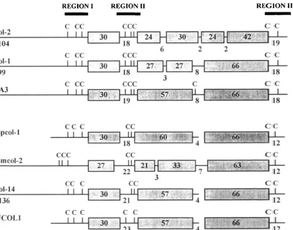
REGION I REGION II REGION HI
COl-2 104 col-1 99 3A3
ccc
c c
Bpcol-1 Bmcol-2 col-14 136 UCOL1 C C I I CCC II I CC C I I I CC C I I I 30 CC 18 CC CC C C 30 » •* 5 7 57. C C 12 C C C C C CFig. 4. Schematic representation of the protein domain organization of collagen molecules Bpcol-1 (Brugia pahangi), Bmcol-2 (B. tnalayi), col-1, col-2, col-14 (Caenorhabditis elegans), 3A3 (Haemonchus contortus), and UCOL1 (Ascaris suum). The proteins are grouped according to the position of cysteine residues (two major groups). The (Gly-X-Y)n
domains are boxed, the non-(Gly-X-Y)n regions are represented by horizontal lines, numerals indicate number of
amino acids in each region, cysteine residues are indicated by vertical lines and a C. Only the cysteine residues of region I are indicated 5' of the first (Gly-X-Y)n region. Gaps have been introduced in Bpcol-1, col-14, and UCOL1 to
maximize alignments of (Gly-X-Y)n domains. Regions I, II, and III, indicated by thick bars, are compared in Fig. 5.
of Bpcol-1 is 29-5 %. These motifs are interrupted by short stretches of 4—18 amino acids that depart from a repeating (Gly-X-Y)n sequence. This protein
structure has been shown t o be typical for collagen proteins of the cuticle of several nematode species (Cox, 1992).
col-1 col-2 3A3 Bpcol-1 col-14 UCOL1 B m c o l - 2 REGION I Y 6 A A P A V 6 G R L S G T G A Q K V V G P G A K G E A E V N P A P N L Q G G G G G G G G G I T S S E E N G G E R Q K R Q A V F Q V E S F N S E G G G G S H S E V P A G G E G | C | L | G P P D G C | | N 'P G P P E S N E ! P G P P
c
c
s
T G :.Q % G I G P V H R G P P G V G E P G V S P P G P P col-1 col-2 3 A 3 Bpcol-1 Bmcol-2 col-14 UC0L1 REGION II "G G G G G G G K K L Q E P E P G P D D P D -P -A -A P A P A P A P V S D % P L A A G ?E«?
P A P P S T P G S C, Q P ' p E E E L T D P P, T T P P P '••P. V T Q P^Pyi
v s i
:p|f;
P T E R D pW; Q P H I E | | H S L ; P P P' P P E F Ec
c
c
c
c
K P Q P A E F?N£
; Cc
:'Gf P, Q G P P G G P P P; A G P P A G P P PI D A P A P Q C P i W C F E G' Bpcol-1 Bmcol-2 c o l - 1 4 UCOL1 col-1 col-2 3 A 3 REGION III G G G G G G G S S S S I I I "C: •C-.c
c.
Gc
c
; •b'H. PUHJ P;''H| D H P K •Vp K..r .P.-;K;:;c
G qc
X*
2Pjc
C;c
P i P R T A P G Y * , | P R T A P G Y ' * F A R L A P G " Y * :P P R T A A G" Y * A; L D G^G. V F F E D G T R R * A I D GijG* V F F E^ D G" T R R R * A; I D, G)G" I -F-.F-.E, D G" T R R *Fig. 5. Comparison of Brugia pahangi Bpcol-1 collagen amino acid sequences of Regions I—III (indicated in Fig. 4) with corresponding regions of col-1, col-2, and col-14 of Caenorhabditis elegans, Bmcol-2 of B. malayi, 3A3 of
Haemonchus contortus, and UCOL1 of Ascaris suutn. The standard one letter code is used. Sequences are grouped
according to cysteine residues within each region. Horizontal lines denote the beginning or end of (Gly-X-Y)n domains. Conserved cysteine residues are boxed and shaded, amino acids that are identical within a group are shaded. Gaps (dashes) have been introduced in Region II to maximize homology. Asterisks indicate termination codons.
Protein domain and sequence comparison of Bpcol-1 with cuticular collagens of other nematodes
The deduced protein domain structure of Bpcol-1 collagen was compared to the structures of collagens
Bmcol-2 of B. malayi (Scott et al. 1995), UCOL1 of
the intestinal parasite A. suum (Kingston et al. 1989), 3A3 of the sheep parasite H. contortus (Shamanski et
al. 1989), col-1, col-2, and col-14 of the free-living
nematode C. elegans (Cox et al. 1989), as shown in Fig. 4. T h e proteins were aligned for maximal similarity by aligning each triple-helical region and
allowing the introduction of gaps to maximize alignment. T h e first and last (Gly-X-Y)n regions of Bpcol-1 are identical in size to those of col-1, col-14, 3A3, and UCOL1, 30 and 66 amino acids, re-spectively. The organization of all domains of Bpcol-1 and col-Bpcol-14 are identical, except that col-Bpcol-14 has three extra amino acids between the first and the second (Gly-X-Y)n region. These three amino acids do not lack i n Bpcol-1, but are part of the second (Gly-X-Y)n region, instead. UCOL1 has two additional amino acids in this domain, but is identical to Bpcol-1 and col-Bpcol-14 with respect to the lengths of the other
151
domains. This and the position of the cysteine residues of Bpcol-1 (boxed in Fig. 3 and vertical lines in Fig. 4) allows the assignment of Bpcol-1, col-14, and UCOL1 to the same group (Fig. 4). In addition, the similar distribution of cysteine residues of
Bmcol-2 also allows this protein to be grouped to the same
family, although the overall domain organization of
Bmcol-2 differs from that of Bpcol-1. Col-1 and col-2
show different organizations of the second (Gly-X-Y)n region featuring 3-6 interrupting amino acids.
col-1, col-2, and 3 A3 were assigned to another group,
because of their obvious similarities of the domain lengths and the distribution of cysteine residues (Fig. 4).
In C. elegans the sequence of the nontriple-helical domains I, II, and III (bold bars in Fig. 4), and the distribution and position of cysteine residues therein are diagnostic for each of the 4 collagen gene families identified in C. elegans (Cox, 1990). The amino acid sequence of the corresponding regions of Bpcol-1 were further analysed in detail (Fig. 5). Bpcol-1 has 7 cysteine residues, 5 of which are at identical positions to cysteine residues of col-14, UCOL1, and
Bmcol-2 (boxed in Fig. 5). The amino acid sequences
in regions I and II of all the listed proteins differ substantially from each other, except for the con-served cysteine residues. Region III, however, is highly conserved between certain collagen proteins, and even identical between Bpcol-1 and Bmcol-2 (Fig. 5).
Immunolocalization of Bpcol-1
The ultrastructural localization of epitopes that share antigenic determinants with Bpcol-1 collagen was determined in immunocytochemical experiments on thin sections of female B. pahangi parasites tested with the affinity-purified anti-Bpcol-1 antibodies isolated from the total polyclonal antiserum raised against the insoluble material of adult B. pahangi. Antigenic determinants recognized by anti-Bpcol-1 antibodies were specifically stained at two different locations: in all layers of the cuticle (Fig. 6 A and B), on basement membranes and secretions in the uteral lumen (Fig. 6C and D). No staining was observed with normal rabbit serum (Fig. 6E).
DISCUSSION
The cuticle of filarial nematodes is a complex, multilayered extracellular structure that features both rigid and elastic properties (Bird & Bird, 1991). Apart from its function as a selective barrier in the flow of nutrition, the most prominent aspect of the filarial cuticle is its proximity to the host defence mechanisms, and hence its role as the locus of the host-parasite interplay (Ogilvie et al. 1980). Insight into the molecular structure of the cuticle is therefore expected to provide a better understanding of this functional aspect.
The original aim of the present study was to identify non-collagenous structural proteins of the external layers of the epicuticle of filarial parasites. These proteins have been termed 'cuticlins' (Politz & Philipp, 1992), and are resistant to collagenase, which specifically cleaves the Gly-X bond within the (Gly-X-Y)n repetitive motif of collagens. Despite the fact that we used antisera raised against such epicuticular components to screen a cDNA library of adult B. pahangi, mainly collagens were identified by this approach. We hypothesize that the insoluble material used for immunization, even following exhaustive chemical and enzymatic treatment, still contained collagens masking any immunological reaction to the non-collagenous components them-selves. This is consistent with earlier studies in the rodent filarial parasite Acanthocheilonema viteae (Betschart et al. 1985), and with the electron microscopical data presented by Selkirk and coworkers on 2-ME-purified cuticles of Brugia parasites (Selkirk et al. 1989«).
The often-mentioned interspecies conservation of the cuticle structure (Politz & Philipp, 1992) is confirmed by the present study that reports the cloning, preliminary characterization, and inter-species comparison of a collagenous molecule of the adult filarial parasite B. pahangi. By means of alignment searches in the GenEMBL and Swissprot databanks of the DNA and the deduced amino acid sequence of a clone isolated from the B. pahangi cDNA libary, we identified a collagen of the cuticle of B. pahangi. We termed this clone Bpcol-1 for B.
pahangi collagen 1, in analogy with collagen genes
BmColl (Caulagi et al. 1991) and Bmcol-2 (Scott et
al. 1992) of B. malayi. The highest homologies were
obtained with cuticular collagens col-14 of C. elegans (Cox et al. 1989), and Bmcol-2 of B.malayi (Scott et
al. 1995). The amino acid sequence of Bpcol-1
revealed the typical domain organization of cuticular collagens. Bpcol-1 consists of regions containing the repetitive motif (Gly-X-Y)n that is responsible for the triple-helical conformation of collagens (Miller & Gay, 1987), separated by non-(Gly-X-Y)n stretches of 4—18 amino acids. The schematic comparison of the deduced protein domain structure of Bpcol-1 with the protein structures of collagen genes Bmcol-2 of B. malayi (Scott et al. 1995), UCOL1 of the intestinal parasite A. suum (Kingston
et al. 1989), 3A3 of the sheep parasite H. contortus
(ShamanskietaZ. 1989), col-1, col-2, and col-14 of the free-living nematode C. elegans (Cox et al. 1989) showed a significant similarity with col-14 and
Bmcol-2. This structure similarity was strengthened
by comparing the amino acid sequences of the nontriple-helical regions I—III, and the position of cysteine residues within these regions. Both para-meters have been used to classify identified collagens of C. elegans into separate groups or families (Cox, 1990). Cysteine residues of nematode cuticular
Fig. 6. Immunoelectron microscopy with the affinity-purified anti-Bpcol-1 antibodies on sections of adult Brugia pahangi. (A) Overview of epicuticle, cuticle, hypodermis, and muscle. (B) Labelling within the cuticle. (C) Labelling of the basal lamina of the uterus and the muscle layer. (D) Labelling on the uterus secretion. (E) Normal rabbit serum on cuticle. BL, basal lamina; E, epicuticle; C, cuticle; H, hypodermis; MC, muscle; O, ovum; PC, pseudocoel; US, uterus secretion; UW, uterus wall; arrows indicate gold particles.
collagens account for approximately 3 % of all amino acids and are involved in the formation of disulfide cross-links. These disulfides presumably form be-tween polypeptides of a triple-helical rod (interchain cross-links), as well as between polypeptides of different collagen molecules (intrachain cross-links), and account for their insolubility in the absence of a reducing agent, such as 2-ME (Cox, 1992).
Affinity-purified anti-Bpcol-1 antibodies were used to identify corresponding antigenic deter-minants in nematode extracts by immunoblot analy-sis. In extracts of adult B. malayi parasites, the anti-Bpcol-1 antibodies detected proteins of molecular sizes corresponding well to collagen molecules identified not only in a previous study on collagens of B. malayi and B. pahangi (Selkirk et al. 1989«), but also of a variety of other nematodes, such as the free-living organisms C. elegans (Cox et al. 1981), Panagrellus silusiae (Leushner, Semple & Pasternak,
1979), and Meloidogyne incognita (Reddigari et al. 1986), the intestinal parasite H. contortus (Fetterer, 1989), and the filarial nematodes, Onchocerca vol-vulus and O. gutturosa, and Dirofilaria immitis (Petralanda & Piessens, 1991). In addition, we have described collagens of similar sizes in the intestinal parasite A. suum (Betschart et al. 1990). The observed cross-reactivity of anti-Bpcol-1 antibodies to collagenous molecules in extracts of A. suum, and in contrast, the lack of immunoreactions to mam-malian collagens confirm that the antigenic structure of nematode collagens is fundamentally different from those of mammalian collagens. Nevertheless, immunological cross-reactivity between nematode-specific and mammalian collagens has been shown to occur in patients infected by filarial nematodes. Such autoreactive antibodies, however, seem to play a minor role in the context of the symptoms associated with filarial infection (Selkirk et al. 1989 a).
A moderate labelling of several layers of the cuticle of adult B. pahangi, including the outer cortical layer, was observed in immunocytochemical experi-ments using affinity-purified Bpcol-1 anti-bodies. The labelling of the basement membrane and secretions of the uterus was a rather interesting finding, which suggests the occurrence of similar epitopes in the basement membrane and the cuticle, although structural analysis of basement collagen genes of C. elegans (Guo & Kramer, 1989), A. suum (Pettitt & Kingston, 1991), and B. malayi (Caulagi et al. 1991; Caulagi & Rajan, 1995), and their encoded products, have revealed a different structure (Cox, 1992). The cross-reactivity between cuticular and basement membrane collagens observed in this study remains to be further analysed.
The authors wish to thank Dr M. E. Selkirk (Imperial College of Science, Technology and Medicine, London, UK) for providing the B. pahangi cDNA library and the polyclonal antiserum. We are grateful to Georg Scheidegger for technical support at the electron
micro-scope. This work was supported by a grant from the Swiss National Research Foundation (No. 31-33961.92). Note: Nucleotide sequence data reported in this paper will appear in the EMBL, GenBank and DDBJ Nucleotide Sequence Databases under the accession number X92099.
R E F E R E N C E S
ALTMANN, M., HANDSCHIN, C. & TRACHSEL, H. (1987).
mRNA cap-binding protein: cloning of the gene encoding protein synthesis initiation factor eIF-4E from Saccharomyces cerevisiae. Molecular Cell Biology 7, 998-1003.
BETSCHART, B. & WYSS, K. (1990). Analysis of the
cuticular collagens of Ascaris suum. Ada Tropica 47, 297-305.
BIRD, A. F. & BIRD, j . (1991). The Structure of Nematodes. Academic Press, San Diego.
BOISVENUE, R. J . , STIFF, M. I., TONKINSON, L. V. & COX, G. N. (1991). Protective studies in sheep immunized with cuticular collagen proteins and peptides of Haemonchus contortus. Parasite Immunology 13, 227-240.
BRADFORD, M. M. (1976). A rapid and sensitive method for the quantitation of microgram quantities of protein utilizing the principle of protein dye binding.
Analytical Biochemistry 72, 248-254.
CAULAGI, v. R. & RAJAN, T. v. (1995). The structural organization of an <x2 (type IV) basement membrane collagene gene from the filarial nematode Brugia malayi. Molecular and Biochemical Parasitology 70, 227-229.
CAULAGI, V. R., WERNER, C. & RAJAN, T. V. ( 1 9 9 1 ) .
Isolation and partial sequence of a collagen gene from the human filarial parasite Brugia malayi. Molecular and Biochemical Parasitology 45, 57-64.
CORDEN, J . , WASVLYK, B., BUCHWALDER, A., SASSONE-CORSI, P . , KEDINGER, C. & CHAMBON, P . ( 1 9 8 0 ) . Promoter sequences of eukaryotic protein-coding genes. Science 209, 1406-1414.
COX, G. N., KUSCH, M. & EDGAR, R. S. (1981). C u t i c l e of
Caenorhabditis elegans: Its isolation and partial characterization. Journal of Cell Biology 90, 7-17. COX, G. N . , FIELDS, C , KRAMER, J. M . , ROSENZWEIG, B. &
HIRSH, D. (1989). Sequence comparison of developmentally regulated collagen genes of Caenorhabditis elegans. Gene 76, 331-344. cox, G. N. (1990). Molecular biology of the cuticle
collagen gene families of Caenorhabditis elegans and Haemonchus contortus. Ada Tropica 47, 269-281. cox, G. N. (1992). Molecular and biochemical aspects of
nematode collagens. Journal of Parasitology 78, 1—15. DEVEREUX, J . , HAEBERLI, P . & SMITHIES, O. ( 1 9 8 4 ) . A
comprehensive set of sequence analysis programs for the VAX. Nucleic Acids Research 12, 387-395.
FEINBERG, A. p. & VOGELSTEIN, B. (1983). A technique for
radiolabeling DNA restriction endonuclease fragments to high specific activity. Analytical Biochemistry 132, 6-13.
FETTERER, R. H. (1989). The cuticular proteins from free-living and parasitic stages of Haemonchus contortus-l. Isolation and partial characterization. Comparative Biochemistry and Physiology 94B, 383-388.
FUJIMOTO, D. & KANAYA, s. (1973). Cuticlin: a noncollagen structural protein from Ascaris cuticle. Archives of Biochemistry and Biophysics 157, 1-6. Guo, x. & KRAMER, j . M. (1989). The two Caenorhabditis
elegans basement membrane (type IV) collagen genes are located on separate chromosomes. Journal of Biological Chemistry 264, 17574-17582.
HO, N . F. H . , GEARY, T. G., RAUB, T. J., BARSUHN, C. L. & THOMPSON, D. P. (1990). Biophysical transport properties of the cuticle of Ascaris suum. Molecular and Biochemical Parasitology 41, 153-166.
JOSSE, j . & HARRINGTON, w. F. (1964). Role of pyrrolidine residues in the structure and stabilization of collagen. Journal of Molecular Biology 9, 269-287.
KIEFER, E., RUDIN, W . , BETSCHART, B . , WEISS, N . & HECKER, H. (1986). Demonstration of anti-cuticular antibodies by immunoelectron microscopy in sera of mice immunized with cuticular extracts and isolated cuticles of Dipetalonema viteae (Filarioidea). Ada
Tropica 43, 99-112.
KINGSTON, I. B., WAINWRIGHT, S. M. & COOPER, D. ( 1 9 8 9 ) . Comparison of collagen gene sequences in Ascaris suum and Caenorhabditis elegans. Molecular and Biochemical Parasitology 37, 137-146.
LEUSHNER, J., SEMPLE, N . L. & PASTERNAK, J. ( 1 9 7 9 ) . Isolation and characterization of the cuticle from the free-living nematode Panagrellus silusiae. Biochimica et Biophysica Ada 580, 166-174.
MANIATIS, T . , FRITSCH, E. F. & SAMBROOK, J. ( 1 9 8 2 ) . Molecular Cloning: A Laboratory Manual, 2nd edn. New York: Cold Spring Harbor Laboratory Press. MCCALL, J. W., MALONE, J. B . , AH, H. S. & THOMPSON, P . E.
(1973). Mongolian jirds (Meriones unguiculatus). infected with Brugia pahangi by the intraperitoneal route: a rich source of developing larvae, adult filariae and microfilariae. Journal of Parasitology 59, 436. MILLER, E. j . & GAY, s. (1987). The collagens: an
overview and update. Methods in Enzymology 144, OGILVIE, B. M . , P H I L I P P , M . , JUNGERY, M . , MAIZELS, R. M . ,
WORMS, M. J. & PARKHOUSE, R. M. E. ( 1 9 8 0 ) . T h e surface of nematodes and the immune response of the host. In The Host Invader Interplay (ed. Van den Bossche, H.), pp. 99-104. Elsevier, Amsterdam. PETTITT, j . & KINGSTON, I. B. (1991). The complete
primary structure of a nematode ct2 (IV) collagen and partial structural organization of its gene. Journal of Biological Chemistry 266, 16149-16156.
PETRALANDA, I. & PIESSENS, w. (1991). Onchocerca volvulus, O. gutturosa, Brugia malayi and Dirofilaria
immitis: A comparative study of the immunochemical properties of the cuticular proteins from filarial parasites. Experimental Parasitology 72, 164—173. POLITZ, s. M. & PHILIPP, M. (1992). Caenorhabditis elegans
as a model for parasitic nematodes: A focus on the cuticle. Parasitology Today 8, 6-12.
PRITCHARD, D. I., McKEAN, P. G. & ROGAN, M . T. (1988).
Cuticular collagens - a concealed target for immune attack in hookworms. Parasitology Today 4, 239—241. PROUDFOOT, N . J. & BROWNLEE, G. G. ( 1 9 7 6 ) . 3 ' N o n
-coding region sequences in eukaryotic messenger RNA. Nature, London 263, 211-214.
REDDIGARI, S. R., JANSMA, P . L., PRMACHANDRAN, D. & HUSSEY, R. H. (1986). Cuticular collagenous proteins of second-stage juveniles and adult females of
Meloidogyne incognita: Isolation and partial
characterization. Journal of Nematology 18, 294—302. SANGER, F . , NICKLEN, S. & COULSON, A. R. ( 1 9 7 7 ) . D N A
sequencing with chain-terminating inhibitors.
Proceedings of the National Academy of Sciences, USA 74, 5463-5467.
SCOTT, A. L., YENBUTR, P . , EISINGER, S. W. & RAGHAVAN, N. (1995). Molecular cloning of the cuticular collagen gene Bmcol-2 from Brugia malayi. Molecular and Biochemical Parasitology 70, 221-225.
SELKIRK, M. E., NIELSEN, L., KELLY, C , PARTONO, F . ,
SAYERS, G. & MAIZELS, R. M. (1989a). Identification, synthesis and immunogenicity of cuticular collagens from the filarial nematodes Brugia malayi and Brugia pahangi. Molecular and Biochemical Parasitology 59, 229-246.
SELKIRK, M. E., DENHAM, D. A., PARTONO, F. & MAIZELS, R. M. (19896). Heat shock cognate 70 is a prominent immunogen in brugian filariasis. Journal of Immunology 143, 299-308.
SHAMANSKY, L. M . , PRATT, D., BOISVENUE, J. & COX, G. N . (1989). Cuticle collagen genes of Haemonchus contortus and Caenorhabditis elegans are highly conserved. Molecular and Biochemical Parasitology 37, 73—86. SULSTON, j . E. & BRENNER, s. (1974). The DNA of
Caenorhabditis elegans. Genetics 77, 95-104. TOWBIN, H . , STAEHELIN, T. & GORDON, J . ( 1 9 7 9 ) .
Electrophoretic transfer of proteins from
polyacrylamide gels to nitrocellulose sheets: Procedure and some applications. Proceedings of the National Academy of Sciences, USA 76, 4350-4354.
WHARTON, D. A. (1986). A Functional Biology of Nematodes. Croom Helm, London & Sydney.
YOUNG, R. A. & DAVIS, R. w. (1983). Efficient isolation of genes using antibody probes. Proceedings of the National Academy of Sciences, USA 80, 1194-1198.