www.elsevier.com / locate / cardiores
Review
Sex hormones and hypertension
a,b ,
*
e b a,c,dRaghvendra K. Dubey
, Suzanne Oparil , Bruno Imthurn , Edwin K. Jackson
a
Center for Clinical Pharmacology, University of Pittsburgh Medical Center, Pittsburgh, PA 15213, USA
b
University Hospital Zurich, Department of Obstetrics and Gynecology, Clinic for Endocrinology, Frauenklinikstrasse 10, 8091 Zurich, Switzerland
c
Department of Medicine, University of Pittsburgh Medical Center, Pittsburgh, PA 15213, USA
d
Department of Pharmacology, University of Pittsburgh Medical Center, Pittsburgh, PA 15213, USA
e
The Vascular Biology and Hypertension Program, University of Alabama, Birmingham, AL 35294, USA Received 8 June 2001; accepted 30 October 2001
Abstract
Gender has an important influence on blood pressure, with premenopausal women having a lower arterial blood pressure than age-matched men. Compared with premenopausal women, postmenopausal women have higher blood pressures, suggesting that ovarian hormones may modulate blood pressure. However, whether sex hormones are responsible for the observed gender-associated differences in arterial blood pressure and whether ovarian hormones account for differences in blood pressure in premenopausal versus postmenopausal women remains unclear. In this review, we provide a discussion of the potential blood pressure regulating effects of female and male sex hormones, as well as the cellular, biochemical and molecular mechanisms by which sex hormones may modify the effects of hypertension on the cardiovascular system. 2002 Elsevier Science B.V. All rights reserved.
Keywords: Blood pressure; Endothelial function; Gender; Growth factors; Hypertension; Smooth muscle
1. Introduction (SHR; [6–9]), Dahl salt-sensitive rats [10,11], deoxy-corticosterone acetate-salt hypertensive rats [12], and New Sexual dimorphism in arterial blood pressure appears in Zealand genetically hypertensive rats [13] have higher adolescence and persists throughout adulthood [1,2]. Aver- blood pressures. In these animal models of hypertension, age systolic and diastolic blood pressures in men less than blood pressure is reduced in males by castration [6– 60 years-of-age are higher than in age-matched women by 9,14,15], but is not increased in females by ovariectomy 6–7 and 3–5 mmHg, respectively [3–5]. After that time, [16,17]. Thus, sex-associated differences in blood pressure blood pressure (particularly systolic blood pressure) in- also may be due to changes in testicular hormones. In the creases in women so that hypertension becomes more sections that follow, we examine the evidence for and prevalent [4] or at least as prevalent in women as men. against the involvement of female and male sex hormones Inasmuch as gender-associated differences in hypertension in the pathogenesis of hypertension.
prevalence either disappear or cross over after women enter menopause, ovarian hormones may be responsible in
part for lower blood pressure in premenopausal women 2. Estrogens and hypertension
and for the increase in blood pressure in postmenopausal
women. Similar to humans, sex-associated differences in 2.1. Effects of estrogens on blood pressure blood pressure also exist in animals. For example,
com-pared with females, male spontaneously hypertensive rats Cross sectional [18–20], but not longitudinal [21–23], studies show a significant increase in systolic and diastolic
*Corresponding author. Clinic for Endocrinology, Department of blood pressure following the onset of menopause. Staessen Obstetrics and Gynecology, University Hospital Zurich, Frauenklinik- et al. [18] reported a four-fold increase in the incidence of strasse 10, CH-8051 Zurich, Switzerland. Tel.: 141-1-2555-383; fax:
141-1-2554-439.
E-mail address: [email protected] (R.K. Dubey). Time to primary review 26 days.
0008-6363 / 02 / $ – see front matter 2002 Elsevier Science B.V. All rights reserved. P I I : S 0 0 0 8 - 6 3 6 3 ( 0 1 ) 0 0 5 2 7 - 2
hypertension in postmenopausal women (40% in post- months to 5 years, four became normotensive from 1 to 7 menopausal women vs. 10% in premenopausal women). In months after they cessation of therapy [46]. Similarly, of a subsequent prospective evaluation of blood pressure 27 women who developed hypertension while taking (conventional and ambulatory) in women who were pre- Premarin (1.25 mg / day), 13 became normotensive within menopausal, perimenopausal or postmenopausal, the au- 3.6 months after Premarin was discontinued, and of five thors reported that postmenopausal women had a higher women who restarted Premarin, blood pressure was again systolic blood pressure (4–5 mmHg) compared with elevated within 6 weeks to 6 months [50]. In a prospective premenopausal or perimenopausal women [20]. Also, the study of 160 postmenopausal women taking either Pre-rise in systolic blood pressure per decade was 5 mmHg marin (n573) or piperazine estrone sulphate (Ogen; n5 greater in perimenopausal and postmenopausal women 87), development of hypertension was observed only in compared with premenopausal women [20]. Because women taking Premarin [51]. It should be noted that in the menopause is associated with decreased synthesis of above studies, conjugated estrogens (Premarin) were ad-estradiol, it is likely that changes in blood pressure induced ministered orally. Since conjugated estrogens administered by menopause may be due in part to reductions in estradiol via patches have been shown to have largely neutral or
production. marginally blood pressure lowering effects, the route of
Support for the conclusion that estradiol has a blood administration may have a decisive role in defining the pressure lowering effect in women is provided by the effects of conjugated estrogens on blood pressure. observation that during the menstrual cycle, blood pressure With regard to contraceptive estrogens, in 22 patients is lower during the luteal phase (when estradiol levels who developed hypertension on oral contraceptive pills, peak) than during the follicular phase [24–26]. Observa- the blood pressure was normalized after oral contraceptives tions made during pregnancy provide additional circum- were discontinued (blood pressures before, during and stantial evidence for a blood pressure lowering effect of after administration of oral contraceptives were 125 / 76, estradiol. Estradiol levels increase 50–180-fold during 183 / 110, and 130 / 82 mmHg, respectively [46]). Similar pregnancy [27], and these increases are associated with reversals in oral contraceptive-induced hypertension were substantial reductions in blood pressure [28]. The timing of observed in eight out of 14 women within 3.6 months of maximal decreases in blood pressure and maximal in- discontinuing the contraceptive estrogen [50], and seven of creases in estradiol levels does not coincide completely, the eight became hypertensive again 3–6 months after however. In the first, second and third trimesters of resuming therapy. In a prospective cohort study of 68 297 pregnancy, estradiol levels increase by 8-, 15- and 186-fold female nurses, compared with women who had never used [27], respectively. In contrast, ambulatory blood pressure oral contraceptives, the age adjusted relative risk of is lowest in the first (systolic 10367 mmHg) and second hypertension was 1.5 (95% CI51.5 to 1.8) for current use (systolic 10169 mmHg) trimesters and rises in the third and 1.1 (95% CI50.9 to 1.2) for past use [59]. After trimester (systolic 11169 mmHg) of pregnancy [28]. This adjusting for age, body mass index, hormones, cigarette suggests that factors in addition to estradiol modulate smoking, family history of hypertension, parity, physical blood pressure in pregnancy [29] and / or that other hor- activity, alcohol intake, and ethinicity, current users of oral mones or local modulators generated during pregnancy contraceptives had an increased risk of development of abrogate the blood pressure lowering effects of estradiol hypertension [relative risk (RR)51.8; 95% CI51.5–2.3]. during the third trimester of pregnancy. The risk of hypertension associated with oral contracep-If endogenous estradiol does lower blood pressure, tives increased with age, duration of use, body mass and administration of estrogenic preparations to women might progestin potency [56]. It is important to note that the also be expected to reduce blood pressure. However, data estrogens used in contraceptive pills (e.g., ethinyl es-on the effects of estrogenic preparaties-ons es-on blood pressure tradiol) are different than the natural estrogen estradiol; are inconsistent, and include reports of blood pressure moreover, the doses for contraceptive estrogens and es-lowering [30–45], blood pressure elevating [46–51], and trogens used for hormone replacement therapy vary con-blood pressure neutral effects [37,52–54]. Evidence for siderably. This underscores the principle that the blood blood pressure elevating effects of estrogen preparations pressure effects of estrogenic preparations cannot be comes primarily from studies conducted between 1970 and equated just because the preparations belong to the same the early 1980s that used conjugated estrogens or contra- pharmacological class.
ceptive estrogens (different from natural estradiol) and More recent studies indicate that postmenopausal es-conventional rather than ambulatory blood pressure mea- trogen replacement therapy either does not affect or surements. For example, in several studies, PremarinE (a reduces blood pressure. For example, tibolone, a synthetic mixture of conjugated estrogens isolated from urine of estrogen with both androgenic and gestagenic properties, pregnant mares) increased blood pressure [46,50,51], and had no effect on blood pressure [61]. In the Postmenopaus-various synthetic contraceptive estrogens increased the risk al Estrogen / Progestin Interventions Trial (PEPI), no of hypertension [55–60]. Of five women who developed change in blood pressure was observed in 875 normoten-hypertension after using Premarin (1.25 mg / day) for 3 sive postmenopausal women receiving conjugated equine
estrogens in combination with a variety of progestins [52]. day), the 24-h ambulatory blood pressure was lowered In smaller placebo-controlled, randomized crossover significantly [31]. A significant decrease in blood pressure studies, administration of conjugated estrogens to nor- was also observed in postmenopausal women with mild to motensive postmenopausal women had blood pressure moderate hypertension who were receiving transdermal lowering or neutral effects (Table 1). estradiol [33]. In a prospective study conducted in 34 Administration of estradiol (oral or transdermal patches) postmenopausal women with treated hypertension, ad-to normotensive postmenopausal women either reduced or ministration of estradiol plus the progestin norgestrel for had no effect on blood pressure (Table 1). For example, in 19 weeks lowered 24-h ambulatory blood pressure [36]. In a placebo-controlled, double-blind, crossover study with 13 postmenopausal women with ongoing treatment for 24-h ambulatory blood pressure measurements, transder- hypertension, acute administration of transdermal estradiol mal estradiol (50 mg / day; achieves physiological levels of reduced 24-h ambulatory day time systolic blood pressure estradiol) treatment for 2 months was reported to decrease and nocturnal systolic and diastolic blood pressure [43]. In nocturnal blood pressure (systolic, diastolic and mean), but a follow up study of 75 hypertensive postmenopausal not daytime (office) blood pressure [40]. Several other women on estradiol replacement therapy for 36 months, no studies utilizing 24-h ambulatory blood pressure measure- significant changes in blood pressure were observed [53], a ment reported blood pressure lowering effects of estradiol negative result that may have been due to the lack of (Table 1). In a non-randomized, non-placebo-controlled ambulatory blood pressure measurements.
study, nocturnal blood pressure was decreased in nor- Estradiol also lowers blood pressure in several animal motensive women treated chronically with estradiol (trans- models of hypertensive, including SHR [63], stroke prone dermal) [41]. Twenty four-hour ambulatory blood pressure SHR (SHRSP; [64]), rats with deoxycortisterone acetate-was also lowered in postmenopausal women who were salt induced hypertension [65], Dahl salt-sensitive rats [66] treated with estradiol sequentially with the progestin and rats with pulmonary hypertension [67,68]. In contrast, dydrogesterone (5 or 10 mg) for 1 year [32]. ovariectomy does not affect the development of hyperten-Blood pressure lowering effects of estradiol were also sion in SHRs [6], suggesting that the effects of estradiol on observed by Seely et al. [42] who evaluated in healthy blood pressure in rats are pharmacological rather than postmenopausal women the effects of transdermal estradiol physiological. Unlike the natural estrogen estradiol, the (0.1 mg patches administered twice per week to attain synthetic estrogen ethinyl estradiol increases blood pres-physiologic estradiol levels) administered in combination sure [55], suggesting that the effects of natural and with intravaginal progesterone (300 mg). Significant de- synthetic estrogens differ markedly and may influence creases in nighttime systolic, diastolic, and mean blood distinct mechanisms involved in the regulation of blood pressure were observed in the group treated with estradiol pressure. For example, even though contraceptive es-and estradiol / progesterone. Similar to the findings of trogens are administered at a lower dose (30–200 mg) than Cagnacci et al. [40] and the data from the PEPI trial [52], estrogens (0.625–2 mg) used for hormone replacement there were trends toward lower daytime or office systolic therapy, contraceptive estrogens increase blood pressure and diastolic blood pressures in patients on estradiol or [55].
estradiol / progestin compared to placebo; however, the In summary, although the literature on the effects of changes were not significant. Since women, in particular estrogens on blood pressure is confusing and inconsistent, may be vulnerable to white coat hypertension [62], it is several general trends can be gleaned from existing reports. possible that nocturnal blood pressure may be more Whether blood pressure is decreased, increased or un-sensitive to estradiol and progesterone. Moreover, this may changed in response to estrogen treatment depends pri-be a potential reason for the lack of decrease in office / marily on three factors: (1) the type of estrogenic prepara-daytime blood pressure observed in the PEPI trial [52]. tion; (2) the dose of estrogens; and (3) how blood pressure This also reaffirms the importance and superiority of 24-h is monitored. Contraceptive estrogenic preparations tend to ambulatory blood pressure measurements for ascertaining increase blood pressure; conjugated equine estrogens ap-blood pressure changes accurately and without missing pear to have little effect on blood pressure, and estradiol some subtle but potentially important effects of estrogen tends to lower blood pressure. The blood pressure lowering replacement therapy on blood pressure. Indeed, decreases effects of estradiol are more readily observed if 24-h in nighttime ambulatory blood pressure in response to ambulatory blood pressure monitoring is employed. The hormone replacement therapy have been observed in other effects of estradiol on the cardiovascular system and clinical studies [32,41]. kidneys are discussed below from the perspective of how Results from several small clinical trials suggest that these effects may contribute to the blood pressure lowering estrogen replacement therapy also lowers blood pressure in actions of estradiol.
hypertensive postmenopausal women (Table 1). In a
randomized, double-blind crossover trial carried out in 30 2.2. Effects of estradiol on vascular tone postmenopausal women with mild hypertension who were
R .K . Dubey et al . / Cardiovascular Research 53 (2002 ) 688 – 708 691
Effects of estrogens on blood pressure
Study Subjects Treatment Outcome Ref.
Normotensive (N) / hypertensive (H)
1 62 PMW (42 no HRT; 20 HRT); N CEE 24 h ambulatory BP was decreased [30] 2 30 PMW (randomized, double blind crossover; mild H) Transdermal 17b-E 24 h BP was decreased [31] 3 29 PMW (15 placebo; 14 treated; N) Oral 17b-E daily plus dydrogesterone Follow-up after 1 year of treatment showed [33]
every 3–4 weeks a significant decrease in BP
4 16 PMW (mild to moderate H) Transdermal 17b-estradiol Ambulatory 24 h BP was decreased [34] 5 12 surgically PMW (N) Oral CEE Auscultatory BP unchanged after 1 week [35]
Oral CE plus MPA Auscultatory BP lowered after 1 week
6 73 PMW (38 oral; 35 transdermal; N) Oral 17b-E1norethindrone 24 h ambulatory BP decreased after 2 and 6 months [36] Transdermal 17b-E1norethindrone 24 h ambulatory BP decreased after 2, but not 6 months
7 34 PMW (prospective study, 34 PMW with treated H) Cyclic estradiol1norgestrel (19 weeks) 24 h ambulatory BP decreased after 19 weeks [37] 8 60 PMW (20 placebo, 20 CEE, 20 17b-E; CAD) Oral CEE6MPA (10 days) Night time ambulatory BP decreased [38]
treatment for 1 year; transdermal Transdermal 17b-E6MPA (10 days) No significant change in BP
9 16 PMW (placebo-controlled, randomized crossover study; N) 17b-E plus cyclic NETA 24 h ambulatory BP decreased and this effect was [39] more pronounced in presence of NETA
10 17 PMW (3 month, placebo-controlled, Oral CEE 24 h ambulatory BP decreased [40] randomized crossover study; N) Oral CEE plus MPA 24 h ambulatory BP decreased
11 18 PMW (placebo-controlled, randomized crossover study, N) Transdermal 17b-E for 2 months 24 h nocturnal, but not day time, BP decreased [41] 12 15 PMW (placebo-controlled, randomized crossover study, N) Transdermal 17b-E (8 weeks)6P 24 ambulatory BP (nocturnal&daytime) decreased by 17b-E [43]
(vaginal; 2 weeks) alone and the effect was enhanced by P
13 107 PMW1ERT; 223 PMW-HRT; Oral CEE, 17b-E and other Es oral No change in BP [55] population based sample; H and N or transdermal
14 PEPI trial-875 PMW; N CEE6various progestins No change in BP [53]
15 13 PMW (with ongoing treatment for hypertension, Transdermal 17b-E 24 h ambulatory daytime diastolic BP decreased at 24 h and 24 h [44] placebo-controlled double blind, crossover study; H) ambulatory nocturnal systolic and diastolic BP decreased at 24 h
16 90 PMW women (oophorectomized, 30–59 yr, Transdermal 17b-E (n540) 24 h ambulatory nocturnal systolic and daytime as well [42] non-randomized, prospective study; N) as night time BP reduced at 3 and 6 months
No change in BP in subjects receiving oral ERT Oral ERT (n550)
17 20 PMW, double blind, cross over study (N) Oral CEE plus oral cyclical MPA Ambulatory day time diastolic and mean BP reduced and [176] MPA lowered BP dose-dependently in presence of conjugated estrogens
PMW, Postmenopausal women; ERT, estrogen replacement therapy; HRT, hormone replacement therapy; CEE, conjugated equine estrogens; 17b-E, 17b-estradiol; MPA, medroxyprogesterone; NETA, norethisterone acetate.
subtypes are expressed in vascular endothelial [60,70] and Additionally, estradiol activates adenylyl cyclase activi-smooth muscle cells [60,70], and it is well established that ty and increases the synthesis of cyclic AMP, a vasodilator estradiol can cause vasodilation by both ER-dependent and second messenger [99]. Moreover, estradiol stimulates the ER-independent mechanisms. Acute administration of es- production of adenosine in vascular smooth muscle cells tradiol in vitro and in vivo induces rapid dilation of via the cyclic AMP-adenosine pathway [99]. Estradiol also coronary arteries of cholesterol-fed ovariectomized animals increases the synthesis of the vasodilator prostacyclin by [69–73]. Exogenous estradiol also dilates coronary and inducing the expression of prostacyclin synthase and brachial arteries in postmenopausal women and men [74– cyclooxygenase [100,101]. Finally, estradiol reduces the 77]. Long-term treatment with estradiol abrogates the synthesis of potent vasoconstrictors such as angiotensin II vasoconstrictor effects of U46619 (thromboxane mimetic), (Ang II), endothelin-1 and catecholamines [102–104]. phenylepinephrine, 5-HT, calcium, potassium and acetyl- In summary, estradiol is a vasodilator that decreases choline on vascular tissues such as aortic rings and vascular resistance by multiple mechanisms. Increased coronary arteries [69,78–81]. Compared with pre- production of NO plays a prominent role, and increased menopausal women, vasodilator effects of estradiol are synthesis of other endogenous vasodilators, decreased decreased in postmenopausal women and are normalized synthesis of endogenous vasoconstrictors and activation of
1
by estrogen replacement therapy [69]. The vasodilator K channels also contribute to the vasodilatory actions of effect of estradiol replacement therapy is diminished by estradiol.
co-administration of synthetic progestins such as
medroxy-progesterone and cyproterone acetate [82,83]. 2.3. Effects of estradiol on vascular growth Both the acute and long-term vasodilator effects of
estradiol are mediated in part via generation of endo- The elevated total peripheral resistance characteristic of thelium-derived NO and are attenuated by NO synthesis hypertension is due in part to accelerated growth of inhibitors [84,85]. Estradiol induces an increase in intracel- vascular smooth muscle cells [105]. Vascular remodeling in lular free calcium concentration in endothelial cells hypertension involves interactions among multiple cell [69,86], which could contribute to the increase in endo- types, such as endothelial cells, smooth muscle cells, thelial-derived NO. Since inhibition of NO synthesis adventitial fibroblasts, monocytes, macrophages and promotes arterial hypertension [87], it is conceivable that leukocytes, and among multiple growth inducers, including estradiol protects against hypertension by increasing NO local growth factors, circulating growth factors and me-synthesis. Long-term administration of estradiol increases chanical forces [106]. The processes that lead to increased acetylcholine-mediated coronary vasodilation in non- vascular resistance involve endothelial cell damage / human primates [71,72], male-to-female transsexuals [88], dysfunction, increased generation of chemotactic and and postmenopausal women [89], particularly those with mitogenic factors at injury sites, migration of smooth angina and normal coronary arteries [90]. The ability of muscle cells into the intima, proliferation of the migrated estradiol to increase endothelium-dependent vasodilation in cells, hypertrophy of smooth muscle cells (increase in cell the forearm of hypertensive postmenopausal women is size) and deposition of extracellular matrix proteins [106]. shared by conjugated equine estrogens [91]. Progestins In addition to smooth muscle cells, migration of adventitial inhibit estradiol-induced synthesis of endothelium-derived fibroblasts into the neointima and fibroblast proliferation NO [92,93], and this may contribute to the diminished also play a major role in the vascular remodeling process vasodilator effects of estrogen observed in postmenopausal [107]. The sequence of events in vascular remodeling may women receiving estradiol plus progestins. vary depending on the type of vascular challenge (me-Other mechanisms also contribute to estradiol-induced chanical, immunologic, lipid-induced), but the abnormal vasodilation. Estradiol causes coronary vasodilation by growth of smooth muscle cells is the final process that
1
opening calcium-activated K channels and relaxes endo- leads to increased vascular resistance. In vivo studies thelium-denuded porcine coronary arteries by opening conducted in several species using various models (balloon large conductance calcium-activated and voltage-activated injury-induced neointima formation, allograft-induced dys-K1 channels [86,94]. Estradiol inhibits voltage-dependent plasia, cholesterol / lipid-induced atherosclerosis and vascu-L-type calcium currents in vascular smooth muscle cells lar narrowing-induced neointima formation) provide con-and has potent stimulatory effects on large-conductance, vincing evidence that estradiol prevents the vascular
1
calcium- and voltage-activated K channels in coronary remodeling processes [69,70].
artery vascular smooth muscle cells [95]. Since the vasodi- Estradiol engages key cellular / molecular components of lator effects of estradiol in intact arteries are abrogated by the vascular remodeling process. Specifically, estradiol: (1)
1
blockers of calcium-activated, large conductance K chan- protects against endothelial damage caused by mechanical nels and inhibitors of cyclic GMP-dependent protein injury, oxidized-low-density lipoprotein (LDL), homocy-kinase, estradiol may exert its vasodilator effects by steine, free radicals and immunological factors; (2) blocks
1
opening calcium-activated, large conductance K channels vascular inflammation and decreases the expression of via NO and cGMP-dependent pathways [94–98]. adhesion molecules (intercellular adhesion molecule-1,
vascular cell adhesion molecule-1, and endothelial women [119]. In women-to-men transsexuals, homocy-leukocyte adhesion molecule-1) at sites of vascular dam- steine levels increase with androgen treatment, whereas in age; (3) inhibits neointima formation by attenuating / inhib- men-to-women transsexuals, homocysteine levels decrease iting the recruitment of circulating macrophages, lympho- with estrogen substitution [120]. Thus, estrogen may cytes and thrombocytes to the site of injury, thus reducing protect the vasculature in part by lowering homocysteine the secretion of cytokines, eicosanoids and growth factors; levels.
(4) inhibits the vascular remodeling processes in athero- Endothelin-1 is a vasoconstrictor and mitogenic peptide sclerosis by stopping the migration of macrophages into that is thought to play a role in the pathogenesis of various the subendothelial spaces and preventing them from ac- forms of vascular disease. The synthesis and biological cumulating LDL and becoming foam cells; (5) prevents activity of endothelin-1 are regulated by estradiol. Es-foam cell-induced inflammatory processes such as platelet tradiol inhibits serum- and Ang II-stimulated synthesis of adhesion and the release of multiple platelet-derived endothelin-1 [121] in endothelial cells via ER-receptor growth factors and cytokines; and (6) inhibits the dependent mechanisms [122]. Estradiol also blocks the mitogenic effects of multiple factors generated at the site mitogenic effects of endothelin-1 on smooth muscle cells of endothelial injury / dysfunction and which trigger a and inhibits endothelin-1 induced MAP kinase activation cascade of biochemical events that stimulate hyperplastic [123]. Compared with premenopausal women, plasma and / or hypertrophic growth of vascular smooth muscle endothelin-1 levels are increased in postmenopausal
cells [69,70,108]. women not taking estradiol, and are reduced following
In summary, there is no doubt that estradiol profoundly estradiol replacement therapy [104,119]. These findings inhibits the vascular response to injury. This important suggest that estradiol may have anti-mitogenic effects on effect of estradiol is mediated by numerous mechanisms, the vasculature in part by reducing endothelin-1 levels. and this is an active area of contemporary research. The Estradiol influences the synthesis of a number of factors vasculoprotective effects of estradiol may attenuate the associated with coagulation and fibrinolysis. For example, development of hypertension and may protect the vascula- estradiol decreases plasma concentrations of clottable ture from the injurious effects of high blood pressure. fibrinogen, soluble thrombomodulin, plasminogen activator inhibitor 1, antithrombin III and protein S [125–127]. 2.4. Effects of estradiol on circulating factors Moreover, most studies report that estradiol decreases levels of von Willebrand factor [128]. Importantly, the Estradiol modulates the synthesis of circulating factors effects of estradiol on coagulation and fibrinolytic factors known to influence vascular tone and structure. For are not the same as those of the oral contraceptive ethinyl example, estradiol increases bradykinin levels and may estradiol [129]. Even though ethinyl estradiol is adminis-lower blood pressure by increasing bradykinin synthesis tered at a much lower dose than is used for estrogen [124]. Estradiol down-regulates the expression of angioten- replacement therapy, it increases coagulation factors such sin converting enzyme (ACE) in serum as well as in the as factor VII, VIII, VIII:Ag, VIII:C, IX and X. Ethinyl vasculature [103,109–111] and decreases renin release and estradiol also increases beta-thromboglobulin, plas-Ang II formation [103]. These effects are in some respects minogen, fibrinogen antigen and euglobulin lysis activity paradoxical since estradiol stimulates angiotensinogen [130–132], and decreases antithrombin levels and the expression in the liver [50]. Ang II is a potent mitogen for sensitivity to activated protein C, a process that could vascular smooth muscle cells [112,113] and induces vascu- increase the risk of thrombosis. Moreover, ethinyl estradiol lar remodeling processes associated with hypertension. has a greater ability to induce hepatic synthesis of an-Therefore, the inhibitory effects of estradiol on the renin– giotensinogen than does estradiol, and ethinyl estradiol angiotensin system may lead to reduced vascular growth. tends to cause sodium and fluid retention [132]. Unlike the Estradiol also down-regulates the expression of Ang II natural estrogens, ethinyl estradiol increases triglycerides type 1 receptors in smooth muscle cells [114]. Since these and total cholesterol and is associated with impaired receptors mediate the mitogenic effects of Ang II, estradiol glucose tolerance [132].
may abrogate the effects of Ang II in part via this The clinical significance of the aforementioned effects mechanism. Our own studies show that estradiol inhibits of estradiol and oral contraceptives on circulating factors is Ang II-induced growth of human smooth muscle cells in underscored by the well established fact that oral contra-vitro [115], thus supporting this concept. Additionally, ceptives induce disorders of coagulation and increase estradiol induces the synthesis of Ang 1–7, a vasodilator blood pressure, whereas estradiol when used for hormone and smooth muscle cell growth inhibitor [116]. replacement therapy tends to lower blood pressure and Homocysteine contributes to vascular disease by induc- have neutral effects on the coagulation system. In this ing endothelial cell damage, inhibiting endothelial cell context, following the early reports in 1960s that blood growth and inducing smooth muscle cell growth [117,118]. pressure is elevated in women on oral contraceptives [55], Clinical studies provide evidence that estradiol reduces multiple clinical studies have provided evidence that circulating levels of homocysteine in postmenopausal contraceptive estrogens increase blood pressure [55–60].
In a study of 83 women using oral contraceptives for 3 ing process that leads to cardiac hypertrophy and abnormal years, increases in both systolic and diastolic blood growth and function of cardiac fibroblasts and myocytes. pressure (9.2 and 5.0 mmHg, respectively) were observed. Cardiac fibroblasts contribute to pathological changes in In a subgroup of these subjects, the mean increases in the hypertensive heart by proliferating, depositing extracel-systolic and diastolic blood pressure were 14.2 and 8.5 lular proteins and replacing myocytes with fibrotic scar mmHg, respectively [133]. Moreover, the blood pressure tissue. Estradiol and progesterone inhibit mitogen-induced returned to pretreatment levels within 3 months of stopping proliferation of cardiac fibroblasts and extracellular matrix the contraceptives [133]. In a longitudinal study of 13 358 (collagen) synthesis by cardiac fibroblasts [138], suggest-women on oral contraceptives, a significant rise in blood ing that these sex hormones may attenuate the structural pressure was observed, and this increase could be reversed changes in the heart that are usually associated with by discontinuing oral contraceptives [134]. The Nurses hypertension. The direct effects of estradiol on the heart Health study found that current users of contraceptive may be amplified by estradiol-induced changes in circulat-estrogens had significantly increased (RR51.8; 95% CI5 ing and local factors such as Ang II, endothelin, NO, 1.5–2.3) risk of hypertension compared with never users prostacyclin, adenosine and bradykinin.
[59]. Many studies provide evidence that estradiol ameliorates
The adverse effects of contraceptive estrogens in the ischemia / reperfusion-induced myocardial injury and ven-past can be attributed to the high doses ($150 mg) of tricular arrhythmias [139–143]. Estradiol may induce these contraceptive estrogens (ethinyl estradiol) used. Indeed, protective effects by: (1) upregulating NO synthesis and with the current regimen of 30 mg, the blood pressure opening calcium-activated potassium channels [139]; (2) elevating effects of contraceptive estrogens have been inhibiting TNF-a production and limiting the deleterious considerably reduced [132]. Briggs and Briggs [135] ICAM-1 mediated binding of leukocytes to injured reported no rise in blood pressure in women taking 30 mg myocardium [140]; (3) regulating endothelin-1 synthesis contraceptive estrogens for 3 years, while there was an and expression of endothelin type-B receptors [141]; and increase in women taking 50 mg contraceptive estrogens. (4) activating mitochondrial ATP-dependent potassium Lack of blood pressure elevating effects of 30 mg contra- channels in myocardium [142]. Importantly, the protective ceptive estrogens was also observed in a three year study effects of estradiol on exercise-induced myocardial is-of 1000 women [136]. Although the lower doses may be chemia in postmenopausal women are enhanced by the safer, contraceptive estrogens still can elevate blood pres- naturally occurring progestin, progesterone, but not by the sure and oral contraceptive-induced hypertension is still synthetic progestin, medroxyprogesterone [143].
the most frequent form of secondary hypertension in Female sex hormones have other protective effects on younger women [137]. cardiac myocytes. For instance, apoptosis causes loss of Apart from the blood pressure elevating effects of cardiac myocytes in heart failure [144], and estradiol contraceptive estrogens, clinical studies provide evidence prevents programmed cell death in cardiac myocytes [144]. that oral contraceptives can disturb glucose metabolism, In addition, estradiol and progesterone, but not testo-and induce glucose intolerance, procoagulant effects, hy- sterone, upregulate the expression of heat shock factor-1, percholesterolemia and unfavorable effects on the plasma and overexpression of this factor attenuates cardiac dam-lipoprotein [high-density dam-lipoprotein (HDL) and LDL] age [145]. Other protective mechanisms induced by es-profile [132]. High, but not low, doses of oral contracep- tradiol in cardiac myocytes include induction of NO tive estrogens were associated with increased risk of synthesis (inducible NOS and endothelial NOS; [146]), ischaemic stroke and venous thromboembolism [132]. reduction in L-type calcium channel current and density
1
Taken together, this once again illustrates the concept that [147] and inhibition of K currents [148].
all estrogenic compounds are not created equal. The above findings indicate that estradiol has favorable In summary, the direct vasodilator and vasoprotective effects on both cardiac myocytes and fibroblasts. The effects of estradiol are likely reinforced by the ability of cardioprotective effects of estradiol may attenuate hy-estradiol to modify several important humoral systems and pertension-induced cardiac damage, as well as ischemia / circulating factors, including the renin–angiotensin system, reperfusion injury of the heart.
homocysteine, endothelin-1, the coagulation cascade and
the fibrinolytic system. As discussed above, in contrast to 2.6. Effects of estradiol on the kidney estradiol, the oral contraceptives, such as ethinyl estradiol,
induce unfavorable effects on coagulation, fibrinolysis, as The kidneys play a major role in the regulation of blood well as on hepatic angiotensinogen synthesis, sodium and pressure, and renal abnormalities are involved in the fluid balance, triglycerides and total cholesterol levels, and development and maintenance of hypertension. As pointed
glucose tolerance. out repeatedly by Guyton et al. [149], the common defect
in hypertension is a shift to the right in the pressure-2.5. Effects of estradiol on the heart natriuresis relationship [150]. This contention is strongly supported by the experimental evidence that transplanta-Hypertension is importantly associated with a remodel- tion of prehypertensive kidneys from SHR to Wistar–
Kyoto rats and from Dahl salt-sensitive to salt-resistant rats TGFb, growth factors implicated in the pathophysiology of produces hypertension [150]. Moreover, blood pressure is progressive renal injury in various experimental models of normalized in hypertensive humans transplanted with kidney disease [160–162]. Moreover, estradiol inhibits kidneys from normotensive donors [151]. serum-induced collagen synthesis in mesangial cells by Gender-associated differences in renal hemodynamics down-regulating TGFb expression [161]. Since endothelin-[single nephron glomerular filtration rate (GFR), glomeru- 1 and Ang II induce proliferation of mesangial cells via
ˆ
lar plasma flow, preglomerular resistance, glomerular TGFa [163], it is likely that estradiol attenuates the pressure] and renal responses to Ang II are well estab- deleterious effects of endothelin-1 and Ang II on the lished. In single nephrons, GFR and glomerular plasma kidney by modulating TGFb. Thus, there is strong evi-flow are higher and preglomerular resistance is lower in dence that estradiol protects the kidney in part by abrogat-male compared with feabrogat-male rats despite similar numbers of ing the mitogenic effects of multiple growth factors that glomeruli per kidney [16,17]. A reduction in preglomerular participate in the pathophysiology of glomerulosclerosis. resistance is associated with increased glomerular injury, In addition to increased production and deposition of particularly if there is a concomitant increase in systemic extracellular matrix proteins, increased proliferation of
BP. mesangial cells play a key role in glomerulosclerosis.
Premenopausal women are protected against the pro- Estradiol inhibits mitogen-induced proliferation of human gression of renal disease [16,152,153], and evidence from glomerular mesangial cells [92], suggesting that it may epidemiological studies, clinical trials and experimental protect against glomerular remodeling by inhibiting studies with animal models of renal injury suggests that mesangial cell growth.
ovarian hormones are responsible for this renoprotection. Other mechanisms that may contribute to the renop-A meta-analysis has shown that men are predisposed to the rotective effects of estradiol include: (1) up-regulation of rapid progression of multiple non-diabetic chronic renal anti-aggregatory pathways by induction of ecto-ADPase in diseases, including idiopathic membranous nephropathy glomeruli and the vessel wall [164]; (2) up-regulation of and autosomal dominant polycystic kidney disease [153]. renal oxytocin receptor gene expression responsible for Further, aged male rats of various strains exhibit decreased regulating renal fluid dynamics [165]; (3) prevention of GFR and develop glomerular injury, including glomerulos- urinary stone formation with inhibition of crystal deposi-clerosis, at an earlier age than females rats [16,152]. tion and calcium content in renal tissue; (4) decrease in Moreover, estradiol treatment reduces glomerulosclerosis, urinary excretion of oxalate; (5) down-regulation of the cellular infiltration, and expression of adhesion and ex- expression of osteopontin mRNA in renal tissue [166]; (6) tracellular matrix molecules and prevents tubular damage up-regulation of the synthesis of anti-mitogenic prosta-in animal models with chronic renal allograft rejection or glandins [100]; (7) attenuation of IgA-induced nephro-unilateral nephrectomy [154,155]. Since healthy glomeruli pathy [167]; (8) up-regulation of adenosine synthesis [99]; are critical for normal renal hemodynamics and blood and (9) increases in NO synthesis by glomerular endotheli-pressure, the above findings suggest that estradiol helps al cells [92].
maintain a normal blood pressure by protecting the kidney. Injury to glomerular endothelial cells (ECs) also contri-Estradiol down-regulates the synthesis of multiple fac- butes to glomerulopathies [168], and growth of glomerular tors known to induce glomerulosclerosis and elevate blood ECs participates in capillary repair in glomerulonephritis. pressure. Of particular relevance to the kidney, estradiol Since estradiol induces growth of aortic ECs, it may also decreases: (1) the local synthesis of Ang II within the facilitate the glomerular repair process by inducing growth kidney [109]; (2) the production of endothelin-1 [121], a of glomerular ECs. It is important to note that estradiol strong mitogen for glomerular mesangial cells [106]; (3) induces VEGF synthesis [169], and VEGF is known to plasma levels of homocysteine [119], another endogenous repair glomerular EC injury [170]. This suggests that factor that causes glomerular damage and induces estradiol may protect the glomeruli by inducing VEGF glomerulosclerosis; (4) IGF-1, a potent mitogen for synthesis in glomerular ECs. Estradiol prevents TNFa and mesangial cells [106] and IGF-1 receptors [156]; (5) PAI- LPS-induced apoptosis of vascular ECs [171]. Thus it is 1, a mesangial cell mitogen that is decreased by almost likely that estradiol also protects against the deleterious 50% by estradiol [126]; (6) free radicals [106,157]; (7) effects of TNFa and LPS on glomerular ECs.
oxidized-LDL [158]; (8) cholesterol [158]; and (9) lipid In summary, estradiol engages multiple mechanisms to
peroxides [159]. protect the kidney from injury. Thus, estradiol-induced
Increased generation and deposition of extracellular renoprotection may importantly contribute to the ability of matrix proteins is an initial step in the development of estradiol to maintain the normotensive state.
glomerular obsolescence and progressive loss of renal
function. Estradiol suppresses collagen (types I and IV) 2.7. Effects of estradiol on the sympathetic nervous synthesis in mesangial cells [70,92], suggesting that it may system
limit the development of glomerulosclerosis by reducing
matrix accumulation after glomerular injury. In particular, In 12 perimenopausal women randomized to receive estradiol inhibits collagen synthesis induced by Ang II and estradiol valerate (n57) or placebo (n55), Sudhir et al.
[172] demonstrated that estrogen supplementation de- administration of progesterone did not alter mean arterial creases norepinephrine-induced vasoconstriction and total blood pressure in rats [185].
body norepinephrine spillover. Vogpatanasin et al. [173] Most studies conducted with synthetic progestins for recently conducted a randomized, crossover, placebo-con- contraception or hormone replacement therapy have shown trolled study in 12 normotensive postmenopausal women a blood pressure elevating effect [132]. Contraceptive who were treated for 8 weeks with either transdermal progestins have androgenic activity, whereas natural pro-estradiol, oral conjugated estrogens or placebo. Measured gesterone are non-androgenic. The blood pressure elevat-parameters included 24-h ambulatory blood pressure and ing effects of contraceptive progestins may therefore sympathetic nerve discharge. Transdermal estradiol, but depend on the androgenicity of the individual progestin not conjugated estrogens, decreased sympathetic nerve [186].
discharge and blood pressure. Hunt et al. [174] examined In theory, synthetic progestins increase blood pressure the effects of conjugated estrogens administered for 6 by stimulating sodium retention [187]. However, data from months to 11 healthy, postmenopausal women on barorefl- small well controlled clinical studies (Table 1) provide ex function. In this study, conjugated estrogens did not evidence that sequential administration of the non-an-affect blood pressure or cardiac-vagal baroreflex gain, but drogenic progestogen medroxyprogesterone acetate (MPA) significantly increased sympathetic baroreflex gain. The or the androgenic progestogen norethisterone acetate effects of estrogen on sympathetic baroreflex reflexes are (NETA) either does not alter or enhances the blood also supported by two other studies in postmenopausal pressure lowering effects of estrogens. In a randomized women [175,176]. Moreover, in menopausal women, acute double-blind crossover study of 53 postmenopausal women administration of estradiol and progesterone attenuated treated for 4 weeks with conjugated equine estrogen (0.625 mental stress-induced cardiovascular responses and in- mg daily) and sequentially with either 2.5, 5 and 10 mg creases in plasma catecholamines [177], and muscle sym- MPA during the last 14 days [186], there was a dose-pathetic nerve activity measured by microneurography is dependent decrease in daytime ambulatory diastolic and reduced in women compared with age-matched men [178]. mean arterial blood pressure with the MPA treatment There is also evidence that estradiol inhibits the sympa- [186]. Similar to MPA, the progestin NETA was shown to thetic nervous system in animal models of hypertension. In significantly enhance the blood pressure lowering effects this regard, Fang et al. recently demonstrated that in SHR of estradiol in a placebo-controlled randomized crossover endogenous estradiol, as well as phytoestrogens (genistein, study in 16 normotensive postmenopausal women [38]. In an ERb ligand), attenuate NaCl-induced hypertension by summary, based on the human data it is evident that at inhibiting NaCl-induced activation of the sympathetic least some progestins given in the cyclic regimens of nervous system [179]. These findings are consistent with hormone replacement therapy have blood pressure neutral the sexually dimorphic response to dietary NaCl supple- or lowering effects.
mentation observed in SHR and WKY rats [180,181]. In summary, it appears that estradiol, but not conjugated
estrogens, decreases basal sympathetic tone, and that 3.2. Effects of progestins on vascular tone estrogenic preparations may augment sympathetic
barorefl-ex gain. These effects would reinforce the antihypertensive Similar to estradiol, progesterone induces endothelium-actions of estradiol. dependent vascular relaxation. In coronary arteries from dogs pretreated with estradiol, endothelium-dependent relaxation is enhanced by progesterone [188]. These
3. Progestins and hypertension findings suggest that natural progesterone has beneficial or neutral, but not adverse, effects on blood pressure. On the 3.1. Effects of progestins on blood pressure other hand, synthetic progestins seem to antagonize the beneficial effects of estradiol on the vasculature. In vitro Data from both human and animal studies indicate that studies provide evidence that estradiol-induced NO syn-progesterone, the natural progestin, has either neutral or thesis is inhibited by synthetic progestins [92,93], and in depressor effects on blood pressure. For example, de- women the vasodilator effects of estradiol are diminished creases in blood pressure with the progression of preg- by co-administration of synthetic progestins such as MPA nancy are positively correlated with increases in progester- and cyproterone acetate [93]. In atherosclerotic monkeys, one [182]. Moreover, oral administration of natural proges- estradiol induces coronary artery dilation and increases terone significantly lowered blood pressure in six men and blood flow reserve, and co-administration of MPA results four postmenopausal women with mild to moderate hy- in a 50% reduction in the dilator response to estradiol pertension who were not receiving antihypertensive drugs [189]. Moreover, estradiol-induced NO synthesis in post-[183]. In contrast, administration of natural progesterone menopausal women is significantly reduced by MPA, as (200 mg, orally) to seven postclimacteric women failed to well as by other synthetic progestins such as norethisterone influence blood pressure [184]. In animal studies, acute acetate and cyproterone acetate [83,84]. Moreover, MPA
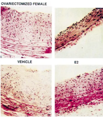
androgens on blood pressure and cardiovascular disease. There are reports of lower circulating testosterone [192– 194] and androstenedione [194] levels in hypertensive men [192–194], and circulating testosterone levels in men with coronary artery disease [195] or myocardial infarction [196] are either unchanged or decreased. These studies suggest that decreased, rather than increased, androgen levels are associated with hypertension, myocardial infarc-tion and coronary artery disease. An important caveat is that the lower testosterone levels observed in the afore-mentioned studies may merely reflect increased stress. Testosterone levels decrease in response to stress induced by recent myocardial infarction, surgery, head trauma, burns, hypoxia, sleep deprivation and psychological stres-sors [196]. Thus, the lower levels of testosterone in men with hypertension and cardiovascular disease may not indicate that testosterone reduces blood pressure and protects the cardiovascular system. In support of this conclusion is the low prevalence of coronary disease among men with hypotestosteronemia plus hyperes-trogenemia [195]. Also, women suffering from chronic anovulation and exhibiting hypertestosteronemia have an increased risk of coronary artery disease and myocardial infarction [196]. Moreover, men with testosterone de-Fig. 1. Representative photomicrographs of right common carotid arteries
ficiency following orchiectomy have a slightly lower from ovarectomized female Sprague–Dawley rats 14 days after balloon
injury. The rats were treated with vehicle, estradiol (E2), medroxyproges- mortality from heart disease [196], suggesting that lower terone (MPA) or E21MPA. The inhibitory effects of estradiol on testosterone may protect against cardiovascular disease. neointima formation were abrogated in the presence of the synthetic
In summary, clinical studies are needed to directly progestin, MPA (with permission from the American Heart Association).
define the effects of testosterone on the cardiovascular system in human subjects. Studies should be conducted in can abrogate the antivasoocclusive effects of estradiol men with Klinefelter disease who have testosterone de-following vascular injury ([190]; Fig. 1). ficiency and require testosterone supplementation. Results In summary, natural progesterone augments and some from these studies could provide data on the dose-depen-synthetic progestins abrogate the beneficial effects of dent effects of testosterone on cardiovascular function. estradiol on vascular tone and the vascular injury response. Studies in animals strongly suggest that testosterone is a Nonetheless, synthetic progestins may augment the anti- pro-hypertensive hormone. Blood pressure is higher in hypertensive effects of estradiol. The aforementioned male SHR, Dahl salt-sensitive rats, deoxycorticosterone studies teach that like estrogens, not all progestins have the acetate-salt hypertensive rats and New Zealand genetically same pharmacological effects. hypertensive rats compared to females [6–13]. The as-sociation between testosterone and high blood pressure in 3.3. Effects of progestins on vascular growth these animals is supported by the observation that castra-tion at a young age (3–5 weeks) attenuates the develop-Similar to estradiol, progesterone inhibits mitogen in- ment of hypertension in SHR and Dahl salt-sensitive rats duced growth and proliferation of cardiac fibroblasts [138], [6–9,14,15]. Furthermore, treatment of ovariectomized vascular smooth muscle cells [191] and glomerular normotensive females or castrated males with testosterone mesangial cells [106], which contribute to the vascular and increases blood pressure to levels similar to those in intact glomerular remodeling associated with hypertension, males [7,9], and testosterone increases blood pressure in atherosclerosis and glomerulosclerosis. Little is know ovariectomized female SHRs [7,9]. Moreover, irreversible regarding the effects of synthetic progestins on vascular increases in arterial blood pressure are observed in
nor-growth. motensive, uninephrectomized female rats treated
chroni-cally with testosterone [197,198].
Use of anabolic steroids that are synthetic derivatives of
4. Androgens and hypertension testosterone is temporally associated with hypertension, ventricular remodeling, myocardial ischemia and sudden 4.1. Effects of androgens on blood pressure cardiac death [199]. Moreover, in a double blind placebo-controlled study in 21 men, treatment with testosterone Relatively little is known regarding the influence of enanthate (3.5 mg / kg body weight) for 12 weeks
sig-nificantly increased systolic blood pressure by 10 mmHg In porcine coronary arteries, testosterone, progesterone [200]. In contrast, administration of testosterone to 20 male and estradiol induce concentration-dependent relaxation of subjects (mean age 70.666.2 years) to achieve physiologic both prostaglandin F -induced and KCl-induced contrac-2a
and superphysiologic serum testosterone concentrations did tion [208,209]. In contrast to estradiol, testosterone and not increase blood pressure [201], and no changes in blood progesterone have greater relaxing effects on vessels pressure were observed in men receiving testosterone precontracted with prostaglandin F . Estradiol inhibits2a
21
undecanoate (120 mg / day for 5 days; [202]). The different Ca entry, and progesterone and testosterone cause outcomes in the above studies may have been due to the coronary relaxation by inhibiting other contractile mecha-age group of the subjects tested (young vs. old) or the nisms [208,209]. Estradiol induces NO synthesis in endo-duration of treatment (acute vs. long-term). Importantly, thelial cells, and this effect is accompanied by transloca-the blood pressure elevating effects were observed in tion of membrane-associated eNOS [210]. In contrast to subjects who were undergoing regular weight training. In estradiol, neither progesterone nor testosterone modulates contrast, the subjects with no increase in blood pressure NO synthesis [210]. Testosterone improves post-exercise did not exercise. Therefore, it is possible that the effects of ST-segment depression in patients with angina testosterone on blood pressure are modulated by exercise. [196,211,212], and short-term administration of testoster-A number of mechanisms by which testosterone can one induces beneficial effects on exercise-induced myocar-elevate blood pressure and damage blood vessels have dial ischemia in men with coronary artery disease, an been elucidated. Testosterone increases circulating levels effect that may be due to the direct coronary-relaxing of homocysteine. Homocysteine induces endothelial dam- actions of testosterone [213].
age, thus leading to the development of atherosclerosis, In contrast to the direct vasodilator and vasorelaxing and may adversely influence renal function by damaging effects discussed above, testosterone may block the effects glomerular endothelial cells [120]. In contrast to estradiol, of other vasodilator agents. In this regard, coronary testosterone increases endothelin-1 levels in subjects un- vascular perfusion with testosterone blocks adenosine-me-dergoing a sex change, providing a humoral mechanism by diated vasodilation via a rapid non-genomic mechanism which it may elevate blood pressure [120]. Finally, in male [214]. Treatment of cultured cells with testosterone, but SHR, elevated levels of catecholamines are associated with not estradiol, significantly increases thromboxane A2 re-high blood pressure [203], and castration reduces catechol- ceptor density. This effect is inhibited by the testosterone amine levels to those observed in normotensive controls receptor blocker hydroxyflutamide, suggesting the in-[204], suggesting that testosterone can also directly induce volvement of androgen receptors. The testosterone pre-catecholamine synthesis [204]. This effect may be ex- cursor androstenedione also increases thromboxane A2
plained by the observation that testosterone stimulates receptor density [215,216]. Several lines of evidence tyrosine hydroxylase, the rate limiting enzyme for cat- support the functional significance of this androgen-in-echolamine synthesis [203,204]. Thus, testosterone may duced increase in thromboxane A receptor density. The2
elevate blood pressure and contribute to the pathogenesis aortas of male rats show increased (compared to females) of cardiovascular disease by altering a number of humoral contractile responses to thromboxane A2 [215]. Further, factors, including homocysteine, endothelin-1 and cat- testosterone enhances coronary artery constriction induced
echolamines. by a thromboxane A mimetic in vitro and in vivo [217].2
Thromboxane A2 is implicated as a risk factor for car-4.2. Effects of androgens on vascular tone diovascular diseases, and it is possible that the contrasting effects of estradiol and testosterone on thromboxane A2
Androgen receptors are expressed in vascular cells signaling may, in part, be responsible for some of their [108], and androgens may have a functional role in differential effects on the cardiovascular system.
modulating vascular tone. Testosterone relaxes precon- Testosterone has additional pro-hypertensive effects. In tracted rabbit coronary arteries and aortas in vitro, with or SHR, testosterone enhances the activity of tyrosine hy-without endothelium [108,205]. Short-term intracoronary droxylase, the rate limiting enzyme in norepinephrine infusions of testosterone dilate male and female canine synthesis, and the resultant increase in norepinephrine coronary arteries in vivo and increase coronary blood flow. levels may contribute to the development of hypertension These vasodilator effects of testosterone are largely me- in male animals with this form of genetically-determined
1
diated via ATP-sensitive K channels [206]. Via similar hypertension [204]. Moreover, testosterone up-regulates mechanisms, short-term intracoronary administration of the release of the potent vasoconstrictor neuropeptide Y physiological concentrations of testosterone induces cor- [218]. Testosterone also enhances, whereas estradiol at-onary artery dilatation and increases corat-onary blood flow tenuates, the contractile effects of endothelin-1 in porcine in men with established coronary artery disease [207]. coronary artery rings [219].
Androgen receptor blockers do not alter these rapid onset These findings indicate that testosterone can activate acute vasorelaxing effects of testosterone. Therefore, the mechanisms that cause both vasoconstriction and vasorela-acute vascular effects of testosterone are most likely xation. The balance between the vasodilating and vasocon-androgen-receptor independent and non-genomic. stricting effects of testosterone determines its net effect on
vascular tone and, perhaps, blood pressure. Thus, the 4.5. Effects of androgens on renal mechanisms inconsistent effects of testosterone on blood pressure
discussed above may be due to its variable effects on Reckelhoff and Granger have described several mecha-vascular tone, in part dependent on the pretreatment nisms by which androgens can induce renal injury and condition of the vasculature. precipitate hypertension [231]. Studies in rat models provide evidence that, compared with females, ageing males exhibit decreased glomerular filtration rate and 4.3. Effects of androgens on vascular growth and develop glomerular injury, glomerulosclerosis, proteinuria
atherogenesis and increased blood pressure [16,152,231]. Castration at an
early age attenuates renal injury and prevents the develop-There is evidence that testosterone can protect against ment of hypertension; whereas, administration of exogen-the response to vascular injury and exogen-the development of ous testosterone to castrated animals increases blood atherosclerosis [205,220,221]. In cholesterol fed rabbits pressure to levels similar to those found in intact males [221], testosterone is anti-atherosclerotic by a mechanism [7,152,231]. These findings suggest testosterone can ad-independent of changes in plasma lipids. Testosterone also versely influence renal function and blood pressure, protects against post-injury-induced plaque development eventually leading to hypertension.
[205]. Despite its anti-atherosclerotic effects in rabbits, The kidneys express androgen receptors [231,232], and testosterone does not inhibit smooth muscle cell prolifer- studies have examined whether the effects of testosterone ation [220]. Testosterone is also ineffective in inhibiting on blood pressure are androgen-receptor mediated. Ad-mitogen-induced smooth muscle cell migration [222] and ministration of flutamide, an androgen receptor antagonist, proliferation in vitro [157,223]. These findings suggest that to intact male SHRs lowers blood pressure to levels found the vasoprotective effects of testosterone are not mediated in female or castrated male SHRs [232], suggesting that by inhibition of vascular smooth muscle cell growth. the effects of androgens are receptor mediated. Further-In contrast, a number of other observations suggest that more, sodium-induced increases in blood pressure in testosterone is pro-atherosclerotic. Testosterone: (1) ex- Wistar–Kyoto and SHR are suppressed by androgen-re-acerbates diet-induced atherosclerosis and has adverse ceptor blockade [233], confirming the androgen-receptor effects on atherosclerosis-related arterial remodeling in dependence of salt sensitive hypertension in this animal female monkeys [224]; (2) induces vascular plaque forma- model.
tion in chicks [225]; (3) increases monocyte adhesion to Disruption of the CYP4A14 gene (arachidonic acid v-1-vascular endothelial cells and increases the expression of hydroxylase) in mice results in hypertension that is more vascular cell adhesion molecule-1 in humans [226]; (4) severe in males and is androgen sensitive, i.e., increases in lowers HDL and raises LDL [227]; (5) up-regulates the blood pressure are reduced upon castration and restored release of neuropeptide Y [219] which can induce pro- upon testosterone supplementation [234]. Renal vascular atherosclerotic actions and local vasoconstriction; (6) has a resistance is dramatically increased in these mice, sug-pro-mitogenic effect on vascular smooth muscle cells gesting that lack of functional CYP4A14 in the kidney [228]; and (7) increases the synthesis of Ang II, a may have several interrelated metabolic and regulatory vasoconstrictor and mitogen known to induce hypertension effects whose functional manifestations are increased renal and atherosclerosis [6]. Thus, whether testosterone inhibits vascular resistance, impaired renal hemodynamics and or accelerates atherosclerosis appears to be model depen- hypertension. It has been suggested that the final mediator
dent. for the blood pressure enhancing effects of androgens in
CYP4A14 knockout mice is 20-HETE [234], which is known to alter renal hemodynamics and function. 4.4. Effects of androgens on cardiac mechanisms Another key mechanism by which androgens tilt the
cardiovascular system toward a pro-hypertensive state is Testosterone and its metabolite dihydrotestosterone by shifting the pressure-natriuresis relationship to the right (DHT) induce cardiac hypertrophy in part by activating [149,150,152]. At comparable renal perfusion pressures, androgen receptors that are expressed in cardiac myocytes intact SHR males and ovariectomized females receiving [229]. In baroreceptor-denervated rats, left ventricular chronic testosterone treatment excrete significantly less hypertrophy is gender-dependent; estradiol inhibits, where- sodium and water than intact females, ovariectomized as testosterone stimulates, cardiac hypertrophy [230]. females or castrated males [7,231]. These effects may be Moreover, in vitro studies provide evidence that androgens mediated by the renin–angiotensin system because testo-induce hypertrophic growth in cultured myocytes [229], sterone increases plasma renin activity (PRA) [7,152,231], suggesting that the growth promoting effect is direct. The castration of male rats decreases PRA, and administration hypertrophic effects of DHT, but not testosterone, are of testosterone to ovariectomzied female rats increases associated with increased synthesis of atrial natriuretic PRA [7,152,231]. The effect of testosterone on PRA is peptide [229], suggesting that these androgens may induce dose-dependent [7,152,231]; suggesting that the blood hypertrophy via different mechanisms. pressure regulating effects of testosterone may be
con-centration-dependent [7,152,231]. Angiotensinogen mRNA Findings from our laboratory provide evidence that levels are also higher in male rats than in females, and sequential metabolism of estradiol to catecholestradiols castration reduces these levels [235,236]. Thus, animal and ultimately methoxyestradiols is responsible for the studies provide convincing evidence that testosterone anti-mitogenic effects of estradiol on vascular smooth diminishes the ability of the kidneys to excrete salt, and muscle cells [238], cardiac fibroblasts [239] and glomeru-thereby predisposes to hypertension. Whether this occurs in lar mesangial cells [240]. Importantly, these effects of
humans is unknown. estradiol on cell growth appear to be ER-independent
[92,238]. Increased proliferation of these cell types leads to hypertension, vascular disease, left ventricular hypertrophy
5. Gender independent factors and role of hormone and glomerulosclerosis. Thus, some of the cardiovascular
metabolism and renal protective effects of both testosterone and estradiol may be mediated via their conversion to methox-Both estradiol and testosterone are present in both sexes, yestradiols, which have anti-mitogenic effects on multiple albeit in different concentrations and ratios [196]. Endog- cell types (Fig. 2). The importance of estradiol metabolites enous androgens (dehydroepiandrosterone, andros- in vasoprotection is further supported by our finding that in tenedione, and testosterone) are readily converted to es- male obese ZSF1 rats that exhibit the metabolic syndrome tradiol by the sequential actions of 17b-hydroxysteroid (i.e., hypertension, obesity, diabetes and hyperlipidemia) dehydrogenase (17b-HSD) and aromatase. Thus, some of and have left ventricular, renal and vascular dysfunction, the beneficial effects of testosterone observed in males treatment with 2-hydroxyestradiol decreases body weight, may be due to its conversion to estradiol and estradiol improves vascular endothelial function, decreases nephro-metabolites. This hypothesis is supported by the recent pathy, exerts antidiabetic actions and lowers blood pres-finding that the inhibitory effects of dehydroepiandros- sure and blood cholesterol [241].
terone, a precursor of androstenedione, on atherosclerosis The hypothesis that estradiol metabolites are responsible are blocked by the aromatase inhibitor fadrozole [237]. for the anti-mitogenic effects of estradiol is supported by This finding suggests that the sequential conversion of several recent findings. Estradiol prevents neointima for-androgens to estradiol is responsible for their anti-atheros- mation in mice lacking functional ER-a and ER-b, sug-clerotic actions, but whether estradiol is the ultimate gesting that the protective effects of estradiol on the mediator remains unclear. cardiovascular system may be ER independent. Estradiol
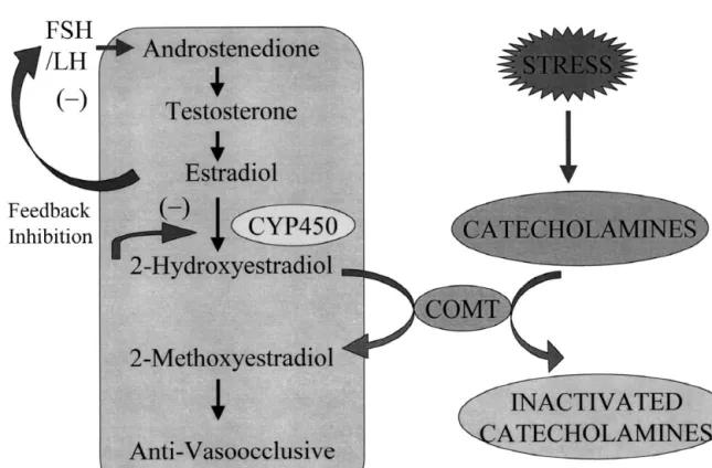
Fig. 2. A schematic representation delineating the possible mechansims via which pathological stimuli such as stress can influence endogenous levels of sex hormones. Catechol-O-methyltransferase, COMT; cytochrome P450, CYP450; follicle stimulating hormone, FSH; luteinizing hormone, LH; inhibition (–).
production of 2-methoxyestradiol may be seriously com-prevents neointima formation in gonadectomized, but not
promised under pathological conditions associated with intact male rats, even though male rats express ER-a and
increased release of catecholamines. As illustrated in Fig. ER-b. In female rats the inhibitory effects of estradiol are
2, under normal conditions, testosterone is metabolized by abrogated by medroxyprogesterone, a synthetic progestin
aromatase to estradiol; estradiol is metabolized by hy-[190]. Since both androgens and medroxyprogesterone are
droxylases (CYP1A1, CYP1A2 and CYP1B1) to cate-potent inhibitors of the enzyme responsible for the
forma-cholestradiols (e.g., 2-hydroxyestradiol), and catecholes-tion of 2-hydroxyestradiol ([242], Dubey et al. unpublished
tradiols are metabolized by COMT to methoxyestradiols data), these steroids may abrogate the anti-vasoocclusive
[245], which induce anti-vasoocclusive effects [238,244]. effects of estradiol by blocking its metabolism to
hydroxy-However, when catecholamine release is increased, cat-estradiols and methoxycat-estradiols. This hypothesis merits
echolamines compete with catecholestradiols and inhibit further direct testing.
their metabolism to methoxyestradiols. This results in Lifestyle factors, such as dietary habits and stress, may
increased accumulation of catecholestradiols, inhibition of participate in the development of cardiovascular diseases.
hydroxylases and therefore accumulation of estradiol. Since life style factors can interfere with estradiol
metabo-Since estradiol inhibits testosterone synthesis by negatively lism, it is feasible that they may induce their adverse
regulating FSH / LH synthesis [246], the net result is a effects in part by preventing methoxyestradiol formation.
decrease in levels of testosterone. The general principle In this regard, it is important to note that metabolism of
illustrated by this hypothesis is that the metabolism of 2-hydroxestradiol to 2-methoxyestradiol is mediated by
estradiol and testosterone may play a critical role in catechol-O-methyltransferase (COMT), the enzyme that
determining the overall effects of sex hormones on the metabolizes catecholamines to methoxy metabolites. Our
cardiovascular system. recent studies show that catecholamines can abrogate the
anti-mitogenic effects of estradiol and 2-hydroxyestradiol on vascular smooth muscle cells, cardiac fibroblasts and
6. Conclusion
glomerular cells, and that these effects are associated with the inhibition of 2-methoxyestradiol formation [243,244].
Compared to men and postmenopausal women, pre-Although catecholamines may not normally interfere
menopausal women are relatively protected against hy-with the metabolism of estradiol to 2-methoxyestradiol,
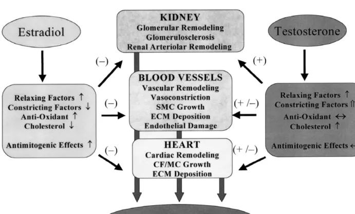
Fig. 3. A schematic representation summarizing and comparing the potential mechansims via which estradiol and testosterone can influence blood pressure and be associated with preventing or inducing hypertension. Smooth muscle cells (SMC); cardiac fibroblast (CF); cardiac myocytes (MC); extracellular matric synthesis (ECM); inhibition, (–); induction (1); increased (↑); decreased (↓); highly increased (⇑); neutral effects (↔).