Cell Tissue Res (2006) 323: 395–404 DOI 10.1007/s00441-005-0112-1
R E G U L A R A RT I C L E
Katharina Matschke . Luis Da Silva-Azevedo . Ruslan Hlushchuk . Valentin Djonov . Oliver Baum
Annexins as cell-type-specific markers in the developing
chicken chorionallantoic membrane
Received: 8 October 2005 / Accepted: 30 October 2005 / Published online: 13 December 2005 # Springer-Verlag 2005
Abstract Between day E8 and E12 of embryonic devel-opment, the chicken chorioallantoic membrane (CAM) undergoes massive structural rearrangement enabling cal-cium-uptake from the eggshell to supply the growing embryo. However, the contribution of the various cell types of the chorionic epithelium including the capillary covering (CC) cells, villus cavity (VC) cells, endothelial-like cells, and basal cells to this developmental program is largely unknown. In order to obtain markers for the different cell types in the chorionic epithelium, we determined the expression patterns of various calcium-binding annexins in the developing chicken CAM. By reverse transcription/ polymerase chain reaction with primers deduced from nucleotide sequences available in various databases, the presence of annexin (anx)–1, anx–2, anx–5, and anx–6 was demonstrated at days E8 and E12. Quantitative immuno-blotting with novel antibodies raised against the recombi-nant proteins revealed that anx–1 and anx–5 were significantly up-regulated at day E12, whereas anx–2 and anx–6 expression remained almost unchanged in compar-ison to levels at day E8. Immunohistochemistry of paraffin-embedded sections of E12 CAM revealed anx–1 in CC cells and VC cells. Anx–2 was localized in capillaries in the chorionic epithelium and in basal cells of the allantoic epithelium, whereas anx–6 was detected in basal cells or endothelial-like cells of the chorionic epithelium and in the media of larger vessels in the mesenchyme. A 2-day exposure of the CAM to a tumor cell spheroid resulted in
strong proliferation of anx–1-expressing CC cells suggest-ing that these cells participate in the embryonic response to experimental intervention. Thus, annexins exhibit comple-mentary expression patterns and represent appropriate cell markers for the further characterization of CAM develop-ment and the interpretation of results obtained when using CAM as an experimental model.
Keywords Chorioallantoic membrane . Calcium resorption . Annexins . Chicken (Brown Leghorn)
Introduction
The chicken chorioallantoic membrane (CAM) is widely used as an experimental model. It has been employed in, for example, tests of the biocompatibility/toxicology of organic and inorganic materials (Spanel-Borowski 1989; Valdes et al.2002), assessments of the ability of bacterial strains to invade epithelial barriers (Adam et al. 2002), determinations of the metastatic and developmental growth potencies of grafted tumor and embryonic tissue samples (Navarro et al. 2003), and the quantitative analysis of the angiogenic response to a defined stimulus (for reviews, see Ribatti et al.2001; Richardson and Singh2003).
The CAM is an extra-embryonic tissue formed by fusion of the chorion and allantois during early avian development in the egg (Patten1950) and consists of three layers: (1) the chorionic epithelium of ectodermal origin, which is in direct contact with the shell membrane, (2) the intermediate mesenchyme consisting of both ecto- and endodermal components, and (3) the endodermal allantoic epithelium facing the allantoic cavity containing urine. The membrane is important for three biological processes (Bellairs and Osmond1998). Because its surface is in close contact to the porous shell membrane, the CAM enables the respira-tory interchange of oxygen and carbon dioxide. Further-more, it is involved in the storage of urea, ammonia, and uric acid produced by the embryo. Finally, Ca2+is taken up to a greater extent from the shell through the chorionic epithelium to be transported into the vasculature of the K. Matschke . R. Hlushchuk . V. Djonov . O. Baum (*)
Institute of Anatomy, University of Berne, Baltzerstrasse 2, CH-3009 Berne 12, Switzerland e-mail: [email protected]
Tel.: +41-31-6313445 Fax: +41-31-6313807 L. Da Silva-Azevedo Institute of Physiology,
Charité-Campus Benjamin Franklin, Free University Berlin,
embryo, which initially obtains its calcium until day E10 from the yolk sac. At this time point, the Ca2+availability in the chorionic epithelium is suddenly strongly increased, which might be correlated with changes in the expression patterns and functions of Ca2+-binding proteins.
Previous studies have demonstrated that both epithelial layers of the CAM consist of distinct cell types. Villus cavity (VC) cells and capillary covering (CC) cells are the two major components of the chorionic epithelium (Coleman and Terepka 1972). Both cell types pass through the chorionic epithelium to face the eggshell and are involved in Ca2+ mobilization and resorption (Akins and Tuan
1993). Two other cell types present in the chorionic epi-thelium layer are the basal cells (Hoshi and Mori 1971; Lusimbo et al.2000) and the endothelial-like (or pericyte-like) cells (Coleman and Terepka1972; Patan et al.1993; Shumko et al. 1988), which are functionally uncharacter-ized to date. Furthermore, capillaries are in close contact with the chorionic epithelium (Narbaitz 1977). The allan-toic epithelium consists of three different cell types in-cluding granule-rich cells, mitochondria-rich cells, and basal cells (Coleman and Terepka1972).
Despite the extensive use of the CAM system in cell biology, most investigators regard the CAM as a uniform structure. In particular, in angiogenesis research, investiga-tions are restricted to determinainvestiga-tions of the quantitative changes within the microvasculature in response to a defined stimulus. The significance of non-specific inflam-matory reactions or experiment-independent proliferation (particularly of the chorionic epithelium in which the capillaries are embedded) is rarely addressed and might lead to the misinterpretation of results (Richardson and Singh 2003). To extend our knowledge of this cellular layer, we have performed a structural characterization of the chorionic epithelium of the developing chicken CAM by focusing on the expression patterns of the various annexins.
The annexins are a family of closely related Ca2+ -binding and membrane--binding proteins expressed in most eukaryotic cell types (for reviews, see Gerke and Moss
2002; Moss and Morgan2004). So far, 12 members of the annexin gene family have been detected in mammals; they are involved in many physiological processes, such as the regulation of cell growth and differentiation, apoptosis, membrane fusion, and exocytosis. They are also involved in cell signaling events and anti-inflammatory processes. In many tissues, the different annexin forms exhibit comple-mentary expression patterns (Raynal and Pollard 1994), whereas in other cases, the annexins are co-expressed and cooperate in function (Draeger et al.2005).
With novel antibodies, we have determined the expression patterns of four annexins in the developing chicken CAM. We have found that these annexins are specific for the different cell types of the chorionic epi-thelium, suggesting their involvement in different cellular functions.
Materials and methods
Tissue preparation
The shell-free culture method for chicken embryos was used to obtain CAM as described by Djonov et al. (2000). After 3 days of incubation, eggs from Brown Leghorn hens were opened and carefully transferred into plastic Petri dishes (8 cm in diameter). The embryos were incubated at 37°C in a humid atmosphere.
CAMs were collected after the time points indicated. The CAM tissue overlying the embryo was mechanically dissected, washed three times in ice-cold phosphate-buffered saline (PBS) pH 7.4 to remove leaking blood and extravascular fluid, and then quickly subjected to biochemical and histological analyses as described below.
RNA isolation and reverse transcription/polymerase chain reaction
Total mRNA was prepared from embryonic day 8 (E8) and E12 chicken CAMs by using the RNeasy Mini kit (Qiagen, Basel, Switzerland), including proteinase K digestion and DNase I treatment according to the manufacturer’s (Qiagen) protocol. The final mRNA concentrations thus obtained were photometrically determined. cDNA was synthesized by reverse transcription (RT) with 2μg total RNA, 1 μM oligo-dT primers, and 4 U Omniscript reverse transcriptase (Qiagen) at 37°C for 1 h according to the manufacturer’s protocol. For the assessment of annexin mRNA expression, cDNA was analyzed by the polymerase chain reaction (PCR) with pairs of primers specific for each of the various chicken annexins (Table 1). In a total volume of 50 μl, 200 ng cDNA template was mixed with 5 μl 10× Taq-polymerase buffer (Qiagen), primers, and dNTP solution at a concentration of 0.2μM, with 2 mM MgSO4. During the
initial denaturing step at 94°C for 4 min, 0.5μl 5 U/μl Taq DNA polymerase (Qiagen) was added (hot start) to the mixture. The cycle profile consisted of a denaturation step at 94°C (5 min) followed by 40 cycles with denaturation at 94°C (30 s), annealing at 58°C (30 s), and elongation at 72°C (90 s).
PCR fragments were visualized by electrophoresis on a 1% (w/v) agarose (Axon, Baden-Dättwil, Switzerland) gel supplemented with 0.5μg/ml ethidium bromide.
Rapid amplification of 3’-cDNA ends
Because the nucleotide sequence of anx–1 was only partially published (comprising the amino-terminal 130 amino acids) at the initiation of this study (Sidis and Horseman1993), we performed rapid amplification of 3 ’-cDNA ends (3’-RACE) for determination of the complete coding sequence of annexin–1 cDNA. Aliquots with 1 μg
total RNA from chicken CAM at E12 were reverse-transcribed in cDNA with Omniscript RT (Qiagen) by using the T17 adapter primer (Table 1) at 37°C for 1 h. Subsequent 3'-RACE was carried out with adapter primer and anx–1–specific forward primers (Table 1). The PCR protocol comprised the following steps: 94°C for 5 min (hot start) followed by 40 cycles with denaturation at 94°C (30 s), annealing at 55°C (60 s), and elongation at 72°C (120 s).
Four different bands were obtained and were cloned into the vector pET-28a (Novagen, Madison, Wis., USA). Plasmid DNA was extracted from bacterial cultures to sequence the inserts by using T17 adapter primer. The plasmid containing the anx–1 insert was transformed into the expression strain (Escherichia coli BL21), and its expression was induced with isopropyl beta-D-thiogalac-topyranoside. The nucleotide sequence obtained for chicken anx–1 was transferred to the NCBI database (accesion no. AY549497).
Preparation of polyclonal antibodies
Recombinant chicken anx–1 and anx–6 proteins and human anx–5 protein were used for the induction of polyclonal antibodies in rabbits. The recombinant proteins were purified from E. coli BL21 cultures by using the expressed His-tag. Frozen pellets were thawed on ice, suspended in lysis buffer (50 mM NaH2PO4, 10 mM
imidazole, 300 mM NaCl, adjusted to pH 8.0), and sonicated. After centrifugation (10,000g for 30 min), the supernatant was incubated with a suspension of Ni-NTA agarose beads (Qiagen) for 1 h at 4°C. After three washes in washing buffer (50 mM sodium phosphate pH 8.0, 300 mM NaCl, 20 mM imidazole), the protein was eluted
in elution buffer (50 mM sodium phosphate pH 8.0, 300 mM NaCl, 250 mM imidazole). Protein concentration was measured by using a BCA protein assay reagent (Pierce, Rockford, Ill., USA), and protein purity was checked by SDS-polyacrylamide gel electrophoresis (PAGE).
Rabbits were immunized by three subcutaneous injec-tions (1× immunization and 2× booster) of 100 μg recombinant protein mixed 1:1 with GERBU Adjuvans (Gerbu, Gaiberg, Germany). Antisera were obtained 2 weeks after the second booster injection.
The preparation and characterization of the polyclonal anti-human anx–2–antibody was as described earlier (Babiychuk et al.2000).
SDS-PAGE and immunoblotting
For the immunoblot detection of annexins, chicken CAM tissue samples were homogenized at 4°C in PBS pH 7.4, containing 1% (v/v) Triton X-100 and 0.1% (v/v) standard protease inhibitor mixture (Sigma, Munich, Germany) containing 10μM E-64, 40 μM bestatin, 20 μM leupeptin, 1μM aprotinin, 2 mM EDTA, and 1 mM 4-(2-aminoethyl) benzenesulfonyl fluoride (final concentrations). Insoluble material was removed by a centrifugation step in a bench centrifuge at 13,000 rpm for 3 min. Subsequently, each homogenate (50μg protein) was subjected to SDS-PAGE (BioRad, Munich, Germany) on 12.5% gels and transferred to nitrocellulose membrane filters. After being blocked with 5% (w/v) fat-free milk powder in washing buffer consisting of 0.1% (v/v) Tween 20 in PBS, the blot matrices were incubated with the antibodies specific for anx–1 (1:5,000), anx–2 (1:2,000), anx–5 (1:2,000), and anx–6 (1:5,000) overnight at 4°C and, subsequently, for 1 h Table 1 Primers used for PCR
analyses Specificity PCR application Nucleotide sequence Annexin–1 Forward primer 5'–ATATCCCATGGCTATGGTATCAGAATTTCTG–3' Reverse primer 5'–ATATACTCGAGTCTTCTATCAGTTCCAAGCC–3' Annexin–2 Forward primer 5'–CGATGCCATGGCTACTGTCCATGAAATTTTA–3' Reverse primer 5'–CTATACTCGAGGTCCTCTCCACCACACAGGTT–3' Annexin–5 Forward primer 5'–CGATGCCATGGCGAAGTATACAAGAGGCA–3' Reverse primer 5'–CTATACTCGAGCTCATCATCTCCACCACAGAG–3' Annexin–6 Forward primer 5'–ATATCCCATGGCACCCAAAGGAAAGGTTTAC–3' Reverse primer 5'–ATATACTCGAGGTCGTCCCCCCCGCACC–3' T17 adapter primer (T17AP) 5' –GACTCGAGTCGACATC-GATTTTTTTTTTTTTTTTT–3' Adapter primer (AP) 5'–GACTCGAGTCGACATCG–3'
with a 1:5,000 dilution of peroxidase-conjugated anti-rabbit secondary antibody (Sigma). Bound antibodies were detected with an enhanced chemiluminescence detection kit (Supersignal, Pierce) as described by Baum et al. (2004).
Two-dimensional PAGE
E8 and E12 chicken CAMs were solubilized in 1% (w/v) SDS in twice-distilled water for 1 h on ice. Insoluble material was removed by centrifugation at 20,000 g for 15 min. The supernatants were subjected to acetone-precipitation with 50% (v/v) acetone cooled to–20°C. The protein pellets obtained were re-dissolved in isoelectric focusing (IEF) sample buffer consisting of 7 M urea, 2 M thiourea, 2% (w/v) CHAPS, 1% (w/v) dithiothreitol (DTT), and 2% (v/v) ampholytes in twice-distilled water, i.e., 60μg protein in a total volume of 300 μl IEF sample buffer was used for re-hydration of 17-cm-long immobilized pH-gradient (IPG) strips covering the pH range of 3–10 (BioRad, München, Germany) overnight. IEF was per-formed at 20°C in a Multiphor II chamber (Pharmacia) at 53,250 Vh in total (500 V for 5 h, 3,500 V for 14.5 h).
After IEF, the IPG strips were stored at –70°C until further use or were directly incubated in equilibration buffer consisting of 6 M urea, 30% (v/v) glycerol, 2% (w/v) SDS in 0.05 M TRIS-HCl pH 8.8. During the first 15-min incubation period, the equilibration buffer was supplement-ed with 1% (w/v) DTT, which was replacsupplement-ed with 2% (w/v) iodoacetamide during the second 15-min incubation period. The electrophoretic separation of proteins in the second dimension was performed on 12.5% polyacrylamide gels overnight at 6 mA per gel by using the Protean II-system (BioRad, München, Germany).
After SDS-PAGE, gels were either subjected to immuno-blotting or silver-stained. For silver-staining, gels were first fixed in 50% (v/v) methanol, 5% (v/v) acetic acid in distilled water for 20 min and then washed with twice-distilled water overnight. After reduction in sensitizer solution consisting of 0.02% (w/v) Na2S2O3×5 H2O in
twice-distilled water for 1 min, the gels were washed with twice-distilled water (2×1 min) and then incubated in 0.1% silver nitrate in twice-distilled water for 20 min at 4°C. Protein patterns were obtained after short washes in twice-distilled water for 1 min and three incubations in developer consisting of 0.04% (v/v) formaldehyde, 2% (w/v) Na2CO3
in twice-distilled water, 5 min each. The staining reaction was stopped by incubation in 5% (v/v) acetic acid in twice-distilled water.
Immunohistochemistry
The embedding of chicken CAM samples in paraffin was performed as described previously (Djonov et al. 2000). After being blocked with 1% (w/v) casein in TRIS-buffered saline (TBS) for 10 min, sections were incubated with the primary antibody (diluted in TBS) at 4°C overnight. The
following antibodies and dilutions were used: anti-anx–1 (1:500), anti-anx–2 (1:500), anx–6 (1:1,000), and anti-sma (alpha-smooth muscle actin; Sigma; 1:200).
Reactions were developed with an avidin-biotin horse-radish-peroxidase complex (Dako, Glostrup, Denmark) and 3–amino–9–ethylcarbazole (Sigma, Buchs, Switzer-land) as the substrate as described earlier (Baum et al.
2005). The sections were then counter-stained with hema-toxylin and cover-slipped in Aquatex (Merck, Darmstadt, Germany). Negative controls were prepared by using non-specific mouse and rabbit sera.
Electron microscopy
For the preparation of sections for electron microscopy, CAM areas were excised and stored in Karnowsky solution consisting of 2.5% (v/v) glutaraldehyde, 2% (v/v) paraform-aldehyde in 0.1 M sodium cacodylate (adjusted with HCl to pH 7.4) at 4°C for several days. The generation of 1 –μm-thick semithin sections and 80–nm to 90-nm ultrathin sections for ultrastructural analysis was performed as reported by Baum et al. (2005).
Generation of spheroids and their grafting onto chicken CAMs
Spheroids as three-dimensional cell aggregates used for grafting experiments were prepared as described (Korff and Augustin 1998). To obtain spheroids, 10,000 trypsinated human breast cancer MDA-MB-231 cells were mixed with 32μl DMEM containing 10% (v/v) fetal calf serum and 8 μl methocel sock solution. The cell suspension was pipetted as drops onto the cover of a Petri dish and then incubated at 37°C in a humid 5% CO2atmosphere. After a
2-day incubation, the spheroids were carefully grafted onto the surface of E10 CAMs and then incubated at 37°C in a humid atmosphere for 3 days.
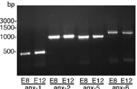
Fig. 1 RT-PCR for the detection of annexin mRNA in chicken CAM. mRNA was isolated from E8 and E12 chicken CAM and reverse-transcribed into cDNA. The presence of the various annexin cDNAs in the CAM samples was shown by PCR with primers specific for each annexin. Calculated sizes for the PCR products were 474 bp for anx–1, 1019 bp for anx–2, 982 bp for anx–5, and 1265 bp for anx–6
Fig. 2 Annexin–1, –2, –5, and –6 are expressed as proteins in the chicken CAM. SDS-PAGE and subsequent immunoblotting of equal amounts of homogenate proteins revealed that anx–1 (a), anx–2 (b), anx–5 (c), and anx–6 (d) are expressed in E8, E10, and E12 CAM at the protein level. Antibody against anx–1 recognized three different bands (38 kDa, 36 kDa, and 30 kDa; a), whereas each of the antibodies against anx–2 (38 kDa), anx–5 (36 kDa), and anx–6 (75 kDa) were mono-specific for one band (b–d). For further discrimination of anx–1 and anx–2 immunoreactivity, immunoblot-ting was performed on CAM E12 samples after two-dimensional
PAGE and demonstrated that the anx–1 antibody bound to anx–1 and anx–2 and to two smaller proteins (f), whereas the antibody against anx–2 was almost specific for anx–2 (g). e Silver-staining of a gel in which the proteins of the CAM day E12 were separated by two-dimensional PAGE showing the area of the immunoblots in insets f, g. Note the variable expression of the bands after SDS-PAGE (a–d) demonstrating that the concentrations of anx–1 and anx–5, but not of anx–2 and anx–6, are strongly up-regulated during CAM development at E8–E12
Fig. 3 Annexins are localized in various cell types in the chicken CAM. Immunohisto-chemistry on paraffin sections of E12 CAM localized anx–1 to a subset of chorionepithelial cells located apically and laterally in the capillaries (a). Immunoreac-tivity for anx–2 was clustered in capillaries in the chorionic epithelial layer and in basal cells of the allantoic epithelial layer (b), whereas anx–6 was detected in the middle part of the chori-onic epithelial layer and in the media of larger arteries and veins (c). Smooth muscle cell actin (sma) as a control was exclusively expressed in the media of larger feeding arteries (d). Note that the anx–1, anx–6, and sma immunohistochemistry was performed on serial sec-tions. Insets Erythrocytes (ar-rows) in capillaries. Bars 100μm (d), 20 μm (inset in a)
Results
The presence of annexins in the developing chicken CAM was first investigated by RT-PCR. This analysis was performed with primers specific for anx–1, anx–2, anx–5, and anx–6, since chicken cDNA sequences were available for these annexins in public databases (SwissProt, NCBI) at
the initiation of the study. As shown in Fig.1, RT-PCR on samples of CAM at E8 and E12 resulted in the amplifi-cation of a single DNA fragment for each of these four annexin cDNAs investigated. In all cases, the sizes of the DNA fragments obtained corresponded to the sizes calcu-lated. These data indicated that anx–1, anx–2, anx–5, and anx–6 were expressed in the chicken CAM at E8 and E12. Fig. 4 Electron-microscopic analysis of ultrathin sections of E12
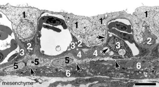
CAM revealed five different cell types in the chorionic epithelium of the CAM. Cytoplasm-rich cells with large nuclei representing CC cells were found apically and between the capillaries (1). Darker VC cells (2) were also interspersed between capillaries consisting of
endothelial cells (3) separated by a basement membrane (arrows) from endothelial-like cells (4) in close contact to basal cells (5). Filipodia-containing cells (6) accumulated in the mesenchyme under a second basement membrane (arrows). Bar 10μm
Fig. 5 Anx–1-expressing CC cells proliferate at the chorion-epithelial layer in response to grafted tumors. a, b Immuno-histochemistry on serial sections demonstrated that the chorionic epithelium of E12 CAM had significantly proliferated (arrows prominent chorionic epithelial cells) after a 2-day exposure to a grafted tumor spheroid (T). Most of the chori-onic epithelial cells expressed anx–1 (a) and only a few anx–6 (b). c–e Further ultrastructural analysis showed that the apical (c), central (d), and basal (e) sites of the enlarged chorionic epithelium were mainly com-posed of cytoplasm-rich cells with large nuclei (1 presumed CC cells) and capillary endo-thelial cells (3). Occasionally, degenerated cells with apoptotic depositions were observed (2). Bars 20 μm (b), 10 μm (e)
The analysis of the annexin expression patterns was continued at the protein level by using antibodies. Because chicken anx–5 and anx–2 both show a high sequence similarity to their human counterparts, we used polyclonal antibodies against human anx–5 and porcine anx–2. We prepared polyclonal antibodies against chicken recombi-nant anx–1 and anx–6. Since, however, the anx–1 nucle-otide sequence had only partially been published, we first performed 3' RACE to obtain a clone containing the com-plete coding sequence of chicken anx–1. Comparison of the cDNA deduced primary structures revealed that chicken anx–1 contained 342 amino acids with a sequence similarity of 76% to human anx–1. Furthermore, amino acid sequence analysis showed that chicken anx–1 was 69% similar to chicken anx–2, 58% to chicken anx–5, and 59% to chicken anx–6.
With the antibodies against the four annexins, immuno-blotting after SDS-PAGE was performed on detergent extracts of E8, E10, and E12 CAMs (Fig. 2a–d). In these samples, the antibodies against anx–2 (band at 38 kDa), anx–5 (band at 36 kDa), and anx–6 (band at 75 kDa) each recognized only one band. In contrast, the anti-anx–1 antibody bound to three bands (38 kDa, 36 kDa, and 30 kDa). The gels were loaded with identical amounts of proteins to render the quantitative expression patterns of the annexins investigated. During CAM development between days E8 and E12, both anx–1 and anx–5 were strongly up-regulated, whereas the anx–6 message was only slightly elevated. The expression of anx–2 was apparently not changed. Control immunoblotting experiments revealed that all annexins were solubilized by Triton X-100 because no immunoreactive bands were detectable in insoluble pellets after being boiled in 1% SDS (data not shown).
Because the size of the anx–1 band detected in immunoblots resembled that of anx–2, we continued to characterize the specificity of these antibodies by immu-noblotting after two-dimensional PAGE (Fig. 2e–g). As shown in Fig. 2f, the antibody against anx–1 recognized four proteins in the two-dimensional immunoblot: two 38-kDa spots with slightly differing pI (6.9 and 7.1), a 36-38-kDa spot, and a 30-kDa spot. The anti-anx–2 antibody bound only to the two 38-kDa spots, with higher specificity for the more acidic protein (Fig.2g). Based on calculations that the pI of anx–1 is 7.1 and that of anx–2 is 6.9, the anti anx–1 antibody recognized anx–1 and anx–2, whereas the anti-anx–2 antibody was almost specific for anx–2.
Immunohistochemistry with anti-annexin antibodies was performed on paraffin-embedded sections of E12 CAM. As shown in Fig. 3, anx–1 immunoreactivity was restricted to a subset of cells in the chorionic epithelium located at apical and lateral sites of the capillaries. Immunoreactivity for anx–2 was found at two sites: in capillaries in the chorionic epithelial layer and in basal cells of the allantoic epithelial layer. Anx–6 was detected in the middle part of the chorionic epithelial layer (below capillaries) and in the media of larger arteries and veins, whereas smooth muscle cell actin (used as a control) was exclusively expressed in the media of larger feeding arteries. The anx–5 antibody only functioned on
immuno-blots and did not produce staining in immunohistochem-istry (data not shown).
Electron-microscopic analysis of ultrathin sections of E12 CAM revealed that the chorioepithelial layer was composed of five different cell types (Fig.4). Apical to and lateral of the capillaries, cytoplasm-rich cells with large nuclei representing CC cells were found at the ultrastruc-tural level. VC cells were recognized as dark cells, although they were cut only basally in many cases. These cells were interspersed regularly between capillaries consisting of thin endothelial cells. Separated by a basement membrane, endothelial-like cells lay in close contact to the capillaries and also to the cytoplasm-rich basal cells in the lowest layer of the chorionic epithelium. Filipodia-containing cells were observed below a second basement membrane and there-fore belonged to the mesenchyme.
Tumor cell spheroids grafted onto the CAM provoked a strong defense reaction detected as significant thickening of the chorionic epithelium (Fig. 5) compared with the non-stimulated CAM (Figs. 3, 4). Immunohistochemistry was performed on serial paraffin-embedded sections from an E12 CAM after a 2-day exposure to tumor cell spheroids (Fig.5). Most of the chorionic epithelial cells expressed anx–1, whereas only a few cells were anx–6-immunoreactive. Fur-thermore, electron-microscopic analysis showed that the thickened chorionic epithelial layer was mainly composed of CC cells. Single cells of unknown origin that clearly had undergone apoptosis were also regularly observed.
Discussion
In the present study, we have determined the expression patterns of four annexins (anx–1, anx–2, anx–5, and anx–6) in the developing chicken CAM between E8 and E12. At this time, the chorionic epithelium of the chicken CAM undergoes massive structural rearrangement. The originally bilayered chorionic epithelium differentiates and prolifer-ates forming a multilayer that subsequently lies in close contact with the acellular inner shell membrane. This spatial proximity allows the cells of the chorionic epithe-lium to mobilize Ca2+ from the eggshell to supply the growing embryo (Akins and Tuan1993). Simultaneously, the capillary network expands, first by sprouting and then by intussusceptive growth to form, at E10, a dense plexus that finally invaginates into the chorionic epithelium (Djonov et al.2003; Patan et al.1993). This movement of the capillary system and the simultaneous thinning of the endothelial cells greatly reduce the thickness of the diffusion barrier.
Twelve different annexin forms have been described in eukaryotic cells (for reviews, see Gerke and Moss 2002; Moss and Morgan 2004), but nucleotide sequences were available for only four chicken annexins at the initiation of this study. Using these sequence data, we performed RT-PCR analysis demonstrating the presence of anx–1, anx–2, anx–5, and anx–6 in the chicken CAM at E8 and E12. For further characterization of the expression patterns of these annexins, polyclonal antibodies were raised against the
recombinant proteins. In immunoblots after SDS-PAGE on CAM homogenates, the anti-anx–5 and anx–6 anti-bodies recognized only one band with sizes corresponding to the theoretical values (75 kDa and 36 kDa, respectively) suggesting that these two antibodies were monospecific for their antigens. The anx–1 antibody bound to three bands (38 kDa, 36 kDa, and 30 kDa). The two smaller im-munoreactive proteins presumably represented proteolytic fragments of anx–1, as has also been described by others (Movitz and Dahlgren 2000). A partial degradation with impact on biological activity has been also observed for anx–2 (Babiychuk et al.2002). However, because anx–1 is more susceptible to this posttranslational modification (Babiychuk et al. 2002), only anx–1 and not anx–2 fragmentation might have been detected in this study.
Because intact anx–1 and anx–2 have a similar size (38 kDa) and show the highest grade of sequence similarities within the chicken annexin gene family, we performed two-dimensional immunoblotting to determine the specificity of the antibodies prepared against these two annexins. Both antibodies recognized anx–1 and anx–2. However, anti-anx–1 antibody binding was greater to anx– 1 than to anx–2, if one considers all immunoreactive anx–1 forms (intact and degraded). The anti-anx–2 antibody bound with significantly higher affinity to anx–2 than to anx–1. Thus, all the antibodies used in this study showed no or only limited cross-reactivity against further chicken CAM proteins including the other annexin forms.
Although the cellular composition of the chorionic epithelium of the CAM has been clarified in several ultrastructural studies (Ganote et al. 1964; Leeson and Leeson 1963; Skalinsky and Kondalenko 1964), the functional relevance of the different cell types is still mostly unknown. VC cells represent an intensively characterized cell type of the chorionic epithelium and contain electron-dense cytoplasm with numerous mito-chondria and sub-apical vesicles (Coleman and Terepka
1972; Owczarzak 1971). These VC cells produce and secrete, into the extrachorionic space, protons that are responsible for dissociation of calcium carbonate com-plexes in the eggshell. Furthermore, VC cells are also important for the acid-base balance of the embryo. To carry out these functions, VC cells contain high concentrations of carbonic anhydrase CAII (Anderson et al. 1981;
Gabrielli et al. 2001; Tuan 1980), vacuolar-type H+-AT Pase (Narbaitz et al.1995), and anion exchanger-1, which is accumulated in the basolateral plasma membrane domain (Gabrielli et al.2004).
The second common cell type of the chorionic epithe-lium, designated the CC cell, covers almost the total apical surface. With their thin cytoplasmic extensions, these almost organelle-free cells are separated from the capillary plexus (Coleman and Terepka1972). CC cells take up Ca2+ from the eggshell and release these ions into the micro-vasculature after transcytosis. Consequently, CC cells express as markers transcalcin (a vitamin K-dependent Ca2+-transporter) and a Ca2+-activated Mg2+-dependent ATPase (Akins and Tuan 1993). To our knowledge, spe-cific biochemical properties or molecular markers of the other cell types of the chorionic epithelium (basal cells, endothelial-like cells, and capillary endothelial cells) have not been described so far. However, endothelial-like cells contribute to intussusceptive-growth angiogenesis (Patan et al.1993).
Immunohistochemistry has revealed anx–1 immunoreac-tive sites regularly in cells apical of the capillary plexus and interspersed between two capillary sinus in the chorionic epithelium. This localization is typical for VC cells and CC cells. Massive proliferation of VC cells and CC cells at E10 (Coleman and Terepka1972) leads to an higher concentra-tion of these two cell types in the CAM and, consequently, the content of their associated proteins in homogenates. Because the level of anx–1 is strongly elevated in immu-noblots on CAM homogenates after E10, we conclude that anx–1 is a marker of CC cells and VC cells.
At all the time points investigated (E8–E12), anx–2-immunoreactive sites are exclusively present in endothelial cells of the chorionic epithelium and basal cells of the allantoic epithelium. These cells do not undergo re-arrangement to any significant degree during embryonic development as is reflected by the almost unchanged levels of anx–2 detected in immunoblots. These data suggest that anx–2 is a marker of the capillary endothelium of the chorionic layer.
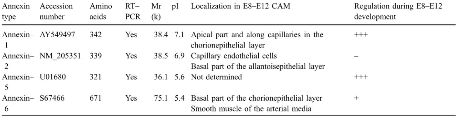
Unfortunately, antibodies against anx–5 worked in immunoblots only. Our analysis revealed that the anx–5 concentration increased after day 8. Because such an up-regulation was also seen for anx–1, we speculate that anx–5 Table 2 Summary of biochemical properties, localization, and developmental regulation (+++ strong, + positive,− none) of annexins in chicken CAM Annexin type Accession number Amino acids RT– PCR Mr (k)
pI Localization in E8–E12 CAM Regulation during E8–E12 development
Annexin– 1
AY549497 342 Yes 38.4 7.1 Apical part and along capillaries in the chorionepithelial layer
+++ Annexin–
2
NM_205351 339 Yes 38.5 6.9 Capillary endothelial cells – Basal part of the allantoisepithelial layer
Annexin– 5
U01680 321 Yes 36.1 5.6 Not determined +++
Annexin– 6
S67466 671 Yes 75.1 5.4 Basal part of the chorionepithelial layer + Smooth muscle of the arterial media
is also expressed in epithelial cells of the chorionic epithelium as these are the only cells significantly pro-liferating at this time period of embryonic development (Coleman and Terepka 1972). However, immunohisto-chemistry data are needed to confirm this suggestion.
Immunohistochemistry has demonstrated strong anx–6 immunoreactivity exclusively in cells located under the capillary plexus. At this site, basal cells and endothelial-like cells are present, neither of which undergo prolifera-tion (Coleman and Terepka 1972). Additional anx–6 immunoreactivity has been observed in the smooth muscle cell layer of the larger vessels in the mesenchyme; these are also not augmented during E8–E12 development. In ac-cordance with these data, immunoblotting levels of anx–6 remain constant. In this study, we have not been able to determine whether anx–6 is expressed in basal cells or in endothelial-like cells. However, because there is no marker for both of these cell types, our detection of anx–6 as being specifically expressed in one of these two cell types of the chorionic epithelium is the first report of their biochemical characterization.
The biochemical properties and immunohistochemically determined expression patterns of chicken annexins in the developing CAM as obtained in this study are summarized in Table 2. For validation, particularly of the exact assignment of immunoreactivity to defined cell types, im-munoelectron microscopy should be applied.
The expression of defined annexins cannot be assigned to special cell types because each annexin might be expressed in multiple cell types. Furthermore, the various functions of all the different annexins are only partially understood. Thus, the functions of the four annexins in the chicken chorionic epithelium during E8–E12 development cannot be deduced by analysis of their cellular expression patterns.
However, anx–1 is often present in cells that are involved in anti-inflammatory and immunomodulation reactions, e.g., in macrophages where it inhibits the inducible form of nitric oxide synthetase and phospholi-pase A2 activity (Parente and Solito 2004). This observa-tion agrees with our results of the strong proliferaobserva-tion of anx–1–expressing CC cells in the chorionic epithelium in response to grafted tumor spheroids; this might represent part of the reaction of the chicken CAM to invading pathogens (Sweeny and Bather 1968) or experimental intervention. Interestingly, degenerated cells in the CAM (similar to those that we have seen) have previously been identified as apoptotic endothelial cells that perish after exposure to pathogens (Sweeny and Bather 1968). We would also like to mention that anx–2 is found in endothelial cells of many tissues (Kim and Hajjar 2002), similar to the findings of the present study. This localiza-tion is in accordance with the funclocaliza-tion of anx–2 as an inhibitor of fibrinolysis and a modulator of angiogenesis (Hayes and Moss2004).
In conclusion, our study shows that anx–1, anx–2, anx– 5, and anx–6 exhibit complementary expression patterns in the chorionic epithelium. This knowledge should aid
further functional characterization of the various cell types in the developing chicken CAM.
Acknowledgements The skilful technical support of Bettina de Breuyn, Regula Buergy, and Christina Sala is gratefully acknowl-edged. We thank Dr. Katia Monastyrskaya for providing the anti-annexin-2 antibody and for helpful discussions.
References
Adam R, Mussa S, Lindemann D, Oelschlaeger TA, Deadman M, Ferguson DJ, Moxon R, Schroten H (2002) The avian chorioallantoic membrane in ovo—a useful model for bacterial invasion assays. Int J Med Microbiol 292:267–275
Akins RE, Tuan RS (1993) Transepithelial calcium transport in the chick chorioallantoic membrane. I. Isolation and characteriza-tion of chorionic ectoderm cells. J Cell Sci 105:369–379 Anderson RE, Gay CV, Schraer H (1981) Ultrastructural localization
of carbonic anhydrase in the chorioallantoic membrane by immunocytochemistry. J Histochem Cytochem 29:1121–1127 Babiychuk EB, Monastyrskaya K, Burkhard FC, Wray S, Draeger A
(2002) Modulating signaling events in smooth muscle: cleav-age of annexin 2 abolishes its binding to lipid rafts. FASEB J 16:1177–1184
Babiychuk VS, Draeger A, Babiychuk EB (2000) Smooth muscle actomyosin promotes Ca2+-dependent interactions between annexin VI and detergent-insoluble glycosphingolipid-enriched membrane domains. Acta Biochim Pol 47:579–589
Baum O, Djonov V, Ganster M, Widmer M, Baumgartner I (2005) Arteriolization of capillaries and FGF-2 upregulation in skeletal muscles of patients with chronic peripheral arterial disease. Microcirculation 12:527–537
Baum O, Da Silva–Azevedo L, Willerding G, Wockel A, Planitzer G, Gossrau R, Pries AR, Zakrzewicz A (2004) Endothelial NOS is main mediator for shear stress-dependent angiogenesis in skeletal muscle after prazosin administration. Am J Physiol Heart Circ Physiol 287:H2300–H2308
Bellairs R, Osmond M (1998) The atlas of chick development. Academic Press, San Diego
Coleman JR, Terepka AR (1972) Fine structural changes associated with the onset of calcium, sodium and water transport by the chick chorioallantoic membrane. J Membrane Biol 7:111–127 Djonov V, Baum O, Burri PH (2003) Vascular remodeling by
intussusceptive angiogenesis. Cell Tissue Res 314:107–117 Djonov V, Schmid M, Tschanz SA, Burri PH (2000) Intussusceptive
angiogenesis: its role in embryonic vascular network formation. Circ Res 86:286–292
Draeger A, Wray S, Babiychuk EB (2005) Domain architecture of the smooth-muscle plasma membrane: regulation by annexins. Biochem J 387:309–314
Gabrielli MG, Cox JV, Materazzi G, Menghi G (2004) Cell type-specific and developmentally regulated expression of the AE1 anion exchanger in the chicken chorioallantoic membrane. Histochem Cell Biol 121:189–199
Gabrielli MG, Materazzi G, Cox JV, Menghi G (2001) Specialised cell types in the chorioallantoic membrane express carbonic anhydrase during chick embryogenesis. J Anat 198:229–238 Ganote CE, Beaver DL, Moses L (1964) Ultrastructure of the chick
chorio-allantoic membrane and its reaction to inoculation trauma. Lab Invest 13:1575–1589
Gerke V, Moss SE (2002) Annexins: from structure to function. Physiol Rev 82:331–371
Hayes MJ, Moss SE (2004) Annexins and disease. Biochem Biophys Res Commun 322:1166–1170
Hoshi H, Mori T (1971) The fine structure of the chorionic epithelium of chick embryo. Arch Histol Jpn 33:45–58 Kim J, Hajjar KA (2002) Annexin II: a plasminogen-plasminogen
Korff T, Augustin HG (1998) Integration of endothelial cells in multicellular spheroids prevents apoptosis and induces differ-entiation. J Cell Biol 143:1341–1352
Leeson TS, Leeson CR (1963) The chorio-allantois of the chick. Light and electron microscopic observations at various times of incubation. J Anat 97:585–595
Lusimbo WS, Leighton FA, Wobeser GA (2000) Histology and ultrastructure of the chorioallantoic membrane of the mallard duck (Anas platyrhynchos). Anat Rec 259:25–34
Moss SE, Morgan RO (2004) The annexins. Genome Biol 5:219 Movitz C, Dahlgren C (2000) Endogenous cleavage of annexin I
generates a truncated protein with a reduced calcium require-ment for binding to neutrophil secretory vesicles and plasma membrane. Biochim Biophys Acta 1468:231–238
Narbaitz R (1977) Structure of the intra-chorionic blood sinus in the chick embryo. J Anat 124:347–354
Narbaitz R, Bastani B, Galvin NJ, Kapal VK, Levine DZ (1995) Ultrastructural and immunocytochemical evidence for the presence of polarised plasma membrane H(+)-ATPase in two specialised cell types in the chick embryo chorioallantoic membrane. J Anat 186:245–252
Navarro M, DeRuiter MC, Carretero A, Ruberte J (2003) Micro-vascular assembly and cell invasion in chick mesonephros grafted onto chorioallantoic membrane. J Anat 202:213–225 Owczarzak A (1971) Calcium-absorbing cell of the chick
chorio-allantoic membrane. I. Morphology, distribution and cellular interactions. Exp Cell Res 68:113–129
Parente L, Solito E (2004) Annexin 1: more than an anti-phospholipase protein. Inflamm Res 53:125–132
Patan S, Haenni B, Burri PH (1993) Evidence for intussusceptive capillary growth in the chicken chorio-allantoic membrane (CAM). Anat Embryol (Berl) 187:121–130
Patten B (1950) Early embryology of the chick. Lewis, London
Raynal P, Pollard HB (1994) Annexins: the problem of assessing the biological role for a gene family of multifunctional calcium-and phospholipid-binding proteins. Biochim Biophys Acta 1197:63–93
Ribatti D, Nico B, Vacca A, Roncali L, Burri PH, Djonov V (2001) Chorioallantoic membrane capillary bed: a useful target for studying angiogenesis and anti-angiogenesis in vivo. Anat Rec 264:317–324
Richardson M, Singh G (2003) Observations on the use of the avian chorioallantoic membrane (CAM) model in investigations into angiogenesis. Curr Drug Targets Cardiovasc Haematol Disord 3:155–185
Shumko JZ, Defouw DO, Feinberg RN (1988) Vascular histodif-ferentiation in the chick chorioallantoic membrane: a morpho-metric study. Anat Rec 220:179–189
Sidis Y, Horseman ND (1993) The hinge region of chicken annexin I contains no site for tyrosine phosphorylation. FEBS Lett 329:296–300
Skalinsky EI, Kondalenko VF (1964) Electron microscopic studies of the chick chorio-allantois during embryogenesis. Acta Morphol Acad Sci Hung 12:247–259
Spanel-Borowski K (1989) The chick chorioallantoic membrane as test system for biocompatible materials. Res Exp Med (Berl) 189:69–75
Sweeny PR, Bather R (1968) An electron microscopic study of the chorioallantoic membrane following infection with Rous sar-coma virus. J Cell Biol 36:299–311
Tuan RS (1980) Calcium transport and related functions in the chorioallantoic membrane of cultured shell-less chick embryos. Dev Biol 74:196–204
Valdes TI, Kreutzer D, Moussy F (2002) The chick chorioallantoic membrane as a novel in vivo model for the testing of biomaterials. J Biomed Mater Res 62:273–282