BRAIN
A JOURNAL OF NEUROLOGYTyrosine hydroxylase deficiency: a treatable
disorder of brain catecholamine biosynthesis
Miche`l A. Willemsen,
1Marcel M. Verbeek,
2Erik-Jan Kamsteeg,
3Johanneke F. de Rijk-van Andel,
4Alec Aeby,
5Nenad Blau,
6Alberto Burlina,
7Maria A. Donati,
8Ben Geurtz,
2Padraic J. Grattan-Smith,
9Martin Haeussler,
10Georg F. Hoffmann,
11Hans Jung,
12Johannis B. de Klerk,
13Marjo S. van der Knaap,
14Fernando Kok,
15Vincenzo Leuzzi,
16Pascale de Lonlay,
17Andre Megarbane,
18Hugh Monaghan,
19Willy O. Renier,
20Pierre Rondot,
21Monique M. Ryan,
22Ju¨rgen Seeger,
23Jan A. Smeitink,
24Gerry C. Steenbergen-Spanjers,
2Evangeline Wassmer,
25Bernhard Weschke,
26Frits A. Wijburg,
27Bridget Wilcken,
28Dimitrios I. Zafeiriou
29and Ron A. Wevers
21 Radboud University Nijmegen Medical Centre, Donders Institute for Brain, Cognition and Behaviour, Department of Paediatric Neurology, 6500 HB, Nijmegen, The Netherlands
2 Radboud University Nijmegen Medical Centre, Donders Institute for Brain, Cognition and Behaviour, Department of Neurology and Department of Laboratory Medicine, 6525 GA, Nijmegen, The Netherlands
3 Radboud University Nijmegen Medical Centre, Department of Human Genetics, 6500 HB, Nijmegen, The Netherlands 4 Amphia Hospital, Department of Paediatric Neurology, 4800 RK, Breda, The Netherlands
5 Hoˆpital Erasme – ULB, Department of Paediatric Neurology, 1070 Bruxelles, Belgium
6 University Children’s Hospital, Division of Clinical Chemistry and Biochemistry and Zu¨rich Center for Integrative Human Physiology (ZIHP), 8032 Zu¨rich, Switzerland
7 University Hospital, Department of Paediatrics, 35128 Padua, Italy 8 AOU Meyer Hospital, Clinic of Paediatric Neurology, 50139 Florence, Italy 9 Sydney Children’s Hospital, Department of Neurology, 2031 Randwick, Australia 10 Fru¨hdiagnosezentrum Wu¨rzburg, 97080 Wu¨rzburg, Germany
11 University Children’s Hospital Heidelberg, Department of General Paediatrics, 69120 Heidelberg, Germany 12 University Hospital Zu¨rich, Department of Neurology, 8091 Zu¨rich, Switzerland
13 Erasmus Medical Centre, Sophia Children’s Hospital, Department of Paediatrics, 3015 GJ, Rotterdam, The Netherlands
14 VU University Medical Centre, Neuroscience Campus Amsterdam, Department of Child Neurology, 1081 HV, Amsterdam, The Netherlands 15 University of Sao Paulo School of Medicine, Department of Paediatric Neurology, 05403-900 Sao Paulo, Brazil
16 La Sapienza University of Rome, Department of Child Neurology and Psychiatry, 00141 Rome, Italy 17 University Paris Descartes, Hospital Necker, Reference Centre of Metabolic Diseases, 75743 Paris, France 18 Saint Joseph University, Medical Genetics Unit, 1107 2180 Beirut, Lebanon
19 Our Lady’s Children’s Hospital, Crumlin, Department of Paediatrics, Dublin 12, Ireland 20 Canisius Wilhelmina Hospital, Department of Neurology, 6532 SZ, Nijmegen, The Netherlands 21 CHU Biceˆtre, Service de Neurologie, 94275 Le Kremlin Biceˆtre, France
22 University of Melbourne, Royal Children’s Hospital, 3052 Melbourne, Australia 23 German Clinic for Diagnostics, Department of Paediatrics, 65191 Wiesbaden, Germany
24 Radboud University Nijmegen Medical Centre, Nijmegen Centre for Mitochondrial Disorders at the Department of Paediatrics, 6500 HB, Nijmegen, The Netherlands
25 Birmingham Children’s Hospital, Neuroscience Department, Birmingham B4 6NH, United Kingdom 26 Charite´ University Medicine Berlin, Department of Neuropaediatrics, 13353 Berlin, Germany 27 Academic Medical Centre, Department of Paediatrics, 1105 AZ, Amsterdam, The Netherlands
28 Children’s Hospital at Westmead, Department of Biochemical Genetics, Westmead, Sydney, NSW 2145, Australia 29 Aristotle University of Thessaloniki, Department of Paediatrics, 54622 Thessaloniki, Greece
Received September 13, 2009. Revised February 14, 2010. Accepted March 22, 2010. Advance Access publication April 29, 2010 ßThe Author (2010). Published by Oxford University Press on behalf of the Guarantors of Brain. All rights reserved.
Correspondence to: Miche`l A. Willemsen, Radboud University Nijmegen Medical Centre, Donders Institute for Brain,
Cognition and Behaviour,
Department of Paediatric Neurology (820 IKNC), PO Box 9101,
6500 HB Nijmegen, The Netherlands E-mail: [email protected]
Tyrosine hydroxylase deficiency is an autosomal recessive disorder resulting from cerebral catecholamine deficiency. Tyrosine hydroxylase deficiency has been reported in fewer than 40 patients worldwide. To recapitulate all available evidence on clinical phenotypes and rational diagnostic and therapeutic approaches for this devastating, but treatable, neurometabolic disorder, we studied 36 patients with tyrosine hydroxylase deficiency and reviewed the literature. Based on the presenting neurological features, tyrosine hydroxylase deficiency can be divided in two phenotypes: an infantile onset, progressive, hypokinetic-rigid syndrome with dystonia (type A), and a complex encephalopathy with neonatal onset (type B). Decreased cerebrospinal fluid concentrations of homovanillic acid and 3-methoxy-4-hydroxyphenylethylene glycol, with normal 5-hydroxyindoleacetic acid cerebrospinal fluid concentrations, are the biochemical hallmark of tyrosine hydroxylase deficiency. The homovanillic acid concentrations and homovanillic acid/5-hydroxyindoleacetic acid ratio in cerebrospinal fluid correlate with the severity of the phenotype. Tyrosine hydroxylase deficiency is almost exclusively caused by missense mutations in theTH gene and its promoter region, suggesting that mutations with more deleterious effects on the protein are incompatible with life. Genotype–phenotype correlations do not exist for the common c.698G4A and c.707T4C mutations. Carriership of at least one promotor mutation, however, apparently predicts type A tyrosine hydroxylase deficiency. Most patients with tyrosine hydroxylase deficiency can be successfully treated with L-dopa.
Keywords:
tyrosine hydroxylase; neurotransmitters; cerebrospinal fluid; dystonia;L-dopaAbbreviations:
5HIAA = 5-hydroxyindoleacetic acid; HVA = homovanillic acid; MHPG = 3-methoxy-4-hydroxyphenylethylene glycol; THD = tyrosine hydroxylase deficiencyIntroduction
The enzyme tyrosine hydroxylase (EC 1.14.16.2) catalyzes the
conversion of L-tyrosine to L-dihydroxyphenylalanine (L-dopa),
which is the rate-limiting step in the biosynthesis of the catechol-amines dopamine, norepinephrine and epinephrine (Fig. 1). Catecholamines are produced in the brain and adrenal medulla, but also in non-neuronal, e.g. renal, intestinal and lymphoid tissues. Their vital functions as neurotransmitters and hormones, and the crucial role of tyrosine hydroxylase in their biosynthesis are demonstrated by the observation that complete loss of tyrosine hydroxylase activity is lethal in knock-out mice (Zhou et al., 1995). Human tyrosine hydroxylase deficiency [THD; Online Mendelian Inheritance in Man (OMIM) number 191290] is an autosomal recessive neurometabolic disorder due to mutations in the tyrosine hydroxylase (TH) gene on chromosome 11p15.5. The first reports of THD described patients with an early onset, progressive
L-dopa-responsive dystonia (Castaigne et al., 1971; Rondot and
Ziegler, 1983; Rondot et al., 1992). Later, neonates were recog-nized with a more severe phenotype described as progressive,
L-dopa-non-responsive encephalopathy (Hoffmann et al., 2003).
THD can be diagnosed by demonstrating decreased CSF levels of the down-stream metabolites of the catecholamine degrad-ation pathway (Fig. 1), i.e. homovanillic acid (HVA) and
3-methoxy-4-hydroxyphenylethylene glycol (MHPG) and by mu-tation analysis of the TH gene.
THD has been reported in fewer than 40 patients worldwide, reviewed in this article. This disorder is known under different names in the literature, namely ‘Segawa syndrome’, ‘infantile
par-kinsonism’ and ‘L-dopa-responsive dystonia’. ‘Segawa syndrome’,
however, is also used to indicate another defect in neurotransmit-ter biosynthesis, caused by GTP cyclohydrolase I mutations.
‘Infantile parkinsonism’ and L-dopa-responsiveness are not found
in all patients with THD. Furthermore, theL-dopa-responsive
dys-tonias encompass a heterogeneous class of movement disorders, THD being only one of them (Muller et al., 1998; Albanese et al., 2006; Tarsy and Simon, 2006; Muller, 2009). Finally, the pheno-type of THD can be so complex that it is not simply associated with an extrapyramidal movement disorder (Hoffmann et al., 2003). Altogether, we prefer to name the disorder after its under-lying enzymatic defect, as is common practice for inborn errors of metabolism.
In this article, we summarize the medical literature on human THD, and include detailed novel clinical, biochemical and genetic data on thus far unpublished patients. This large review on THD recapitulates all available evidence on clinical phenotypes and ra-tional diagnostic and therapeutic approaches of this severe, but potentially treatable disorder.
Methods
Our laboratory has a longstanding tradition of neurotransmitter ana-lysis, and we have been performing TH gene mutation analysis since the genetic basis of THD was elucidated. This history supplies us with a unique database including patients with biochemically and genetically proven THD from many different countries.
The laboratory methodologies used for CSF neurotransmitter analysis have previously been reported in detail (Brautigam et al., 1998; Verbeek et al., 2008) Reference values for HVA and 5-hydroxyindoleacetic acid (5HIAA; end-product of serotonin degrad-ation) in CSF decrease with age, and there is a rostrocaudal gradient for the concentrations of both metabolites, necessitating analysis of a standardized CSF fraction (Brautigam et al., 1998) Mutation analysis of the TH gene was performed as previously described (van den Heuvel et al., 1998) Numbering of coding sequence mutations was according to GenBank reference sequence NM_199292.1 (tyrosine hydroxylase isoform A) in which the A of the ATG transcription initi-ation codon is designated position 1. Mutiniti-ations were named according to the guidelines of the Human Genome Variation Society (www.hgvs.org).
A questionnaire was sent to all physicians who have referred (sam-ples from) THD patients to our centre. In this way we collected de-tailed information on demographic data, pregnancy and perinatal period, presenting clinical features, mode of treatment, follow-up during treatment and results of cerebral imaging studies. The results of CSF and mutation analysis were available in our database. In those few patients in whom CSF analysis was performed elsewhere, the results were obtained together with the appropriate reference values.
A Pubmed search was performed for reports in English, using the terms: ‘tyrosine hydroxylase recessive’ and ‘tyrosine hydroxylase dys-tonia’. The reference lists of all relevant papers were checked for other citations, especially those reports from the era before THD was recog-nized as a separate disease entity.
This study was approved by the ethics committee of the Radboud University Nijmegen Medical Centre, The Netherlands. The require-ment for additional local ethical approval differed between participat-ing countries and was obtained if required.
Results
We had the names of 36 patients with THD in our database. Questionnaires were completed by the referring physicians of all patients.
Literature
Besides reports concerning patients who had been diagnosed in our laboratory (Table 1) (Castaigne et al., 1971; Rondot and Ziegler, 1983; Rondot et al., 1992; van den Heuvel et al., 1998; Brautigam et al., 1999; Wevers et al., 1999; de Lonlay et al., 2000; de Rijk-Van Andel et al., 2000; Dionisi-Vici et al., 2000; Janssen et al., 2000; Swaans et al., 2000; Haussler et al., 2001; Grattan-Smith et al., 2002; Hoffmann et al., 2003; Schiller et al., 2004; Verbeek et al., 2007; Zafeiriou et al., 2009), we only found 14 other THD patients from 12 families in whom the diagnosis was genetically proven (Ludecke et al., 1995, 1996; Knappskog
Figure 1 Simplified scheme of the biosynthesis and catabolism of serotonin and the catecholamines dopamine, norepinephrine and epinephrine. TPH = tryptophan hydroxylase; AADC = aromatic amino acid decarboxylase; PAH = phenylalanine hydroxylase; TH = tyrosine hydroxylase; BH4 = tetrahydrobiopterin; DOPAC = 3,4-dihydroxyphenylacetic acid.
Table
1
Clinical
characteristics,
demographic
data
and
results
of
mutation
analysis
in
36
patients
with
THD
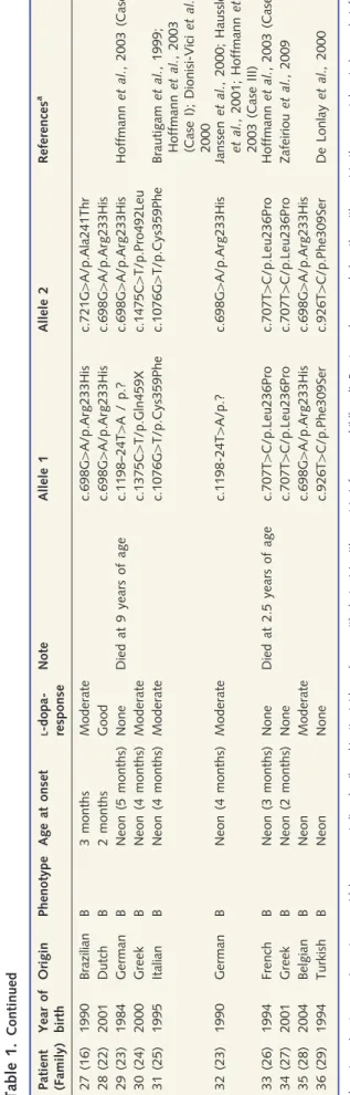
Patient (Family) Year of birth Origin Phenotype Age at onset L -dopa- response Note Allele 1 Allele 2 References a 1 (1) 1962 French A 5 years Good c.1010G 4 A/p.Arg337His c.1481C 4 T/p.Thr494Met Castaigne et al. , 1971 (Case 2); Rondot and Ziegler, 1983 (Case 1); Rondot et al. , 1992 (Case 2); Swaans et al. , 2000 (Case 2) 2 (2) 1950 Swiss A 3 years Good c.1127C 4 T/p.Ala376Val c.1493A 4 G/p.Asp498Gly Schiller et al. , 2004 (Case 1) 3 (2) 1961 Swiss A 3 years Good c.1127C 4 T/p.Ala376Val c.1493A 4 G/p.Asp498Gly Schiller et al. , 2004 (Case 2) 4 (1) 1965 French A 2 years Good c.1010G 4 A/p.Arg337His c.1481C 4 T/p.Thr494Met Castaigne et al. , 1971 (Case 1); Rondot and Ziegler, 1983 (Case 2); Rondot et al. , 1992 (Case 1); Swaans et al. , 2000 (Case 1) 5 (3) 1965 Belgian A 1.5 year Good c.826A 4 C/p.Thr276Pro c.941C 4 T/p.Thr314Met Swaans et al. , 2000 (Case 3) 6 (4) 1989 Turkish A 4 1 year Good c. 70G 4 Ac . 70G 4 A Verbeek et al. , 2007 7 (5) 2002 Lebanese A 5 12 months Good c. 70G 4 Ac . 70G 4 A Verbeek et al. , 2007 8 (6) 2004 German A 9 months Good c.680A 4 G/p.Asp227Gly c.698G 4 A/p.Arg233His 9 (7) 1990 Lebanese A 8 months Moderate c.698G 4 A/p.Arg233His c.698G 4 A/p.Arg233His 10 (8) 1993 Italian A 8 months Good c. 69T 4 Ac . 69T 4 A Verbeek et al. , 2007 11 (9) 2004 Pakistan A 8 months Good Sudden onset c.1181T 4 C/p.Ile394Thr c.1181T 4 C/p.Ile394Thr 12 (4) 1999 Turkish A 6 months Good c. 70G 4 Ac . 70G 4 A Verbeek et al. , 2007 13 (10) 1997 Irish A 6 months Good c.620G 4 A/p.Cys207Tyr c.698G 4 A/p.Arg233His 14 (11) 1992 Dutch A 6 months Good c.295delC/p.Leu99fs b c.698G 4 A/p.Arg233His Brautigam et al. , 1998; Wevers et al. , 1999; de Rijk-Van Andel et al ., 2000 15 (12) 1989 Dutch A 6 months Good c. 71C 4 T c.1159C 4 A/p.Leu387Met Verbeek et al. , 2007 16 (13) 1993 Dutch A 5 months Good c.698G 4 A/p.Arg233His c.698G 4 A/p.Arg233His Brautigam et al. , 1998; van den Heuvel et al. , 1998; Wevers et al. , 1999; de Rijk-Van Andel et al ., 2000 17 (7) 1987 Lebanese A 4 months None c.698G 4 A/p.Arg233His c.698G 4 A/p.Arg233His 18 (14) 2003 Fijian A 4 months Good c. 70G 4 A c.1475C 4 T/p.Pro492Leu Verbeek et al. , 2007 19 (5) 2004 Lebanese A 4 months Good c. 70G 4 Ac . 70G 4 A Verbeek et al. , 2007 20 (15) 2000 Greek A 3 months Unknown Lost from follow-up c.707T 4 C/p.Leu236Pro c.707T 4 C/p.Leu236Pro 21 (16) 2003 Brazilian A 3 months Good c.698G 4 A/p.Arg233His c.721G 4 A/p.Ala241Thr 22 (17) 1992 Dutch A 3 months Good c.698G 4 A/p.Arg233His c.698G 4 A/p.Arg233His Brautigam et al. , 1998; van den Heuvel et al. , 1998; Wevers et al. , 1999; de Rijk-Van Andel et al ., 2000 23 (18) 1993 Dutch A 3 months Good c.698G 4 A/p.Arg233His c.698G 4 A/p.Arg233His Brautigam et al. , 1998; van den Heuvel et al. , 1998; Wevers et al. , 1999; de Rijk-Van Andel et al ., 2000 24 (19) 1998 Lebanese A 2 months Good c.698G 4 A/p.Arg233His c.698G 4 A/p.Arg233His Grattan-Smith et al. , 2002 25 (20) 2005 Italian A 2 months Moderate c.776A 4 G/p.Glu259Gly c.1529T 4 A/p.Leu510Gln 26 (21) 1997 Dutch B 3 months Good Sudden onset at 3 months and sudden deterioration at 22 months during infection c.698G 4 A/p.Arg233His c.698G 4 A/p.Arg233His (Continued)et al., 1995; Surtees and Clayton, 1998; Furukawa et al., 2001; Diepold et al., 2005; Moller et al., 2005; Yeung et al., 2006; Giovanniello et al., 2007; Ribases et al., 2007; Wu et al., 2008; Clot et al., 2009; Doummar et al., 2009). The key data on these patients, and type A/B classification (see next paragraph) based on available clinical descriptions, are summarized in Table 2.
Clinical features
After careful evaluation of the detailed case histories in the litera-ture and the questionnaires used in this study, it was possible to class the different phenotypes at presentation into two major groups. Most patients (n = 25) suffered from a disorder that can be summarized as a progressive hypokinetic-rigid syndrome with dystonia. The onset of symptoms was generally in the first year of life (age range: 2 months and 5 years). This is the first phenotype described in the literature, further referred to as type A in this article (Castaigne et al., 1971; Rondot and Ziegler, 1983; Rondot et al., 1992; Knappskog et al., 1995; Ludecke et al., 1995; Swaans et al., 2000; Furukawa et al., 2001; Diepold et al., 2005; Yeung et al., 2006; Giovanniello et al., 2007; Ribases et al., 2007; Wu et al., 2008; Clot et al., 2009). The other eleven patients suffered from a more ‘complex encephalop-athy’ with earlier onset (age range: 0–3 months), as described by Hoffmann et al. (2003), designated type B. These two phenotypes are defined in detail in Boxes 1 and 2. Although, we found no difficulties in designating individual patients into type A or B THD, it was obvious that the phenotype of THD is a spectrum with overlap of clinical features between both groups. Patient 24, previously described by Grattan-Smith et al. (2002), is a good example of a type A patient with a phenotype very close to type B.
Table 1 summarizes the key demographic data, and clinical characteristics, as well as the results of mutation analysis. The 36 patients came from 29 families with their roots in 13 different countries. THD is a movement disorder with very early onset: all type B patients had an age at onset within the first months of life, and 19 out of 25 (76%) of type A patients presented in the first year of life. The majority of patients (69%) suffered from type A
THD. While responsiveness to L-dopa was absent (36%),
moder-ate (45%) or good (18%) in the type B patients, it was good in 84% of the type A patients. Table 3 provides further details on the prevalence of other clinical features as well as the response to
L-dopa treatment in type A and B patients as a group. Most
add-itional clinical features are not unique to either type A or B patients. Nevertheless, there is a clear predominance of extra features in type B patients, especially with regard to perinatal abnormalities, diurnal fluctuations, autonomic disturbances and body length and weight at presentation. None of the patients in this series had clinical features suggesting systemic deficiency of catecholamines, such as abnormalities in the maintenance of blood pressure. These systemic phenomena were not, however, formally studied.
Type A patients generally showed a beneficialL-dopa response
within the first 2 weeks of treatment, while positive effects in type B patients always occurred later. Selegiline, an inhibitor of dopa-mine degradation, as well as the dopadopa-mine-agonists bromocriptine and pramipexole, were prescribed in one type A and five type B
Table
1.
Continued Patient (Family) Year of birth Origin Phenotype Age at onset L -dopa- response Note Allele 1 Allele 2 References a 27 (16) 1990 Brazilian B 3 months Moderate c.698G 4 A/p.Arg233His c.721G 4 A/p.Ala241Thr 28 (22) 2001 Dutch B 2 months Good c.698G 4 A/p.Arg233His c.698G 4 A/p.Arg233His 29 (23) 1984 German B Neon (5 months) None Died at 9 years of age c.1198–24T 4 A / p.? c.698G 4 A/p.Arg233His Hoffmann et al. , 2003 (Case II) 30 (24) 2000 Greek B Neon (4 months) Moderate c.1375C 4 T/p.Gln459X c.1475C 4 T/p.Pro492Leu 31 (25) 1995 Italian B Neon (4 months) Moderate c.1076G 4 T/p.Cys359Phe c.1076G 4 T/p.Cys359Phe Brautigam et al. , 1999; Hoffmann et al. , 2003 (Case I); Dionisi-Vici et al. , 2000 32 (23) 1990 German B Neon (4 months) Moderate c.1198-24T 4 A/p.? c.698G 4 A/p.Arg233His Janssen et al. , 2000; Haussler et al. , 2001; Hoffmann et al. , 2003 (Case III) 33 (26) 1994 French B Neon (3 months) None Died at 2.5 years of age c.707T 4 C/p.Leu236Pro c.707T 4 C/p.Leu236Pro Hoffmann et al. , 2003 (Case IV) 34 (27) 2001 Greek B Neon (2 months) None c.707T 4 C/p.Leu236Pro c.707T 4 C/p.Leu236Pro Zafeiriou et al. , 2009 35 (28) 2004 Belgian B Neon Moderate c.698G 4 A/p.Arg233His c.698G 4 A/p.Arg233His 36 (29) 1994 Turkish B Neon None c.926T 4 C/p.Phe309Ser c.926T 4 C/p.Phe309Ser De Lonlay et al., 2000 Phenotype A = ‘progressive extrapyramidal movement disorder (hypokinetic-rigid syndrome with dystonia) with onset in infancy or childhood’; B = ‘c omplex encephalopathy w ith onset in the n eonatal period o r e arly infancy’; Neon = n eonatal. Response to L -dopa: ‘none’ means that there was n o b eneficial response at all, often reflecting the occurrence o f such severe dyskinesia that treatment w as impossib le. a References: papers in which (clinical, biochemical or genetic) data of the p atient h ave been published previously. b T he c.295delC mutation w as previously designated c.291delC, but renamed according to the guidelines of the Human G enome V ariation Society (www.hg vs.org).patients, with some, although limited, additional effects.
Treatment with L-dopa dramatically improved motor outcome in
type A patients: one patient was lost from follow-up, Patients 17 and 25 remained wheelchair bound, and all others (22 out of 25, 88%) were able to walk independently during follow-up. Type B patients 26, 28 and 35 finally learned to walk independently,
although the initial response to L-dopa had been less impressive
than in most type A cases. The majority (67%) of type A patients had normal cognitive capacities during follow-up, while 10 out of 11 (91%) type B patients in our series were mentally retarded. Two type B patients (cases 29 and 33) died during follow-up due to infectious and respiratory complications.
Cerebral imaging
Most subjects (29 out of 36) underwent cerebral MRI studies. The images were not systematically reviewed for the purpose of this study. Non-specific, mild white matter signal changes and increased volume of extra-cerebral CSF spaces were reported in nine patients, while the majority of patients (n = 20) had normal images. The number of patients with abnormalities on MRI was 4 out of 21 (19%) and 5 out of 8 (63%) in Types A and B patients, respectively. Importantly, gross structural abnormalities of the brain and signal changes in the basal ganglia were never observed.
Biochemical findings
Based on the metabolic pathway involved, it can be predicted that CSF levels of the down-stream metabolites of the dopamine deg-radation pathway will be low, while the serotonin pathway is not affected (Fig. 1). Age-dependent reference values for the different metabolites have been published in detail (Brautigam et al., 1998). As an example, we give here the reference values (P2.5–P97.5) in childhood (2–5 years): HVA 384–769 nmol/l, 5HIAA 110–265 nmol/l, MHPG 35–64 nmol/l and HVA/5HIAA ratio 1.8–4.4.
Pre-treatment CSF results were lacking from seven patients [1–5, 17 (Family 7) and 29 (Family 23)]. Patients 1–5 were initially
clinically diagnosed as having aL-dopa-responsive movement
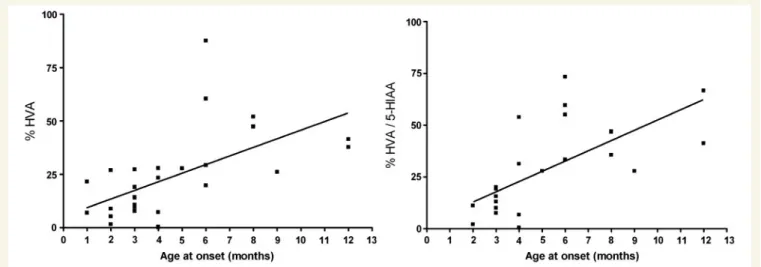
dis-order in the 1970s, and never underwent lumbar puncture. In Families 7 and 23, a lumbar puncture was performed in only one of the two affected siblings. Decreased CSF concentrations of HVA and MHPG, with decreased HVA/5HIAA ratios were demonstrated in all patients in whom CSF was analysed (Fig. 2). To enable comparison of patients of different ages, with different references values, CSF HVA concentrations and HVA/5HIAA ratios were expressed as percentage of the lower reference limit (2.5th percentile) (Brautigam et al., 1998). The mean CSF HVA concen-tration in type B patients was significantly lower (P50.005) than in type A patients, namely 8.8% (SD 8.9) and 32.7% (SD 19.8), respectively. As a group, type B patients also had a significantly lower (P50.005) mean CSF HVA/5HIAA ratio than type A pa-tients, namely 5.6 (SD 4.7) and 36.7 (SD 18.9), respectively. After ranking the patients in order of age of onset of the disease (as in Table 1), we were able to show a positive correlation between age of onset and CSF HVA concentrations as well as HVA/5HIAA ratio (Fig. 3). Urinary concentrations of catecholamines and their
Table
2
Patients
reported
in
the
literature
(not
diagnosed
in
our
laboratory)
Family, Patient Pheno- type Age at onset Cognitive impairment L -dopa- response Presenting clinical features Allele 1 Allele 2 References I A 5 years N.r. Good Spastic paraplegia c.296delT c.1493A 4 G Furukawa et al. , 2001 II A 3 years N.r. Good Delayed walking and rigidity, wheelchair bound at 10 years c.698G 4 A c.739G 4 AW u et al. , 2008 III A Childhood Mild Good Progressive dystonia c.956G 4 C c.1240G 4 A Clot et al. , 2009 IV A 2 years Mild Good Spastic paraplegia from age 1 year, deterioration at 11 years c.1240G 4 A c.1529T 4 A Giovanniello et al. , 2007 V A 14 months Mild Good Normal until 14 months, then deterioration c.736C 4 T c.1493A 4 G Diepold et al. , 2005 VI-1 A N.r. N.r. N.r. Progressive L -dopa-responsive dystonia c.1141C 4 A c.1141C 4 A Ludecke et al ., 1995; Knappskog et al. , 1995 VI-2 A N.r. N.r. N.r. Progressive L -dopa-responsive dystonia c.1141C 4 A c.1141C 4 A Ludecke et al ., 1995; Knappskog et al. , 1995 VII A 6 months N.r. Moderate L -dopa-responsive encephalopathy c. 71C 4 Tc . 71C 4 T Ribases et al. , 2007 VIII-1 A 4 months Yes Moderate Mental retardation, ‘mixed type cerebral palsy’, galactorrhea (p.Arg169X) b c.698G 4 A Yeung et al. , 2006 VIII-2 A N.r. Yes Moderate As sibling but without galactorrhea (p.Arg169X) b c.698G 4 A Yeung et al. , 2006 IX B 9 months No Good Hypokinesia, hypotonia, dystonia, ptosis; adopted child c.1125C 4 G c.1399A 4 G Clot et al. , 2009; Doummar et al. , 2009 X B 5 months Yes Good Complex movement disorder, abnormal eye movements, diurmal fluctuation c.901C 4 G c.901C 4 G Clot et al. , 2009 XI B 5 5 months Yes Moderate Complex movement disorder, ptosis, irritability c.982C 4 T c.1196C 4 T M oller et al. , 2005 XII B 3 months No Good Complex movement disorder, ptosis c.614T 4 C c.614T 4 C Ludecke et al. , 1996; Surtees and Clayton, 1998 a R eferences: papers in which (clinical, biochemical or genetic) data of the p atient h ave been published previously. b The cDNA mutation w as not reported b y the authors, only the protein change was g iven. N .r. = not reported.degradation products were available in a minority of the patients (data not shown) and were generally found to be non-informative.
Mutation analysis
We identified 24 different TH gene mutations (Tables 1 and 4; Fig. 4), located in the promoter sequence, exons 3, 5–14, and in intron 11. Six mutations were not reported previously. The novel c.1375C4T mutation was predicted to lead to a stop codon
(p.Gln459X). The other five novel mutations (c.620G4A,
c.680A4G, c.721G4A, c.776A4G and c.1181T4C) were all con-sidered pathogenic since they were (i) not encountered in 200 control alleles; (ii) not reported as polymorphisms in databases and the literature (Haavik et al., 2008); and (iii) affecting amino acids within the tyrosine hydroxylase protein that are highly con-served among various species. Additionally, the program SIFT (Sorting Intolerant From Tolerant) (Ng and Henikoff, 2001) pre-dicted ‘not tolerated’, i.e. a deleterious effect for all five mutations.
The total number of mutated alleles reported in THD is 100 (Table 4). Five out of these 100 alleles harboured (four different) mutations that lead to protein truncation [c.295delC, c.296delT,
p.Arg169X (c.DNA change not reported) and c.1375C4T], while all other 95 alleles were affected by less deleterious missense mu-tations. Homozygosity for the common c.698G4A mutation was found in six type A and three type B patients. Homozygosity for the c.707T4C mutation occurred in one type A and two type B patients. Promoter mutations were only encountered in type A
THD with good L-dopa responsiveness.
Discussion
In general, the results of the present study and available data in the literature are in perfect agreement. In total, reports on 50 THD patients (from 41 families) are now available in the literature, and are reviewed in this article. Type A THD (n = 35) is more often diagnosed than type B THD (n = 15).
Clinical features at presentation
The many different neurological features of THD (hypokinesia, bradykinesia, rigidity, dystonia, chorea, tremor, oculogyric crises, ptosis and hypersalivation, among others) are caused by cerebral Box 1. THD type A: ‘Progressive extrapyramidal movement disorder (hypokinetic-rigid syndrome with dystonia) with onset in infancy or childhood’
These patients are born after uncomplicated pregnancies and develop normally during the first months of life. In rather exceptional cases (including the first patients described with THD), psychomotor development is even normal or only slightly delayed during the first 2 to 5 years of life (Castaigne et al., 1971; Rondot and Ziegler, 1983; Rondot et al., 1992). Thereafter, however, progressive motor signs appear. Affected individuals become hypokinetic and rigid, and dystonia develops. In early stages, generally only one leg is involved, but with time both legs and also the arms, trunk, face and oropharyngeal musculature become affected. Initial complaints thus encompass abnormal posturing and walking difficulties, or frequent falls in those who already learned to walk before onset of symptoms. These children become wheelchair bound within some years.
Most patients with this type A THD are younger than those described above. In these infants, hypokinesia, bradykinesia and rigidity may dominate the neurological picture while dystonia tends to be less prominent. Initial motor symptoms are generally symmetric and involve arms as well as legs. The ability to walk is not achieved unless children are treated. Severity of dystonia may fluctuate during the day (generally worse in the afternoon), but can also fluctuate within days, giving the impression of a paroxysmal dystonia especially in the early stages of the disease. Mild, non-progressive mental retardation can be found in patients with relatively early onset of motor symptoms, while cognitive functions appear unaffected in patients who develop symptoms after the first year of life. Besides the hypokinetic-rigid syndrome with dystonia, other features like tremor, chorea, oculogyric crises and ptosis, as well as behavioural or autonomic disturbances are absent or—if present—are found as a mild feature and in a minority of patients.
In almost all patients with type A THD, treatment withL-dopa results in an excellent response, sometimes even a miraculous improvement of the neurological condition. During follow-up, all patients continue to be asymptomatic or display only mild motor or cognitive impairment while on a low dose ofL-dopa. They show no evidence of progressive disease, and tolerateL-dopa well during many years. Extensive, very readable clinical descriptions of type A THD were, for example, provided by Castaigne et al. (1971), Rondot and Ziegler, (1983), Rondot et al. (1992), de Rijk-Van Andel et al. (2000) and Schiller et al. (2004).
Box 2. THD type B: ‘Complex encephalopathy with onset in the neonatal period or early infancy’
Immediately after birth, or after a symptom-free interval of only weeks, these patients rapidly develop a complex disorder. In most patients, the presenting signs are initially contributed to their complicated perinatal history, which makes estimation of age of onset difficult (Table 1; Patients 29–34). The initial signs may differ between infants, but they all develop a varied neurological disorder that generally includes marked hypokinesia, bradykinesia and hypotonia, mixed with focal or generalized dystonic features and (often excessive) jerky movements like tremor and myoclonus, and that can also encompass bilateral ptosis and oculogyric crises. Diurnal fluctuation of symptoms may be present to a minor degree but is generally absent. However, especially in the most severely affected infants dystonic crises occur within regular intervals of 4–5 days. Mental retardation is generally present, but—as far as can be judged in these severely handicapped children—cognitive functions seem stable over time. Autonomic functions are often disturbed, especially during periods of dystonia or so-called ‘lethargy-irritability crises’, leading to excessive drooling, sweating, body temperature instability and marked periods of ‘pyrexia of unknown origin’. True epileptic seizures and non-epileptic paroxysms may further complicate the clinical picture.L-dopa treatment does not improve all signs equally, and it may take months before all effects of treatment become clear. Hypersensitivity toL-dopa is an important management problem in many of these patients, necessitating (extremely) lowL-dopa doses at start, divided over four to six doses per day, and only increased over periods of weeks or months. Compared to type A THD, prognosis with regard to final outcome is worse for motor as well as cognitive functions. Very readable case histories and videotapes have been provided on patients with this type B phenotype, for example by Surtees and Clayton (1998), de Lonlay et al. (2000), Hoffmann et al. (2003) and Zafeiriou et al. (2009).
dopamine and norepinephrine deficiency, as nicely explained and discussed previously (Grattan-Smith et al., 2002). THD leads to symptoms early in life, generally in infancy, but sometimes as early as the neonatal period. Presentation in childhood was very
rare, and no patients identified to date have presented in adoles-cence or adulthood.
Since THD is rare and its features overlap with many other neurological disorders, the diagnosis will generally not be made
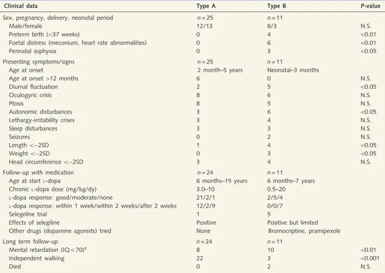
Table 3
Prevalence of additional clinical features and response to treatment in type A and B patients as a group
Clinical data Type A Type B P-value
Sex, pregnancy, delivery, neonatal period n = 25 n = 11
Male/female 12/13 8/3 N.S.
Preterm birth (537 weeks) 0 4 50.01
Foetal distress (meconium, heart rate abnormalities) 0 6 50.01
Perinatal asphyxia 0 3 50.05
Presenting symptoms/signs n = 25 n = 11
Age at onset 2 month–5 years Neonatal–3 months
Age at onset 412 months 6 0 N.S.
Diurnal fluctuation 2 5 50.05 Oculogyric crisis 8 6 N.S. Ptosis 8 5 N.S. Autonomic disturbances 3 6 50.05 Lethargy-irritability crises 3 4 N.S. Sleep disturbances 3 3 N.S. Seizures 0 2 N.S. Length 5 2SD 1 4 50.05 Weight 5 2SD 0 3 50.05 Head circumference 5 2SD 3 4 N.S.
Follow-up with medication n = 24 n = 11
Age at startL-dopa 6 months–15 years 6 months–7 years
ChronicL-dopa dose (mg/kg/dy) 3.0–10 0.5–20
L-dopa response: good/moderate/none 21/2/1 2/5/4
L-dopa response: within 1 week/within 2 weeks/after 2 weeks 12/2/9 0/0/7
Selegeline trial 1 5
Effects of selegiline Positive Positive but limited
Other drugs (dopamine agonists) tried None Bromocriptine, pramipexole
Long term follow-up n = 24 n = 11
Mental retardation (IQ570)a 8 10 50.01
Independent walking 22 3 50.001
Died 0 2 N.S.
The differences in the occurrence of clinical features in the two patient categories were studied by using Fisher’s exact test. N.S. = not significant. a See ‘Discussion’ section for details.
Figure 2 Concentrations of HVA (left panel) and MHPG (middle panel), and HVA/5HIAA ratio (right panel) in CSF of patients with THD at diagnosis (i.e. without treatment) according to THD subtype. Concentrations are given in percentage of the lower reference limit (2.5th percentile) in controls (see text). For comparisons between the two groups the Student’s t-test was used. ***P50.005.
on clinical grounds alone. The differential diagnosis of THD in neonates or very young infants with type B presentation initially encompasses a long list of progressive as well as stable, hereditary as well as acquired disorders. Type B THD is often accompanied by perinatal complications (Table 3), which may further distract
the attention in the direction of common infectious or
hypoxic-ischaemic encephalopathies. Type B THD can also mimic genetic disorders like catastrophic epileptic encephalopathies or mitochondrial disorders. Only extensive work-up, including cere-bral imaging and screening for inborn errors of metabolism, including CSF analysis, will lead to the correct diagnosis. In type A patients on the other end of the spectrum, the children with
‘parkinsonian’ features and L-dopa-responsive dystonia, clinical
recognition of the diagnosis might be easier. Importantly, THD with a relatively mild course can strongly mimic cerebral palsy, which may lead to serious diagnostic delay. Besides cerebral palsy, the differential diagnosis of ‘juvenile parkinsonism’ also in-cludes various other acquired as well as genetic disorders, among which GTP cyclohydrolase deficiency and other defects (like sepiapterine reductase deficiency) in the synthesis of the tyrosine
hydroxylase co-factor tetrahydrobiopterin (BH4) (Muller et al., 1998; Albanese et al., 2006; Tarsy and Simon, 2006; Muller, 2009). The lack of abnormalities on cerebral imaging studies and
a marked responsiveness to L-dopa are clues to the disorders of
neurotransmitter biosynthesis as a group.
In GTP cyclohydrolase deficiency, the most common defect of tetrahydrobiopterin biosynthesis, diurnal fluctuation of dystonia, can be a prominent hallmark. Both THD and GTP cyclohydrolase
deficiency are considered L-dopa-responsive dystonias, and have
been named Segawa syndrome and DYT5 in the past (see ‘Introduction’) (Muller et al., 1998; Albanese et al., 2006; Tarsy and Simon, 2006; Muller, 2009). THD, however, is generally more severe and characterized by an earlier onset of symptoms. Furthermore, the CSF profile of neurotransmitter and pterin metabolites discriminates between the two disorders. It has recently been proposed to designate these conditions into GTP cyclohydrolase deficiency and THD ‘dystonia 5a’ and ‘dystonia 5b’, respectively, illustrating their clinical and biochemical relation-ship and reflecting their different molecular basis (Muller et al., 1998).
Figure 3 HVA concentration (left panel) and HVA/5HIAA ratio (right panel) concentrations in CSF of THD patients at diagnosis (i.e. without treatment), in relation to age at onset of disease. Concentrations are given in percentage of the lower reference limit
(2.5th percentile) in controls. Regression coefficients are: HVA r2= 0.52 (P50.0001), HVA/5HIAA ratio r2= 0.66 (P50.0001).
Figure 4 Overview of all known pathogenic mutations in the TH gene. The cyclic adenosine monophosphate response element of the TH
Dopamine plays an important regulatory role in the neuro-endocrine system. Since dopamine suppresses the release of prolactin, THD may lead to hyperprolactinaemia. Indeed, serum prolactin may be increased in THD. In the literature, one patient with THD has been described who presented with galac-torrhoea due to hyperprolactinaemia before the neurological features appeared (Yeung et al., 2006). Dopamine is also known to play an important role in growth hormone secretion. Nevertheless, height and pubertal development are generally normal in THD.
Treatment and clinical course
The natural course of THD is—in sensu strictu—unknown. However, the observations in all patients reported here and in the literature indicate that the severe neurological features will never reverse as long as patients are not treated properly.
Since THD leads to dopamine deficiency in the CNS, treatment
with L-dopa is by far the strategy of first choice. In fact, the
lacking metabolite can simply be supplemented (Fig. 1). Drugs
containing L-dopa generally also contain a peripheral L-dopa
de-carboxylase inhibitor (benserazide or carbidopa) to prevent loss of
L-dopa in the circulation. Without further specification, we write
‘treatment withL-dopa’, meaningL-dopa combined with a
decarb-oxylase inhibitor.
L-dopa dosages commonly used in paediatric neurology range
from 3 to 10 mg/kg bodyweight per day, given in three doses.
Although L-dopa-responsive THD patients were usually treated
with an ongoingL-dopa dose in this range, many types A and B
patients started with lower doses, tolerating only very gradual in-creases in dosage over weeks or even months (see below). Type B
patients especially were often extremely sensitive toL-dopa. This
hypersensitivity necessitated initial dosages below 0.5 mg/kg bodyweight per day, in four (or even six) divided doses, and
impeded some patients from being treated with L-dopa at all
(Brautigam et al., 1999; de Lonlay et al., 2000; Dionisi-Vici et al., 2000; Janssen et al., 2000; Haussler et al., 2001; Grattan-Smith et al., 2002; Hoffmann et al., 2003; Zafeiriou et al., 2009).
Alternatively and in addition to L-dopa, THD patients can
ra-tionally be treated with inhibitors of dopamine degradation like selegiline, assuming that some dopamine is still formed in tyrosine hydroxylase-deficient neurons. Selegiline as well as dopamine agonists, anticholinergic drugs and benzodiazepines available for the treatment of dystonia (Albanese et al., 2006; Tarsy and Simon, 2006) were, however, hardly ever used in this series.
Patients who toleratedL-dopa well generally showed a good or
at least moderate response with dramatic improvement of move-ments due to disappearance of hypokinesia, tremor, rigidity and dystonia, and subsequent impressive gain in motor functions. Children who had been in a wheelchair for years started to walk again. Good responders generally also responded early (Table 3).
These patients only developed L-dopa induced dyskinesia when
dosages were increased too quickly or in too great increments. To date, only one subject (Patient 23), developed dyskinesia prob-ably as the consequence of the long-term, i.e. 13 year, treatment
with L-dopa. All others tolerated L-dopa very well and remained
without serious side effects after more than one (Cases 6, 9, 10, 13, 14, 16, 22, 24, 25, 27, 31) or even two or three (Cases 1–5)
decades of treatment. In a limited number of L-dopa-responsive
THD patients, lumbar punctures were performed to monitor treat-ment effects at the biochemical level. In those cases, clinical re-sponses were good irrespective to the fact that HVA never reached normal levels.
Despite appropriate treatment, long-term cognitive develop-ment was subnormal in many patients with THD. We did not systematically study cognitive profiles in this series but based on available test reports, school performance, social functioning and
Table 4
Mutations in the tyrosine hydroxylase gene that
lead to THD
Number Exon Mutation Protein
change Number of alleles affected 1 Promotor c. 71C4T 3 2 Promotor c. 70G4A 9 3 Promotor c. 69T4A 2 4 3 c.295delC p.Leu99fs 1 5 3 c.296delT p.Leu99fs 1 6 3 n.r. p.Arg169X 2 7 5 c.614T4C p.Leu205Pro 2 8 5 c.620G`A p.Cys207Tyr 1 9 6 c.680A`G p.Asp227Gly 1 10 6 c.698G4A p.Arg233His 28 11 6 c.707T4C p.Leu236Pro 6 12 6 c.721G`A p.Ala241Thr 2 13 6 c.736C4T p.His246Tyr 1 14 7 c.739G4A p.Gly247Ser 1 15 7 c.776A`G p.Glu259Gly 1 16 8 c.826A4C p.Thr276Pro 1 17 8 c.901C4G p.Pro301Ala 2 18 8 c.926T4C p.Phe309Ser 2 19 9 c.941C4T p.Thr314Met 1 20 9 c.956G4C p.Arg319Pro 1 21 9 c.982C4T p.Arg328Trp 1 22 9 c.1010G4A p.Arg337His 2 23 10 c.1076G4T p.Cys359Phe 2 24 10 c.1125C4G p.Phe375Leu 1 25 10 c.1127C4T p.Ala376Val 2 26 11 c.1141C4A p.Gln381Lys 4 27 11 c.1159C4A p.Leu387Met 1 28 11 c.1181T`C p.Ile394Thr 2 29 11 c.1196C4T p.Thr399Met 1 30 Intron 11 c.1198–24T4A p.? 2 31 12 c.1240G4A p.Gly414Arg 2 32 13 c.1375C`T p.Gln459X 1 33 13 c.1399A4G p.Ser467Gly 1 34 14 c.1475C4T p.Pro492Leu 2 35 14 c.1481C4T p.Thr494Met 2 36 14 c.1493A4G p.Asp498Gly 4 37 14 c.1529T4A p.Leu510Gln 2
This table includes all mutations reported to date in THD, and six novel mutations (in bold) reported in this article. In case of affected sib-pairs, both patients are included. The total number of mutated alleles is 100. For all mutations: see Tables 1 (our series of patients) and 3 (all patients reported by others). n.r. = not reported.
clinical impression by experienced neurologists, it was concluded that mild to moderate mental retardation occurred in 33 and 91% of Types A and B patients, respectively (Table 3). In none of the patients we encountered signs of cognitive decline under treatment.
Based on shared experiences, but impossible to prove due to the low number of patients, we think that early diagnosis and treat-ment of THD improves the final outcome with regard to motor as well as cognitive functions. On the other hand, it has extensively been demonstrated in animal studies that dopamine plays a vital role in foetal brain development (Zhou et al., 1995; Araki et al., 2007). Dopamine deficiency might therefore cause, already before birth, irreversible structural brain abnormalities at the microscopic level and leading to mental retardation.
Taking all data together, THD can best be treated with an initial
L-dopa dose of 0.5–1 (for type B patients) to 3 (for type A
pa-tients) mg/kg bodyweight per day, divided over three or four doses. When tolerated well, the dose can slowly be increased until the desired clinical response is observed or the occurrence of adverse effects forces dose reduction. It should be kept in mind that some patients respond only in the course of months: one should thus wait long enough to see the final effects in ap-parent moderate or non-responders. We think that type B patients
with extremeL-dopa hypersensitivity might be ideal candidates for
continuous duodenal administration of a soluble formulation of
L-dopa (duodopa), although we did not encounter this approach
in our series and the literature.
Biochemistry and genetics
Cathecholamine biosynthesis was long thought to occur only in specific neuronal cell populations in the CNS, sympathetic periph-eral nervous system, adrenal glands and the kidneys. Interestingly, however, the presence of the tyrosine hydroxylase protein, its mRNA and its enzymatic activity, have been demonstrated in other non-neuronal tissues capable of catecholamine biosynthesis, such as lymphoid tissues, exocrine pancreas and the gastrointes-tinal tract (Mezey et al., 1996; Eisenhofer et al., 1997). Human THD leads to a neurological disorder, apparently leaving the other organs unaffected. An explanation for this observation might be that the brain is the most vulnerable organ, already severely affected by relatively minor changes in the TH gene (see below). A second explanation might be found in other enzymes, expressed in non-neuronal tissues, which can hydroxylate tyrosine. Tyrosinase (E.C. 1.14.18.1), for example, is expressed in the epidermis and plays a crucial role in melanin biosynthesis from tyrosine (Rios et al., 1999). In humans, tyrosinase deficiency leads to oculocutaneous albinism type 1 (OMIM 606933) (Oetting et al., 2003). In mice, tyrosinase substantially contributes to peripheral, tyrosine hydroxylase-independent dopamine produc-tion (Eisenhofer et al., 2003).
The biochemical diagnosis of THD would ideally rely on direct measurement of enzyme activity in tissue samples, blood cells or cultured fibroblasts. Since it was traditionally thought that tyro-sine hydroxylase was only expressed in tissues that would not be available for enzymatic analyses, we and others have always relied on CSF metabolites (Fig. 1) and TH gene
mutation analysis to diagnose THD. The collected data of the large series of patients described here clearly demonstrate that the most severely affected patients have the lowest concentrations of neurotransmitter metabolites in CSF (Fig. 3). The HVA concen-tration and HVA/5HIAA ratio in CSF showed overlap between Types A and B patients, however this overlap was only minor for the HVA/5HIAA ratio (Fig. 2). These parameters (especially the HVA/5HIAA ratio in CSF), most probably reflecting the degree of residual tyrosine hydroxylase activity in the brain, may
thus be used to predict L-dopa responsiveness and overall
outcome.
Measurements of phenylalanine and tyrosine in body fluids, and urinary concentrations of catecholamines, HVA and MHPG, are non-informative in patients with THD (Brautigam et al., 1998; Wevers et al., 1999; Hoffmann et al., 2003). The surprisingly often normal urinary dopamine excretion in THD is hypothetically attributed to residual tyrosine hydroxylase enzyme activity in per-ipheral non-neuronal tissues or alternative enzymes with the cap-acity to hydroxylate tyrosine, as discussed above. The fact that profound cerebral dopamine deficiency due to defective dopamine biosynthesis can be accompanied by normal urinary dopamine ex-cretion makes analysis of urine unreliable, even potentially mis-leading, in the diagnostic work-up for dopamine biosynthesis disorders.
The human TH gene (mRNA type 1) contains 14 exons with an open reading frame of 1491 bp encoding for a protein with 497 amino acids that is highly conserved among various species (Nagatsu and Ichinose, 1991) Tables 1, 2 and 4, and Fig. 4, give an overview of the 37 different TH mutations in THD pa-tients. Out of 100 alleles, 95 were affected by missense mutations leading to amino acid substitutions in the protein with subsequent partial loss of enzyme activity. The pathogenic effects of some missense mutations on the protein have been confirmed, (Ludecke et al., 1995, 1996; Knappskog et al., 1995; Royo et al., 2005; Haavik et al., 2008) but most can only be designated pathogenic based on indirect evidence. Interestingly, we and others have recently identified pathogenic mutations in the pro-moter region of the TH gene as a rare underlying genetic mech-anism (Ribases et al., 2007; Verbeek et al., 2007). A founder effect has been described for the c.698G4A mutation in the Dutch population (van den Heuvel et al., 1998). Except for this and two other mutations (c. 70G4A and c.707T4C), almost all other pathogenic changes in the TH gene are so-called ‘private’ mutations (Table 4). Based on the latter finding, it can be ex-pected that THD occurs in all parts of the world, irrespective of the fact that up to now most patients were diagnosed in Western Europe.
The relatively high number of patients in this paper allows us to discuss possible genotype–phenotype correlations in THD. Only five patients, from four families, harboured deleterious mutations that lead to protein truncation: namely c.295delC and c.296delT, which both lead to p.Leu99fs; p.Arg169X (the c.DNA change was not reported in the two affected siblings); and c.1375C4T, which causes p.Gln459X. Heterozygosity for each of these mutations was found in type A as well as type B patients. Until now, no patient has been reported with homozygosity or compound heterozygos-ity for two truncating TH mutations. This observation most
probably indicates that complete loss of tyrosine hydroxylase ac-tivity is incompatible with human life, comparable with the find-ings in knockout mice (Zhou et al., 1995). Homozygosity for the common c.698G4A mutation and homozygosity for the c.707T4C mutation was found in both THD phenotypes (Table 1), leaving these genotypes without predictive value with regard to THD phenotype. Importantly, however, all patients carrying at least one promoter mutation (n = 8, Tables 1 and 2) suffered from
type A THD with goodL-dopa responsiveness. As discussed
pre-viously, reduced TH gene transcription apparently leaves
signifi-cant residual tyrosine hydroxylase enzyme activity, likely
corresponding to a relatively mild type A phenotype (Ribases et al., 2007; Verbeek et al., 2007).
The differential diagnosis in children and adults with movement disorders is essentially different. This explains the remarkable di-versity in the diagnostic approach depending on the patient’s age. Adult patients with dystonia can be investigated at the genetic level after dedicated neurological classification (Muller et al., 1998; Albanese et al., 2006; Tarsy and Simon, 2006; Wu et al., 2008; Clot et al., 2009; Muller, 2009), while children often have to be exposed to more extensive and invasive diagnostic procedures to illuminate the underlying cause of their disorder (Assmann et al., 2003). In contrast to what is advocated for adult patients, we strongly advise performance of a lumbar puncture in children with otherwise unexplained (simple and complex) movement disorders, to diagnose or rule out potentially treatable conditions like THD.
In conclusion, THD is a severe but often very treatable neurometabolic disorder resulting from cerebral catecholamine deficiency. The diagnosis of THD relies on clinical suspicion and the analysis of CSF metabolites. Importantly, the CSF concentra-tion of HVA and HVA/5HIAA ratio correlate with the severity of the clinical phenotype. THD can be proven by demonstrating mutations in the TH gene or its promoter region. The disorder is almost exclusively caused by missense mutations, suggesting that mutations with more deleterious effects on the protein will be incompatible with life. Genotype–phenotype correlations do not exist for the common c.698G4A and c.707T4C mutations. Carriership of at least one promotor mutation, however, apparent-ly predicts type A THD. Most patients with THD, but not all, can
successfully be treated withL-dopa.
Acknowledgements
We thank Prof. Robert Surtees, London, UK, who sadly died during the preparation of this article. Prof. Robert Surtees was well-known for his expertise in the field of paediatric movement disorders, and contributed to the manuscript in its early phases by sharing his clinical experiences with us, and by offering helpful suggestions.
Funding
The Swiss National Science Foundation (Grant No.
3100A0-119982/1) (to N.B.); Zon-MW Innovational Research Grant (Vidi Program No. 917.46.331 to M.M.V.).
References
Albanese A, Barnes MP, Bhatia KP, Fernandez-Alvarez E, Filippini G, Gasser T, et al. A systematic review on the diagnosis and treatment of primary (idiopathic) dystonia and dystonia plus syndromes: report of an EFNS/MDS-ES Task Force. Eur J Neurol 2006; 13: 433–44. Araki KY, Sims JR, Bhide PG. Dopamine receptor mRNA and protein
expression in the mouse corpus striatum and cerebral cortex during pre- and postnatal development. Brain Res 2007; 1156: 31–45. Assmann B, Surtees R, Hoffmann GF. Approach to the diagnosis of
neurotransmitter diseases exemplified by the differential diagnosis of childhood-onset dystonia. Ann Neurol 2003; 54(Suppl 6): S18–24. Brautigam C, Steenbergen-Spanjers GC, Hoffmann GF, Dionisi-Vici C,
van den Heuvel LP, Smeitink JA, et al. Biochemical and molecular genetic characteristics of the severe form of tyrosine hydroxylase defi-ciency. Clin Chem 1999; 45: 2073–8.
Brautigam C, Wevers RA, Jansen RJ, Smeitink JA, de Rijk-van Andel JF, Gabree¨ls FJ, et al. Biochemical hallmarks of tyrosine hydroxylase defi-ciency. Clin Chem 1998; 44: 1897–904.
Castaigne P, Rondot P, Ribadeau-Dumas JL, Said G. Progressive extra-pyramidal disorder in 2 young brothers. Remarkable effects of treat-ment with L-dopa. Rev Neurol (Paris) 1971; 124: 162–6.
Clot F, Grabli D, Cazeneuve C, Roze E, Castelnau P, Chabrol B, et al. Exhaustive analysis of BH4 and dopamine biosynthesis genes in patients with Dopa-responsive dystonia. Brain 2009; 132: 1753–63. de Lonlay P, Nassogne MC, van Gennip AH, van Cruchten AC, Billatte
de Villemeur T, Cretz M, et al. Tyrosine hydroxylase deficiency unre-sponsive to L-dopa treatment with unusual clinical and biochemical presentation. J Inherit Metab Dis 2000; 23: 819–25.
de Rijk-Van Andel JF, Gabreels FJ, Geurtz B, Steenbergen-Spanjers GC, van Den Heuvel LP, Smeitink JA, et al. L-dopa-responsive infantile hypokinetic rigid parkinsonism due to tyrosine hydroxylase deficiency. Neurology 2000; 55: 1926–8.
Diepold K, Schutz B, Rostasy K, Wilken B, Hougaard P, Gu¨ttler F, et al. Levodopa-responsive infantile parkinsonism due to a novel mutation in the tyrosine hydroxylase gene and exacerbation by viral infections. Mov Disord 2005; 20: 764–7.
Dionisi-Vici C, Hoffmann GF, Leuzzi V, Hoffken H, Bra¨utigam C, Rizzo C, et al. Tyrosine hydroxylase deficiency with severe clinical course: clin-ical and biochemclin-ical investigations and optimization of therapy. J Pediatr 2000; 136: 560–2.
Doummar D, Clot F, Vidailhet M, Afenjar A, Durr A, Brice A, et al. Infantile hypokinetic-hypotonic syndrome due to two novel mutations of the tyrosine hydroxylase gene. Mov Disord 2009; 24: 943–5. Eisenhofer G, Aneman A, Friberg P, Hooper D, Fa˚ndriks L, Lonroth H,
et al. Substantial production of dopamine in the human gastrointestinal tract. J Clin Endocrinol Metab 1997; 82: 3864–71.
Eisenhofer G, Tian H, Holmes C, Matsunaga J, Roffler-Tarlov S, Hearing VJ. Tyrosinase: a developmentally specific major determinant of peripheral dopamine. FASEB J 2003; 17: 1248–55.
Furukawa Y, Graf WD, Wong H, Shimadzu M, Kish SJ. Dopa-responsive dystonia simulating spastic paraplegia due to tyrosine hydroxylase (TH) gene mutations. Neurology 2001; 56: 260–3.
Giovanniello T, Leuzzi V, Carducci C, Carducci C, Sabato ML, Artiola C, et al. Tyrosine hydroxylase deficiency presenting with a biphasic clin-ical course. Neuropediatrics 2007; 38: 213–5.
Grattan-Smith PJ, Wevers RA, Steenbergen-Spanjers GC, Fung VS, Earl J, Wilcken B. Tyrosine hydroxylase deficiency: clinical manifestations of catecholamine insufficiency in infancy. Mov Disord 2002; 17: 354–9. Haavik J, Blau N, Thony B. Mutations in human monoamine-related
neurotransmitter pathway genes. Hum Mutat 2008; 29: 891–902. Haussler M, Hoffmann GF, Wevers RA. L-dopa and selegiline for tyrosine
hydroxylase deficiency. J Pediatr 2001; 138: 451–2.
Hoffmann GF, Assmann B, Brautigam C, Dionisi-Vici C, Ha¨ussler M, de Klerk JB, et al. Tyrosine hydroxylase deficiency causes progressive encephalopathy and dopa-nonresponsive dystonia. Ann Neurol 2003; 54(Suppl 6): S56–65.
Janssen RJ, Wevers RA, Haussler M, Luyten JA, Steenbergen-Spanjers GC, Hoffmann GF, et al. A branch site mutation leading to aberrant splicing of the human tyrosine hydroxylase gene in a child with a severe extrapyramidal movement disorder. Ann Hum Genet 2000; 64: 375–82.
Knappskog PM, Flatmark T, Mallet J, Ludecke B, Bartholome K. Recessively inherited L-DOPA-responsive dystonia caused by a point mutation (Q381K) in the tyrosine hydroxylase gene. Hum Mol Genet 1995; 4: 1209–12.
Ludecke B, Dworniczak B, Bartholome K. A point mutation in the tyr-osine hydroxylase gene associated with Segawa’s syndrome. Hum Genet 1995; 95: 123–5.
Ludecke B, Knappskog PM, Clayton PT, Surtees RA, Clelland JD, Heales SJ, et al. Recessively inherited L-DOPA-responsive parkinsonism in infancy caused by a point mutation (L205P) in the tyrosine hydro-xylase gene. Hum Mol Genet 1996; 5: 1023–8.
Mezey E, Eisenhofer G, Harta G, Hansson S, Gould L, Hunyady B, et al. A novel nonneuronal catecholaminergic system: exocrine pancreas synthesizes and releases dopamine. Proc Natl Acad Sci USA 1996; 93: 10377–82.
Moller LB, Romstad A, Paulsen M, Hougaard P, Ormazabal A, Pineda M, et al. Pre- and postnatal diagnosis of tyrosine hydroxylase deficiency. Prenat Diagn 2005; 25: 671–5.
Muller U. The monogenic primary dystonias. Brain 2009; 132: 2005–25. Muller U, Steinberger D, Nemeth AH. Clinical and molecular genetics of
primary dystonias. Neurogenetics 1998; 1: 165–77.
Nagatsu T, Ichinose H. Comparative studies on the structure of human tyrosine hydroxylase with those of the enzyme of various mammals. Comp Biochem Physiol C 1991; 98: 203–10.
Ng PC, Henikoff S. Predicting deleterious amino acid substitutions. Genome Res 2001; 11: 863–74.
Oetting WS, Fryer JP, Shriram S, King RA. Oculocutaneous albinism type 1: the last 100 years. Pigment Cell Res 2003; 16: 307–11.
Ribases M, Serrano M, Fernandez-Alvarez E, Pahisa S, Ormazabal A, Garcı´a-Cazorla A, et al. A homozygous tyrosine hydroxylase gene promoter mutation in a patient with dopa-responsive encephalopathy: clinical, biochemical and genetic analysis. Mol Genet Metab 2007; 92: 274–7.
Rios M, Habecker B, Sasaoka T, Eisenhofer G, Tian H, Landis S, et al. Catecholamine synthesis is mediated by tyrosinase in the absence of tyrosine hydroxylase. J Neurosci 1999; 19: 3519–26.
Rondot P, Ziegler M. Dystonia–L-dopa responsive or juvenile parkinson-ism? J Neural Transm Suppl 1983; 19: 273–81.
Rondot P, Aicardi J, Goutieres F, Ziegler M. [Dopa-sensitive dystonia]. Rev Neurol (Paris) 1992; 148: 680–6.
Royo M, Daubner SC, Fitzpatrick PF. Effects of mutations in tyrosine hydroxylase associated with progressive dystonia on the activity and stability of the protein. Proteins 2005; 58: 14–21.
Schiller A, Wevers RA, Steenbergen GC, Blau N, Jung HH. Long-term course of L-dopa-responsive dystonia caused by tyrosine hydroxylase deficiency. Neurology 2004; 63: 1524–1526.
Surtees R, Clayton P. Infantile parkinsonism-dystonia: tyrosine hydroxy-lase deficiency. Mov Disord 1998; 13: 350.
Swaans RJ, Rondot P, Renier WO, van den Heuvel LP, Steenbergen-Spanjers GC, Wevers RA. Four novel mutations in the tyrosine hydro-xylase gene in patients with infantile parkinsonism. Ann Hum Genet 2000; 64: 25–31.
Tarsy D, Simon DK. Dystonia. N Engl J Med 2006; 355: 818–29. van den Heuvel LP, Luiten B, Smeitink JA, de Rijk-van Andel JF,
Hyland K, Steenbergen-Spanjers GC, et al. A common point mutation in the tyrosine hydroxylase gene in autosomal recessive L-DOPA-responsive dystonia in the Dutch population. Hum Genet 1998; 102: 644–6.
Verbeek MM, Blom AM, Wevers RA, Lagerwerf AJ, van de GJ, Willemsen MA. Technical and biochemical factors affecting cerebrosp-inal fluid 5-MTHF, biopterin and neopterin concentrations. Mol Genet Metab 2008; 95: 127–32.
Verbeek MM, Steenbergen-Spanjers GC, Willemsen MA, Hol FA, Smeitink J, Seeger J, et al. Mutations in the cyclic adenosine monopho-sphate response element of the tyrosine hydroxylase gene. Ann Neurol 2007; 62: 422–6.
Wevers RA, de Rijk-Van Andel JF, Brautigam C, Geurtz B, van den Heuvel LP, Steenbergen-Spanjers GC, et al. A review of biochemical and molecular genetic aspects of tyrosine hydroxylase deficiency including a novel mutation (291delC). J Inherit Metab Dis 1999; 22: 364–73.
Wu ZY, Lin Y, Chen WJ, Zhao GX, Xie H, Murong SX, et al. Molecular analyses of GCH-1, TH and parkin genes in Chinese dopa-responsive dystonia families. Clin Genet 2008; 74: 513–21.
Yeung WL, Lam CW, Hui J, Tong SF, Wu SP. Galactorrhea-a strong clinical clue towards the diagnosis of neurotransmitter disease. Brain Dev 2006; 28: 389–91.
Zafeiriou DI, Willemsen MA, Verbeek MM, Vargiami E, Ververi A, Wevers R. Tyrosine hydroxylase deficiency with severe clinical course. Mol Genet Metab 2009; 97: 18–20.
Zhou QY, Quaife CJ, Palmiter RD. Targeted disruption of the tyrosine hydroxylase gene reveals that catecholamines are required for mouse fetal development. Nature 1995; 374: 640–3.