during mouse embryonic germ cell development
The MIT Faculty has made this article openly available.
Please share
how this access benefits you. Your story matters.
Citation
Gura, Megan A. et al. "Dynamic and regulated TAF gene expression
during mouse embryonic germ cell development." PLoS Genetics
16, 1 (January 2020): e1008515 © 2020 Gura et al
As Published
http://dx.doi.org/10.1371/journal.pgen.1008515
Publisher
Public Library of Science (PLoS)
Version
Final published version
Citable link
https://hdl.handle.net/1721.1/125273
Terms of Use
Creative Commons Attribution 4.0 International license
RESEARCH ARTICLE
Dynamic and regulated TAF gene expression
during mouse embryonic germ cell
development
Megan A. GuraID1, Maria M. MikedisID2, Kimberly A. Seymour1, Dirk G. de Rooij2, David C. PageID2,3,4, Richard N. FreimanID1*
1 Brown University, MCB Graduate Program and Department of Molecular Biology, Cell Biology and
Biochemistry, Providence, RI, United States of America, 2 Whitehead Institute, Cambridge, MA, United States of America, 3 Department of Biology, Massachusetts Institute of Technology, Cambridge, MA, United States of America, 4 Howard Hughes Medical Institute, Whitehead Institute, Cambridge, MA, United States of America
*Richard_Freiman@Brown.edu
Abstract
Germ cells undergo many developmental transitions before ultimately becoming either eggs or sperm, and during embryonic development these transitions include epigenetic repro-gramming, quiescence, and meiosis. To begin understanding the transcriptional regulation underlying these complex processes, we examined the spatial and temporal expression of TAF4b, a variant TFIID subunit required for fertility, during embryonic germ cell develop-ment. By analyzing published datasets and using our own experimental system to validate these expression studies, we determined that both Taf4b mRNA and protein are highly germ cell-enriched and that Taf4b mRNA levels dramatically increase from embryonic day 12.5–18.5. Surprisingly, additional mRNAs encoding other TFIID subunits are coordinately upregulated through this time course, including Taf7l and Taf9b. The expression of several of these germ cell-enriched TFIID genes is dependent upon Dazl and/or Stra8, known regu-lators of germ cell development and meiosis. Together, these data suggest that germ cells employ a highly specialized and dynamic form of TFIID to drive the transcriptional programs that underlie mammalian germ cell development.
Author summary
Assisted reproductive therapy and fertility preservation are increasingly used to improve human reproduction across the world, yet there are still many unanswered questions regarding what factors govern the development of eggs and sperm and how these factors work together. We previously identified a subunit of the general transcription factor TFIID, TAF4b, that is essential for fertility. However, many basic characteristics of how Taf4b and its associated TFIID family members contribute to the formation of healthy sperm and eggs in mice and humans remain unknown. In this study, we find that mouse Taf4b and several closely related TFIID subunits become highly abundant during mouse embryonic gonad development, specifically in the cells that ultimately become eggs and a1111111111 a1111111111 a1111111111 a1111111111 a1111111111 OPEN ACCESS
Citation: Gura MA, Mikedis MM, Seymour KA, de
Rooij DG, Page DC, Freiman RN (2020) Dynamic and regulated TAF gene expression during mouse embryonic germ cell development. PLoS Genet 16 (1): e1008515.https://doi.org/10.1371/journal. pgen.1008515
Editor: Paula E. Cohen, Cornell University, UNITED
STATES
Received: June 21, 2019 Accepted: November 11, 2019 Published: January 8, 2020
Copyright:© 2020 Gura et al. This is an open access article distributed under the terms of the
Creative Commons Attribution License, which permits unrestricted use, distribution, and reproduction in any medium, provided the original author and source are credited.
Data Availability Statement: All sequencing data
are available from NCBI GEOhttps://www.ncbi.nlm. nih.gov/geounder accession number GSE139005 and NCBI SRA (https://www.ncbi.nlm.nih.gov/sra) under accession number SRP226023. The RNA-seq data used in this research is from the following accession numbers: the mouse data from E9.5 to E18.5 in female and male sorted Oct4-EGFP gonads used for Figs1,2,4,5, and10was obtained through ArrayExpress: E-MTAB-4616; the human data comparing female germ cells versus somatic cells used forFig 3Awas obtained through
sperm. Here, we analyzed data from public repositories and isolated these developing cells to examine their gene expression patterns throughout embryonic development. Together these data suggest that the dynamic expression ofTaf4b and other TFIID family members are dependent on the well-established reproductive cell regulatorsDazl and Stra8. This understanding ofTaf4b gene expression and regulation in mouse reproductive cell devel-opment is likely conserved during develdevel-opment of human cells and offers novel insights into the interconnectedness of the factors that govern the formation of healthy eggs and sperm.
Introduction
Healthy development and maintenance of germ cells is essential for the continuation of all sex-ually reproducing species. In the United States, approximately 10% of individuals face fertility issues, and worldwide, at least 1% of females experience primary ovarian insufficiency (POI), which is associated with infertility [1,2]. However, in most instances of POI and infertility, the underlying molecular causes are unknown [3]. Work from our laboratory has shown that a TBP-Associated Factor 4b (Taf4b)-deficient mouse model recapitulates many aspects of POI, including a premature depletion of the ovarian reserve and female infertility [4,5]. Taf4b-defi-ciency in the mouse also leads to progressive male infertility in early adulthood that is associ-ated with exhaustion of their adult spermatogonial stem cell (SSC) pool [6]. Since human studies implicate TAF4B as important for fertility and oocyte quality [7–9], our efforts to understand the molecular mechanisms underlying the expression and function of TAF4b in mouse germ cell development may contribute to our increased understanding of human fertil-ity and infertilfertil-ity.
TAF4b is a gonad-enriched subunit of the basal transcription factor TFIID, which is a com-plex of TATA-binding protein (TBP) and 13–14 TBP-associated factors (TAFs) [5,10]. TFIID, as part of a larger ensemble of the basal transcription machinery, recruits RNA Polymerase II (RNAPII) to the core promoters of genes. However, unlike its paralogTaf4a, which is expressed ubiquitously,Taf4b is more highly expressed in the mouse ovary and testis com-pared to other tissues [5]. Most recently, we demonstrated that TAF4b is a critical regulator of female meiosis I in the mouse [11].Taf4b mRNA expression is also highly correlated with the expression of many important germline genes during human fetal ovary development, such as Deleted in Azoospermia-like (Dazl), which encodes a germ cell-specific RNA-binding protein that promotes translation and/or prevents the degradation of its target mRNAs [11–13]. In female mice as early as E13.5, when germ cells initiate meiosis,Taf4b-deficiency results in reduced expression of meiotic transcripts, includingStimulated by Retinoic Acid 8 (Stra8), which is a master regulator of meiosis [14]. This reduced meiotic gene expression is followed by defective meiotic progression, and excessive perinatal oocyte loss [11].Stra8 mRNA has also been found to be greatly reduced inTaf4b-deficient male newborn testes compared to wild-type andTaf4b-heterozygous testes, indicating that this disruption of meiosis is likely shared between female and maleTaf4b-deficient mice [15].
We previously detected TAF4b occupancy at the proximal promoters ofDazl and Stra8 in E18.5 embryonic ovaries, which suggests that TAF4b directly regulates these genes [11].Stra8 was recently identified as a major transcriptional regulator of meiotic initiation [14]. However, it was postulated thatStra8 alone is not sufficient to induce meiosis, and other epigenetic and transcriptional regulators likely work together withStra8 to orchestrate meiotic initiation. It is NCBI SRA: SRP057098; the human data
comparing male germ cells versus somatic cells used forFig 3Bwas obtained through NCBI SRA: SRP045294; the human timeline data from female and male cKit sorted gonads used forFig 3C–3D
was obtained through NCBI SRA: SRP049981; the mouse data using E14.5 WT, Dazl-KO, and Stra8-KO whole ovaries used for Figs6–7was obtained through NCBI SRA: SRP059601 and SRP059599; the mouse data using PND6 WT and Dazl-KO spermatogonia used forFig 6was obtained through NCBI SRA: SRP128645; the mouse data using male WT and Stra8-KO preleptotene germ cells used forFig 7was obtained through NCBI SRA: SRP150721, the mouse data from E9.5 to E15.5 in female and male sorted gonads, in vitro PGCLC culture, and Stra8 knockdown used for
S2A–S2B FigandS6B–S6F Figwas obtained through NCBI SRA: SRP098051, and the mouse data from E11.5 to E16.5 female and male sorted Oct4-EGFP gonads used forS2C–S2D Figwas obtained through NCBI SRA: ERP001953. The ChIP-seq used for this research, where Stra8-FLAG/FLAG postnatal testis synchronized for preleptotene germ cells used forFig 9andS6A Fig
were obtained from NCBI SRA: SRP150721.
Funding: MAG received funding from National
Institutes of Health award 1F31HD097933 and 5T32GM007601. MMM was supported by a Lalor Foundation Postdoctoral Fellowship (http:// lalorfound.org) and a NRSA Postdoctoral Fellowship, Grant Number F32HD093391 from NICHD. RNF received funding from National Institutes of Health awards 2R56HD065445 and 1R01HD091848. DCP was funded by the Howard Hughes Medical Institute (https://www.hhmi.org/). The Brown University Flow Cytometry and Sorting Facility has received generous support in part by the National Institutes of Health (NCRR Grant No. 1S10RR021051) and the Division of Biology and Medicine, Brown University. The funders had no role in study design, data collection and analysis, decision to publish, or preparation of the manuscript.
Competing interests: The authors have declared
unknown what transcriptional program coordinates withStra8 in germ cells to produce gametes.
While female germ cells initiate meiosis at E13.5, male germ cells remain mitotically active until ~E16.5 [16]. During this time,Taf4b-deficient male mice display deficient embryonic gonocyte numbers, as well as defective postnatal germ cell proliferation and development [6]. Therefore, during early postnatal development, a paucity of germ cells leads to a narrow win-dow of fertility that may result from an inability to establish a healthy embryonic germ cell and/or postnatal SSC pool. One common theme in both adult females and males is that their Taf4b-dependent fertility deficits can be traced back to defects in embryonic germ cell development.
Despite the knowledge thatTaf4b function is essential for embryonic germ cell develop-ment in both sexes, the precise expression profile and mechanism of action for this fertility transcription factor remains unknown. Thus, it is uncertain whetherTaf4b uses similar molec-ular mechanisms in female and male germ cells. This gap in knowledge makes understanding the role ofTaf4b in embryonic gametogenesis more challenging. To develop a better under-standing ofTaf4b, we implemented complementary computational and experimental tools to assess the sex-specific timing and localization ofTaf4b expression in the embryonic mouse gonad. We found thatTaf4b mRNA and protein are highly enriched in the germ cells of the embryonic gonad and that strikingly similar expression patterns were observed for other TFIID subunits, such asTaf7l, which is essential for male germ cell maturation and fertility [17,18]. In addition, we demonstrate thatTaf4b is the only component of the TFIID and SAGA complexes that is directly regulated by both DAZL and STRA8 during meiosis. Based upon these data,Taf4b functions in an interdependent gene regulatory network with DAZL and STRA8 during meiosis.
Results
Taf4b is significantly enriched and dynamically expressed in the germ cells
of the embryonic ovary and testis
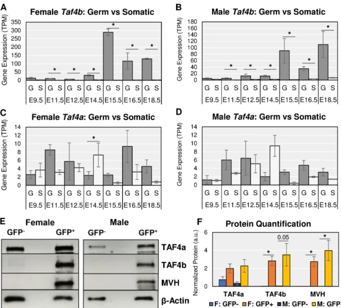
To evaluateTaf4b expression in the embryonic gonad, we reprocessed an RNA-sequencing (RNA-seq) dataset that used Oct4-EGFP mice covering 7 developmental time points, which range from embryonic day 9.5 (E9.5) to E18.5, wherein each time point had appropriate female and male replicates [19,20]. This sex-specific time course was further broken down into germ cell (GFP+) and gonadal somatic (GFP-) cells by performing fluorescence activated cell sorting (FACS). Principal component analysis (PCA) (Fig 1) indicates distinct grouping of germ cells and somatic cells on opposite sides of the PC1 dimension and further grouping of germ cells based on their developmental milestones.
We first examined in which embryonic gonadal cell typeTaf4b mRNA is expressed since the embryonic ovary and testis are heterogeneous tissues composed of germ and somatic cells. In both tissues from E11.5 to E18.5,Taf4b mRNA is significantly and consistently enriched in the GFP+germ cells (Fig 2A and 2B, log2FC > |0.25|, p-adj. < 0.05,S1 Table).Taf4b
expres-sion, shown in transcripts per million (TPM,S2 Table), remains relatively low in all gonadal somatic cells throughout the time course. The slight increase inTaf4b mRNA in male somatic samples at E18.5 (Fig 2B) is not significant. Thus,Taf4b mRNA expression is preferentially expressed in the germ cells of the embryonic gonad. We hypothesize thatTaf4b plays a special-ized role in germ cell development and fertility in contrast to its more ubiquitously expressed paralogTaf4a. We thus compared Taf4a mRNA to Taf4b expression levels in this dataset (Fig 2C and 2D). In contrast toTaf4b, Taf4a mRNA expression was in general not significantly dif-ferent between germ cell and somatic cell types. Furthermore,Taf4a mRNA was detected at
lower TPMs over the entire time course, which indicates thatTaf4a mRNA is reduced com-pared toTaf4b mRNA.
To test if the enrichment ofTaf4b mRNA within germ cells is reflected at the protein level, we performed western blots on whole cell protein extracts of E13.5 germ and somatic cells derived from a similar Oct4-EGFP mouse line. At E13.5, the signal for mouse vasa homolog (MVH), a germ cell-specific marker, is only found in the GFP+protein samples of female and male cells (Fig 2E). This marker expression indicates that we successfully separated the germ cells from the somatic cells. Based on quantification of western blot signal from 3 biological replicates, TAF4b protein expression is predominantly detected in the GFP+germ cells from both female and male samples, whereas TAF4a is expressed in both GFP+germ cells and GFP -somatic cells (Fig 2E and 2F,S1A–S1D Fig). Even at higher GFP-somatic cell numbers, a TAF4b protein signal is barely detectable (S1E Fig), indicating that TAF4b protein is strongly enriched in the germ cells of E13.5 mouse gonads and suggesting that the expression of both Taf4b mRNA and protein during embryonic reproductive development is primarily within the germ cells.
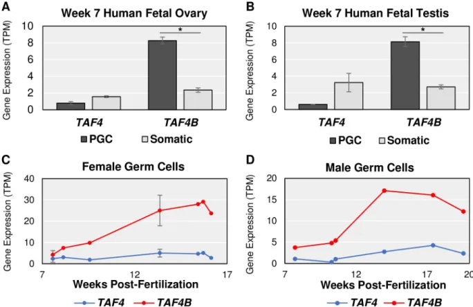
Given its expression in germ cells, we more closely examined the mRNA expression of Taf4b versus Taf4a in the E9.5-E18.5 time course. Taf4b dramatically increases at E15.5 in both female and male germ cells whileTaf4a expression is relatively constant and much lower (Fig 2A–2D, dark gray bars). However, the female expression ofTaf4b reaches greater TPM than the males, as indicated by the differences in the scale of the y-axes. SimilarTaf4b and Taf4a mRNA expression profiles can be reliably found in independent RNA-seq datasets that isolate germ cells from the embryonic gonad (S2 Fig). These data suggest that as germ cells progress through development, and enter meiosis in female germ cells,Taf4b mRNA and pro-tein are available to play a role in these processes. In addition, when the human embryonic gonad is sorted into germ and somatic cell populations,TAF4B mRNA expression is similarly
Fig 1. A comprehensive RNA-seq time course dataset from Sangrithi et al. in the cells of the embryonic gonad [19]. PCA plot of the
reprocessed mouse RNA-seq data. Somatic and germ cell lineages (dot size) are separate on the plot, germ cells group together depending on stage of development (dot color) and later by sex (dot shape).
germ cell-enriched (Fig 3A and 3B), and increases over comparable time points in the avail-able human datasets (Fig 3C and 3D,S3–S5Tables), indicating that mouse and human
TAF4B may play analogous roles during embryonic germ cell development [21–23]. While we identified many differentially expressed genes in pairwise comparisons of germ versus somatic cell gene expression across time (S1 Table), we used the program ImpluseDE2 [24] for time course differential expression analysis in female and male germ cells. ImpulseDE2 determined thatTaf4b, but not Taf4a mRNA expression was significant (p-adj. < 0.05) across
Fig 2.Taf4a vs Taf4b enrichment in the germ cells of the mouse embryonic gonad. Gene mRNA expression levels of Taf4b (A-B) and Taf4a
(C-D) in female and male embryonic germ cells (“G”) and somatic cells (“S”) from E9.5 to E18.5 indicate thatTaf4b is significantly (�= log2FC > |
0.25|, p-adj. < 0.05) and consistently enriched in germ cells from E11.5 to E18.5 in comparison to somatic cells. Error bars indicate± standard error of the mean (SEM). (E) Western blot protein signal of TAF4a and TAF4b in cells sorted from E13.5 Oct4-EGFP gonads. Mouse Vasa Homology (MVH) is a germ cell marker, which is only detected in the GFP+lanes of the female and male samples, indicating that the GFP-lane
contains only somatic cells. TAF4a protein signal is detected in all lanes (both germ and somatic cell types), whereas TAF4b is only detected in the GFP+lane (germ cells).β-Actin is a protein loading control. Two female mice and two male mice were used to obtain roughly 35,000 pooled cells for each lane. (F) Western blot experiment performed in 3 replicates was quantified for relative protein signal. TAF4b protein levels between GFP+ and GFP-cells in female E13.5 ovary are significantly (�= p < 0.05, unpaired t-test) different. MVH is significantly different for both females and
males. Error bars indicate± SEM.
https://doi.org/10.1371/journal.pgen.1008515.g002
both female and male germ cell timelines, referred to as “significantly dynamic” (S6 Table). Together these data suggest thatTaf4b expression is nearly exclusive to the germ cells of the embryonic gonad and in the female this corresponds to our time period of interest, when mei-osis and primordial follicle development have initiated.
Many TFIID subunits are dynamically expressed in embryonic germ cells
Using these ImpulseDE2 data, we asked whether other subunits of the TFIID complex were co-regulated withTaf4b. To our surprise, many other TFIID subunits were also found to be significantly dynamic in the female timeline, male timeline, or both (Fig 4A). One especially notable TAF variant that was found to be significantly dynamic in both female and male embryonic germ cells wasTaf7l. Taf7l is X-linked and known to be essential for male fertility [17,18]. Interestingly,Taf7l is a paralog of Taf7 and has an autosomal retrotransposed copy calledTaf7l2 (previously known as “4933416C03Rik” [14]). ImpulseDE2 identifiedTaf7l2 and Taf7 as significantly dynamic in the female and male germ cells, respectively. In female germ cells,Taf7l mRNA expression drastically increases starting at E14.5 and remains high (Fig 4B). Taf7l2 appears to mimic the female gene expression trajectory of Taf7l albeit at a lower TPM level (S3A Fig). In male germ cells,Taf7 is the most abundant of the three, with Taf7lFig 3. Human embryonic data resemblesTaf4a and Taf4b characteristics in mouse. (A-B) At 7 weeks post-fertilization, female gonadal cells
sorted for alkaline phosphatase-positive, CD117-positive germ cells (PGC) and male gonadal cells sorted for cKit-positive, TNAP-positive PGCs have significantly (�= log
2FC > |0.25|, p-adj. < 0.05) greaterTAF4B mRNA in comparison to the gonadal somatic cells (Somatic) in both
females and males.TAF4 mRNA levels are not significantly different in the two cell populations in both sexes, like the mouse. (C-D) Using
cKit-positive germ cells to perform FACS, from ~8 weeks fertilization to ~16 weeks fertilization in females and to ~20 weeks post-fertilization in males, humanTAF4B mRNA expression increases more so than TAF4. Also, TAF4B TPMs is greater in the female human germ
cells than the male germ cells, making the expression patterns ofTAF4B over time very similar between mice and humans. Error bars
indicate± SEM.
increasing in expression at E14.5, like its female germ cell expression (Fig 4C). When looking more closely at onlyTaf7l and Taf7l2, we see that Taf7l begins to increase in expression at E14.5, like in females, butTaf7l2 appears to increase in expression around E18.5 and does not mimic the male trajectory ofTaf7l (S3B Fig). These data suggest that both embryonic female and male germ cells shareTaf7l expression but express Taf7l2 and Taf7 in fe and male-specific ways, respectively.
Furthermore, the germ cell enrichment or specificity ofTaf7l, Taf7l2, and Taf7 depends on the sex of the germ cells. In females,Taf7l and Taf7l2 are significantly and consistently germ cell-specific starting at E14.5 whereas in males it isTaf7 and Taf7l that are significantly and consistently germ cell-enriched (S3C–S3H Fig). This implicatesTaf7l as a shared TFIID com-ponent of importance between female and male germ cells whileTaf7l2 and Taf7 display a female or male germ cell-specific preference, respectively. However, in the available human data,TAF7L is not detectable in the embryonic gonad 8–20 weeks post fertilization, TAF7 shows a non-significant preference for somatic cell expression at 7 weeks post-fertilization, andTaf7l2 is a rodent-exclusive retrogene which altogether might indicate that this trio is less important in human prenatal germ cell development (S3I–S3L Fig).
Since bothTaf4b and Taf7l are germ cell-enriched, and several TFIID subunits were identi-fied as significantly dynamic by ImpulseDE2 in the mouse, we asked if any other components
Fig 4. Many TFIID components changing during embryonic germ cell development. (A) Venn diagram of TFIID subunits identified by
ImpulseDE2 as significantly dynamic in the female and/or male germ cell time course data. (B-C) Gene expression levels forTaf7, Taf7l, and Taf7l2 in female and male germ cells, respectively. Taf7l is most highly expressed in female germ cells, but Taf7 is expressed more in male germ
cells. Error bars indicate± SEM.
https://doi.org/10.1371/journal.pgen.1008515.g004
of TFIID are also germ cell-enriched likeTaf4b and Taf7l. If individual TFIID components had significantly (log2FC > |0.25|, p-adj. < 0.05) higher mRNA expression in the germ cells
over the somatic cell samples for at least 3 of the 7 measured time points in either sex, it was considered “germ cell-enriched”. We found that 13 additional TFIID-encoding genes were germ cell-enriched (S3M Fig), demonstrating that germ-cell enrichment of TFIID complex genes is not unique toTaf4b and Taf7l.
Taf4b, Taf7l and Taf9b cluster with critical germ cell development and
meiotic genes
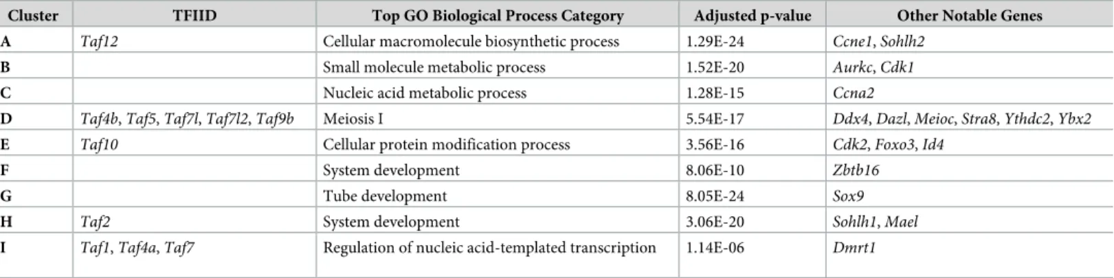
The enrichment and dynamic expression of TFIID subunits in both female and male germ cells led us to question if these TFIID components were coordinately expressed. We performed k-means clustering on the top 10,000 most variable genes across all germ cell samples in the reprocessed mouse time course dataset (Fig 5). Nine clusters were chosen based on the elbow method of determining k clusters (S4 Fig). Using this clustering method, 11 TAFs were found in our 9 clusters (Table 1). Cluster D (Fig 5, red box) is particularly interesting because 5 TAFs (Taf4b, Taf5, Taf7l, Taf7l2, and Taf9b) clustered with genes highly relevant to overall germ cell development (Dazl, Ddx4, Ybx2) and meiosis I (Stra8, Meioc, Ythdc2). Furthermore, gene ontology of Cluster D indicates that there is significant enrichment of meiosis I and germ cell development genes in this group (Table 1,S7 Table).
It is very interesting to find thatTaf4b, a gene with known connections to meiotic genes, clusters withTaf5, Taf7l, Taf7l2, and Taf9b, which have not been previously associated with meiosis. Furthermore, all butTaf5 are alternative TAFs, meaning that their place in the TFIID complex is not guaranteed but could instead be occupied by their counterpart. For example, both TAF4a and TAF4b are capable of replacing each other in TFIID.
Loss of
Dazl and Stra8 regulation affect proper expression of many TFIID
components, including
Taf4b
To further examine the potential relevance of TFIID in germ cells and what could be connect-ing their expression patterns, we reprocessed three RNA-seq datasets fromDazl- and Stra8-KO female and male mice [13,14,25]. BothDazl and Stra8 are germ cell-specific regulators of gene expression that are essential for meiosis and fertility.Dazl encodes an RNA-binding pro-tein that may promote translation of its target mRNAs and/or stabilize transcripts to prevent their degradation [12,13].Stra8 encodes a sequence-specific transcriptional activator that amplifies meiotic and cell cycle genes to initiate meiosis I [14].
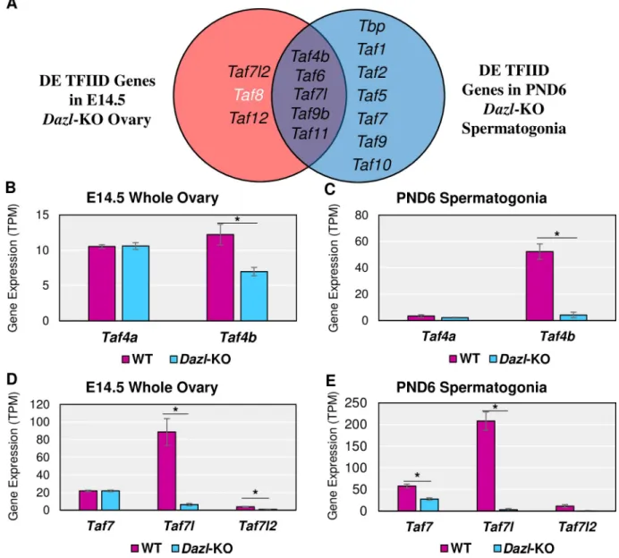
To determine which TAFs, if any, were deregulated whenDazl expression is disrupted, we reprocessed twoDazl-KO RNA-seq datasets. For the female Dazl-KO, Soh et al. performed RNA-seq on E14.5 whole ovaries with a global disruption of theDazl gene (S8 Table).Dazl is expressed in a germ cell-specific manner, so noise from the somatic cells of the ovary is mini-mized. For the maleDazl-KO, Zagore et al. used the same Dazl mutation but combined its usage with aStra8-Cre; IRG+system, which expresses RFP before Cre recombination and EGFP after, so that EGFP+spermatogonia could be collected via FACS at PND6 despite low germ cell numbers (S9 Table) [26]. Even with different female and male biological contexts in these RNA-seq datasets, we found many components of the TFIID complex to be significantly affected (log2FC > |0.25|, p-adj < 0.05), withTaf4b, Taf7l, and Taf9b significantly reduced in
both sexes of theDazl-KO mouse (Fig 6A). Closer examination of these data indicates that Taf4b, but not Taf4a, is significantly decreased in the female and male Dazl-KO mouse (Fig 6B and 6C). Additionally,Taf7l and Taf7l2 are significantly decreased in the female Dazl-KO data whileTaf7 and Taf7l are significantly decreased in the male Dazl-KO (Fig 6D–6E).
To determine which TAFs, if any, might be dependent onStra8 expression, we reanalyzed twoStra8-KO RNA-seq datasets. For the female Stra8-KO, Soh et al. also performed RNA-seq on E14.5 whole ovaries in a global disruption in theStra8 gene (S8 Table). For the male Stra8-KO, Kojima et al. used the sameStra8-KO crossed to a Ddx4-Cre; Rosa26-tdTomato mouse line and isolated preleptotene male germ cells (S10 Table). Like in theDazl-KO data, Taf4b andTaf7l were significantly reduced in the female and male Stra8-KO gonads along with many other TAFs, includingTaf9b in the females (Fig 7A).
Fig 5. K-means clustering of female and male germ cell samples in Oct4-EGFP mouse time course. Heatmap
generated based on clustering the top 10,000 most variable genes in the dataset into 9 clusters. Red box around Cluster D to identifyTaf4b-containing group of genes.
https://doi.org/10.1371/journal.pgen.1008515.g005
The mRNA dynamics ofTaf4a and Taf4b in the Stra8-KO data are much like the Dazl-KO data, withTaf4b being significantly decreased in the Stra8-KO female and male samples (Fig 7B and 7C). Interestingly,Taf4a is significantly increased in the Stra8-KO preleptotene germ cells. BothTaf7l and Taf7l2 are significantly decreased in the Stra8-KO but Taf7 is unchanged (Fig 7D and 7E). These data suggest that not only do DAZL and STRA8 regulate several TFIID components not traditionally thought of as part of the germ cell development gene pro-gram, but also thatTaf4b and Taf7l are consistent, active members of both the female and male germ cell-specific programs of gene expression.
DAZL and STRA8 directly bind to many of the TFIID components
improperly expressed in
Dazl- and Stra8-KO mice
To inquire as to whether DAZL may directly regulate any TFIID components, we identified DAZL-bound transcripts via individual-nucleotide resolution cross-linking and immunopre-cipitation (iCLIP) (S5A Fig) [27]. Given that we have previously shown thatTaf4b impacts meiotic prophase I in females and males [11], we sought to understand whetherTaf4b is regu-lated by DAZL in a meiotic context, focusing on the leptotene stage of meiotic prophase I. To obtain the large number of leptotene spermatocytes needed for iCLIP, we developmentally synchronized spermatogenesis by chemically regulating the levels of retinoic acid, which is required for spermatogonial differentiation [28,29]. This synchronized development allowed us to collect testes enriched for leptotene spermatocytes. The successful accumulation of lepto-tene spermatocytes, without contamination from other spermatocyte stages, was verified in a testis biopsy via histological analysis [30].
DAZL iCLIP peaks in 3 biological replicates were identified in 3’ UTRs (Fig 8A and 8B), where DAZL binds to facilitate post-transcriptional regulation [12,13,31–34]. Defining DAZL binding sites as those iCLIP peaks in 3’ UTRs present in at least 2 of 3 biological replicates, we identified a total of 1,652 DAZL binding sites corresponding to 1,281 genes (Fig 8B). This is far fewer binding sites and targets than identified by Zagore et al. and Li et al. [12,13]. These differences may be due to the tissue used, as these studies used whole testes which contain mul-tiple spermatogenic stages that express DAZL [12,13]. The 3’ UTR binding sites identified here were strongly enriched for the GUU motif with which DAZL preferentially interacts (Fig 8C) [12,13,35]. Replicated peaks were also identified in other genomic regions (S5B Fig,S11 Table), but the number of replicated peaks in these genomic regions was small and/or these peaks showed only moderate enrichment for DAZL’s GUU motif relative to the 3’ UTR peaks (Fig 8B and 8C,S5B and S5C Fig), suggesting that peaks outside of 3’ UTRs originate from nonspecific DAZL:RNA interactions.
Table 1. Top gene ontology category in each cluster, TFIID component found in each, and other notable genes found in cluster.
Cluster TFIID Top GO Biological Process Category Adjusted p-value Other Notable Genes
A Taf12 Cellular macromolecule biosynthetic process 1.29E-24 Ccne1, Sohlh2
B Small molecule metabolic process 1.52E-20 Aurkc, Cdk1
C Nucleic acid metabolic process 1.28E-15 Ccna2
D Taf4b, Taf5, Taf7l, Taf7l2, Taf9b Meiosis I 5.54E-17 Ddx4, Dazl, Meioc, Stra8, Ythdc2, Ybx2
E Taf10 Cellular protein modification process 3.56E-16 Cdk2, Foxo3, Id4
F System development 8.06E-10 Zbtb16
G Tube development 8.05E-24 Sox9
H Taf2 System development 3.06E-20 Sohlh1, Mael
I Taf1, Taf4a, Taf7 Regulation of nucleic acid-templated transcription 1.14E-06 Dmrt1
Based on the 3’ UTR binding sites, DAZL targets 6 TFIID component genes, including Taf4b but not Taf4a (Fig 8D–8J,S11 Table). The observation that all of these genes exhibit reduced expression inDazl-KO spermatogonia may reflect that DAZL protects these tran-scripts from degradation. Alternatively, the reduced TFIID gene expression in theDazl-KO may reflect indirect regulation by DAZL, which may increase the protein expression of other transcription factors that are required for the expression of TFIID component genes.
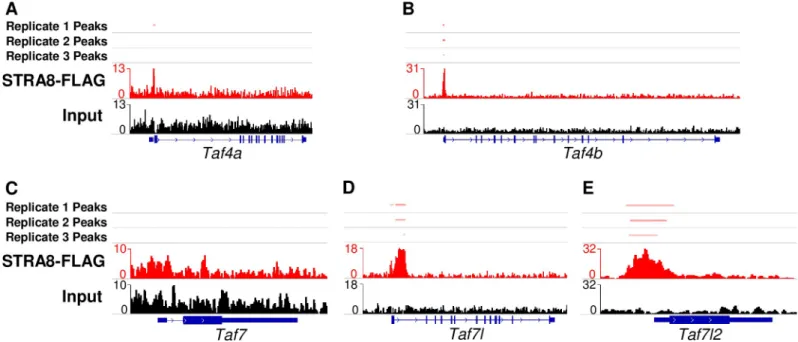
To determine whether STRA8 directly promotes the gene expression of TFIID components, we reprocessed the ChIP-seq data of Kojima et al. [14] on preleptotene synchronized whole testis expressing a FLAG-tagged STRA8 protein. We found peaks in two of the three replicates at many of the same genes with reduced expression inStra8-KO RNA-seq experiments,
Fig 6. TFIID subunits inDazl-KO female and male mice. (A) Venn diagram of TFIID subunits found to be significantly (log2FC > |0.25|, p-adj < 0.05) different between the WT andDazl-KO of the E14.5 ovary and/or PND6 spermatogonia. Black text indicates decreased mRNA
expression in theDazl-KO and white text indicates increased mRNA expression in the Dazl-KO compared to WT. Gene expression levels of Taf4a versus Taf4b in female (B) and male (C) WT versus Dazl-KO RNA-seq experiments. Gene expression levels of Taf7, Taf7l, and Taf7l2 in
female (D) and male (E) WT versusDazl-KO experiments (�= log
2FC > |0.25|, p-adj. < 0.05). Error bars indicate± SEM.
https://doi.org/10.1371/journal.pgen.1008515.g006
includingTaf4b, Taf7l, and Taf7l2. By contrast, STRA8 does not bind to Taf4a or Taf7, which are not differentially expressed in the RNA-seq analysis (Fig 9A–9E,S6A Fig,S12 Table). Interestingly, STRA8-FLAG ChIP signal reliably localized to the 5’ UTR regions ofTaf4b and Taf7l2 (Fig 9A and 9E), but localized after the first exon ofTaf7l (Fig 9D). In addition to ChIP-seq data, inin vitro differentiated primordial germ cell-like cells (PGCLCs), treatment of PGCLCs with retinoic acid and bone morphogenetic protein 2 (BMP2) inducesTaf4b, but not Taf4a, expression on culture day 9 (S6B and S6C Fig) [36]. However, this induction ofTaf4b fails to occur ifStra8 is knocked down in the cell culture system, which is also the case for Taf7l and Taf7l2, but not Taf7 (S6D–S6F Fig). These combined lines of evidence including
Fig 7. TFIID subunits inStra8-KO female and male mice. (A) Venn diagram of TAFs found to be significantly (log2FC > |0.25|, p-adj < 0.05) different between the WT andStra8-KO of the E14.5 ovary and/or male preleptotene germ cells. Black text indicates decreased
mRNA expression in theStra8-KO and white text indicates increased mRNA expression in the Stra8-KO compared to WT. Gene expression
levels ofTaf4a versus Taf4b in female (B) and male (C) WT versus Stra8-KO RNA-seq experiments. Gene expression levels of Taf7, Taf7l, and Taf7l2 in female (D) and male (E) WT versus Stra8-KO experiments (�= log
2FC > |0.25|, p-adj. < 0.05). Error bars indicate± SEM.
RNA-seq, ChIP-seq, andin vitro Stra8 knockdown demonstrate a direct link between Stra8 and several TFIID components in and around the time of meiotic initiation.
SAGA complex subunit
Supt3 mirrors Taf4b regulation
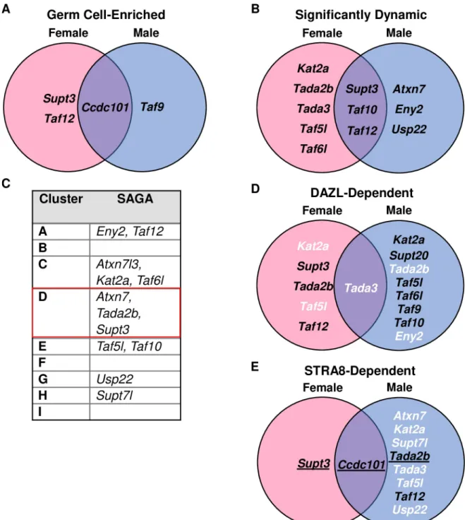
As TFIID and Spt-Ada-Gcn5-acetyltransferase (SAGA) are closely related complexes, even sharing TAF9, TAF10, and TAF12 as subunits, we investigated whether these trends in germ cells were unique to TFIID or also applied to SAGA [10,37]. In the mouse time course data, we found four SAGA subunits (includingTaf12) to be germ cell-enriched (Fig 10A) by the criteria of higher mRNA expression in the germ cells over the somatic cell samples for at least 3 of the 7 measured time points in either sex. ImpulseDE2 identified eleven SAGA subunits as signifi-cantly dynamic in female and/or male germ cells (Fig 10B) andAtxn7, Tada2b, and Supt3 were found in Cluster D of our k-means clustering (Fig 10C, red box). When examining our data regardingDazl, we found eleven subunits differentially expressed between WT and Dazl-KO samples in female and/or male mice, but only two of these genes (Usp22 and Trrap) were identified as DAZL-bound in our iCLIP data and neither were found to be differentially expressed (Fig 10D,S11 Table). Similarly, we found ten SAGA genes to be differentially expressed between WT andStra8-KO samples in female and/or male mice, but in the ChIP-seq data, only three of these genes display STRA8-FLAG peaks in their promoter regions
Fig 8. Identification of DAZL targets via iCLIP in testes synchronized for leptotene spermatocytes. (A) Genomic distribution of DAZL
iCLIP peaks identified in 3 biological replicates (p < 0.001). Peaks in each type of genomic region were called via ASPeak. (B) Venn diagram showing overlap of DAZL iCLIP peaks in 3’ UTRs among 3 biological replicates. 1,670 replicated peaks (present in at least 2 of 3 replicates; highlighted in blue) were identified. After merging replicated peaks falling on consecutive nucleotides, 1,652 binding sites were identified. These binding sites correspond to 1,281 genes, which were designated as DAZL-bound genes. (C) GUU motif is enriched at DAZL 3’ UTR peaks. AME from the MEME Suite was used to identify motif enrichment at crosslinked nucleotides in replicated peaks relative to shuffled control sequences. (D-J) DAZL and IgG iCLIP gene tracks showing 3’ UTRs forTaf1 (D), Taf2 (E), Taf4a (F), Taf4b (G), Taf7 (H), Taf9b
(I), andTbp (J). Each iCLIP track represents the crosslinked sites from the sum of unique reads from 3 biological replicates, with DAZL
iCLIP reads in red and IgG iCLIP reads in black. The horizontal blue lines on top mark the replicated peaks.
https://doi.org/10.1371/journal.pgen.1008515.g008
Fig 9. STRA8 ChIP-seq signal shows STRA8 binding at or near the promoters of TFIID subunits. (A-E) ChIP-seq signal from STRA8-FLAG samples and the peaks
called (red) in comparison to input DNA (black) forTaf4a (A), Taf4b (B) Taf7 (C), Taf7l (D), and Taf7l2 (E).
Fig 10. SAGA complex components not particularly germ cell relevant. (A) Venn diagram of SAGA subunits identified as significantly
germ cell-enriched in the female and/or male germ cell time course data. (B) Venn diagram of SAGA subunits identified by ImpulseDE2 as significantly dynamic in the female and/or male germ cell time course data. (C) SAGA complex subunits that were part of the top 10,000 most variable genes in the k-means clustering. (D) Venn diagram of SAGA subunits found to be significantly (log2FC > |0.25|,
p-adj < 0.05) different between the WT andDazl-KO of the E14.5 ovary and/or PND6 spermatogonia. Black text indicates decreased mRNA
expression in theDazl-KO and white text indicates increased mRNA expression in the Dazl-KO compared to WT. Underlined text
indicates the gene was found to have DAZL binding in its 3’ UTR in 2 out of 3 DAZL iCLIP replicates. (E) Venn diagram of SAGA found to be significantly (log2FC > |0.25|, p-adj < 0.05) different between the WT andStra8-KO of the E14.5 ovary and/or male preleptotene
germ cells. Black text indicates decreased mRNA expression in theStra8-KO and white text indicates increased mRNA expression in the Stra8-KO compared to WT. Underlined text indicates peaks at transcription start sites detected in two of three ChIP-seq replicates.
https://doi.org/10.1371/journal.pgen.1008515.g010
(Fig 10E) and three more (Taf5l, Taf9, Trrap) were not differentially expressed. Altogether, only expression ofSupt3 is most similar to Taf4b, but its mRNA is not found to be DAZL-bound (S11 Table), which suggests that there are differences in how they are regulated during embryonic germ cell development.
Discussion
The underlying transcriptional programs that drive germ cell development during mammalian embryogenesis are just coming into focus. In this study, we reprocessed publicly available RNA-seq datasets to understand the dynamics and potential regulators of the TFIID complex. We discovered thatTaf4b is germ cell-enriched, significantly dynamic, clusters with meiotic genes, and isDazl- and Stra8-regulated in female and male mice. Furthermore, our DAZL iCLIP data demonstrates that the 3’ UTRs of six TFIID components (Taf1, Taf2, Taf4b, Taf7, Taf9b, and Tbp) are directly bound by DAZL in leptotene spermatocytes. Moreover, reprocess-ing of STRA8-FLAG ChIP-seq data indicates that six TAFs (Taf3, Taf4b, Taf7l, Taf7l2, Taf9, andTaf11) have STRA8 bound to their promoters in preleptotene spermatocytes [13,14]. Of all these genes that were either DAZL- or STRA8-regulated, onlyTaf4b, Taf7l, and Taf9b were also germ cell-enriched, significantly dynamic in their expression, and clustered with meiotic genes. Taken together, these data indicate that several TAFs may participate in germ cell devel-opment, particularly during early meiosis, and potentially implicate a germ cell-specific TFIID complex that is preferentially stabilized to integrate proper germ cell-specific transcription and development.
Although basal transcription components such as TFIID are not widely studied in the con-text of reproduction, Sisakhtnezhad and Heshmati [38] found thatTaf7l (with Dazl, Mael, etc.) was one of the 30 most significantly SSC-enriched genes in comparison to mesenchymal stem cells in PND7 mice. In addition,Taf1, Taf2, Taf4b, Taf7, Taf7l2, and Taf9b were also significantly enriched in SSCs along with other basal transcription machinery such as RNA-PII subunits [38]. Soh et al. found thatTaf4b, Taf5, Taf7l, Taf7l2, and Taf9b were “ovarian germ cell enriched”, of whichTaf7l, Taf7l2, and Taf9b are also highlighted as “meiotic pro-phase” genes [25]. It is already known thatTaf7l is essential for male fertility: Taf7l-KO male mice are infertile due to defects in spermiogenesis [18].Taf7l2 was only recently uncovered as a rodent-specificTaf7l retrogene [14] and it is not known if there is a fertility defect in a Taf7l2 mutant. Taf7l interacts with Trf2 (also known as Tbpl1) to bind to promoters of genes in mouse testis. Interestingly,Trf2/Tbpl1 was found to be significantly dynamic in female germ cells, but not germ cell-enriched nor significantly affected byDazl or Stra8 in our data. It might be thatTaf7l interacts with different proteins to perform a different role in the embryonic ovary or postnatal testis. E13.5 germ cells have been found to have much higher transcription levels than their somatic cell counterparts, and this has been proposed to be a phase of hypertranscription in the germ cells [39]. However, this phase was found to end at E15.5, which contrasts with many of the TAFs we observed that increase in gene expression at E15.5.
Taf9b participating in the Cluster D in the k-means clustering was surprising out of the five TAFs found in that cluster.Taf9b is known to play a critical role in proper differentiation of motor neurons, but it has no known role in germline development, asTaf9b-KO mice are via-ble and fertile [40].Taf9b is not the only gene to be associated with cell type-specific differenti-ation.Taf7l has also been found to play a role in adipocyte formation [41,42]. When the germ cell samples were split into female- or male-only for individual analysis,Taf4b, Taf7l, Taf7l2, andTaf9b remained clustered together and with other meiotic genes in female germ cells. However, in male germ cells, these four TAFs did not cluster together, butTaf4b and Taf7l2
did, as well asTaf7l and Taf9b (S7 Table). Hill et al. also groupedTaf7l, Taf7l2, and Taf9b together as germline reprogramming responsive (GRR) genes, which is a group of 45 genes that includesDazl, Hormad1, and thirteen other genes that were also found in Cluster D [43]. These genes were designated GRR because their promoters were highly methylated at E10.5 but this DNA methylation was reduced during germline epigenetic programming, and the genes became progressively more transcribed in both sexes. This consistent association with essential gamete generation and meiotic genes in both our clustering analysis and other pub-lished research strongly implicatesTaf4b, Taf7l, Taf7l2, and Taf9b as genes that warrant fur-ther study. Only TAF4b and TAF7l are being studied for their germ cell-specific function. It is unknown to what extent, if any, these other TAFs regulate the expression of each other. Future research should investigate if genes such asTaf7l are differentially expressed in the Taf4b-defi-cient mouse. Our finding that, among the SAGA complex genes, onlySupt3 (also known as Spt3) is germ-cell enriched, significantly dynamic, in Cluster D, and STRA8-regulated suggests that this gene may function in germline-specific gene expression. Despite not being DAZL-regulated, it is possible that the SAGA complex could play a role in germ cell development in a similar way toTaf4b.
Our findings also reinforce the role ofTaf4b in germ cell development, but now as part of a larger, coordinated effort with additional TFIID components. Given that previous research has shown that TAF4b functions during meiosis and localizes to the promoters ofDazl and Stra8 at E18.5, it is possible that TAF4b, DAZL, STRA8, and perhaps other factors work interdepen-dently. Identifying DAZL and STRA8 as regulators ofTaf4b is an important step forward in understandingTaf4b, but nevertheless, there are likely more regulators yet to be found. This is hinted at in our data, where maleTaf4b expression rises at E15.5 (Fig 2A and 2B) in the absence of STRA8. It is plausible that another transcription factor facilitates this increase. As STRA8 is considered a transcriptional amplifier rather than an activator, it is likely that this unknown transcription factor(s) is shared between female and male germ cells. Future work in embryonic germ cells, particularly in males, should add more context to the role TAF4b may be playing there.
The specialized role for TAF4b as a germ cell relevant TFIID component is bolstered by single-cell RNA-seq analyses in mice and humans that associateTaf4b with essential develop-mental steps inId4-GFP adult mouse germ cells and adult human spermatogonia [44]. Alter-native Taf4 subunits have been identified in bothDrosophila melanogaster (known as nht) andArabidopsis thaliana (known as taf4b) [45,46]. The alternative Taf4 genes in vertebrates, Drosophila, and Arabidopsis arose independently [10]. Interestingly,Drosophila nht is testis-specific and required for male fertility, and Arabidopsis Taf4b is directly implicated in regu-lating meiotic crossover rates and germline transcription, similar to mouseTaf4b [47]. This mechanism of an alternative Taf4 in TFIID, particularly for the purpose of germ cell develop-ment and function, is an intriguing instance of convergent evolution across plants, inverte-brates, and vertebrates.
TFIID has been typically viewed as static and generic in composition as a basal transcrip-tion factor. These dynamic gene expression data, particularly forTaf4b, challenge this view and suggest that there are subunits in TFIID that are preferred and regulated over others in mammalian germ cells. This may also be the case for other cell type-specific differentiation programs, as some of the same variant TFIID subunits shown to be expressed in a germ cell-specific manner here have also been shown to regulate somatic cells, including neurons and adipocytes. Though initially unexpected, such research indicates that there is more informa-tion to be garnered from studying TFIID in the context of germ cells and other specialized cell types.
Materials & methods
Ethics statement
This study was approved by Brown IACUC protocol #1803000344. For the iCLIP experiment, the MIT IACUC approved this research (#067-059-20). The primary method of euthanasia is CO2inhalation and the secondary method used is cervical dislocation both as per AVMA
guidelines on euthanasia.
Mice
Mice that were homozygous for anOct4-EGFP transgene (The Jackson Laboratory: B6;129S4-Pou5f1tm2Jae/J) were mated for mRNA and protein collections. Timed matings were estimated to begin at day 0.5 by evidence of a copulatory plug. To confirm the sex of E12.5 and younger embryos, genotyping for the presence or absence of theSry gene was completed. Male and female embryos were identified at E13.5 and older by confirming the presence or absence of testicular cords. Genomic DNA from tails was isolated using ThermoFisher PureLink Geno-mic DNA isolation kit (Cat #: K182001) for PCR genotyping assays. All animal protocols were reviewed and approved by Brown University Institutional Animal Care and Use Committee and were performed in accordance with the National Institutes of Health Guide for the Care and Use of Laboratory Animals. Gonads were dissected out of embryos into cold PBS. Wild-type C57BL/6N mice (Taconic Biosciences) were mated to produce postnatal males for iCLIP collection. All experiments involving mice were performed in accordance with the guidelines of the Massachusetts Institute of Technology (MIT) Division of Comparative Medicine, which is overseen by MIT’s Institutional Animal Care and Use Committee (IACUC). The animal care program at MIT/Whitehead Institute is accredited by the Association for Assessment and Accreditation of Laboratory Animal Care, International (AAALAC), and meets or exceeds the standards of AAALAC as detailed in the Guide for the Care and Use of Laboratory Animals. The MIT IACUC approved this research (no. 0617-059-20).
Embryonic gonad dissociation and fluorescence-activated cell sorting
To dissociate gonadal tissue into a single-cell suspension, embryonic gonads at specified times were harvested and placed in 0.25% Trypsin/EDTA and incubated at 37˚C for 15–20 minutes. Eppendorf tubes were flicked to dissociate tissue halfway through and again at the end of the incubation. Trypsin was neutralized with 75μL of FBS. Cells were pelleted, the supernatant was removed, and cells were resuspended in 100μL PBS. The cell suspension was strained through a 35μm mesh cap into a FACS tube (Gibco REF# 352235). Propidium iodide (1:500) was added to the cell suspension as a live/dead distinguishing stain. Fluorescence-activated cell sorting (FACS) was performed using a Becton Dickinson FACSAria III in the Flow Cytometry and Cell Sorting Core Facility at Brown. A negative control of a non-transgenic mouse gonad was used for each experiment to establish an appropriate GFP signal baseline. Dead cells were discarded based on propidium iodide signal and then cells were sorted at 4˚C in PBS based on GFP signal into GFP+or GFP-samples. Cells for western blots were pelleted, PBS wasremoved, flash-frozen with liquid nitrogen, and stored at -80˚C.
Western blotting
Protein to be loaded for western blots was estimated based on cell numbers reported during FACS. Thawed cells were mixed with laemmli sample buffer andβ-mercaptoethanol, pipetted thoroughly, incubated on ice for 1 hour, and stored at -80˚C. Western blotting was performed by loading roughly similar amounts of protein (estimated based on cell number) into a BioRad
Mini-PROTEAN TGX 4–15% gel (Cat. #456–1083). Protein was transferred to nitrocellulose membrane (0.45μm). Nitrocellulose was cut, blocked in 5% milk in PBS with 0.1% Tween (PBS-T) or blocked in 5% Amersham ECL Prime Blocking Reagent (RPN418) for 30 minutes at room temperature. Incubation in primary antibody in 1% milk in PBS-T or only PBS-T was performed in 4˚C overnight and secondary antibody in 1% milk in PBS-T or only PBS-T for 1 hour at room temperature. In between the block/primary and primary/secondary incubations, nitrocellulose was washed in PBS-T at least three times for 5 minutes.β-Actin or GAPDH was used as a loading control. GE Healthcare Amersham ECL Prime Western Blotting Detection Reagent (RPN2232) was used for horseradish peroxidase-tagged (HRP) secondary antibody detection. For stripping antibodies, Restore Western Blot Stripping Buffer (ThermoFisher: 21059) was used for 10 minutes at room temperature. For relative protein quantification, Ima-geJ was used to normalize protein signal toβ-Actin in each cell type.
Antibodies used were as follows: mouse monoclonal anti-TAFII135 (BD Transduction
Lab-oratories: 612054), polyclonal rabbit anti-mouse TAF4B (as described previously [11]), mouse monoclonal TAF7 (Novus Biologicals: H00006879-M01), rabbit polyclonal Taf7l (a generous gift from Dr. Haiying Zhou and Dr. Robert Tjian, rabbit polyclonal MVH (Abcam: ab13840); mouse monoclonalβ-Actin (Ambion: AM4302).
RNA-seq data analysis
All computational scripts regarding RNA-seq used in this publication are available to the pub-lic:https://github.com/mg859337/TAF_Manuscript_MG/tree/master/RNAseq. Datasets SRP059601, SRP059599, SRP057098, SRP045294, SRP049981, SRP128645, and SRP150721 were from NCBI SRA, and dataset E-MTAB-4616 was obtained from ArrayExpress. Kojima et al. performed RNA-seq on four Stra8-WT samples, two labeled “high-Stra8” and two “low-Stra8”, but only the “high-Stra8” were used as WT in these data because the “low-Stra8” were in early preleptotene and not representative of full meiotic initiation in male germ cells. All accessed raw fastq files were initially processed on Brown University’s high-performance com-puting cluster at the Center for Computation and Visualization. They were analyzed using FastQC (v0.11.4 or 0.11.5) for quality and then aligned to the mm10 or hg38 genomes using HiSat2 (v2.1.0) with the optional -dta setting enabled (S12 Table) [48,49]. Resulting sam files were converted to bam files using Samtools (v1.9) [50].
To obtain transcripts per million (TPMs) for each sample, StringTie (v1.3.3b) was used with the optional parameters -A and -e [49]. A gtf file for each sample was downloaded and, using RStudio (R v3.5.1), TPMs of all samples were aggregated into one comma separated (csv) file using a custom R script. To create interactive Microsoft Excel files for exploring the TPMs of each dataset: the csv of aggregated TPMs was saved as an Excel spreadsheet, colored tabs were added to set up different comparisons, and a flexible Excel function was created to adjust to new gene name inputs. To explore the Excel files, please find the appropriate tab and type in the gene name of interest into the highlighted yellow boxes. There is an Excel file for each dataset analyzed in the supplementary tables.
To obtain count tables, HTSeq (v0.9.1) and the count tables for each dataset were merged using a custom RStudio script [51]. Metadata files for dataset were created manually in Excel and saved as a csv. These count tables were used to create PCA plots by variance-stabilizing transformation (vst) of the data in DESeq2 (v1.22.2) and plotting by ggplot2 (v3.1.0) [52,53]. DESeq2 was also used for differential gene expression analysis, where count tables and meta-data files were used as input.
ImpulseDE2 (v1.6.1), a Bioconductor R package that can identify differentially expressed genes in longitudinal count datasets, was used to identify “significantly dynamic” genes from
E9.5 –E18.5 female and male Oct4-EGFP+germ versus Oct4-EGFP-somatic cell time course datasets [24]. ImpulseDE2 was used in case-only mode and no batch effects settings were added to the parameters.
For k-means clustering, iDEP (v.9) was used. It is a webpage hosted by South Dakota State University that is a simple and fast resource to perform typical RNA-seq data analysis [54]. The Sangrithi et al. (2018) mouse time course merged counts table was uploaded to iDEP. Counts were filtered out by the criteria of at least 0.5 counts per million in one of the samples. The data was then transformed using vst, in the “Heatmap” module the blue-white-red color scheme was chosen and in the “k-Means” clustering module the top 10,000 most variable genes across the time course were selected for clustering. The setting of 9 clusters was chosen using the within-sum-of-squares (wss) elbow method displayed under the “How many clus-ters?” button. The heatmap of gene clustering and associated list of clustered genes were down-loaded. Gene ontology is another part of the “k-Means” module, the GO category of Biological Process was chosen, and the enrichment details were downloaded.
iCLIP library construction, sequencing, and computational analysis
To obtain testes enriched for the leptotene stage of meiotic prophase I, spermatogenesis was chemically synchronized via a protocol originally developed by Hogarth et al. and modified by Romer et al. [55,56]. Briefly, on PND2-8, male mice were injected daily subcutaneously with WIN 18,446 (Santa Cruz Biotechnology) at 0.1 mg/gram body weight. On PND9, the mice were injected once subcutaneously with retinoic acid (RA; MilliporeSigma) at 0.0125 mg/gram body weight. Mice were euthanized 8.0 days after the RA injection to obtain testes enriched for leptotene spermatocytes. From each pup, one half of a testis was collected as a biopsy for histo-logical analysis to verify enrichment of leptotene spermatocytes, and the remaining 1.5 testes were collected for iCLIP. For histological analysis, the testis biopsy was fixed in Bouin’s solu-tion for 3h at room temperature. Testis secsolu-tions were stained with anti-STRA8 (rabbit poly-clonal; Abcam ab49405) and then counterstained with hematoxylin, as previously described [14]. At least 40 seminiferous tubule cross-sections per mouse were analyzed for morphology [30] and STRA8 expression, found in preleptotenes and early leptotenes [57,58]. For all sam-ples analyzed via iCLIP, 100% of the tubule cross-sections contained leptotene spermatocytes but not preleptotene or zygotene spermatocytes.After synchronization of spermatogenesis, testis tubules were dissociated by pipetting in ice-cold PBS and irradiated three times at 200 mJ/cm2at 254 nm in a Stratalinker 2400 before pelleted, flash frozen, and stored at -80˚C until library preparation. DAZL and IgG iCLIP libraries were prepared as previously described [27]. For each biological replicate, 1.5 testes from a single mouse were lysed in 640μl lysis buffer. Immunoprecipitations were carried out via anti-DAZL (rabbit polyclonal; Abcam ab34139; validated for CLIP in studies [12,13]) or polyclonal rabbit IgG (Abcam ab27478) using 12μg antibody bound to 120 μl Dynabeads Pro-tein G (ThermoFisher Scientific 10003D) with 600μl lysate. iCLIP libraries were prepared in two batches, with each batch containing an equal number of DAZL iCLIP and IgG iCLIP libraries. A total of 3 DAZL iCLIP and 3 IgG iCLIP libraries were generated. The libraries were pooled and sequenced with 50bp single-end reads on the Illumina HiSeq 2500 machine. The 5’ end of each iCLIP read contained (in 5’ to 3’ orientation), a 3-nt random barcode, a 4-nt sam-ple-specific barcode, and a 2-nt random barcode.
All computational scripts used in this publication for the iCLIP analysis are available at: https://github.com/mmikedis/Gura_et_al_2019_PLOS_Genetics. Reads were quality-trimmed with cutadapt v1.8 (options: -q 20 -m 24) [59]. PCR duplicates were collapsed using the FAS-TX-Toolkit v0.0.14 (http://hannonlab.cshl.edu/fastx_toolkit/index.html), and the 5’-most 3-nt
random barcode was trimmed from the 5’ end of each read with the fastx_trimmer tool from FASTX-Toolkit v0.0.14 (options: -f 4). Reads were demultiplexed using the fastx_barcode_s-plitter.pl tool from FASTX-Toolkit v0.0.14 (options:—bol), and the sample-specific barcodes with remaining random barcodes were trimmed from the 5’ end of each read with the fas-tx_trimmer tool (options: -f 7). iCLIP libraries were mapped to the mouse genome (mm10 assembly) via STAR v2.5.4b [60] (options:—outFilterMultimapNmax 1—alignEndsType Extend5pOfRead1—outFilterMismatchNmax 2—outSAMattributes None—outReadsUn-mapped Fastx). All other parameters were set to default. The iCLIP None—outReadsUn-mapped reads were then converted to crosslinked nucleotides (i.e., the nucleotide immediately preceding the first nucleotide of the mapped read) using a custom script [61]. Within each library batch, DAZL crosslinked peaks were called using the DAZL iCLIP crosslinked nucleotides via ASPeak v2.0.0 [62] with the IgG iCLIP crosslinked nucleotides for the -control parameter. All other parameters were set to default. The UCSC RefSeq transcript annotations and Retrogenes V6 annotations for the mm10 assembly were used to call peaks. Given that DAZL binds the 3’ UTR of coding transcripts, peaks were called via the following hierarchy: 3’ UTR exon > 5’ UTR exon > coding exon > ncRNA > retrogene > intron > intergenic region. Peaks were fil-tered for p < 0.001, and then replicated peaks present in at least 2 out of 3 biological replicates were identified. Replicated peaks identified in consecutive nucleotides were merged into a sin-gle peak. iCLIP sequencing data and analysis have been deposited in NCBI GEO under acces-sion number GSE139005 and NCBI SRA under accesacces-sion number SRP226023.
Enrichment of the GUU motif at replicated peaks from each genomic region was assessed via the MEME Suite’s AME v4.11.2 [63] (options:—scoring avg—method ranksum) using the replicated crosslinked nucleotides±2 nt and shuffled control sequences.
ChIP-seq data analysis
All computational scripts used in this publication regarding ChIP-seq are available to the pub-lic:https://github.com/mg859337/TAF_Manuscript_MG/tree/master/ChIPseq. ChIP-seq samples from dataset SRP150721 was obtained from NCBI SRA and all raw fastq files from genotype FLAG/FLAG were initially processed on Brown University’s high-performance com-puting cluster at the Center for Computation and Visualization. They were analyzed using FastQC (v0.11.5) for quality and trimmed for very low-quality reads using Trim Galore! (v0.5.0) (https://www.bioinformatics.babraham.ac.uk/projects/trim_galore/).
These trimmed reads were then aligned to the mm10 genome using Bowtie2 (v 2.3.0) (S12 Table) [64]. Resulting sam files were converted to bam files, then unmapped and duplicated reads were removed using Samtools (v1.9) [50]. Bedtools (v2.26.0) was used to removed regions in bam files that mapped to ChIP-seq blacklisted regions in the mm10 genome [65]. For visualization, Samtools was used to merge replicates and Integrative Genomics Viewer (IGV) was used to visualize the gene tracks [66]. For calling peaks MACS (v 2.1.1) was used, comparing the FLAG replicate to its IgG control [67].
Supporting information
S1 Fig. Replicates of western blots and protein signal of TAF4b in cells sorted from female E13.5 Oct4-EGFP gonads. (A-B) Entire blots of images displayed inFig 2Eof TAF4a (135 kDa), TAF4b (105 kDa), andβ-Actin (42 kDa) (A) and MVH (76 kDa) after antibody stripping and reprobing (B). (C) Entire blot of second western blot replicate of E13.5 Oct4-EGFP sorted gonads. Four female mice and three male mice were used to load roughly 40,000 cells. (D) Entire blot of third western blot replicate of E13.5 Oct4-EGFP sorted gonads. Four female mice and three male mice were used in the experiment to load roughly 25,000 cells. (E)
Approximate numbers of female GFP-and GFP+cells loaded into western blot are as indi-cated. TAF4b protein signal is detected in only the GFP+lane (germ cell), despite 5X more cells being loaded into the GFP-lane.β-Tubulin is a protein loading control. S1 Fig is associ-ated withFig 2.
(PDF)
S2 Fig. Independent RNA-seq datasets replicate increase inTaf4b mRNA expression after
E13.5 in both female and male mouse germ cells and low expression ofTaf4a. (A-B) RNA-seq from Miyauchi et al. [Supporting Information References 1] of cells sorted from Stella-EGFP E9.5-E13.5 and MVH-RFP E14.5-E15.5 female (A) and male (B) mice. For E9.5-E11.5, sexes were pooled together and the same data appear in both plots. (C-D) RNA-seq from Sei-senberger et al. [Supporting Information References 2] of cells sorted from Oct4-EGFP female (C) and male (D) mice. For E11.5, sexes were pooled together, and the same data point appears in both plots. S2 Fig is associated withFig 2.
(PDF)
S3 Fig. Germ cell-enriched TFIID subunits and closer examination ofTaf7, Taf7l, and Taf7l2. (A) Removal of Taf7l expression to more closely examine female germ cell expression ofTaf7 and Taf7l2 over time. (B) Removal of Taf7 expression to more closely examine male germ cell expression ofTaf7l and Taf7l2 over time. Expression of female (C, E, G) and male (D, F, H) mRNAs ofTaf7, Taf7l, and Taf7l2 in germ cells (“G”) and somatic cells (“S”) from E9.5 to E18.5 (�= log2FC > |0.25|, p-adj. < 0.05). (I-J)TAF7 and TAF7L expression in human
gonads indicate thatTAF7L is barely detectable at 7 weeks post-fertilization. Taf7l2 does not have a human homolog. (K-L) From ~8 to ~16 weeks post-fertilization in females and to ~20 weeks post-fertilization in males, humanTAF7L mRNA expression is low and unchanging whileTAF7 expression is variable over time. This is dissimilar to the mouse TAF7L RNA-seq data. Error bars indicate± SEM. (M) Venn diagram of TFIID subunits identified as signifi-cantly germ cell-enriched in the female and/or male germ cell time course data. S3 Fig is asso-ciated withFig 2,Fig 3andFig 4.
(PDF)
S4 Fig. Within sum of squares (wss) graph for evaluating how many clusters to set in k-means clustering. Nine clusters were chosen. S4 Fig is associated withFig 5andTable 1. (PDF)
S5 Fig. DAZL iCLIP in testes synchronized for leptotene spermatocytes. (A) Radioblot of
DAZL:RNA complexes from postnatal testes synchronized for leptotene spermatocytes. DAZL:RNA complexes are larger than 37 kDa, the approximate molecular weight of DAZL. One of three biological replicates used to prepare iCLIP libraries reported here. (B) Venn dia-gram showing overlap of DAZL iCLIP peaks among 3 biological replicates in genomic regions other than the 3’ UTR. (C) Enrichment of DAZL’s GUU motif at replicated iCLIP peaks from genomic regions other than the 3’ UTR. AME from the MEME Suite was used to identify motif enrichment at crosslinked nucleotides in replicated peaks relative to shuffled control sequences. S5 Fig is associated withFig 8.
(TIF)
S6 Fig. STRA8 peaks in preleptotene germ cells and knockdown experiments in primordial germ cell-like cells (PGCLCs). (A) STRA8-FLAG peaks called by MACS2 at the transcription
start sites of TFIID components in comparison to the DNA input control. (B-F) mRNA expression levels in WT andStra8-knockdown in vitro differentiated PGCLCs from repro-cessed Miyauchi et al. RNA-seq using retinoic acid and BMP2. Expression ofTaf4a (B) and
Taf7 (D) do not see a strong induction at culture day 9 (c9) nor are differentially expressed in theStra8-knockdown. Taf4b (C), Taf7l (E), and Taf7l2 (F) are highly expressed at c9 but fail to be induced inStra8-knockdown cells (�= log
2FC > |0.25|, p-adj. < 0.05). S6 Fig is associated
withFig 7. (PDF)
S1 Table. DESeq2 output for comparisons between germ and somatic cells at different time points. Sheets contain DESeq2 results for germ versus somatic cell gene expression for
every time point and sample type. S1 Table is associated withFig 1,Fig 2,Fig 4, andS3 Fig. (XLSX)
S2 Table. Transcripts per million (TPM) of all reprocessed time course data from Sangrithi et al. with tabs for asking specific questions about the data. Sheet 2 (“All_TPMs”) contains a
TPM values table for all samples and genes. Sheets 3–10 contain search and plotting tools to quickly evaluate the data. S2 Table is associated withFig 1,Fig 2,Fig 4, andS3 Fig.
(XLSX)
S3 Table. Transcripts per million (TPM) of all reprocessed time course data from Gkoun-tela et al. with tabs for asking specific questions about the data. Sheet 2 (“All_TPMs”)
con-tains a TPM values table for all samples and genes. Sheets 3–5 contain search and plotting tools to quickly evaluate the data.S3 Tableis associated withFig 3andS3 Fig.
(XLSX)
S4 Table. Transcripts per million (TPM) of all reprocessed time course data from Tang et al. with tabs for asking specific questions about the data. Sheet 2 (“All_TPMs”) contains a
TPM values table for all samples and genes. Sheet 3 (“Quick_Calc”) contains search and plot-ting tools to quickly evaluate the data. Sheet 4 (‘DESeq_PGCs_Somatic’) contains DESeq2 results for human female PGC versus gonadal somatic gene expression. S4 Table is associated withFig 3andS3 Fig.
(XLSX)
S5 Table. Transcripts per million (TPM) of all reprocessed time course data from Irie et al. with tabs for asking specific questions about the data. Sheet 2 (“All_TPMs”) contains a TPM
values table for all samples and genes. Sheet 3 (“Quick_Calc”) contains search and plotting tools to quickly evaluate the data. Sheet 4 (‘DESeq_PGC_Somatic’) contains DESeq2 results for human male PGC versus gonadal somatic gene expression. S5 Table is associated withFig 3andS3 Fig.
(XLSX)
S6 Table. ImpulseDE2 output for individual sex and cell type data. Sheets 2–5 correspond
to the ImpulseDE2 output for female germ cells, male germ cells, female somatic cells, and male somatic cells, respectively. S6 Table is associated withFig 4.
(XLSX)
S7 Table. k-means clustering and gene ontology of clusters. Sheet 2 (‘Kmeans_10000’)
con-tains k-means clustering output for the 10,000 most variable genes across all germ cells samples in the Sangrithi et al. reprocessed time course data, 9 clusters total. Sheet 3 (‘GO_Biological_-Process) contains the significant gene ontology enrichment for all clusters. Sheets 4 and 6 (‘Kmeans_10000_Females_Only’ and ‘Kmeans_10000_Males_Only’) are the k-means cluster-ing output for the 10,000 most variable genes in germ cell samples, 9 clusters total from female and male samples, respectively. Sheets 5 and 7 (‘GO_Biological_Process_Females’ and ‘GO_Biological_Process_Males’) are the gene ontology for the female- and male-only
![Fig 1. A comprehensive RNA-seq time course dataset from Sangrithi et al. in the cells of the embryonic gonad [19]](https://thumb-eu.123doks.com/thumbv2/123doknet/14753892.581505/5.918.164.863.114.480/fig-comprehensive-course-dataset-sangrithi-cells-embryonic-gonad.webp)