Addressing the Risks of Diagnostic Radiology: What Should Be Done About the Increasing Use of Computed Tomography in the United States
by Gary Eastwick
SUBMITTED TO THE DEPARTMENT OF NUCLEAR SCIENCE AND ENGINEERING IN PARTIAL FULFILLMENT OF THE REQUIREMENTS FOR THE DEGREE OF
BACHELOR OF SCIENCE IN NUCLEAR SCIENCE AND ENGINEERING AT THE
MASSACHUSETTS INSTITUTE OF TECHNOLOGY JUNE 2010
0 2010 Gary Eastwick. All rights reserved.
MASSACHUSETTS INSTITUTE
I
OF TECHNOLOGYDEC
0
9 2010
LIBRARIES
The author hereby grants MIT permission to reproduce and to distribute publicly paper and electronic copies of this thesis document in whole or in part in any
medium now known or hereafter created.
ARCHIVES
Signature of Author: ...
Departmept"of Nuclear Science and Engineering
May 1 4th 2010
/1,1
/ Certified by: ... I/I
Professor of Nuclear Jacquelyn C. Yanch Science and Engineering Thesis SupervisorAccepted by: ... -...- --.-. -...- ----.-.-. Dennis G.Whyte Associate Professor of Nuclear Science and Engineering Chairman, Department Committee on Undergraduates
Addressing the Risks of Diagnostic Radiology: What Should Be Done About the Increasing Use of Computed Tomography in the United States
by Gary Eastwick
Submitted to the Department of Nuclear Science and Engineering on May 14th 2010 In Partial Fulfillment of the Requirements for the Degree of
Bachelor of Science in Nuclear Science and Engineering
ABSTRACT
Computed tomography (CT) is a prominent procedure in the US with larger radiation doses than traditional radiology. CT is a powerful tool in the diagnosis of a wide variety of conditions and its use has grown quickly because of its power. CT contributes a significant portion of annual per capita dose in the US. The risk of this additional dose is poorly understood. The risks of low doses of radiation are estimated through models, primarily the linear no-threshold (LNT) model. Epidemiological evidence from atomic bomb survivors provides some understanding of the risk of low doses of radiation, but not on the order of doses from typical CT procedures. This paper explores the evidence of the risk of low doses of radiation and discusses some of the models proposed. Recommendations for improving these models are made including experimental and epidemiological studies. Recommendations for reducing radiation exposure through the
intelligent use of CT are also presented including: using CT only when it produces a clear clinical benefit, reducing dose per scan, and tracking total patient dose. Finally, a case is made that a thorough understanding of the risk versus dose relationship at doses relevant to CT is not necessary to use CT appropriately. The culture of evidence-based medicine will achieve this result without conscious efforts to reduce patient radiation exposure.
Thesis Supervisor: Jacquelyn C. Yanch
Table of Contents 1. Introduction 4 2. Computed Tomography 5 2.1. Use 5 2.2. Concerns 6 2.3. Measuring Dose 8 3. Radiation Risk 9 3.1. Radiological Damage 9 3.2. Contextualizing CT Doses 10 3.3. Epidemiological Data 11 4. Models 13
4.1. Linear No-Threshold Model 13
4.2. Threshold Model 14
4.3. Alternative Models 14
5. Improving Risk Models 15
5.1. Experimental Data 15 5.2. Epidemiological Data 16 5.3. Dose Registry 17 6. Reducing Dose 18 6.1. ALARA 18 6.2. CT Efficacy 19 6.3. Decision Criteria 20
6.4. Reducing Dose Per Scan 21
6.5. Dose Registry 21
7. The Appropriateness of ALARA 22
8. Evidence-Based Medicine 24
9. Conclusion 27
10. Acknowledgements 28
1. Introduction
Ionizing radiation is an unavoidable part of the natural environment. In the US, per capita exposure to radiation is evenly divided between natural and man-made sources.
Epidemiological data, primarily from atomic bomb survivors and nuclear industry employees, has shown a significant link between radiation exposure and subsequent cancer induction [1]. The atomic bomb survivor data provide a good sense of the risk of radiation doses above
100 mSv. For comparison, 100 mSv is equivalent to about 50 chest x-rays. No epidemiological studies have shown a significant risk of cancer for doses less than 100 mSv. Risk estimates for doses below this level are therefore derived from experimental data combined with models of risk as a function of dose. An accurate and useful model of the risk of low doses of radiation is
necessary for the consideration of societal health consequences in a variety of situations, including medical uses of radiation and contamination scenarios.
Computed tomography (CT) is a prominent procedure in the US with larger radiation doses than traditional radiology. CT contributes a significant portion of annual per capita dose in the US. The risk of this additional dose is poorly understood. A better understanding of the risk of low doses of radiation will allow for scientifically-justified protocols for dealing with a variety of radiological situations. Independent measures with the sole purpose of reducing societal dose from diagnostic radiology are probably not necessary because evidence-based medicine practices will achieve this result in an efficient manner.
Computed tomography is a powerful tool in the diagnosis of a wide variety of conditions. Its use in hospitals has become ubiquitous because of its power. The cost has decreased considerably over the past decade, which has opened up the technology for more procedures and in more situations. Non-invasive imaging with the resolution of CT is invaluable
as exploratory surgeries carry large inherent risk and are expensive. The rapid increase in CT use is not surprising. The exponential growth in the use of CT in the US has far outpaced
scientific knowledge of the effects of CT-relevant radiation doses on the human body. Scientists are working hard to catch up in this respect, but there is much progress to be made.
This paper explores the current trends in diagnostic radiology use in the United States. A review of biological damage from radiation and of the current knowledge about the risks of low doses of radiation follows. Several models of the risk verses dose relationship are introduced. Suggestions are made for ways to help improve these models. An analysis of the current
methods being developed for the systematic reduction of excessive CT use is presented. Finally, the concerns surrounding the increasing dose from diagnostic radiology are addressed.
2. Computed Tomography 2.1. Use
Radiation is an integral component of modern medicine in the United States. More than 400 million procedures involving radiation, excluding dental, were performed in the US in 2006 [2]. These procedures include diagnostic and interventional radiology, radiation therapy in the treatment of cancers, and nuclear medicine. Of particular concern is the number of CT scans
administered. CT scans are generally considered high exposure procedures especially compared to traditional planar radiography, and, for several reasons to be discussed, can lead to substantial doses in many cases. In 2006 alone, 62 million CT scans were performed in the
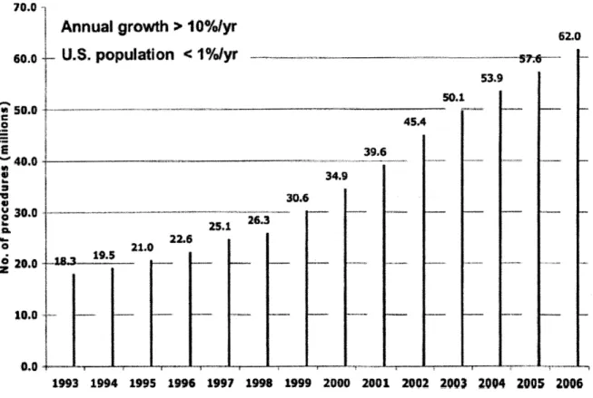
US-approximately one scan for every five people [2]. This represents an annual 10% growth over the past decade in the number of procedures as shown in Figure 1 [2]. CT scans account for a quarter of the total radiation dose to the US population-about 1.5 mSv per capita [2].
CT procedures by year (millions)
70.0
Annual growth > 10%/yr 62.
60.0 U.S. population <I10/yr .7._
53. 50.0 0 45.4 #400 _______ ______39.6 * 34.9 W 30.6 30 .03 26.3 ao22.6 ~ 118.319.5 1993 1994 1995 1996 1997 1998 1999 2000 2001 2002 2003 2004 2005 2006
Figure 1. Number of CT procedures per year in the US from 1993 to 2006. Figure courtesy of Mettler et al.
2.2. Concerns
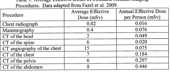
An average CT scan delivers a much higher dose than traditional radiography-about 100 times more. Common CT scans deliver doses that range from 2 to 15 mSv as shown in Table 1. These doses are below levels with clear evidence of elevated cancer rates, but several factors can quickly cause the true dose received by a patient to approach or even surpass this threshold. Record keeping for medical imaging is generally very poor. Usually, the record
indicates that a patient received a procedure, and includes the instrumentation settings used. Many patients receive repeat scans in one sitting, for a variety of reasons, but this is rarely
least 3 scans, 7% have at least five, and 4% have at least nine scans [3]. These numbers of scans represent the total number received in an acute treatment episode, including repeats and follow-up procedures. A 6 mSv procedure can quickly become 12, 18, or even 24 mSv simply because
the first few attempts were not useful. These dose estimates are based off of a standardized phantom patient, Reference Man, who weighs approximately 75 kg and is about 6' tall. This model is simply not reflective of the majority of Americans. Simulated research on the dose delivered during planar radiography to overweight patients suggests that patients can receive several times the expected dose because of the attenuation in adipose tissue [4]. This study involved planar radiography only, and does not necessarily reflect dose scaling factors for CT procedures. This discourse is meant to illuminate the severity of the situation. However, little research has been done with these complications in mind. For the remainder of this discussion, the doses delivered to Reference Man will be considered.
Table 1. Average Effective Doses of Common Medical Imaging Procedures. Data adapted from Fazel et al. 2009.
Average Effective Annual Effective Dose
Procedure Dose (mSv) per Person (mSv)
Chest radiograph 0.02 0.016
Mammography 0.4 0.076
CT of the head 2 0.049
CT of the spine 6 0.020
CT angiography of the chest 15 0.075
CT of the chest 7 0.184
CT of the pelvis 6 0.297
CT of the abdomen 8 0.446
The quickly growing nature of CT exposure in the US has prompted researchers to examine the negative health consequences resulting from these procedures. Questions are being raised by members of the medical community about the appropriateness of the increasing
number of CT scans administered. Some studies have been done to determine the efficacy of medical images in some diseases [5][6]. These concerns are raised because of the fear of the adverse health effects of ionizing radiation. One group estimates that between 1.5 and 2% of all
cancers in the US are the result of CT scans [7]. Brenner's estimate places the number of excess deaths due to CT scans at 12,000 per year. This number is sensitive to the current understanding of risk as a function of dose.
2.3. Measuring Dose
Determining the actual dose received by a patient from a procedure is a difficult task. As previously discussed, the dose delivered varies substantially between patients or even for the same patient over multiple scans for the same procedure. Currently, software programs are used to estimate the dose to a patient as a function of the machine's parameters, patient age, and gender [8]. Sophisticated software programs use Monte Carlo simulations to estimate organ equivalent doses. However, these programs still use Reference Man as the simulated patient, and fail to capture the true dose received for patients whose body types do not resemble Reference Man's body type.
Because of this convention, all epidemiological studies to date use estimates which may not accurately represent the dose received by all patients. These studies are not able to correlate the true dose received with cancer induction and may miss significant relationships because of this weakness. Improving data collection of doses delivered would enable more accurate epidemiological studies and is an important part of other strategies to be discussed.
3. Radiation Risk
3.1. Radiological Damage
Radiation causes damage at the molecular level directly, which can then manifest as cellular and eventually tissue damage. In the case of low doses of radiation, the cellular damage is either the induction of cancerous phenotypes or the formation of an inheritable mutation. The cancerous traits then present at the tissue level if the affected cells are able to grow unchecked. This mass may then metastasize and become damage at the whole organism level. This hierarchy of molecular to cellular to tissue to organism damage is the path through which radiation harms people. There are defensive mechanisms at each level to repair damage and to prevent the damage from becoming a more serious problem.
Radiation changes molecules by ionizing atoms. Since water is the primary component of cells, it is most often the target of radiation. The ionized water molecule reacts with other water molecules to form hydroxyl and hydrogen radicals. These radicals are an example of reactive oxygen species (ROS). ROS are constantly produced during metabolic processes in cells; radiation does not introduce a unique chemical species to the cell. ROS can then interact with DNA to form altered DNA chemistry. This situation can lead to a permanent mutation if the damage is not repaired and is misread during replication. Alternatively, the ROS, or radiation directly, may cause a single or double strand break (DSB) in DNA. These strand breaks are sensed by the cell and are repaired. Especially for the case of DSBs, the repair
process is subject to error and may lead to point mutations or more serious chromosomal damage.
If the damage is not repaired, and the changes result in a cell with the potential to become carcinogenic, there are mechanisms to prevent further expansion of the damage. Cells
undergoing division stop at a couple of points during the division cycle to assess their DNA and check for damage. If the damage is found here, a cell may attempt to repair this damage, or if it is too great, the cell may destroy itself through apoptosis. If these and other checkpoints fail, the cell can continue to divide and could eventually become cancer.
3.2. Contextualizing CT Doses
Most conversations about the risk of CT do not put the relevant doses in the context of natural background exposure. Cosmic x-rays, radon, potassium, and other sources contribute to
the natural background exposure. The annual dose can vary considerably between locations, but is on average 2.8 mSv per year. There is considerable variability in the annual background dose due to geography [9].
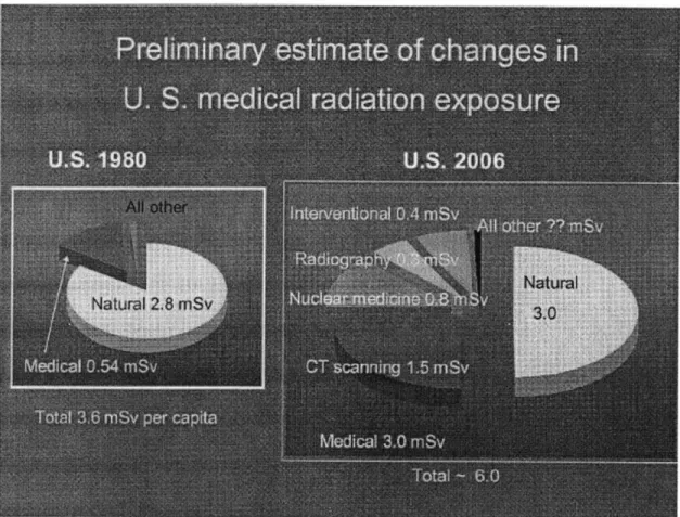
Human technologies can contribute significant doses as well. People who smoke a pack and a half of cigarettes a day receive an extra 8 rem per year to the bronchial epithelium from lead and polonium. A flight from Frankfurt to Singapore is approximately 40 ptSv, because cosmic radiation is more intense at high altitudes. Nuclear power plant workers receive about
1.6 mSv annually. These numbers should help contextualize the typical CT doses, which are on the order of millisieverts to tens of millisieverts. The most significant technology in terms of radiation exposure is CT, which accounts for about 1.5 mSv per person per year in the US. A comparison of the breakdown of natural and artificial dose contributions from 1980 and 2006 is shown in Figure 2.
Figure 2. Breakdown of sources of annual per capita radiation exposure in the US for 1980 and 2006. Image courtesy of Mettler et al. 2008.
3.3. Epidemiological Data
Models of the risk of low levels of radiation exposure attempt to relate the risk of an adverse event to the dose received. For doses of radiation more than about a Gray, the effects of radiation are deterministic, in that they confer a certain amount of observable damage as a function of dose. For example, a dose of about 0.5 Gy will induce nausea and vomiting. A dose of about 20 Gy will send the victim into a coma almost immediately. These deterministic effects are inherently different from the risks associated with smaller doses of radiation. The risk of a low dose of radiation is primarily the induction of cancer, but also includes inheritable genetic changes, both of which are essentially binary in their nature-either cancer appears or it does
not. Therefore, the severity of the risk associated with low doses of radiation is independent of dose, but the probability of such an effect is a function of dose. These probabilities are
exceedingly difficult to measure directly with any certainty; recall that out of 62 million CT scans, approximately 12,000 people contracted cancer, for a probability of about 0.0002 cancers per scan. These numbers are simply estimates obtained through the use of models and are highly suspect. Considering the natural cancer rate in the US is approximately 25%, deducing the additional risk of diagnostic radiology is very difficult.
Most of the knowledge of the dangers of low doses of radiation comes from the atomic bomb survivor studies in Japan. The survivors were acutely exposed to radiation, primarily gamma rays and neutrons, and received doses ranging from less than 0.005 Gy to 4 Gy. One study looked at the risk of cancer at low doses in 50,000 survivors who received less than 0.5 Gy [10]. A statistically significant dose response is seen in survivors who received less than 150 mGy [10]. For those who were exposed at age 30, the increase in solid cancer rates at age 70 were about 35% per Gy (90% CI: 28%; 43%) for men and 58% per Gy (43%; 69%) for women [10]. There is not a statistically significant increase in cancer rates if the analysis is restricted to groups of lower doses. The relative risk of cancer is defined as the ratio of cancer rates in two populations. Preston et al. calculated solid cancer relative risks as a function of age at exposure comparing those who received between 5 and 500 mGy to those who received less than 5 mGy. The relative risks in each age group ranged from 0.91 to 1.18,
corresponding to males in their twenties and females over fifty at time of exposure [10]. These data indicate that there is a small observable increase in solid cancer rate from doses similar to those from diagnostic radiology in some populations; in other populations, such as males exposed before the age of ten in this study, the rate of solid cancers actually decreases. These data do not reveal a trend in relative risk as a function of age for atomic bomb survivors.
The atomic bomb survivor studies are comprehensive in that they include a large number of people of all ages. However, the studies do not directly measure the effects of diagnostic radiology. A
recent German study [11] observed cancer incidence rates in children who received x-rays for diagnostic purposes in the late 1970s. The authors did not observe an increase in cancer risks for these children.
The standardized incidence ratio for all cancers was 0.99 (95% CI: 0.79; 1.22) [11]. The study may not be perfectly relevant to the discussion of risks from CT, because the doses involved in planar radiography are much lower than those in CT, but it does demonstrate that such studies are feasible, and that some people are paying attention to this concern. Large scale studies like this one of patients undergoing CT could help fill in the gaps in knowledge pertaining to the risk of low doses of radiation.
Except for the Hammer study, there is not much evidence of the effects of very low doses of radiation, like those in diagnostic radiology. The atomic bomb survivor data provide a clear picture of the response to doses above about 100 mSv, but not for those below this number [12]. To determine the risks below 100 mSv, models of the dose response must be used. A variety of models have been proposed, and conflicting models have been adopted by different international organizations [12]. These models
influence regulatory and accident-scenario policy. Therefore, it is important that these models accurately reflect the dose response.
4. Models
4.1. The Linear No-Threshold Model
The linear no-threshold (LNT) model suggests that the risk from radiation is directly proportional to the dose received. The dose-risk relationship is achieved by drawing a line connecting the origin with doses with solid epidemiological evidence. This model implies that there is no safe amount of radiation, and that any single molecular event has a constant
probability of inducing carcinogenesis. The primary type of damage within the cell which will give rise to cancer is a DNA double-strand break (DSB). These are dangerous because they are difficult to repair with high fidelity and can lead to chromosomal instability. The LNT model suggests that five DSBs are exactly five times more likely to induce cancer than a single DSB.
This model makes intuitive sense on some levels, however biological responses including DNA repair call into question the accuracy of the LNT model. The LNT model has been adopted by the American Academy of Sciences [1] and by the International Commission on Radiological Protection [13].
4.2. Threshold Model
An alternate theory is the threshold model. This model acknowledges that over a certain range of dose, the risk is proportional to dose. However, there is a threshold below which the actual risk is either zero or at least much lower than predicted by a purely linear model. The current atomic bomb surivivor data are consistent with a threshold model in which the minimum dose required to increase the risk of cancer is 40 mSv [10]. This model is as consistent as the LNT model with the atomic bomb survivor data and other epidemiological studies in that the two models predict the observed cancer rates with about the same accuracy. The biological
motivation for this model is that the cell is able to fully deal with, either through repair or apoptosis, a certain amount of DNA damage. Metabolic processes contribute to a steady state level of reactive oxygen species (ROS), the intermediate damaging agent in radiation, so it is expected that the cell can survive normally with some non-zero ROS concentration.
4.3. Alternative Models
There are also models which predict non-linear behavior over certain dose ranges. A supra-linear model suggests that low doses are more dangerous than predicted by the LNT model. Biological mechanisms such as the bystander effect lead to the prediction of this response. The bystander effect is the observation that cells which experience radiation damage
send a signal to other cells which prompts the recipient cells to respond almost as if they had suffered damage themselves. Other evidence suggests that the bystander effect is protective. A quadratic model suggests that the risk of a dose is actually less than the risk predicted by the LNT model. The adaptive response and DNA repair mechanisms suggest that this model may be appropriate for a certain dose range. The adaptive effect is the phenomenon observed when small doses of radiation prime against a second dose; the negative response is reduced by the priming dose compared to previously unirradiated cells. Ultimately, the interactions of these and other phenomena and their relative degrees of importance determine the true shape of the dose-response curve.
5. Improving Risk Models
The risk models currently in use are not inspired by experimental evidence. Experiments designed to elucidate the biological response to radiation injury at the molecular, cellular, tissue,
and organism level should be used to inform the choice of risk model. By determining the responses and their effectiveness under various conditions, scientists can attempt to justify the use of one model over another.
5.1. Experimental Data
These risk models will be improved through further experimentation and additional epidemiological studies. Experiments using cell culture should be continued with the intention of gaining a more complete understanding of radiation responses at the cellular level-especially the adaptive response and the bystander effect. These experiments could also be scaled for tissue models to measure these responses in a more relevant environment. Large-scale animal studies
may also be conducted in mice and monkeys. Such studies have proven valuable in accessing the risk of radiation [14], though more studies using low doses of radiation would be beneficial.
The interactions between competing effects-such as the adaptive response and the bystander effect-complicate the assessment of various risk models. An understanding of the relative importance of these effects under different conditions and at different doses and dose rates is essential for choosing an accurate model of risk as a function of dose. Experimental data will help determine the appropriateness of competing models, but epidemiological data may provide the best picture of the risks of diagnostic radiology.
5.2. Epidemiological Data
With more than 67 million CT scans performed in the US annually, there is a lot of potential for epidemiological studies to determine the risk of these procedures. Some researchers have attempted to do just this, though there are several complicating factors. Few healthy people receive CT scans, which may strongly bias the sample population. It is difficult to separate the disease which prompted the examination from the CT scan itself in terms of contribution to a subsequent malignancy. However, since CT is used in a wide variety of settings and disease states, it may be plausible to perform such epidemiological studies with appropriate comparisons. A study could follow patients who received CT scans for a variety of reasons in an attempt to minimize the complications of a single disease. Alternatively, researchers could choose a CT prompt with very little likelihood of inducing cancer, such as trauma. A study could follow patients admitted to trauma units at multiple hospitals and compare the subsequent cancer incidence in those who received CT scans versus those who did not. This study assumes that
there will be a large enough population of patients who do not receive CT scans, which may be a non-trivial obstacle.
Despite these limitations, there have been attempts to systematically study cancer
induction in patients who undergo diagnostic radiology procedures. As previously mentioned, a recent study [11] of nearly 93,000 German children who received x-ray exams in the seventies found that there was not an increase in cancer rates in this population. Since doses from x-rays are considerably lower than those from CT, this example is presented simply to show that such studies are possible. CT technology is too young to have a study of this magnitude with a thirty-year follow up, but these studies will soon become possible.
5.3. Dose Registry
There is a way to facilitate such studies which may prove invaluable in understanding the risk of diagnostic radiology while at the same time serving to more fully inform the physician's decision-making process when ordering a CT scan. A national registry of diagnostic radiology procedures should be created to track the number and type of diagnostic procedures for all US patients. Such a registry would enable retrospective studies to be performed using the data from millions of patient records. There are several barriers to the creation of this registry, but a strong
effort should be made to overcome these obstacles.
Patient records in the United States are currently unorganized. A registry would require a complete overhaul of the patient record system. Such reform is currently being promoted on the national scale and in some hospital systems, such as the Geisinger Medical Center. The
digitalization of patient records would provide numerous benefits to several aspects of health care, and, if done correctly, would provide the single greatest database for low dose radiation
exposures. Creating a database for radiological procedures will be difficult without a digital patient records system.
The creation of a registry of radiology exams requires a change in the culture surrounding radiology today. Patient records rarely contain sufficient detail to properly determine the dose a patient received as a result of an examination. The actual number of scans performed would need to be recorded for this registry to be maximally useful; this does not often happen today. Along with the implementation of the registry, there must be a conscious effort to change the culture among technicians and doctors that the recording of this information is important.
The final major obstacle for a national dose registry is the estimation of the doses from diagnostic procedures themselves. As previously discussed, the actual dose delivered is a strong function of body weight and shape [4]. The dose for a given procedure can vary considerably between hospitals. These inter-hospital variations are probably best attributed to differences in protocol and instruments. Record keeping of the settings and model used would help minimize this complication.
If a registry is created, includes these relevant data, and is accepted and promoted by the medical community, future estimates of the risk of diagnostic radiology would be much more reliable than those in use today. This system should be implemented as soon as possible.
6. Reducing Dose 6.1. ALARA
The US Nuclear Regulatory Commission has adopted a policy known as ALARA as part of the consideration of acceptable dose limits for nuclear industry employees and the general public. ALARA stands for "as low as reasonable achievable." This policy implies that any dose
of radiation is hazardous to one's health. The ALARA principle is also applied within the medical field for both patients and health care providers. In the absence of a good understanding
of the risks involved in diagnostic radiology, this philosophy makes sense. However, the
extraordinary growth in the number of CT scans over the past decade calls into question whether or not the ALARA principle is being obeyed properly.
Several physicians have begun to question the near-ubiquitous use of CT scans. In an article in Radiology [15], Ginsberg questions the appropriateness of the "if clinically indicated" clause in radiology protocols. Fear of litigation, Ginsberg claims, forces radiologists to order CT scans when the scans would not offer any clinically relevant information. This is especially true when a second type of study is called for, for example a CT is ordered when the results of an MRI are ambiguous, even though the radiologist knows that the CT will not offer a clearer picture. Ginsberg encourages radiologists to take an active role in determining when additional
studies are beneficial by taking into account a patient's overall condition. Ginsberg's opinion piece is an example of the growing sentiment among physicians that they are the ones who need to act responsibly in curtailing the growth of CT usage.
6.2. CT Efficacy
Studies have been undertaken to directly examine the benefits of diagnostic CT scans in specific settings. A study of the use of CT to determine appendicitis prior to operation found that the patients who underwent pre-operatory CT scans exhibited fewer negative appendectomies
[5]. This was observed for women 45 years of age and younger, but not for any other patient group. This decrease in negative appendectomy rates in women 45 years of age and younger cannot be definitively attributed to the use of CT. Confirmatory CT scans are used in over 90%
of cases of suspected appendicitis. This study by Coursey et al. suggests that these CT scans may not provide a clinical benefit. Similar studies have been performed for various other
diagnostic uses of CT. More of these studies should be conducted to determine which uses of CT are inappropriate. Measures should then be taken to eliminate these inappropriate uses of CT.
Studies on the efficacy of diagnostic CT uses will not immediately translate into the elimination of inappropriate uses. The appendectomy studies have sparked a considerable controversy within the medical field. Other studies have been released which indicate that CT scans do reduce the rates of negative appendectomies [16]. These conflicting results prevent reform. Even in the face of clear evidence, which is not yet available, there will be resistance within the medical profession which must be overcome.
6.3. Decision Criteria
Other doctors are recommending policies to systematically reduce the quantity of diagnostic scans ordered through the use of decision trees and exclusion criteria. Such criteria exist for the use of head CT in minor trauma cases [17] and in many other settings. These criteria are formed following studies like those by Coursey. The criteria describe situations in which CT scans are beneficial and when they are inappropriate. These decision trees help reduce unnecessary doses to patients without compromising the potential for positive outcomes.
The creation of these criteria alone will not be enough to properly reduce the number of CT scans administered. A study of the appropriateness of outpatient CT and MRI referrals from primary care clinics showed that a quarter of referrals did not meet the relevant evidence-based
appropriateness criteria [18]. The rates of compliance with such criteria can be improved by physician education and electronic order entry systems with built-in inclusion criteria.
6.4. Reducing Dose Per Scan
Another approach for limiting radiation exposure has been to reduce the dose delivered per scan. The image quality obtained in a CT scan is proportional to the dose delivered. Some
CT procedures deliver much more dose than is required to achieve the clinically relevant information. A recent study on the effects of using reduced radiation protocols in the detection of renal calculi (kidney stones) showed that it is possible to maintain the same diagnostic
specificity and sensitivity while decreasing patient dose by up to 70% [6]. This dose reduction is achieved by reducing the current by about a third compared to the current protocol. The study has the benefit of using cadavers instead of live patients, and does not involve the problems that accompany epidemiological studies. Studies such as this open up the possibility of greatly reducing patient dose without compromising outcomes.
6.5. Dose Registry
A registry of patient doses would also serve as a tool for physicians to more consciously obey the ALARA principle. Physicians would be able to identify patients who have a history of repeated CT scans and could more strongly consider alternative methods of diagnosis if
applicable. For example, consider the case of an 18 year old male who presents with symptoms consistent with appendicitis. This patient received several abdominal CT scans when he was 6 due to a pair of fractured ribs. The study which found that CT screening does reduce the number of negative appendectomies found that most of the negative appendectomies occurred in patients
under 20 years old [16]. The combined history of multiple CT scans and the weak diagnostic predictive power of CT in the case of juvenile appendicitis might cause the physician to forego a pre-operatory CT scan for this patient.
7. The Appropriateness of ALARA
Applying the ALARA principle to the diagnostic radiology practices is a difficult task, because determining the benefit to risk ratio is challenging. The ALARA principle has been chosen because there is not sufficient evidence to suggest the actual risk of radiation, and it may be overly-cautious. The results of the investigations suggested here may indicate that different thresholds may more effectively protect against the risks of radiation.
Researchers have known that the dose rate of radiation is an important factor in biological responses for quite some time [19]. The notion that damage is proportional to the dose rate for a given dose is consistent with a model of repair at the sub-cellular level. As long as the body has sufficient time to repair itself between assaults, it is reasonable that damage may not accumulate. This model would lead to the adoption of a policy in which the frequency of radiological
procedures is regulated, and not the aggregate number of procedures. This model would relieve the fear of excessive CT scans over the course of a patient's life; however, many patients receive multiple studies over a brief period in response to an acute disease state. This practice may be more harmful than annual head CTs for the tracking of a tumor in remission. Adjusting to this frequency-threshold policy would discourage repeat studies and require physicians to make the most out of a single study in the analysis of a patient.
The atomic bomb survivor data are consistent with a threshold of 40 mSv [10]. That is to say that there is not a statistically significant increase in cancer risk for survivors who received a
dose less than 40 mSv. This threshold model fits the data as well as the linear no-threshold model [10]. This may imply that CT procedures with an effective dose below 40 mSv do not increase a patient's cancer risk. Alternatively, the data may imply that as long as a patient's cumulative dose remains below 40 mSv, his or her cancer risk will not increase. Since the atomic survivor population was exposed in a single event, the data cannot distinguish between these two possibilities. Most CT procedures have average effective doses below 40 mSv [2]; if the dose from a single scan is the relevant parameter, diagnostic CT procedures should not increase the risk of cancer at all.
A different type of question, but an important one, asks for which populations are these concerns relevant. Children have a greater risk from radiation than adults. This is because their cells are more actively dividing, the absorbed dose per procedure is higher, and there is a longer period for cancers to appear following the procedure. Perhaps the concerns surrounding CT use are only relevant for children, or perhaps for children and adults under 30, 40, or 50 years old. There may be an age at which it is safe to administer as many CT scans as desired. Considering the disproportionate allocation of care at the end of life, this question may be quite relevant to the discussion of CT risks. A recent study of CT procedure rates in an 18 to 64 years old population found that 86% of patients 60-64 years old underwent one or more CT procedure compared to 50% in the 18-34 years old population [20]. In this study, most of the cumulative dose was received by patients over 50 years old [20]. This recent study can be compared to a study from 2000 which shows an age distribution of CT scans with a younger bias, with patients aged 40 to 55 being the most common, as shown in Figure 3 [3]. These data and the notion that
consequences from radiation are inversely related to age suggest that the CT exposure problem in the US is not as serious as many believe.
Total
Bod
0-5 6-14 II-is 14-20 21-231" ? 3 S-3s
Figure 3. Age distribution of of Mettler et al. 2000.
34-40 41-4 4540 145 344 614 "-1# 71-75 764
Age Group
CT scans in the US. Figure courtesy
8. Evidence-Based Medicine
This paper has introduced several studies concerning the appropriateness of CT in certain situations. The doctors who initiate these studies are generally not motivated by the risk
of cancer induction from the radiation. These doctors are concerned more with the excessive cost of CT, both in terms of monetary cost and the opportunity cost of using a limited resource for a potentially inappropriate procedure. These concerns highlight the sentiment of the medical field with respect to diagnostic radiology. Those that are worried about the excess risk of cancer from diagnostic radiology are primarily scientists, not clinicians. Unless physicians can be convinced that the risk of diagnostic radiology is serious, effecting large-scale change with the intention of systematically reducing the dose received by the population will be difficult.
The evidence presented highlights the incoherency of the knowledge of the risk of very low doses of radiation. Some models suggest that approximately 15,000 people are developing cancer each year as a result of CT use in the US. Another model suggests that CT use is
completely safe. This lack of a consensus prevents well-guided organized action with respect to protocols for CT use and patient dose. This helps to explain why there is not a concerted effort to reduce the use of CT in the US. Examining what has caused the rapid increase in CT use is enlightening, and important for this discussion.
The argument is often proposed that in almost any individual case, the benefits of a CT procedure far outweigh the risk of cancer. However, on a societal scale, the risk is very real, and should not be ignored. The societal risk argument requires systematic changes to the use of CT,
along the lines of those presented previously. These changes are beginning to be realized, or at least recognized, by the medical community. They are not, however, being driven primarily by a concern for patient safety. This reveals a very important point about the ways in which the patterns of CT use will evolve over the next decade.
This paper has until now focused on the lack of knowledge concerning the health risks of CT use in the US, and the ways in which this issue can be addressed. Knowledge of the risks
of very low doses of radiation is imperative for several reasons. Large scale epidemiological studies, such as those that would be made possible by a dose registry, will provide a much clearer picture of the risk-dose relationship for doses on the order of millisieverts. This information should confirm which approach to controlling CT use is correct. It will be
particularly useful for children, who experience greater risks for a given procedure, and are likely to receive more subsequent CT studies over the course of their lifetimes. Additionally, the data could be used to craft a scientifically-justified set of responses to various accident scenarios
involving radiation. The current EPA protocols do not reflect any scientific evidence, and could prove to be problematic. The societal risk argument is useful for thinking about the risk in these situations.
Apart from these situations, the societal risk argument fails. If the benefit from a given procedure far outweighs the risk of radiation, then this scales to the societal level as well. The net benefit to society will be positive as long as the procedures provide relevant and accurate information in an efficient way. This is likely true in most cases unless the risk from small doses is much greater than currently estimated. As long as a procedure is efficient with respect to radiation risk, that is to say the benefit of the procedure outweighs the risk due to radiation, the health effects of the procedure on the societal scale, though real, are justified. This does not mean that the effects should be ignored, or that efforts to reduce exposure should be suspended. This logic simply counters the notion that the number of CT scans needs to be reduced simply because of the magnitude of the dose delivered to society at large.
A thorough understanding of the risk of low doses of radiation is not required to select for efficient uses of CT. Some research has already been undertaken to determine the
effectiveness of certain uses of CT. These investigations are not motivated by the desire to reduce radiation exposure, but instead are the results of the evidence-based culture in American
medicine. The ethos of evidence-based medicine requires physicians to continually review their practices to show that they improve a measurable endpoint [21]. One artifact of this culture is that there can be a time lag between when a procedure is introduced or becomes popular and when this assessment begins.
The use of CT in the US rose too quickly for scientific knowledge about the health effects of radiation or for evidence-based assessment to keep up. Now that CT use is so
ubiquitous, there is ample opportunity for physicians to perform controlled trials to determine the efficacy of CT in different situations. These trials will select for the uses of CT which provide a benefit for the patient. This selection should be sufficient with respect to reducing dose in an
intelligent manner for most patient populations.
There is one additional drawback to relying on the culture of evidence-based medicine to limit the amount of unnecessary dose patients receive. This method fails to incentivize the development of alternative technologies with smaller doses to patients, while achieving the same clinical information. Unless scientists discover that CT doses are more harmful than anticipated, there may not be a need for alternative technologies.
9. Conclusion
The use of computed tomography in the United States has risen dramatically over the past decade. This has led to a considerable rise in the annual per capita radiation dose in the US. The risks of the additional doses are not well-understood, and much needs to be done to improve this knowledge. This knowledge would enable the formation of scientifically-justified protocols for CT use and patient dose, for the response to accident scenarios involving elevated radiation levels, and for workers in elevated-dose industries, and potentially lead to new strategies for the prevention and treatment of radiological damage. However, until that goal is realized, scientists and physicians should recognize that CT is providing a valuable service and that the standards of evidence-based medicine will promote efficacious uses of CT and discourage inappropriate ones. On the societal scale, the adverse health effects of ubiquitous CT use might be real, but
10. Acknowledgements
I would like to thank Dr. Yanch for her inspiration, support, and flexibility throughout this process. I would like to thank my family for their support and confidence. Finally, I would like to thank Dwight Chambers and Tom McKrell for their mentorship throughout my
11. References
[1] National Research Council (U.S.). Committee to Assess Health Risks from Exposure to Low Level of Ionizing Radiation, Health Risks from Exposure to Low Levels of Ionizing Radiation:
BEIR VII Phase 2. Washington, D.C.: National Academies Press, 2006.
[2] F. A. Mettler Jr, B. R. Thomadsen, M. Bhargavan, D. B. Gilley, J. E. Gray, J. A. Lipoti, J. McCrohan, T. T. Yoshizumi and M. Mahesh, "Medical radiation exposure in the U.S. in 2006: preliminary results," Health Phys., vol. 95, pp. 502-507, Nov, 2008.
[3] F. A. Mettler Jr, P. W. Wiest, J. A. Locken and C. A. Kelsey, "CT scanning: patterns of use and dose," J. Radiol. Prot., vol. 20, pp. 353-359, Dec, 2000.
[4] J. C. Yanch, R. H. Behrman, M. J. Hendricks and J. H. McCall, "Increased Radiation Dose to Overweight and Obese Patients from Radiographic Examinations1," Radiology, vol. 252, pp.
128-139, July, 2009.
[5] C. A. Coursey, R. C. Nelson, M. B. Patel, C. Cochran, L. G. Dodd, D. M. DeLong, C. A. Beam and S. Vaslef, "Making the Diagnosis of Acute Appendicitis: Do More Preoperative CT
Scans Mean Fewer Negative Appendectomies? A 10-year Studyl," Radiology, vol. 254, pp. 460-468, February, 2010.
[6] D. H. Jin, G. R. Lamberton, D. R. Broome, H. P. Saaty, S. Bhattacharya, T. U. Lindler and D. D. Baldwin, "Effect of Reduced Radiation CT Protocols on the Detection of Renal Calculil,"
Radiology, vol. 255, pp. 100-107, April, 2010.
[7] D. J. Brenner and E. J. Hall, "Computed Tomography -- An Increasing Source of Radiation Exposure," N. Engl. J. Med., vol. 357, pp. 2277-2284, November 29, 2007.
[8] E. K. Osei and R. Barnett, "Software for the estimation of organ equivalent and effective doses from diagnostic radiology procedures," J. Radiol. Prot., vol. 29, pp. 361-376, Sep, 2009.
[9] M. K. Nair, K. S. V. Nambi, N. S. Amma, P. Gangadharan, P. Jayalekshmi, S. Jayadevan, V. Cherian and K. N. Reghuram, "Population Study in the High Natural Background Radiation Area in Kerala, India," Radiat. Res., vol. 152, pp. S145-S148, Dec., 1999.
[10] D. L. Preston, E. Ron, S. Tokuoka, S. Funamoto, N. Nishi, M. Soda, K. Mabuchi and K. Kodama, "Solid Cancer Incidence in Atomic Bomb Survivors: 1958-1998," Radiat. Res., vol.
168, pp. 1-64, 2007.
[11] G. P. Hammer, M. C. Seidenbusch, K. Schneider, D. Regulla, H. Zeeb, C. Spix and M. Blettner. (2010, Mar 16). Cancer incidence rate after diagnostic X-ray exposure in 1976 -2003 among patients of a university children's hospital. Rofo 182(5), pp. 404-414.
[12] M. Tubiana, A. Aurengo, D. Averbeck and R. Masse, "The debate on the use of linear no threshold for assessing the effects of low doses," J. Radiol. Prot., vol. 26, pp. 317-324, Sep, 2006.
[13] ICRP (International Commission on Radiological Protection). (2004, ICRP draft of committee I/Task group, low dose extrapolation of radiation related cancer risk.
[14] G. V. Dalrymple, I. R. Lindsay and J. J. Ghidoni, "The Effect of 2-Mev Whole-Body X-Irradiation on Primates," Radiat. Res., vol. 25, pp. 377-400, Jun., 1965.
[15] L. E. Ginsberg, ""If Clinically Indicated:" Is It?1," Radiology, vol. 254, pp. 324-325, February, 2010.
[16] C. Harswick, A. A. Uyenishi, M. F. Kordick and S. B. Chan, "Clinical guidelines, computed tomography scan, and negative appendectomies: a case series," Am. J. Emerg. Med., vol. 24, pp.
68-72, Jan, 2006.
[17] M. J. Haydel, C. A. Preston, T. J. Mills, S. Luber, E. Blaudeau and P. M. C. DeBlieux, "Indications for Computed Tomography in Patients with Minor Head Injury," N. Engl. J. Med., vol. 343, pp. 100-105, July 13, 2000.
[18] B. E. Lehnert and R. L. Bree, "Analysis of appropriateness of outpatient CT and MRI referred from primary care clinics at an academic medical center: how critical is the need for improved decision support?" J. Am. Coll. Radiol., vol. 7, pp. 192-197, Mar, 2010.
[19] G. W. Barendsen, "Dose fractionation, dose rate and iso-effect relationships for normal tissue responses," Int. J. Radiat. Oncol. Biol. Phys., vol. 8, pp. 1981-1997, Nov, 1982.
[20] R. Fazel, H. M. Krumholz, Y. Wang, J. S. Ross, J. Chen, H. H. Ting, N. D. Shah, K. Nasir, A. J. Einstein and B. K. Nallamothu, "Exposure to Low-Dose Ionizing Radiation from Medical Imaging Procedures," N. Engl. J. Med., vol. 361, pp. 849-857, August 27, 2009.
[21] J. A. Claridge and T. C. Fabian, "History and development of evidence-based medicine,"