HAL Id: hal-01943657
https://hal.archives-ouvertes.fr/hal-01943657
Submitted on 4 Dec 2018
HAL is a multi-disciplinary open access
archive for the deposit and dissemination of
sci-entific research documents, whether they are
pub-lished or not. The documents may come from
teaching and research institutions in France or
abroad, or from public or private research centers.
L’archive ouverte pluridisciplinaire HAL, est
destinée au dépôt et à la diffusion de documents
scientifiques de niveau recherche, publiés ou non,
émanant des établissements d’enseignement et de
recherche français ou étrangers, des laboratoires
publics ou privés.
Preparation of self-healing acrylic latex coatings using
novel oil-filled ethyl cellulose microcapsules
Mojtaba S. Mirabedini, Isabelle Dutil, Lucile Gauquelin, Ning Yan, Ramin R.
Farnood
To cite this version:
Mojtaba S. Mirabedini, Isabelle Dutil, Lucile Gauquelin, Ning Yan, Ramin R. Farnood. Preparation
of self-healing acrylic latex coatings using novel oil-filled ethyl cellulose microcapsules. Progress in
Organic Coatings, Elsevier, 2015, 85, pp.168-177. �10.1016/j.porgcoat.2015.03.024�. �hal-01943657�
OATAO is an open access repository that collects the work of Toulouse
researchers and makes it freely available over the web where possible
Any correspondence concerning this service should be sent
to the repository administrator: tech-oatao@listes-diff.inp-toulouse.fr
This is an author’s version published in:
http://oatao.univ-toulouse.fr/20341
To cite this version:
Mirabedini, Mojtaba S. and Dutil, Isabelle and Gauquelin, Lucile
and Yan, Ning and Farnood, Ramin R. Preparation of self-healing
acrylic latex coatings using novel oil-filled ethyl cellulose
microcapsules. (2015) Progress in Organic Coatings, 85. 168-177.
ISSN 0300-9440
Preparation of self-healing acrylic latex coatings using novel oil-filled
ethyl cellulose microcapsules
S.M. Mirabedini
a,b,∗, I. Dutil
c, L. Gauquelin
d, N. Yan
e, R.R. Farnood
baColor, Resin & Surface Coatings Department, Iran Polymer and Petrochemical Institute, Tehran, Iran bDepartment of Chemical Engineering and Applied Chemistry, University of Toronto, Toronto, ON, Canada cDepartment of Materials Science and Engineering, University of Toronto, Toronto, ON, Canada
dDepartment of Chemical Engineering, Institute National Polytechnique de Toulouse ENSIACET, Toulouse Cedex, France eFaculty of Forestry, University of Toronto, ON, Canada
a r t i c l e
i n f o
Keywords: Coatings Self-healing Microcapsule Mechanical properties Latex Encapsulationa b s t r a c t
Novel oil-filled microcapsules were prepared by introducing a phase separation method using ethyl cellulose as a shell-forming containing rapeseed oil. The prepared oil-filled microcapsules were eval-uated by optical microscopy, scanning electron microscopy and particle size analysis. Results showed that spherical microcapsules with a diameter of 10 to 45 mm and a rough porous shell were obtained. Carboxylated styrene/butadiene copolymer latex films containing various levels of these microcapsules were subjected to various levels of pre-elongation and their tensile properties were examined. The addi-tion of oil-filled microcapsules resulted in a significant improvement in the modulus, strain-to-break, and toughness of the films. The self-healing mechanism of latex films was examined through the col-orimetric measurements of the release of dye-containing following the pre-elongation of the samples. These measurements confirmed that pre-elongation of samples resulted in the release of oil within the latex films, hence plasticizing the surrounding polymeric network and partly restoring the mechanical properties of the pre-elongated films.
1. Introduction
Water-based polymeric coatings are used in a wide variety of applications ranging from decorative wall paints to packaging materials. However, due to the low strain-to-failure exhibited by these materials, unexpected damages and cracks often incur dur-ing their service life[1]. In many cases, physical and mechanical damages of polymeric coatings occur within the manufacturing and converting processes. In addition, water-based coatings usually provide relatively poor barrier properties against gases, high sen-sitivity to water and moisture, and inferior mechanical properties
[2]. Some of conventional methods for improving these properties include; increasing the coating thickness [3], applying multi-ple coating layers[1], adding functionalized nanoparticles[4–6], increasing the crosslinking density of polymer[7]and introducing functionalized core–shell latexes into the coating formulation[8].
Among the aforementioned available methods[3–8], application of healable materials in coating formulations shows great poten-tial for improving the durability and enhancing bulk mechanical properties of the coating layers. Such self-healing coatings possess the ability to repair in response to damage in the material[9].
In 2001, White and co-workers[10]introduced a novel method to repair the mechanical properties of thermosetting polymers following crack propagation through the micro-encapsulation of self-healing materials. Repairing has been obtained via the addition of urea-formaldehyde microcapsules containing dicy-clopentadiene (DCPD) healing agent and through the ring opening polymerization of DCPD in the presence of Grubbs’ catalyst[11]. Micro-encapsulation is an approach that consists of isolating a dis-persed phase from an external medium by surrounding or coating it with a protective sell material having sizes typically ranging from submicron up to 1 mm[12]. The microcapsules containing healing agents have the capability of self-repairing upon crack propaga-tion[13,14]. As the healing agent flows within the crack lines, it comes into contact with the active materials inducing poly-merization and hence sealing up the crack[15]. The repairing of mechanical damage is particularly useful to coatings or compos-ites where subsurface damage occurs that is difficult to detect and fix[15–18].
∗ Corresponding author at: Iran Polymer & Petrochemical Institute, Color, Resin and Surface Coatings, Pajouhesh Blv., Exit 15, Tehran-Karaj, Tehran 14965-115, Iran. Tel.: +98 21 4866 2401; fax: +98 21 4458 0023..
E-mail addresses: m.mirabedini@utoronto.ca,sm.mirabedini@ippi.ac.ir
(S.M. Mirabedini).
A variety of self-healing chemistries have been introduced to enhance the interaction between the healing agent and a polymeric matrix. These include epoxy[10,19,20], isocyanate[21]and solvent welding[22]siloxane chemistries for vinyl ester matrices[23]and poly dimethyl siloxine (PDMS)[24].
An alternative to self-healing using chemical bonding agents is self-healing by plasticizing mechanism. This approach is a promis-ing method for enhancpromis-ing mechanical properties of polymeric coatings via increasing tensile strength and elongation at break.
In the current study, rapeseed oil was encapsulated in ethyl cellulose microcapsules using a robust phase separation route and the mechanical properties of the water-based latex coatings containing these oil-filled microcapsules were investigated. Latex films containing 1–3 wt% microcapsules and two microcapsule sizes were studied. The deformation and disruption of latex films containing microcapsules filled with colored rapeseed oil were examined under applied tensile load using color coordinate measurements.
2. Experimental
2.1. Materials
Ethyl cellulose (EC), rapeseed oil (RO), sodium dodecyl sulfate (SDS), Sudan Red 7B solvent dyestuff, and ethyl acetate were pur-chased from Sigma Aldrich (Oakville, Ontario, Canada). A non-ionic surfactant, Dynol 604, was provided by Air Products USA. A com-mercial carboxylated styrene–butadiene latex, Styronal ND 656 was supplied by BASF Corporation. This latex is an emulsion-type latex commonly used for paper coating applications for its film-forming properties, resistance to water and moisture, and relatively rapid drying with low porosity[25]. All chemicals were analytical reagent grade and used as received.
2.2. Microcapsules preparation
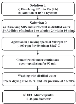
Microcapsules with oil to ethyl acetate ratio of 70:30 were pre-pared via a two-stage solvent evaporation method[2]following
Solution 1
Dissolving EC in EA (2 h)
Addition of RO + Dye stuff to the solution 1
Solution 2
a) Dissolving SDS and surfactant in distilled water
Agitation in a mixing speed of 800 rpm or 1400 rpm for 60 min at 58±2°C
Concentration by continuous open-top stirring for 90 min.
Washing with distilled water
Freeze drying at 60±5 °C and low pressure of 6.5 mPa
RO:EC Microcapsules 10-45μm diameter
Solution
1
a) Dissolving EC into EA (2 h)b) Addition of RO + Dyestuff
Concentrated under continuous open-top stirring for 90 min
b) Addition of solution 1 to solution 2 (within 10 min)
Fig. 1. Microencapsulation procedure.
the methodology reported elsewhere[26,27]. The flowchart for the synthesis of microcapsules is provided inFig. 1. In brief, in the first stage, ethyl cellulose powder was dissolved in ethyl acetate (5 wt%) under magnetic stirring for at least 2 h at ambient temper-ature. RO and 0.005 wt% of red solvent dyestuff were then added to the above-mentioned solution and system was magnetically stirred for further 30 min. In the second stage, the solution was added drop wise to an aqueous solution of 1 wt% SDS and 0.05 wt%
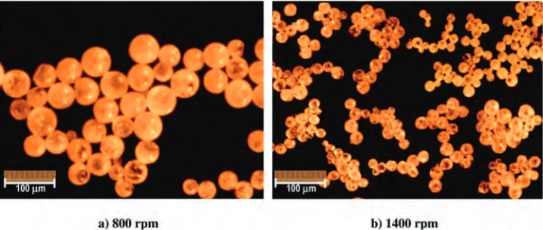
Fig. 3. Optical micrographs of microcapsules prepared at mixing rate of: (a) 800 rpm and (b) 1400 rpm.
Dynol 604 surfactant saturated with ethyl acetate, under mechani-cal mixing (BDC6015 Stirrer, Caframo Ltd., Wiarton, ON, Canada) at either 800 rpm or 1400 rpm at 25 ± 2◦C. The dispersion container
was immersed in a temperature-controlled water bath on a pro-grammable hotplate with external temperature probe (Canlab Co. New York, USA). The dispersion was stirred at a constant speed of either 800 rpm or 1400 rpm for 30 min, and the temperature was slowly increased up to 58 ± 2◦C. The dispersion was stabilized and
concentrated by continuous open-top magnetic stirring at a tem-perature of 58 ± 2◦C for about 90 min. These microcapsules were
washed with deionized water and freeze dried at −60 ± 5◦C and
6.5 mPa for characterization. In addition, to examine the effects of oil encapsulation, a control sample was prepared where no oil was added in the synthesis procedure, producing solid ethyl acetate particles.
2.3. Microcapsules characterization
Multisizer 3 particle size analyzer (Coulter Counter, FL, USA), equipped with a 280 mm aperture tube, was used to determine the average particle size and size distribution of microcapsules. Microcapsules (0.05 g) were dispersed into 250 ml deionized water using a stirring bar for about 10 min before particle size measurement.
The morphology and shape of microcapsules were evaluated using a Leica DMLA optical microscope (Leica Microsystems, Germany). The surface and morphology of microcapsules were also evaluated using a scanning electron microscope (HITACHI S-2500, Japan). The vacuum freeze-dried microcapsules[26]were mounted on a conductive stage and sputter coated with a thin layer (∼10 nm) of gold for 90 s under Ar atmosphere and SEM micrographs were recorded in the secondary electron mode at 10 kV.
2.4. Mechanical properties
Various wt% of oil-filled microcapsules were added to the latex suspension and magnetically stirred for 30 min. The suspension was then applied on the degreased glass plate using a standard coat-ing rod with a wet film thickness of 200 ± 10 mm. the latex samples were then dried at 50 ± 5◦C for about 30 min.
Tensile properties of free-standing latex films containing various levels of RO:EC microcapsules were evaluated accord-ing to ASTM D 638 standard. Rectangular tensile specimens (0.15 mm × 15 mm × 100 mm) were prepared by cutting the latex film to shape. The experiments were carried out on a Universal computerized system for material testing machine (Sintech 1).
Fig. 4. Optical micrographs of latex film containing microcapsules with different morphologies.
The crosshead moved with a constant speed of 50 mm min−1 at
23 ± 2◦C. At least seven individual replicates were tested for each
formulation.
2.5. Color measurements
To visualize the oil released during the mechanical testing of latex films, after being subjected to different elongation percent-ages (0, 25, 50, 100 and 150%), color measurements were performed according to ASTM D 65 standard using a X-Rite 530 spectroden-sitometer. Total color change, 1E, as a function of pre-elongation %
Fig. 5. Scanning electron micrographs of RO:EC microcapsules synthesized at mixing rate of (a) 800 and (b) 1400 rpm.
Table 1
Summarized mechanical properties of latex films containing oil-filled microcapsules.
Sample codinga Elongation at break (%) Young modulus (MPa) Yield stress (MPa) Yield strain (MPa) Toughness (MPa)
±14 ±0.25 ±0.15 ±0.17 ±0.23 NLa 191 4.77 0.74 2.70 3.84 0.5R-800 222 5.59 1.40 3.03 4.36 1.0R-800 204 5.47 2.19 4.38 5.85 2.0R-800 154 4.75 1.15 2.00 2.68 3.0R-800 145 4.08 0.87 1.49 1.55 1.0EC-800 98 5.00 2.35 3.80 3.15 0.5R-1400 187 5.08 1.82 3.28 4.18 1.0R-1400 201 5.74 2.05 3.38 4.65 2.0R-1400 211 5.42 2.23 3.08 3.10 3.0R-1400 153 3.79 1.80 2.60 2.61 1.0EC-1400 117 5.16 2.40 3.85 3.05
aNLa, R and EC in sample coding stand for Neat latex film, RO:EC microcapsules, non-oil filled microcapsules, respectively. The first digit symbol represents the wt% of each type of microcapsule embedded in latex film and the second digit corresponds to mixing speed of either 800 or 1400 rpm. 1.0EC-800 and 1.0EC-1400 show latex films containing 1 wt% oil free microcapsules prepared at mixing speed of 800 and 1400 rpm, respectively.
Fig. 6. Tensile properties of latex films containing various wt% of RO filled micro-capsules prepared at mixing speed of: (a) 800 and (b) 1400 rpm.
was calculated from CIE (Commission International de l’e´ı clairage) (L*a*b*) 1976[28]:
1E∗ ab=
q
(1L∗)2+(1a∗)2+(1b∗)2 (1)
here L* is on the black–white (L* = 0 for black, L* = 100 for white) axis, a* is on the red–green (positive values are red, negative values are green) axis, and b* is on the yellow–blue (positive values are yellow, negative values are blue) axis.
3. Results and discussion
3.1. Microcapsules characterization
It has been reported that functionality, mechanical properties and appearance of microcapsules are affected by their size[29–31]. Also, in the case of self-healing coatings, the amount of healing
0.0 1.0 2.0 3.0 4.0 0 50 100 150 200 250 S tr e s s (M P a ) Strain (%) NLa 1.0R-1400 1.0R-800 NLa-100% 1.0R-800-100% 1.0R-1400-100%
Fig. 7. Stress–strain plots for latex films containing 1 wt% of RO microcapsules pre-pared at 800 and 1400 rpm mixing speeds, before and after 100% pre-elongation.
agent available for delivery to the crack area depends on the micro-capsule size[31].
The particle size distribution plots of the RO microcapsules and the volumetric average (dm) and the coefficient of
varia-tion of particle size (CV) are shown in Fig. 2. With increasing mixing rate from 800 rpm to 1400 rpm, the average diameter of microcapsules decreased from 34.0 mm to 18.8 mm while the coefficient of variation (CV) of particle size remained almost con-stant at about 8.1–8.5%. These results reveal that the average diameter of microcapsule can be controlled by varying mixing speed.
Shell wall integrity, aggregation phenomena, and microcapsule shape and size were also evaluated by optical microscopy and scanning electron microscopy (SEM). The optical micrographs of microcapsules prepared at mixing speed of 800 rpm and 1400 rpm are shown in Fig. 3. The prepared microcapsules were poly-dispersed spherical particles ranging in size from about 5 to 50 mm without any inter-capsule bonding. No free oil was visible in these micrographs, suggesting that oil was well encapsulated in the shell material[1,26].
Fig. 4 shows optical micrographs of latex films representing the range of morphologies of prepared microcapsules observed in this study. The morphology of microcapsules mainly depends on the core material and the deposition process of the shell. Based on their morphology, microcapsules may be classified as mononuclear (or core-shell), multinuclear and matrix [32]. In mononuclear microcapsules core material is surrounded by a continuous shell. Multinuclear microcapsules on the other hand contain a number of small droplets of core material embed-ded within a single shell. Finally, in matrix microcapsules, the core material is distributed uniformly in the shell material. It is expected that a more efficient self-healing property of microcap-sules within the coating film can be achieved by mononuclear morphology.
SEM micrographs of the freeze-dried microcapsules are shown in Fig. 5. With increasing mixing speed, shell structure became more porous and the number of “holes” appeared on the sur-face of microcapsules increased. The porous sursur-face structure of microcapsules is expected to affect the self-healing properties of the latex films. Microcapsules with higher porosity and sur-face holes, could release the oil under a relatively lower level of mechanical stress. However, to avoid premature oil release, it is necessary that oil-filled microcapsules withstand mechan-ical stresses occurred during the processing of latex films. On the other hand, for non-porous microcapsules, breakage of the
Table 2
P-value results obtained from t-test statistical evaluation for mechanical properties of latex films con-taining various amounts of microcapsules.
A) Microcapsules synthesized at mixing speed of 800 rpm
- NLa 0.5R-800 1.0R-800 2.0R-800 3.0R-800 1.0EC-800 NLa - 0.004 0.15 0.001 3.7 ×10⁻⁵ 2.6×10⁻⁸ 0.5R 0.003 - 0.10 8.3×10⁻⁶ 3.0 ×10⁻⁶ 1.7 ×10⁻⁸ 1.0R 0.14 0.38 - 0.001 6.3 ×10⁻⁸ 1.5 ×10⁻⁸ 2.0R 0.46 0.006 0.005 - 0.41 1.1 ×10⁻⁸ 3.0R 0.02 3.7×10⁻⁴ 2.3×10⁻⁴ 0.03 - 2.7 ×10⁻⁸ 1.0EC 0.22 0.02 0.03 0.34 0.008
-B) Microcapsules synthesized at mixing speed of 1400 rpm
- NLa 0.5R-1400 1.0R-1400 2.0R-1400 3.0R-1400 1.0EC-1400 NLa - 0.62 0.14 0.03 0.001 1.0×10⁻⁷ 0.5R 0.06 - 0.10 0.02 0.001 1.8×10⁻⁶ 1.0R 0.01 0.07 - 0.22 2.5×10⁻⁵ 3.4×10⁻⁸ 2.0R 0.01 0.11 0.57 - 2.3×10⁻⁵ 1.7×10⁻⁷ 3.0R 0.13 0.02 0.003 0.004 - 0.001 1.0EC 0.05 0.47 0.23 0.44 0.01
-*: Elongation at break point (horizontal), Young’s modulus (vertical)
microcapsule is the initiating event for oil release and the repair-ing process. Therefore, it is essential to synthesize microcapsules with appropriate shell morphology, wall thickness and mechanical properties[33].
3.2. Mechanical properties
Summary of the mechanical properties of latex films sam-ples are given inTable 1. The results plotted inFig. 6show that with the addition of microcapsules, yield stress and yield strain initially increased before decreasing again at high microcapsule contents. However, statistical analysis revealed no significant dif-ference between the mechanical properties of the samples up to 2 wt% microcapsule addition levels (Table 2).
Stress–strain curves for latex films containing 1 wt% RO micro-capsules with and without pre-elongation are plotted inFig. 7. These samples exhibited a three-part stress–strain plot: an elastic region, a plastic region, and a strain-hardening region. Com-pared to the neat latex films (NLa), the addition of oil-containing microcapsules resulted in a more extended plastic region. Fur-thermore, the latex films containing microcapsules prepared at 800 rpm exhibited a better mechanical properties compared to those containing microcapsules prepared at 1400 rpm. Modulus, toughness and elongation at break of microcapsule containing sam-ples after 100% and 150% pre-elongation are plotted inFig. 8. As expected, increased levels of pre-elongation generally deteriorated the mechanical properties of samples. However, this effect was less pronounced for the latex films containing RO microcapsules.
In particular, microcapsules prepared at 800 rpm better restored the mechanical properties of latex films after pre-elongation. The dependency of mechanical properties on the microcapsule size has been reported for polyurethane composite[34,35], where smaller particles had less of an effect on the tensile properties of the com-posite film[34].
To better examine the oil release within the latex films during the elongation process, optical micrograph images were taken at various elongation levels of latex films. As discussed before, in order to better visualize the oil release in these experiments, a red dyestuff was added to the rapeseed oil. According to Fig. 9, as the specimen was elongated, the oil was released and spread in the surroundings polymeric matrix. After removing the tensile force, the specimen retracted to its initial dimensions but the released oil; as characterized by the spreading of darker regions in the image area, remained within the polymer matrix. Close examination of the samples showed that the oil release mainly occurred via oil penetration through the porous wall of microcapsules and without capsule rupture.
3.3. Colorimetric analysis
The oil release properties from microcapsules were character-ized by evaluation of changes in the color coordinates of samples. The results of color measurements (Table 3) show that for all microcapsules embedded samples, a* values (redness) increased significantly after pre-elongation up to about 50%, then followed
Fig. 8. Tensile strength test results for latex films containing 1 wt% EC:RO microcapsules (A) before elongation, (B) After 100% pre-elongation, (C) after 150% pre-elongation.
by a gradual increase at higher elongation %. Specimens contain-ing 2 wt% of microcapsules prepared at mixcontain-ing speed of 800 rpm had a higher increase in a* value, due to more colored oil released from the bigger microcapsules within the polymeric matrix. The smallest 1a* and the highest L* value were observed for un-filled latex specimens. This is attributed to the fact that the latex film is colorless, transparent and has no particles. For all specimens,
L* values were decreased by addition of microcapsules.
Speci-mens containing 2 wt% of capsules revealed higher reduction in L* value, due to more light scattering by the particles. L* values were decreased with increasing elongation %, resulting of microcapsules rupture, possible stress-whitening and better orientation of poly-meric chains, therefore more lights are scattered through the latex film.
Fig. 10shows that the total color change (1E) of various latex samples increased rapidly with increasing the pre-elongation from
25% to 50%. The color change, 1E had a similar trend compared to redness, a*, and increased after pre-elongation. Specimens contain-ing 2 wt% of microcapsules prepared at mixcontain-ing speed of 800 rpm had a higher increase in 1E value. A higher value of 1E is indicative of more oil being released within the sample. The specimens con-taining 2 wt% of larger capsules revealed maximum color changes (1E = 1.15) after 150% pre-elongation, while the 1E for the speci-men containing 2 wt% of smaller capsules was 1.01. Unfilled coating revealed the lowest color change (1E = 0.323) after 150% pre-elongation.
These color measurements confirm the release of oil was released among the polymeric network and the surrounding latex matrix with increasing the % elongation. The released oil could act as a healing agent and restore mechanical properties of the latex film by filling the micro-cracks generated during the tensile testing of the sample.
Table 3
Color coordinates changes of specimens before and after various elongation percentages.
Sample coding Pre-elongation (%) Average color coordinates Total color changes
L* a* b* 1E STD NLa 0 90.13 −1.43 3.90 0 0 25 90.05 −1.40 3.80 0.132 0.061 50 90.04 −1.37 3.85 0.119 0.037 100 89.98 −1.39 3.84 0.166 0.073 150 89.85 −1.37 4.05 0.323 0.081 1.0EC-800 0 73.00 −1.15 2.10 0 0 25 72.85 −1.18 2.04 0.166 0.012 50 72.77 −1.17 1.98 0.262 0.07 100 72.67 −1.20 2.13 0.330 0.065 150 –1 – – – – 1.0R-800 0 79.80 12.35 −0.70 0 0 25 79.65 12.98 −0.72 0.643 0.1 50 79.48 13.13 −0.68 0.842 0.052 100 79.26 13.21 −0.67 1.016 0.034 150 79.20 13.27 −0.472 1.121 0.1 2.0R-800 0 82.50 10.70 −0.09 0 0 25 82.47 11.29 −0.13 0.595 0.116 50 82.30 11.53 −0.17 0.854 0.112 100 82.08 11.61 −0.12 1.027 0.149 150 81.92 11.69 −0.13 1.149 0.121 1.0R-1400 0 72.79 25.80 −4.40 0 0 25 72.71 26.26 −4.33 0.474 0.052 50 72.60 26.45 −4.32 0.683 0.056 100 72.51 26.54 −4.27 0.801 0.174 150 72.35 26.59 −4.23 0.926 0.109 2.0R-1400 0 73.90 25.05 −4.10 0 0 25 73.81 25.47 −4.09 0.433 0.075 50 73.71 25.75 −4.06 0.726 0.106 100 73.55 25.84 −4.13 0.860 0.169 150 73.39 25.93 −4.11 1.013 0.133
1Specimens disrupted for the elongation grater that 100%.
0.0 0.2 0.4 0.6 0.8 1.0 1.2 0 25 50 75 100 125 150
Pre-elangated (%)
D
e
lt
a
E
1.0EC-800 NLa 1.0R-800 2.0R-800 1.0R-1400 2.0R-1400Fig. 10. The color difference, 1E, versus the pre-elongation for latex films containing RO:EC microcapsules.
4. Conclusion
In this study, new self-plasticizing microcapsules with enhanced mechanical and healing properties of latex film were pre-pared by introducing a robust solvent evaporation method using ethyl cellulose as a shell-forming polymer containing rapeseed oil. These results suggest that the oil was released from the cap-sules under mechanical stress into the microcapcap-sules surrounding through capillary force, and sliding of molecular chains on each
other, resulting in improved mechanical properties through plasti-cization of the latex films.
The results also reveal that latex films containing larger micro-capsules, prepared at mixing rate of 800 rpm, showed slightly better tensile strength properties in second and third tensile measure-ments runs, compared with those counterparts having smaller microcapsule.
The improvement in the mechanical properties of latex film was due to the release of oil within the latex network that plasticized
the surrounding polymeric network thus restoring the integrity of the film.
Acknowledgments
Authors wish to gratefully acknowledge support from Pulp & Paper Centre at the University of Toronto as well as Iran Polymer & Petrochemical Institute.
References
[1]X.M. Tong, T. Zhang, M.Z. Yang, Q. Zhang, Preparation and characterization of novel melamine modified poly (urea-formaldehyde) self-repairing microcap-sules, Colloids Surf. A: Physicochem. Eng. Aspects 371 (2010) 91–97.
[2]C. Andersson, L. Järnström, A. Fogden, I. Mira, W. Voit, S. Zywicki, A. Bartkowiak, Preparation and incorporation of microcapsules in functional coatings for self healing of packaging board, Packag. Technol. Sci. 22 (2009) 275–291.
[3]P.J. Burnett, D.S. Rickerby, The relationship between hardness and scratch adhe-sion, Thin Solid Films 154 (1987) 403–416.
[4]S.M. Mirabedini, M. Sabzi, J. Zohuriaan-Mehr, M. Atai, M. Behzadnasab, Weathering performance of the polyurethane nanocomposite coatings con-taining silane treated TiO2 nanoparticles, Appl. Surf. Sci. 257 (2011) 4196–4203.
[5]S.M. Mirabedini, M. Behzadnasab, K. Kabiri, Effect of various combinations of zirconia and organoclay nanoparticles on mechanical and thermal proper-ties of an epoxy nanocomposite coating, Composites A: Appl. Sci. 43 (2012) 2095–2106.
[6]A.M. Díez-Pascual, M. Naffakh, C. Marco, G. Ellis, Mechanical and electrical prop-erties of carbon nanotube/poly(phenylene sulphide) composites incorporating polyetherimide and inorganic fullerene-like nanoparticles, Composites A: Appl. Sci. 43 (2012) 603–612.
[7]D.K. Chattopadhyay, S.S. Panda, K.V.S.N. Raju, Thermal and mechanical prop-erties of epoxy acrylate/methacrylates UV cured coatings, Prog. Org. Coat. 54 (2005) 10–19.
[8]E.P. Pedraza, M.D. Soucek, Effect of functional monomer on the stability and film properties of thermosetting core–shell latexes, Polymer 46 (2005) 11174–11185.
[9]B.J. Blaiszik, S.L.B. Kramer, S.C. Olugebefola, J.S. Moore, N.R. Sottos, S.R. White, Self-healing polymers and composites, Annu. Rev. Mater. Res. 40 (2010) 179–211.
[10]S.R. White, N.R. Sottos, P.H. Geubelle, J.S. Moore, M.R. Kessler, S.R. Sriram, E.N. Brown, S. Viswanathan, Autonomic healing of polymer composites, Nature 409 (2001) 794–797.
[11]J.D. Rule, E.N. Brown, N.R. Sottos, S.R. White, J.S. Moore, Wax protected cat-alyst microspheres for efficient self healing materials, Adv. Mater. 17 (2005) 205–208.
[12]Y. Hennequin, N. Pannacci, C.P. Torres, G. Tetradis-Meris, S. Chapuliot, E. Bouchaud, P. Tabeling, Synthesizing microcapsules with controlled geometri-cal and mechanigeometri-cal properties with microfluidic double emulsion technology, Langmuir 25 (14) (2009) 7857–7861.
[13]E.N. Brown, S.R. White, N.R. Sottos, Retardation and repair of fatigue cracks in a microcapsule toughened epoxy composite—Part II: In situ self-healing, Compos. Sci. Technol. 65 (2005) 2474–2480.
[14]E.B. Murphy, F. Wudl, The world of smart healable materials, Prog. Polym. Sci. 35 (2010) 223–251.
[15]M.R. Kessler, N.R. Sottos, S.R. White, Self-healing structural composite materi-als, Composites A: Appl. Sci. 34 (2003) 743–753.
[16]A.J.R. Patel, N. Sottos, E.D. Wetzel, S.R. White, Autonomic healing of low-velocity impact damage in fiber-reinforced composites, Composites, A: Appl. Sci. 41 (2010) 360–368.
[17]J.L. Moll, S.R. White, N.R. Sottos, A self-sealing fiber-reinforced composite, J. Compos. Mater. 44 (2010) 2573–2585.
[18]H. Jin, G.M. Miller, N.R. Sottos, S.R. White, Fracture and fatigue response of a selfhealing epoxy adhesive, Polymer 52 (2011) 1628–1634.
[19]D.A. Mc Ilroy, B.J. Blaiszik, M.M. Caruso, S.R. White, J.S. Moore, N.R. Sottos, Microencapsulation of a reactive liquid-phase amine for self-healing epoxy composites, Macromolecules 43 (2010) 1855–1859.
[20]Y.C. Yuan, M.Z. Rong, M.Q. Zhang, J. Chen, G.C. Yang, X.M. Li, Self-healing poly-meric materials using epoxy/mercaptan as the healant, Macromolecules 41 (2008) 5197–5202.
[21]J. Yang, M.W. Keller, J.S. Moore, S.R. White, N.R. Sottos, Microencapsulation of isocyanates for self-healing polymers, Macromolecules 41 (2008) 9650–9655.
[22]M.M. Caruso, D.A. Delafuente, V. Ho, N.R. Sottos, J.S. Moore, S.R. White, Solvent promoted self-healing epoxy materials, Macromolecules 40 (2007) 8830–8832.
[23]S.H. Cho, H.M. Andersson, S.R. White, N.R. Sottos, P.V. Braun, Polydimethylsiloxane-based self-healing materials, Adv. Mater. 18 (2006) 997–1000.
[24]M.W. Keller, S.R. White, N.R. Sottos, Torsion fatigue response of self-healing poly(dimethylsiloxane) elastomers, Polymer 49 (2008) 3136–3145.
[25]T. Schuman, M. Wikström, M. Rigdahl, Dispersion coating with carboxylated and cross-linked styrene–butadiene latices: 2. Effects of substrate and poly-mer characteristics on the properties of coated paperboard, Prog. Org. Coat. 51 (2004) 228–237.
[26]S.M. Mirabedini, I. Dutile, R.R. Farnood, Preparation and characterization of ethyl cellulose-based core–shell microcapsules containing plant oils, Colloids Surf. A: Physicochem. Eng. Aspects 394 (2012) 74–84.
[27]H. Es-haghi, S.M. Mirabedini, M. Imani, R.R. Farnood, Preparation and character-ization of pre-silane modified ethylcellulose-based microcapsules containing linseed oil, Colloids Surf., A: Physicochem. Eng. Aspects 447 (2014) 71–80.
[28]K. McLaren, The development of the CIE 1976 (L*a*b*) uniform colour space and colour difference formula, J. Soc. Dyers Colour. 92 (1976) 338–341.
[29]E.N. Brown, N.R. Sottos, S.R. White, Fracture testing of a self-healing polymer composite, Exp. Mech. 42 (2002) 372–379.
[30]J.D. Rule, N.R. Sottos, S.R. White, Effect of microcapsule size on the performance of self-healing polymers, Polymer 48 (2007) 3520–3529.
[31]M. Behzadnasab, M. Esfandeh, S.M. Mirabedini, M.J. Zohuriaan-Mehr, R.R. Farnood, Preparation and characterization of linseed oil-filled ureaformalde-hyde microcapsules and their effect on mechanical properties of an epoxy-based coating, Colloids Surf. A: Physicochem. Eng. Aspects 457 (5) (2014) 16–26.
[32]E.B. Murphy, F. Wudl, The world of smart healable materials, Prog. Polym. Sci. 35 (2010) 223–251.
[33]P. Samyn, M. Deconinck, G. Schoukens, D. Stanssens, L. Vonck, H. Van den Abbeele, Modifications of paper and paperboard surfaces with a nanostructured polymer coating, Prog. Org. Coat. 69 (2010) 442–454.
[34]G. Landon, G. Lewis, G.F. Boden, The influence of particle size on the tensile strength of particulate-filled polymers, J. Mater. Sci. 12 (1977) 1605–1613.
[35]B.J. Blaiszik, N.R. Sottos, S.R. White, Nanocapsules for self-healing materials, Compos. Sci. Technol. 68 (2008) 978–986.