HAL Id: inserm-00140534
https://www.hal.inserm.fr/inserm-00140534
Submitted on 25 May 2007
HAL is a multi-disciplinary open access
archive for the deposit and dissemination of
sci-entific research documents, whether they are
pub-lished or not. The documents may come from
teaching and research institutions in France or
abroad, or from public or private research centers.
L’archive ouverte pluridisciplinaire HAL, est
destinée au dépôt et à la diffusion de documents
scientifiques de niveau recherche, publiés ou non,
émanant des établissements d’enseignement et de
recherche français ou étrangers, des laboratoires
publics ou privés.
Semantic description of brain MRI images
Ammar Mechouche, Christine Golbreich, Bernard Gibaud
To cite this version:
Ammar Mechouche, Christine Golbreich, Bernard Gibaud. Semantic description of brain MRI images.
SWAMM 2006 Workshop, 2006, Edinburgh, Ireland. �inserm-00140534�
Semantic description of brain MRI
images
Ammar Mechouche1, Christine Golbreich2, Bernard Gibaud1
1Unit / Project Visages U746, INSERM/INRIA/CNRS/IRISA/U. of Rennes I,
2, Av. du Pr. L´eon Bernard, CS 34317, 35043 Rennes Cedex {Ammar.Mechouche, Bernard.Gibaud}@irisa.fr
2LIM, University of Rennes I, Av du Pr. L´eon Bernard, 35043 Rennes, France
[email protected] Abstract
Labelling brain images content at the semantic level is important for decision support in the context of neuroimaging and neurosurgery, as well as for providing images annotations that may support future retrieval. This paper shows how symbolic methods can be used for the semantic description of the images, and the interest of combining ontologies and rules for it. A simplified example illustrates the method proposed for assisting the labelling of some brain structures in Magnetic Resonance Imaging images.
1
Introduction
Identifying the anatomical structures in brain Magnetic Resonance Images (MRI) is an important aspect of the preparation of a surgical intervention in neuro-surgery, especially when the lesion is located in eloquent cortex. Particularly, the precise labelling or cortical structures (gyri, sulci) surrounding the lesion is necessary to determine the optimal surgical strategy, i.e. a strategy lead-ing to the complete resection of the lesion while preservlead-ing normal brain tissue and function. Currently, this identification is solely based on the neurosur-geon’s anatomical knowledge and experience. In practice, it may be more or less difficult, depending on whether the region of interest is located near ma-jor anatomical landmarks (e.g. lateral sulcus, central sulcus), and whether the normal anatomy is modified because of the presence of the presence of a lesion. The general objective of the work presented here is to assist the surgeon in this identification task, based on an ontology about the brain cortex anatomical structures, represented in OWL, and on a rule base capturing the dependencies between the properties of the brain cortex structures. Another interest of the
HAL author manuscript inserm-00140534, version 1
HAL author manuscript
proposed approach is to partially automatize the annotation of the semantic content of anatomical images. This capability paves the way of new applications such as similar case retrieval for decision support, or statistical studies to model the inter-individual variability of anatomical structures. The paper describes a simplified example, illustrating how graphical features denoting sulcus and gyrus parts (called here ‘patches’) can be automatically labelled, based on the symbolic knowledge available in the ontology and the rule base. Figure 1 shows patches as issued from the segmentation of MRI images. The segmented image (Figure 1), shows items without names or labels. For example, we do not know whether the patch P1 is a part of PreCentralGyrus, PostCentralGyrus, ... or of another gyrus, or what segment S10 belongs to either. The goal is to query the system about the possible parts the patches may belong to.
The paper is organized as follows : in section 2 we present the knowledge base and the rule base as well as some results obtained with a simple example, in sec-tion 3 we give the idea of how the reasoning process might be done in the general case, in section 4 we compare our approach with other methods developed in the literature, and give some conclusions.
Figure 1: The MRI segmented image
2
Method
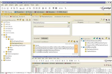
The method used is based on reasoning with an OWL ontology of the brain structures extended by Horn rules representing the dependencies between their properties. Figure 2 shows the brain ontology in OWL DL [1] edited with Prot´eg´e [2]. The knowledge consists of :
• TBox : the TBox provides the logical definitions of concepts (classes), roles (properties) and the asserted axioms. For example PreCentralGyrus1
is defined by (figure 2): PreCentralGyrus ≡ Gyrus u ∃ hasAnatomical-Part.InferiorParsOfPreCentralGyrus u ∃
hasAnatomicalPart.SuperiorPa-1It is not the precise definition of PreCentralGyrus, but just a simplification for the example
rsOfPreCentralGyrus u ∃ isMAEBoundedBy.CentralSulcus u ∃ isMAE-BoundedBy.PreCentralSulcus.
• RBox : The RBox provides rules extending the ontology, for example the rule R1 (figure 2) expressing that a boundary is propagated from parts to whole: isMAEBoundedBy(?x, ?y) ∧ hasSegment(?z, ?y) ∧ SulcalFold(?z) ∧ SulcalFold(?y) ∧ MAE(?x) → isMAEBoundedBy(?x, ?z)
• ABox : The Abox contains the individuals (instances of classes) and the instances of relations between them. In this example, there are several individuals defined e.g. P1, P2 ..P6, are instances of the class Patch, Su-periorPreCentralSulcus 0 is the instance of the class SuperiorPreCentral-Sulcus representing the SuperiorPreCentralSuperiorPreCentral-Sulcus of this patient image. Therefore, the Abox includes the following individuals and relations : (h Gyrus : Gyrus 1 i, h Patch : P1 , P2, ..., P6 i,h SuperiorPreCentralSulcus : SuperiorPreCentralSulcus 0 i, h PreCentralSulcus : PreCentralSulcus 0 i,h CentralSulcus : CentralSulcus 0 i, h InferiorParsOfPreCentralGyrus : InferiorParsOfPreCentralGyrus 0 i, h SuperiorParsOfPostCentralGyrus : SuperiorParsOfPostCentralGyrus 0 i, h isAnatomicalPartOf : P1, Gyrus 1 i, h isAnatomicalPartOf : P3, Gyrus 1 i, h isAnatomicalPartOf : P4, Gyrus 1 i, h isMAEBoundedBy : Gyrus 1, SuperiorPreCentralSulcus 0 i, h hasDirectAnatomicalPart : Gyrus 1, InferiorParsOfPreCentralGyrus 0 i, h hasDirectAnatomicalPart : Gyrus 1, SuperiorParsOfPreCentralGyrus 0 i, h isMAEBoundedBy : Gyrus 1, CentralSulcus 0 i, h hasSegment : Pre-CentralSulcus 0, SuperiorPrePre-CentralSulcus 0 i).
A DL reasoner cannot classify Gyrus 1 as a PreCentralGyrus from the on-tology knowledge alone. Indeed Gyrus 1 is PreCentralGyrus iff it satisfies the definition of PreCentralGyrus class, given in the TBox above, but considering the facts given in the Abox, it can be noticed that, as Gyrus 1 is not connected by the isMAEBoundedBy property to any object that is a PreCentralSulcus one condition is missing in the body of the rule to infer that Gyrus 1 is a PreCen-tralGyrus. However, this fact can be derived from the rule R1 of the RBox. Applying the rule R1 to the facts of the Abox : h isMAEBoundedBy : Gyrus 1, SuperiorPreCentralSulcus 0 i, and h hasSegment : PreCentralSulcus 0, Superi-orPreCentralSulcus 0 i, it comes : h isMAEBoundedBy : Gyrus 1, PreCentral-Sulcus 0 i. Therefore, from the Tbox, Rbox, and ABox it can be inferred that Gyrus 1 is an instance of the PreCentralGyrus class.
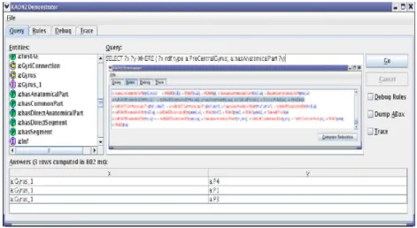
This example has been tested using the reasoner Kaon2 [3] which accepts an ontology extended with rules[4]. The brain ontology is represented in OWL DL and has been edited with Prot´eg´e OWL. Figure 3 shows the answers obtained with Kaon2, for the query below which asks to find all the individuals which are the anatomical part of PreCentralGyrus.
• SELECT ?x ?y WHERE { ?x rdf:type a: PreCentralGyrus; a: hasAnatom-icalPart ?y }
Figure 2: Ontology and rules
This result (P1, P3, P4) precisely illustrates the reasoning described above. Indeed, P1, P3, and P4 are defined in the ABox as anatomical parts of Gyrus 1. As the property ‘isAnatomicalPartOf ’ is the inverse of ‘hasAnatomicalPart’, it is derived from isAnatomicalP artOf = (hasAnatomicalP art)−1 that : h
hasAnatomicalPart : Gyrus 1, P1 i, h hasAnatomicalPart : Gyrus 1, P3 i, and h hasAnatomicalPart : Gyrus 1, P4 i. Then, as Gyrus 1 fulfills all the requested conditions to be a PreCentralGyrus, it is inferred to be a PreCentralGyrus, hence, the patches P1, P3, and P4 are answered to the query since they are parts of a PreCentralGyrus. This example illustrates the benefits of reasoning with symbolic knowledge for image interpretation. However, as at the moment Kaon2 does not allow daraypes nor nominals, this example was necessarily simpified to cope with these restrictions.
3
Labelling Process
Our approach is local in the sense that the user is interested in a particular part of the image. The labeling process will be done gradually in interaction with the user : based on information available in the ABox provided by image processing tools together with information introduced explicitly by the user, the reasoner will infer the labels of the structures using the ontology and the rule base. The labeled structures are then used by the reasoner to infer other labels of other
Figure 3: The result obtained with Kaon2
entities.
4
Related Works
Other approaches exist in the literature, e.g. the SPAM[05] approach (Statisti-cal/Probabilistic Anatomy Maps), in which anatomical knowledge is represented in an implicit way, in 3D object maps obtained from large sets of brain data that were manually labeled, or segmented into sub-volumes, after mapping individuals datasets into the stereotaxic space. Probability maps were then constructed for each segmented structure, by determining the proportion of subjects that were assigned a given anatomic label at each voxel position in stereotaxic space[06]. The disadvantages of these methods are primarily : a poor modeling of the inter-individual variability, and their inefficacity when the brain presents a lesion. Our approach tries to overcome these limitations. The first idea of this approach for the semantic description of images based on ontology and rules has been pre-sented at the W3C workshop on Rule Languages for Interoperability [07] and [08]. The current paper describes some results of implementing this idea using the recent prototype reasoner Kaon2.
5
Conclusion
This paper has described a simple example aiming at illustrating how a sym-bolic method can be implemented for the description of images thanks to recent reasoning techniques. The experiment which is related is a first step. The goal is to apply this approach to the real application. It is now needed to investigate
how to iterate the reasoning for labelling entire regions. It requires the comple-tion of the brain ontology under development in OWL, and overcoming present language and tools limitations. Combining numerical and symbolic methods is a promising direction to identify more effectively the semantic content of im-ages. These results are promising for knowledge-based multimedia processing applications.
6
References
1. http://www.w3.org/TR/owl-features/ 2. http://protege.stanford.edu/
3. http://kaon2.semanticweb.org/
4. Motik, B., Sattler, U., Studer, R.: Query answering for OWL DL with rules, ISWC 2004, LNCS 3298 Springer.
5. Collins, D.L., A.P., Zijdenbos, W.F.C., Baar, A.C. Evans: - ANI- MAL+INSECT : Improved cortical structure segmentation. - Proc. of the Annual Sympo-sium on Information Processing in Medical Imaging IPMI99, LNCS 1613, pp. 210-223, 1999.
6. Thompson P.: Probabilistic brain Atlases. http://www.loni.ucla.edu/ thomp-son/prob atlas.html
7. Golbreich, C., Bierlaire, O., Dameron, O., Gibaud, B.: What reasoning support for Ontology and Rules? the brain anatomy case study. Galway, Ireland, In : OWL Experience and Directions, Nov 11-12, 2005.
8. Golbreich, C., Bierlaire, O., Dameron, O., and Gibaud, B. : Use Case: On-tology with rules for identifying brain anatomical structures, W3C Work-shop on Rule Languages for Interoperability, Washington, April 2005.