HAL Id: inserm-00842753
https://www.hal.inserm.fr/inserm-00842753
Submitted on 9 Jul 2013HAL is a multi-disciplinary open access archive for the deposit and dissemination of sci-entific research documents, whether they are pub-lished or not. The documents may come from teaching and research institutions in France or abroad, or from public or private research centers.
L’archive ouverte pluridisciplinaire HAL, est destinée au dépôt et à la diffusion de documents scientifiques de niveau recherche, publiés ou non, émanant des établissements d’enseignement et de recherche français ou étrangers, des laboratoires publics ou privés.
Animal Toxins
Jean-Marc Sabatier, Michel de Waard
To cite this version:
Jean-Marc Sabatier, Michel de Waard. Animal Toxins: Toxins in Biotechnology. Abba J. Kastin. Handbook of biologically avtive peptides, Elsevier, pp.407-415, 2013. �inserm-00842753�
Animal Toxins in the World of Modern
Biotechnology
JEAN-MARC SABATIER
1& MICHEL DE WAARD
2,31ERT 62 Ingénierie des peptides à visée thérapeutique, Université de la Méditerranée Faculté de Médecine Nord, Boulevard Pierre Dramard, 13916 Marseille Cedex 20, France.
2
Inserm U836, Grenoble Neuroscience Institute, University Joseph Fourier, 38042 Grenoble Cedex 09, France.
3
Smartox Biotechnologies, Bâtiment Biopolis, 5, avenue du Grand Sablon, 38700 La Tronche, France.
Tel.: +33-4-56520563; Fax: +33-4-56520637; E-mail address: michel.dewaard@ujf-grenoble.fr
Abstract
Animal toxins are renowned for their ability to regulate ion channel activity and hence cell function. Therefore, they have been investigated as lead compounds for the development of analogues with toxic activity or conversely with therapeutic potential. Besides the pharmacological usefulness of animal toxins, there is a second emerging field in which toxins may soon reveal all their potential. The advent of modern biology witnesses the development of novel biotechnological applications of interest for which toxins present tremendous advantages. In this review, we will illustrate how toxins can be derived to carry on novel functions that make them become tools of choice for diagnostic, imaging and therapeutic applications. Among the examples developed, the case of maurocalcine will be detailed. This toxin, discovered for its activity on the ryanodine receptor, a calcium channel, is now entering a new phase of development for in vivo drug or imaging agent cell delivery.
Key Words: animal toxin, biotechnology, cell penetrating peptide, ryanodine receptor, drug delivery, in vivo imaging
Animal toxins as candidate drugs
Animal venoms represent an extremely rich source of pharmacological peptides. These peptides are generally of a length comprised between 10 and 70 amino acids and come with a variety of folds. Proteomic profiling of animal venoms with MALDI-TOF mass spectrometry techniques is increasingly used and provides important information regarding peptide mass, disulfide bridging and minimum number of toxins within given venom. According to many pioneering studies, venoms should contain no less than 100 peptides on average. More precise information on the identity of the peptides can be gained when proteomic analyses are combined to transcriptomic studies based on the isolation of venom gland mRNA. In one such study, it has been estimated that spider venoms may well contain 1,000 different peptides [9]. Of course, many of these peptides could differ by simply differences in post-translational modifications, but these numbers remain representative of the important biodiversity encountered in various species. Considering for instance that there are about 80,000 spider species and assuming the idea that one spider venom contains no less than 200 unique peptides, then one may estimate the biodiversity in peptides to close to 16 millions. Adding to this diversity, the ones encountered in cone snails, scorpions and snakes, to name a few, provide an estimate of the richness of these natural bio-libraries. Besides the numbers in venom peptides, these are three advantages to consider when deriving a drug from a natural toxin. First, the natural bio-libraries made by animal venoms contain exclusively bioactive drugs contrary to libraries of drugs of most pharmaceutical industries. Peptide toxins are tailored for efficient bioactivity in vivo by targeting crucial receptors (generally cell surface ion channels and G protein coupled receptors). While some toxins are evidently toxic in vivo, many others, present in the same venom, may have beneficial effects. Second, venoms
are generally injected subcutaneously and reach the blood stream for efficient dissemination in vivo. Some deleterious effects can take hours to come into play demonstrating an encouraging half-life of the active peptides in vivo. Third, gene duplication, extensive splicing and post-translational modifications have ensured that venoms contain toxins with exquisite affinity and selectivity for their pharmacological targets. There is therefore no doubt whatsoever that animal venom constitute an infinite source of biologically active peptides of pharmaceutical interest and therefore an excellent basis for screening programs. While the initial motivation in studying animal venoms was to identify active principles (at best) or active HPLC fractions (at worst) involved in toxicity, combined with the production of therapeutic sera for treating envenoming, current efforts are also now devoted to identifying pharmacological drugs for therapeutics or pesticides for industrial use. While identifying drugs of interest from animal venom is technically eased by combined proteomic and transcriptomic approaches, once a peptide of interest has been isolated, one also needs to overcome the difficulties inherent to its synthetic production. This can be performed by using recombinant techniques or by chemical synthesis, but this task is not always straightforward as the peptides need to undergo oxidative folding, a complex process in which the peptide acquires the correct disulfide bridge pairing pattern and adopts the right fold. Once this task is mastered however, access is gained to the possibility to create a large number of peptide analogues, overcoming the tedious task of toxin purification from natural sources that always come with limited availability and quantity. Analogues can be worked out in order to optimize certain characteristics, including greater affinity for a given target, improved selectivity or enhanced potency. In addition, the increasing development of bioinformatics has steadily improved the functional classification of the growing list of orphan toxins and their potential use in pharmacological applications.
With these considerations in mind, it is therefore not so surprising that several peptides, originating from animal venoms, made their way to the clinics and the drug market. Figure 1 illustrates a non exhaustive list of peptides or analogues thereof that are in various clinical phases or already on the market. For instance, several Conus peptides, present in predatory cone snails, are now used for medication in clinics for pain (Ziconotide, the synthetic form of -conotoxin MVIIA, a Ca2+ channel N-type blocker, from Elan Pharmaceuticals), epilepsy and other neuropathic disorders [34]. Natural peptides are therefore growing the list of peptides (438 in total in 2009) being considered by the pharmaceutical industry in their development programs. Seventy two of these 438 peptides reached phase III clinical trial and 48 were on the market. Four of them reached global sales over 500 millions $ each in 2007: Copaxane ($3.33 billion), Lupron ($1.88 billion), Byetta ($967 million), and Forteo ($709 million). The majority of these peptides target G protein coupled receptors, although other targets are increasingly common such as ion channels. A complete report on the development of peptides as therapeutic drugs is available from http://www.peptidetherapeutics.org. While the pharmacological potential of animal toxins is progressively unravelled and that exploitation of the natural resources for therapeutic aims is only in its infancy, this trend shouldn’t hide the fact that toxins are also increasingly used in biotechnological applications.
Animal toxins in biotechnological applications
In 2004, the group of Wonnacott demonstrated that the subcellular distribution of alpha7 nicotinic acetylcholine receptors can be investigated by coupling alpha bungarotoxin to gold nanoparticles [13]. In that respect, toxins appear as perfect tools to target anti-cancer agents directly to the site of tumour in vivo as far their targets are over-expressed in tumours. For instance, the G-protein coupled receptors (BBR1, BBR2 and BBR3) of bombesin, a 14 amino acid peptide from frog skin, are over-expressed in small cell carcinoma of lung, gastric cancer, neuroblastoma [21] and human prostate cancer. This property has been used to prepare bombesin derivatives harboring lutetium-1777 for prostate cancer targeting, in vivo imaging and therapeutic intervention [24]. Another successful example for tumour applications includes chlorotoxin [16, 29], a 36-mer peptide with four disulfide bridges initially isolated from the venom of the Israeli scorpion Leiurus quinquestriatus. Although initially developed for the diagnosis and treatment of glioma, chlorotoxin was found to specifically label cancer cells from other solid tumors as well (melanoma, small cell lung carcinoma, neuroblastoma, medulloblastoma, Ewing’s sarcoma and pheochromacytoma). The identity of the biomarker on which it binds is still under debate (initially a chloride channel, then matrix metalloprotein 2, and now, which seems more likely, annexin 2A). Currently, a 131iodinated version of the toxin from Eisai (TM601) has successfully ended clinical phase II for the treatment of recurrent glioma and has obtained FDA approval to go to phase III clinical trials. Besides it has also obtained FDA approval to investigate the effect of TM601 on newly occurring glioma. TM601 is extremely stable, presents no immunogenicity and produces no toxicity in humans. Several derivative molecules (termed TM602, TM604, etc) have been produced to facilitate phenotyping and histological staining, and patient treatment. The door is now open for the use of other toxins in cancer diagnosis and treatment. Recently, BmKCT, which presents 68% amino acid sequence identity with chlorotoxin, was also shown to target glioma in vivo and prevent its progression. Similarly, the non-toxic B subunit of the pathogen-produced Shiga toxin, known to bind to the glycosphingolipid Gb3 which is over-expressed in some tumors, was found to specifically label human colorectal carcinoma in nude mice [33]. Other technological applications are possible with toxins. The 12 amino acid peptide Tet1, derived from tetanus toxin, is an efficient vector for the delivery of plasmid DNA in complex to polyethylenimine [22]. A similar fragment of tetanus toxin, when placed in fusion to the reporter protein GFP, allows mapping of synaptic connections of the mammalian central nervous system [17].
Most animal toxins hit cell surface receptors. This is by far the most straightforward means to interfere with signalling pathways for peptides as these molecular entities are reputed to be unable to cross the cell plasma membrane. There is however some noticeable exceptions to this rule as some animal toxins have been discovered to target intracellular ion channel receptors. This is the case of toxins targeting the ryanodine receptor, an intracellular calcium channel that is located in the membrane of the endoplasmic reticulum and that controls cytosolic Ca2+ release. These toxins present intriguing peptide sequences that efficiently favour their entry into the cytoplasm. We will see that ion channel
recognition and cell permeation can easily be dissociated in order to retain the latter property. Also, cell penetration can be derived for the cell entry of other compounds of pharmaceutical or imaging interests. We will illustrate this point extensively by reviewing the case of maurocalcine, the first cell penetrating peptide of toxin origin to be discovered.
Maurocalcine, an animal toxin that targets an intracellular receptor
Maurocalcine is a scorpion toxin from Scorpio maurus palmatus that was discovered in 2000 [19]. Based on its sequence homology with imperatoxin A from the scorpion Pandinus imperator, it was presumed to be active on the ryanodine receptor. This turned out to be indeed the case as maurocalcine i) triggers Ca2+ release from purified sarcoplasmic reticulum [12], ii) induces both an increase in channel opening and the appearance of a long-lasting subconductance state [7], and iii) stimulates the binding of [3H]-ryanodine on its binding site [11]. All these effects occur with an EC50 of 10-20 nM. Maurocalcine interaction with the ryanodine receptor is direct as demonstrated by skeletal muscle ryanodine receptor pull down by a biotinylated derivative of maurocalcine bound to streptavidin-coated beads [1]. Maurocalcine obviously belongs to a larger family of toxins with high sequence homology comprising opicalcine 1 and 2 from Opistophthalmus carinatus scorpion, hemicalcin from
Hemiscorpius lepturus scorpion and hadrucalcin from Hadrurus gertschi scorpion (Figure 2). All these
toxins similarly act on the ryanodine receptor with only minor functional differences. At the structural level, maurocalcine is a 33-mer peptide containing three disulfide bridges. The pairing motif has been assigned to Cys1-Cys4, Cys2-Cys5 and Cys3-Cys6 and the peptide folds according to an inhibitor cystine knot (ICK motif). Although ICK motifs have been found on other toxins acting on cell surface receptors, maurocalcine and its analogues all present interesting distinguishing features. A close examination of the 3D structure of maurocalcine reveals that i) the peptide is heavily charged (1/3 of amino acids are basic, net charge of +12) and ii) the charge distribution of the peptide is strongly asymmetric creating an important dipole moment. One of the most amazing properties of the peptide is that upon external application to skeletal muscle myotubes, the peptide triggers Ca2+ release from internal stores within only a few seconds [11]. A similar observation was made in 2010 on cardiac myotubes with imperatoxin A. This observation points to the fact that the peptide is able to reach its pharmacological target by rapid translocation through the plasma membrane rather than by endocytosis. Also, two evidences indicate that maurocalcine should accumulate into the cytoplasm to regulate the ryanodine receptor. First, maurocalcine regulates channel activity from ryanodine receptors incorporated into artificial lipid bilayers only when applied to the cytoplasmic face of the channel [12]. Second, maurocalcine binding site on the ryanodine receptor has been mapped to a domain that is localized within the cytoplasm according to the membrane topology of this receptor [1].
Altogether, these findings led to the conclusion that maurocalcine possesses the unique feature to rapidly cross the plasma membrane and accumulate within the cytoplasm to concentrations above 20 nM in order to effectively activate the ryanodine receptor. The possibility to indirectly image the cell penetration of maurocalcine by the release of internal Ca2+ is unique to the peptide world. As we will
see, cell penetration of maurocalcine is not limited to itself but also to cargoes attached to the peptide underscoring the huge technological potential of this uncommon toxin.
Maurocalcine is a competitive cell penetrating peptide
Investigating the cell penetration properties of a peptide without labelling it is a tedious task. Therefore, the first evidence that maurocalcine may enter cells came from the use of a biotinylated analogue. This analogue coupled to fluorescent streptavidine was shown to enter into a variety of cell types [10]. This study demonstrated not only that maurocalcine enters into cells but also that it can be used as a peptide vector to facilitate the cell entry of a variety of cargoes, many of them being of a size in considerable excess than the vector itself. It was also the first report that a peptide toxin being a member of the growing family of cell penetrating peptides (CPP). As expected, imperatoxin A was also shown later on to cross the plasma membrane along with a cargo. In parallel, crotamine, a 42 amino acid toxin from the rattlesnake Crotalus durissus terrificus, was shown to penetrate into dividing cells and to carry on plasmid DNA [20]. Reputed CPP include Tat from the HIV-1 virus, the Drosophila transcription factor ANTP (encoded by the antennapedia gene), also called penetratin, and the herpes simplex virus type 1 (HSV-1) VP22 transcription vector. These peptides possess a number of common functional features in spite of impressive sequence divergences:
1- CPP are generally small peptides that rarely exceed 20 amino acids. However, this issue is not mandatory since many CPP originate from larger proteins. The relationship between CPP size and cargo penetration efficacy is not known.
2- Many CPP are enriched in basic amino acids. This led to the discovery that arginine-rich peptides are efficient CPP.
3- CPPs lack clear cell selectivity although this issue is not well investigated. They can thus enter numerous cell types.
4- CPP do not require specific membrane receptors for cell penetration. Indeed, optical CPP stereoisomers, made of D-amino acids instead of the natural L-amino acids, are at least as efficient as their L-counterparts in crossing the plasma membrane [23]. Nevertheless, CPP do interact with cell surface components such as glycoaminoglycans (GAGs) and negatively charged lipids on the basis of electrostatic interactions [8].
5- CPPs enter rapidly and efficiently into cells. Commonly, cell penetration occurs within a few minutes, although quantitatively saturation may take longer times. An important property hardly matched by other vectors is that CPP can penetrate into 100% of a given cell type in vitro. We will see that in vivo this might not be the case.
6- True CPPs do not require metabolic energy for cell entry [31]. CCP cell entry should be preserved in cells maintained at 4°C or in the presence of metabolic inhibitors. This is however
not always true since for many peptides and depending of the cargo being transported, CPP enter via endocytosis which is an energy-consuming process.
7- CPP trigger much interest for their ability to transport a great variety of cell-impermeable cargoes. The type of cargoes that have been coupled to CPPs and shown to enter into cells is astounding. So far, cell entry has been reported for DNA plasmids or mimics [18], oligonucleotides, siRNA or shRNA [14], peptide nucleic acids (PNA) [35], peptides, proteins [5], drugs [28], and nanoparticles [32]. The versatility in cargo that can be coupled to a given CPP opens an unprecedented number of applications that can’t be matched by any other cell delivery system.
The discovery of new CPP sequences is steadily increasing and there is enough choice now on the market to develop an application without worrying about the nature of the CPP. Why then would it be interesting to use maurocalcine as CPP considering the fact that it is more complex (presence of disulfide bridges and difficulties for cargo grafting) and expensive (size) to synthesize than other CPP? We summarize hereunder the advantage that maurocalcine has over other CPP.
The pharmacological activity of maurocalcine is easy to neutralize
Maurocalcine is notorious for its effects on the ryanodine receptor. Ca2+ release from the endoplasmic reticulum is a major signalling event which precludes the use of the original maurocalcine sequence as vector for the cell penetration of compounds of interest. Strategies had to be investigated to circumvent the bioactivity of the peptide. Four strategies turned out to be successful in that respect. They are all based on the fact that cell penetration of the peptide requires less stringent structural integrity than pharmacology. First, the peptide can be mutated in order to lose its ability to bind and stimulate the ryanodine receptor. Arg23 and Arg24 turn out to be two positions that are critical for maurocalcine activity. Analogues mutated on any one of these positions yield efficient CPP devoid of pharmacological activity [15]. Interestingly, the alanine scan of maurocalcine also reveals other interesting amino acid positions where mutations can improve the pharmacology and/or the cell penetration. Mutation of Glu12 is particularly interesting in that respect. Second, maurocalcine can be produced in its D-diastereomer conformation with D-amino acids rather than L amino acids. Interestingly, the peptide folds normally and produces its normal disulfide bridging pattern. It is also lacking recognition of the ryanodine receptor but, as expected, preserves intact its cell penetration efficacy since its affinity for the plasma membrane is not based on a receptor / ligand type interaction [23]. Obviously, this peptide is a very interesting lead compound since it has all the desired advantages as CPP (protease resistance and efficacy). In the future, mutated D-maurocalcine peptides will be designed that further enhance the efficacy of cell penetration. Third, maurocalcine can be mutated in such a way that it lacks disulfide bridges. Disulfide bridging contributes to the acquisition of the 3D structure of maurocalcine. Therefore, a disulfide-less peptide is unable to fold and loses its ability to recognize the ryanodine receptor [27]. Nevertheless, this disulfide-less maurocalcine preserves good cell penetration properties albeit with a
reduced efficacy. This altered potency can nevertheless be partially overcome by point mutations in the amino acid sequence. Needless to say, a similar disulfide-less maurocalcine can be prepared in the future by incorporating D-amino acids that may render it protease resistant and increase the half-life in
vivo. Since in its non-folded version, maurocalcine still acts as a CPP, our group is presently designing
a number of mini-maurocalcine analogues with reduced peptide length. The preliminary results demonstrate that several analogues can be designed with significantly reduced lengths (9 amino acids). The completion of these four strategies has led to three benefits: (i) the in depth understanding of the structural determinants of maurocalcine that preside to pharmacology and cell penetration, (ii) the identification of essential factors that contribute to an efficient cell penetration of maurocalcine, and (iii) the production of several new maurocalcine analogues dedicated only to cell penetration.
Maurocalcine produces cell delivery into the cytoplasm
The list of discovered CPP is getting impressive. However, the definition of a CPP remains too tolerant. A peptide is considered as CPP if it enters into cells. The problem is that any kind of peptide that binds to the external face of the plasma membrane is susceptible to enter into the cell merely by a process of endocytosis. Once this has occurred, organizing the escape of this peptide from the endosome is far from an easy endeavour. For most applications however, the delivery of the cargo in the cytoplasm is simply mandatory. In this registry, maurocalcine appears as a particularly competitive peptide. The native peptide (unmodified and uncoupled to any cargo) presents the following distinguishing features: i) the ability to follow its cytoplasmic accumulation on line (by the release of internal Ca2+), ii) a minimal cytoplasmic accumulation of 10-20 nM (to observe Ca2+ release), and iii) a cytoplasmic accumulation against the concentration gradient (owing to the net positive charge of the peptide). The natural tendency of maurocalcine to accumulate into the cytoplasm ensures high cytoplasmic concentrations of the peptide. This tendency is greatly helped by its structural characteristics. The predominant orientation of basic residues on one face of the peptide, the presence of a dipole moment and the existence of a hydrophobic face ensures that maurocalcine can accumulate against its concentration gradient by electrochemical attraction in normally polarized cells. This wouldn’t be feasible if the peptide first accumulated within endosomes as the cytoplasmic accumulation factor would be kinetically limited by the endocytosis process itself and second by the endosomal escape. This being said, it would be inexact to state that maurocalcine does not also enter into cells by endocytosis. In fact, this is even the preferred route of entry when maurocalcine or one of its analogues is coupled to some cargoes. Macropinocytosis is the main route of entry with streptavidine as cargo [10], or when maurocalcine is coupled to nanoparticles covered with streptavidine molecules [26]. The nature and/or size of the cargo therefore is susceptible to inhibit the normal balance in cell entry modes (translocation versus endocytosis) probably by increasing the residency time of the peptide at or within the plasma membrane. Obviously, smaller cargoes still allow a normal translocation process to occur as witnessed in the case of a fluorescent indicator [23] or a small drug like doxorubicin [4]. This is also the case for a maurocalcine-maleimide-cys-nanoparticle complex, suggesting that in nanotechnology, size is not
necessarily a limitation for cytoplasmic targeting [27]. In conclusion, while it may seem desirable to get rid of the pharmacological activity of maurocalcine, it may in first instance represent an asset when it comes to evaluate the correct cytoplasmic targeting of the cargo transported. This is rendered possible since it was observed that N-terminal modification of the native maurocalcine (biotinylation and coupling to streptavidine for example) does not change dramatically its ability to interact with the ryanodine receptor [10]. Upon completion of this evaluation, it is then easy to turn towards maurocalcine analogues that structurally resemble maurocalcine itself but are mutated for their interaction with the ryanodine receptor.
Maurocalcine has an excellent cell entry / toxicity ratio
A drawback of many CPP is their relative toxicity. This wouldn’t be a major problem if these CPP entered into cells at doses much lower than their toxic concentration. Unfortunately, the concentration ratio between cell penetration efficiency and cell toxicity is often not that encouraging. For reasons that are unknown, maurocalcine is not toxic upon intravenous injection in spite of its pharmacological activity. This is not the case for intracerebroventricular injections in mice. However, this issue is easily circumvented with the numerous pharmacologically-inert maurocalcine analogues that are now available. These analogues have all been assessed for cell toxicity most often for long periods of time
in vitro (24 hrs) and on sensitive cells (neurons). It was systematically observed an almost complete
lack of toxicity at concentrations as high as 10 µM. In contrast, some efficient analogues show significant levels of cell penetration at 10 nM, suggesting that reaching a concentration ratio penetration / toxicity of 1,000 for maurocalcine analogues is feasible. Of course, these values are relevant for the maurocalcine vectors, but ought to be reconsidered upon grafting a cargo on them. This favorable ratio stems from the fact that maurocalcine has been optimized by nature to invade cells with a minimal disturbance which is not necessarily the case of other CPP that originate most often as fragments of larger non cell-penetrating proteins or are designed de novo. One additional feature that needs to be emphasized is the excellent concentration-dependence of maurocalcine’s cell penetration. In its folded conformation, the peptide enters at lower concentrations than the popular CPP (penetratin, Tat or poly-R). Further optimizing these values appears as a reachable objective in spite of the fact that the cell membrane factors that contribute to the efficacy of cell penetration of CPP are ill-defined. In an attempt to define these factors, it was found that maurocalcine binds with micromolar affinity onto cell surface glycoaminoglycans (GAG) [25]. Interaction was evident with heparin, heparin sulphate, and chondroitin sulphate. Soluble GAG inhibit close to 80% of maurocalcine cell uptake because they screen maurocalcine positive charges required for cell entry and compete for its interaction with cell surface GAG. However, maurocalcine’s cell penetration is well conserved (close to 50%) in GAG-deficient cells indicating that GAG mainly contribute to cell penetration at the quantitative level by acting as low-affinity reservoirs of CPP. More importantly, maurocalcine interacts with several negatively charged lipids at suprananomolar affinities [6]. Maurocalcine interacts with gangliosides, such as GD3 (disialoganglioside NeuAcα2-8NeuAcα2-3Galβ1-4Glcβ1-Cer), phosphatidylinositol (PtdIns)(3)P,
PtdIns(4)P, and PtdIns(5)P), phosphatidic acid and sulfatide, and more weakly with lysophosphatidic acid, PtdIns(3,5)P2 and phosphatidylserine. It is difficult to envision that lipids are not directly concerned by the mechanism of membrane translocation of CPP, while on the contrary GAG could be involved in endocytosis. The mutant maurocalcine analogue Ala12 has improved cell penetration and lipid interaction. Conversely, the mutant analogue Lys20 that has poor cell penetration has also limited lipid interaction. The problem is to identify which lipid is the most important for membrane translocation. Solving this question would tremendously help the design of better CPP.
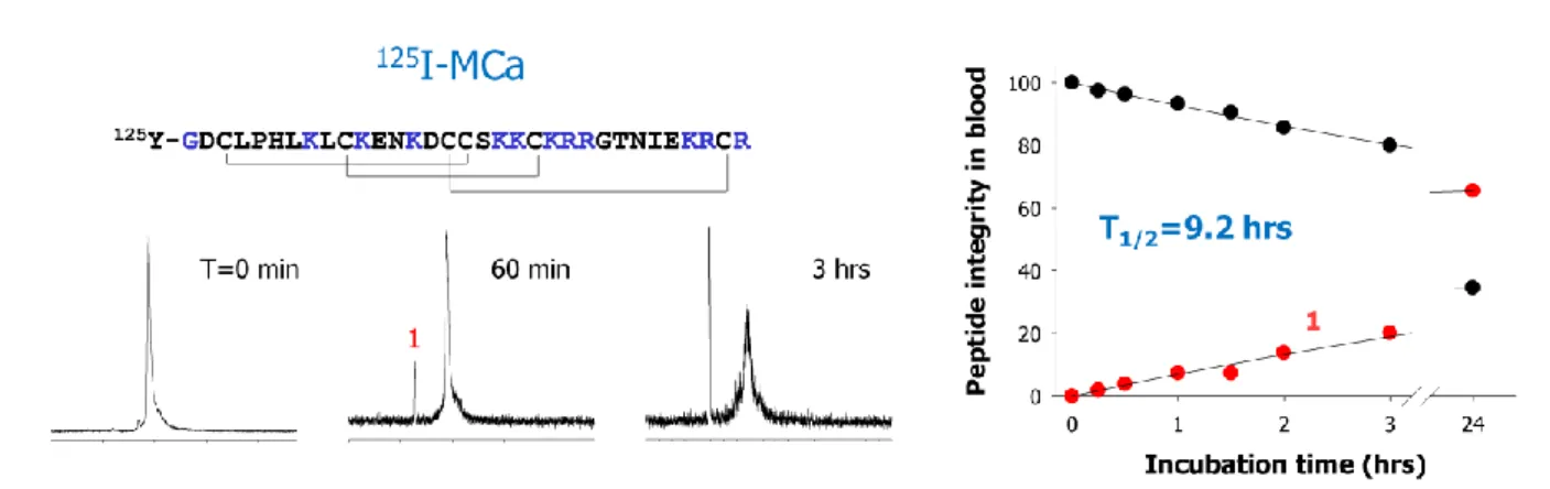
Maurocalcine is a stable CPP vector in vivo
Venom peptides have been designed by nature to be stable upon injection in vivo. The disulfide bridging and the compact fold is most likely also an advantage in that respect. We have investigated this question in the case of maurocalcine. A first analogue was synthesized, tyr-maurocalcine, and iodinated with 125I on the extra N-terminal tyrosine residue. Next, the peptide was incubated with mice blood and its stability followed with time by HPLC. As shown in Figure 3, the folded maurocalcine analogue with its disulfide bridges has excellent blood stability since one third of the peptide is still not degraded after 24 hrs incubation. A single metabolite is evident suggesting that maurocalcine can further be optimized in terms of in vivo stability. The same type of experiment was conducted with a similar analogue but in which all cysteine residues were replaced by isosteric 2-aminobutyric acid residues. Such a peptide lacks disulfide bridges and also secondary structures. In that case, we found that the peptide has a two-fold reduced stability and also that more metabolites are present as well (not shown). This study illustrates that disulfide bridges bring in a significant competitive advantage in terms of peptide stability in vivo. For instance, Tat, a CPP that lacks disulfide bridges, degrades rapidly even in the extracellular culture medium of epithelial cells.
Maurocalcine preferentially targets some cell types and organs in vivo
While it is true that CPP penetrate into all kinds of cell types, emerging evidence suggest that there are cell-specific differences in the efficiency of the penetration. We found for instance that maurocalcine penetrates better into glial cells than in neurons. Also, the snake venom CPP crotamine presents selective cell penetration into actively dividing cells. A close examination of FAM-D-maurocalcine distribution in human blood cells indicates that monocytes and lymphocytes NK are almost all invaded by maurocalcine, whereas a small percentage (less than 10%) of lymphocytes T and B take up maurocalcine. The reasons behind a selective cell-type cell penetration are unclear. One may assume that since CPP have the ability to interact with different glycosaminoglycans species, changes in glycoaminoglycan patterns at the cell surface may explain differences in CPP cell selectivity [8]. It illustrates that CPP sequences may evolve in such a way to penetrate only into desired cell targets. After intravenous injection, 30% of iodinated tyr-maurocalcine is associated to circulating cells, whereas 40% and 26% are associated to plasma and protein-free plasma, respectively. Combined
nanoSPECT/CT imaging gives an indication of where the toxin accumulates in vivo (Figure 4). Additional organ quantification of the radioactive peptide demonstrates strong kidney accumulation and urine elimination. Other organs that light up quite well are: stomach, spleen, skin, lung, liver, heart and duodenum. No brain accumulation is evident even at longer periods of observation suggesting that the peptide does not cross the blood brain barrier. These first observations give a hint to what organs would be best fitted for a therapy based on the use of maurocalcine sequence. It will be interesting to observe if cell and tissue selectivity can be altered by selective point mutations in maurocalcine sequence. If this could work, the door would be open for tissue-targeting cell penetrating peptides for enhanced therapeutic potential.
Concluding remarks
The existence of natural structural analogues of maurocalcine opens the route to new research programs aimed at discovering novel CPP molecules in the incredible rich sources of peptides that are venoms. The added benefit of increasing the number of known toxin-derived CPP is in their potential cell- and organ-specific targeting properties and in their usefulness for in vivo applications. Several discovery strategies may be pursued: (i) determining novel toxins active on intracellular ion channels by affinity column purification, (ii) screening for peptides that can enter into cells, and (iii) purifying peptides from venoms based on their basic or aliphatic nature. As for maurocalcine, these peptides will need optimization in order to remove any potential toxic or undesirable pharmacological effect. On the therapeutic side, what applications are best suited for maurocalcine or toxin-derived CPP? The field of CPP is blossoming with interesting and innovative applications at an exponential growth rate. While most applications have focused on the cell entry of cargoes that are unable to enter into cells, there is also room for applications in which the CPP contributes not to the entry of a drug but to force it to stay into cells by limiting its passive diffusion or expulsion by multi-drug resistance proteins for instance. Two such applications have been designed for maurocalcine. In one application, it was found that coupling maurocalcine to near-infrared emitting nanoparticles charged with MRI-detectable contrast agents allows for greater tissue retention time of the MRI signal in brain at the injection point, at least in much better proportions than the clinically-used DOTAREM [30]. This kind of approach holds promises for theragnostic applications in which the therapeutic drug is combined to the imaging agent. In a second application, it was found that coupling maurocalcine to an anti-tumor agent, such as doxorubicin, a drug used for the treatment of solid tumors in clinics, finds useful applications for fighting chemoresistance [2-4]. Chemoresistance often occurs as the result of cancer cells that start over-expressing multidrug resistance proteins. Freely entering doxorubicin is readily expulsed by these cells and tumors can then continue propagating. Increasing the therapeutic doses is not an option because of the secondary effects on healthy tissues, including neuropathies and cardiotoxicity. The coupling to maurocalcine turns a viable option, at least in vitro for the moment, as multidrug resistance proteins are no longer able to take into charge the complex molecule that keeps its anti-tumor activity. Coupled to cell penetrating peptides that would be able to target cancer cells in vivo, such an application would
hold great promises in the future. Now that maurocalcine is a proven and efficient CPP, it is on the track for biological, diagnostic and technological applications.
References
[1] Altafaj X, Cheng W, Esteve E, Urbani J, Grunwald D, Sabatier JM, et al. Maurocalcine and domain A of the II-III loop of the dihydropyridine receptor Cav 1.1 subunit share common binding sites on the skeletal ryanodine receptor. J Biol Chem 2005;280:4013-6.
[2] Aroui S, Brahim S, De Waard M, Breard J, Kenani A. Efficient induction of apoptosis by doxorubicin coupled to cell-penetrating peptides compared to unconjugated doxorubicin in the human breast cancer cell line MDA-MB 231. Cancer Lett 2009;285:28-38.
[3] Aroui S, Brahim S, Hamelin J, De Waard M, Breard J, Kenani A. Conjugation of doxorubicin to cell penetrating peptides sensitizes human breast MDA-MB 231 cancer cells to endogenous TRAIL-induced apoptosis. Apoptosis 2009;14:1352-65.
[4] Aroui S, Ram N, Appaix F, Ronjat M, Kenani A, Pirollet F, et al. Maurocalcine as a Non Toxic Drug Carrier Overcomes Doxorubicin Resistance in the Cancer Cell Line MDA-MB 231. Pharm Res 2009;26:836-45.
[5] Bleifuss E, Kammertoens T, Hutloff A, Quarcoo D, Dorner M, Straub P, et al. The translocation motif of hepatitis B virus improves protein vaccination. Cell Mol Life Sci 2006;63:627-35.
[6] Boisseau S, Mabrouk K, Ram N, Garmy N, Collin V, Tadmouri A, et al. Cell penetration properties of maurocalcine, a natural venom peptide active on the intracellular ryanodine receptor. Biochim Biophys Acta 2006;1758:308-19.
[7] Chen L, Esteve E, Sabatier JM, Ronjat M, De Waard M, Allen PD, et al. Maurocalcine and peptide A stabilize distinct subconductance states of ryanodine receptor type 1, revealing a proportional gating mechanism. J Biol Chem 2003;278:16095-106.
[8] Console S, Marty C, Garcia-Echeverria C, Schwendener R, Ballmer-Hofer K. Antennapedia and HIV transactivator of transcription (TAT) "protein transduction domains" promote endocytosis of high molecular weight cargo upon binding to cell surface glycosaminoglycans. J Biol Chem 2003;278:35109-14. [9] Escoubas P, Sollod B, King GF. Venom landscapes: mining the complexity of spider venoms via a combined cDNA and mass spectrometric approach. Toxicon 2006;47:650-63.
[10] Esteve E, Mabrouk K, Dupuis A, Smida-Rezgui S, Altafaj X, Grunwald D, et al. Transduction of the scorpion toxin maurocalcine into cells. Evidence that the toxin crosses the plasma membrane. J Biol Chem 2005;280:12833-9.
[11] Esteve E, Smida-Rezgui S, Sarkozi S, Szegedi C, Regaya I, Chen L, et al. Critical amino acid residues determine the binding affinity and the Ca2+ release
efficacy of maurocalcine in skeletal muscle cells. J Biol Chem 2003;278:37822-31.
[12] Fajloun Z, Kharrat R, Chen L, Lecomte C, Di Luccio E, Bichet D, et al. Chemical synthesis and characterization of maurocalcine, a scorpion toxin that activates Ca(2+) release channel/ryanodine receptors. FEBS Lett 2000;469:179-85.
[13] Jones IW, Barik J, O'Neill MJ, Wonnacott S. Alpha bungarotoxin-1.4 nm gold: a novel conjugate for visualising the precise subcellular distribution of alpha 7* nicotinic acetylcholine receptors. J Neurosci Methods 2004;134:65-74.
[14] Juliano RL. Intracellular delivery of oligonucleotide conjugates and dendrimer complexes. Ann N Y Acad Sci 2006;1082:18-26.
[15] Mabrouk K, Ram N, Boisseau S, Strappazzon F, Rehaim A, Sadoul R, et al. Critical amino acid residues of maurocalcine involved in pharmacology, lipid interaction and cell penetration. Biochim Biophys Acta 2007;1768:2528-40.
[16] Mamelak AN, Rosenfeld S, Bucholz R, Raubitschek A, Nabors LB, Fiveash JB, et al. Phase I single-dose study of intracavitary-administered iodine-131-TM-601 in adults with recurrent high-grade glioma. J Clin Oncol 2006;24:3644-50.
[17] Maskos U, Kissa K, St Cloment C, Brulet P. Retrograde trans-synaptic transfer of green fluorescent protein allows the genetic mapping of neuronal circuits in transgenic mice. Proc Natl Acad Sci U S A 2002;99:10120-5.
[18] Morris MC, Gros E, Aldrian-Herrada G, Choob M, Archdeacon J, Heitz F, et al. A non-covalent peptide-based carrier for in vivo delivery of DNA mimics. Nucleic Acids Res 2007;35:e49.
[19] Mosbah A, Kharrat R, Fajloun Z, Renisio JG, Blanc E, Sabatier JM, et al. A new fold in the scorpion toxin family, associated with an activity on a ryanodine-sensitive calcium channel. Proteins 2000;40:436-42. [20] Nascimento FD, Hayashi MA, Kerkis A, Oliveira V, Oliveira EB, Radis-Baptista G, et al. Crotamine mediates gene delivery into cells through the binding to heparan sulfate proteoglycans. J Biol Chem 2007;282:21349-60.
[21] Ohlsson B, Fredang N, Axelson J. The effect of bombesin, cholecystokinin, gastrin, and their antagonists on proliferation of pancreatic cancer cell lines. Scand J Gastroenterol 1999;34:1224-9.
[22] Park IK, Lasiene J, Chou SH, Horner PJ, Pun SH. Neuron-specific delivery of nucleic acids mediated by Tet1-modified poly(ethylenimine). J Gene Med 2007;9:691-702.
[23] Poillot C, Dridi K, Bichraoui H, Pecher J, Alphonse S, Douzi B, et al. D-Maurocalcine, a pharmacologically inert efficient cell-penetrating peptide analogue. J Biol Chem 2010;285:34168-80.
[24] Pujatti PB, Santos JS, Couto RM, Melero LT, Suzuki MF, Soares CR, et al. Novel series of (177)Lu-labeled bombesin derivatives with amino acidic spacers for selective targeting of human PC-3 prostate tumor cells. Q J Nucl Med Mol Imaging 2011;55:310-23. [25] Ram N, Aroui, S., Jaumain, E., Sadoul, R., Mabrouk, K., Ronjat, M., Lortat-Jacob, H., De Waard, M. Direct peptide interaction with surface glycosaminoglycans contribute to the cell penetration of maurocalcine. submitted.
[26] Ram N T-NI, Pernet-Gallay K, Poillot C, Ronjat M, Andrieux A, Arnoult C, Daou J, De Waard M. In vitro and in vivo cell delivery of quantum dots by the cell penetrating peptide maurocalcine. International Journal of Biomedical Nanoscience and Nanotechnology 2011;2:12-32.
[27] Ram N, Weiss N, Texier-Nogues I, Aroui S, Andreotti N, Pirollet F, et al. Design of a disulfide-less, pharmacologically-inert and chemically-competent analog of maurocalcine for the efficient transport of impermeant compounds into cells. J Biol Chem 2008. [28] Sethuraman VA, Bae YH. TAT peptide-based micelle system for potential active targeting of anti-cancer agents to acidic solid tumors. J Control Release 2007;118:216-24.
[29] Soroceanu L, Gillespie Y, Khazaeli MB, Sontheimer H. Use of chlorotoxin for targeting of primary brain tumors. Cancer Res 1998;58:4871-9. [30] Stasiuk GJ, Tamang S, Imbert D, Poillot C, Giardiello M, Tisseyre C, et al. Cell-Permeable Ln(III) Chelate-Functionalized InP Quantum Dots As Multimodal Imaging Agents. ACS Nano 2011.
[31] Thoren PE, Persson D, Karlsson M, Norden B. The antennapedia peptide penetratin translocates across lipid bilayers - the first direct observation. FEBS Lett 2000;482:265-8.
[32] Tseng YL, Liu JJ, Hong RL. Translocation of liposomes into cancer cells by cell-penetrating peptides penetratin and tat: a kinetic and efficacy study. Mol Pharmacol 2002;62:864-72.
[33] Viel T, Dransart E, Nemati F, Henry E, Theze B, Decaudin D, et al. In vivo tumor targeting by the B-subunit of shiga toxin. Mol Imaging 2008;7:239-47. [34] Wang CZ, Chi CW. Conus peptides--a rich pharmaceutical treasure. Acta Biochim Biophys Sin (Shanghai) 2004;36:713-23.
[35] Zielinski J, Kilk K, Peritz T, Kannanayakal T, Miyashiro KY, Eiriksdottir E, et al. In vivo identification of ribonucleoprotein-RNA interactions. Proc Natl Acad Sci U S A 2006;103:1557-62.
Figures
FIGURE 1. Examples of toxins or analogues thereof which are in various clinical phases. Notice the diversity of indications.
FIGURE 2. Sequence alignment of calcin toxins according to ClustalW2. Negatively charged residues are in blue, while positively charged amino acids are in purple. Sequence identities are given with maurocalcine as reference entry.
FIGURE 3: HPLC elution profile of iodinated tyr-maurocalcine analogue as a function of time of incubation in mice blood. The peptide was added at a concentration of 1 mCi per ml of blood and incubated at 37°C. HPLC profile are from the protein-free plasma fraction. The half-life of maurocalcine exceeds 9 hours in these conditions.
Imperatoxin A --G
D
C
LP
H
L
KR
C
K
A
D
N
D
CCG
KK
C
KRR
GTN
A
E
KR
C
R
33
Hemicalcin
--G
D
C
LP
H
L
K
L
C
K
A
D
K
D
CCS
KK
C
KRR
GTN
P
E
KR
C
R
33
Maurocalcine
--G
D
C
LP
H
L
K
L
C
K
E
N
K
D
CCS
KK
C
KRR
GTN
I
E
KR
C
R
33
Opicalcine 1 --G
D
C
LP
H
L
KR
C
K
E
NN
D
CCS
KK
C
KRR
GTN
P
E
KR
C
R
33
Opicalcine 2 --G
D
C
LP
H
L
KR
C
K
E
NN
D
CCS
KK
C
KRR
G
A
N
P
E
KR
C
R
33
Hadrucalcine
SE
K
D
C
I
K
H
L
Q
R
C
R
E
N
K
D
CCS
KK
CS
RR
GTN
P
E
KR
C
R
35
**: **: *: ::***.***.***:* *****
Score (%)
90.0
87.0
81.0
90.0
-75.0
FIGURE 4: In vivo tomographic whole-body imaging of [125I]-Tyr-maurocalcine biodistribution in CD-1 mice 15 min tail i.v. post-injection of 37 MBq. From left to right, 3D rendering, sagital, coronal, and transverse views of tracer activity.

![FIGURE 4: In vivo tomographic whole-body imaging of [ 125 I]-Tyr-maurocalcine biodistribution in CD-1 mice 15 min tail i.v](https://thumb-eu.123doks.com/thumbv2/123doknet/14216075.482723/17.892.71.815.93.538/figure-vivo-tomographic-body-imaging-tyr-maurocalcine-biodistribution.webp)
