HAL Id: tel-01376802
https://tel.archives-ouvertes.fr/tel-01376802
Submitted on 5 Oct 2016
HAL is a multi-disciplinary open access archive for the deposit and dissemination of sci-entific research documents, whether they are pub-lished or not. The documents may come from teaching and research institutions in France or abroad, or from public or private research centers.
L’archive ouverte pluridisciplinaire HAL, est destinée au dépôt et à la diffusion de documents scientifiques de niveau recherche, publiés ou non, émanant des établissements d’enseignement et de recherche français ou étrangers, des laboratoires publics ou privés.
Controlled release of dexamethasone to the inner ear
from silicone-based implants
Maria Gehrke
To cite this version:
Maria Gehrke. Controlled release of dexamethasone to the inner ear from silicone-based implants. Human health and pathology. Université du Droit et de la Santé - Lille II, 2016. English. �NNT : 2016LIL2S004�. �tel-01376802�
Ecole Doctorale Biologie-Santé
Controlled Release of Dexamethasone to the Inner Ear from silicone-based Implants
Ear
Libération contrôlée de dexaméthasone à partir des implants en silicone pour l’oreille interne
THESE DE DOCTORAT
Soutenue le 29 Janvier 2016 à Lille
MARIA GEHRKE
Dirigée par:
Dr. Florence Siepmann – Directrice Prof. Christophe Vincent – Co-directeur
Composition du Jury :
Monsieur SIEPMANN Juergen Président de Jury
Professeur à l’Université de Lille 2
Monsieur VERVAET Chris Rapporteur
Professeur à l’Université de Gand
Madame BOCHOT Amélie Rapporteur
Professeur à l’Université Paris-Sud
Madame PIEL Géraldine Examinatrice
PhD à l’Université de Liège
Madame SIEPMANN Florence Directrice de Thèse
Remerciements
Je tiens à remercier vivement tous les personnes qui m’ont accompagné durant mes trois ans de thèse.
Je tiens à remercier Dr. Florence Siepmann pour avoir accepté d’encadrer ce travail. Merci beaucoup pour l’encouragement et les discussions très ouvertes (p.ex. pour détourner l’utilisation du matériel de laboratoire) et pour m’avoir laissé beaucoup de liberté au sein du laboratoire. Merci de m’avoir encouragée à toujours garder un équilibre entre la recherche scientifique et la vie de famille.
Je remercie aussi le Professeur Christophe Vincent qui m’a encadré comme co-directeur de thèse pour m’avoir donné toujours beaucoup d’idées. De plus, il m’a donné l’opportunité de travailler avec plusieurs étudiants de son équipe qui ont grandement aidé à l’avancement de ce projet de thèse. Merci beaucoup!
Je voudrais remercier le Professeur Jürgen Siepmann pour m’avoir accueilli dans son équipe internationale. Dès le premier jour de mes six mois d’Erasmus à Lille, et jusqu’à la fin de ma thèse, j’ai beaucoup apprécié sa gentillesse d’une part, et ses connaissances scientifiques d’autre part. Merci mille fois!
J’exprime toute ma reconnaissance envers les Professeurs Amélie Bochot de l’Université Paris-Sud et Chris Vervaet de l’Université de Gand qui m’ont fait le grand plaisir de juger ce travail en tant que rapporteurs et membres de mon jury de thèse.
Je tiens également à remercier Dr. Géraldine Piel de l’Université de Liège pour avoir accepté d’évaluer ma thèse en tant que membre de jury.
Je remercie beaucoup les personnes avec qui j’ai eu la possibilité de travailler pendant ma thèse et qui m’ont chacune et chacun grandement aidé : particulièrement le Professeur Stefan Plontke de l’Université de Halle de m’avoir accueillie dans son laboratoire pour apprendre les techniques d’implantation, mais aussi Florence Danède et Jean-François Willart de l’Université de Lille 1 pour l’aide concernant la réalisation et l’évaluation des analyses de calorimétrie différentielle à balayage et de diffraction des rayons X.
tout ira bien! Sanja Puric, Emmely Lacante, Jérémy Verin et Michaël Risoud m’ont appris beaucoup pendant leurs stages et étaient toujours très patients.
Je souhaite aussi remercier le Professeur Anne Gayot pour son accueil au laboratoire.
Je voudrais exprimer mes remerciements envers toute l’équipe du laboratoire de Pharmacotechnie Industrielle, notamment Susi Muschert, Youness Karrout, Mounira Hamoudi, Hugues Florin et Muriel Deudon pour leurs conseils, leur volonté de donner de l’aide et pour avoir instauré une bonne ambiance dans le laboratoire.
J’ai beaucoup apprécié l’ambiance amicale dans le laboratoire! J’aimerais remercier mes camarades de thèse pour m’avoir accueilli avec une solidarité incroyable : Céline pour m’avoir expliqué la vie et la langue française ; Carine pour son accueil chaleureux ; Emilie pour les conversations au café ; Phuong et Huong pour m’avoir montré la vie intérieure des HPLCs et pour leur patience ; Bérengère pour son grand sourire ; Susana pour son goût exceptionnel de musique et ses perles de la langue française ; Hanane pour ses délicieux gâteaux ; Golf pour sa créativité et sa gentillesse ; Petra qui m’aidé à ne pas perdre ma langue maternelle et qui m’a donnée de nombreux conseils ; Oriane pour son sens de l’humour ; Julie pour rassembler tout le monde dans son appartement ; Ting pour sa bonne humeur et ses efforts pour parler Français ; Rapee pour sa curiosité ; Corinna pour avoir ramené une atmosphère berlinoise au bureau et, last but not least, Esther pour ses chansons et sa confiance.
Je tiens à remercier tout particulièrement ma famille et mes amis qui m’ont toujours encouragé et envoyé des colis remplis de gâteaux. Je suis très chanceuse de vous avoir!
i
Table of Contents
1.
Introduction ... 1
1.1. Anatomy and Physiology of the Ear ... 2
1.1.1. Barriers of the Inner Ear ... 6
1.1.2. Auditory perception ... 8
1.1.3. Sense of balance ... 10
1.2. Diseases of the Inner Ear ... 11
1.2.1. Hearing Loss ... 13
1.3. Drug delivery to the Inner Ear ... 15
1.3.1. Systemic drug delivery ... 15
1.3.2. Local drug delivery... 17
1.3.2.1. Intratympanic drug delivery ... 18
1.3.2.2. Intracochlear drug delivery ... 22
1.4. Drug release from silicone matrices ... 24
1.5. Objectives ... 26
2.
Materials and Methods ... 27
2.1. Dexamethasone mobility in thin films ... 27
2.1.1. Materials ... 27
2.1.2. Preparation of drug loaded films ... 27
2.1.3. Preparation of drug loaded extrudates ... 28
2.1.4. Drug release measurements ... 28
2.1.5. Scanning electron microscopy ... 29
2.2. Ear Cube implants for Controlled Drug Delivery to the Inner Ear ... 30
2.2.1. Materials ... 30
2.2.2. Preparation of drug-loaded silicone matrices ... 30
ii
2.2.6. Scanning electron microscopy ... 34
2.2.7. Thermal analysis (DSC) ... 34
2.2.8. X-ray diffraction ... 34
2.3. Trans-Oval-Window Implants: Extended Dexamethasone Release ... 35
2.3.1. Materials ... 35
2.3.2. Preparation of drug-loaded Matrices ... 35
2.3.3. Drug Release Measurements ... 36
2.3.4. Gerbil Study ... 37
2.3.5. Implantation Procedure ... 38
2.3.6. Cochleae Preparation for Further Analysis ... 39
2.3.7. Immunohistochemistry ... 40
3.
Results and Discussion ... 42
3.1. Dexamethasone mobility in thin films ... 42
3.1.1. Effects of PEG addition ... 42
3.1.2. Effects of the type of silicone ... 51
3.1.3. Impact of the initial drug loading ... 55
3.1.4. Theoretical predictions for cylindrical extrudates ... 58
3.2. Ear Cube implants for Controlled Drug Delivery to the Inner Ear ... 61
3.2.1. Physico-chemical key properties of the Ear Cubes ... 61
3.2.2. Characterization of thin films of identical composition ... 63
3.2.3. Drug release from Ear Cubes ... 67
3.2.4. Absence of Ear Cube swelling ... 71
3.3. Trans-Oval-Window Implants: Extended Dexamethasone Release ... 74
3.3.1. Results ... 74
3.3.1.1. In vitro studies ... 74
iii 3.3.2. Discussion ... 80
4.
Conclusion ... 83
References ... 85
Résumé ... 94
List of Publications ... 108
Curriculum Vitae ... 111
iv AIED Autoimmune Inner Ear Disease
BDNF Brain-derived neurotrophic factor DNQX 6,7-dinitroquinoxaline-2,3-dione
DXM Dexamethasone
GOM Glycerolmonooleate
LSR Liquid Silicone Rubber NIHL Noise-Induced Hearing Loss
OWM Oval window membrane
PEG Polyethyleneglycol
PLGA Poly(lactic-co-glycolic acid)
RWM Round window membrane
SNHL Sensorineural Hearing Loss
SPION Superparamagnetic iron oxide nanoparticle SSNHL Sudden Sensorineural Hearing Loss
INTRODUCTION
1
1.
Introduction
The ear is responsible for the perception of sound and the sense of balance. In 2015, the WHO estimated that worldwide 360 million people (over 5 % of the population) are suffering from disabling hearing loss, meaning a loss of 40 or 30 dB in the better hearing ear in adults and children respectively (1). In the USA 15 % of the population over 18 reported at least minor changes in hearing capacities (classification from “a little bit of trouble hearing” to “deaf”) (2).
Hearing loss can have several causes: The loss before or soon after the birth of a child is one of the most frequent birth defects since 0.1 to 0.3 % of all neonates are born with congenital hearing loss (3,4). Nevertheless, hearing impairment nowadays can also be related to the certain employments of people: It has been reported that especially professional soldiers often suffer from hearing loss, tinnitus or other noise-related comorbidities following their service in the armed forces (5–7).
Additionally, a lot of employees in the manufacturing sector suffer from occupationally induced hearing loss. In 2010, about 16 million people have been working in the manufacturing sector in the USA (8). Those 16 million people have reported 42 700 cases of nonfatal occupational illness in 2013, therein - representing the majority - 13 400 cases of hearing loss in 2013 (9). That means that nearly one third of the reported illnesses in the manufacturing sector is related to hearing loss.
Importantly, hearing impairment can not only be related to the working situation but also to free time activities. The WHO states that 1.1 billion people have a high risk to suffer from hearing loss in the future due to excessive consumption of loud music in their free time, referred to as “recreational noise” (10). The use of audio devices or the attendance in a night club can lead to high noise levels over 85 dB that can damage the inner ear. E.g., the attendance to one single rock/pop concert with an average of 98.5 dBA resulted in a threshold shift of 10 dB or greater in 33.3 % of the examined persons in at least one ear compared to the data collected before the concert (11).
The treatment of diseases of the inner ear remains a challenging topic: People from all over the world are affected by hearing loss, tinnitus or other diseases related to inner ear disorders. The impact on the personal lives of patients is tremendous: They might suffer from social exclusion which could lead to psychological, educational and economic problems. Furthermore, the patients might experience violence due to stigmatization or prejudices regarding this invisible illness (12).
2 Despite the personal challenge, the overall costs for the society should not be underestimated: Higher unemployment rates in combination with lower income of patients who receive insufficient treatment of their disorder are estimated to cause lost taxes of over 18 billion US dollar annually in the USA (13). Especially the governments of developing countries sometimes seem to have difficulties providing the public with sufficient material and trained staff to treat hearing related illnesses. Therefore, children often receive appropriate treatment too late, e.g., in the LAUTECH Teaching Hospital (Osogbo, Nigeria), 109 (48.9 %) cases of hearing impairment in children could have been prevented by an appropriate treatment (14).
The examples cited above make it obvious why research on inner ear diseases remains a global challenge. To understand the underlying processes and find matching strategies to treat and help people whose daily lives are strongly affected by inner ear diseases will be a major topic in the upcoming years.
Before describing current strategies to deliver drugs to the inner ear (section 1.2.), a brief introduction of the anatomy and physiology of the ear will be given in the following chapter.
1.1. Anatomy and Physiology of the Ear
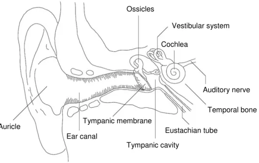
The ear is divided into three main parts: the outer, the middle and the inner ear (Figure 1.1.). The outer ear consists of the auricle which is the visible part of the ear and the 2.5 cm long ear canal that connects the outer ear with the tympanic membrane, also called ear drum (15).
The middle ear is limited by the tympanic membrane which is connected to the malleus, the incus and the stapes, the tiny chained up ossicles in the tympanic cavity. The stapes at the end of the ossicular chain stays in connection with the oval window. The air filled tympanic cavity has a volume of 1 to 2 cm3 and is connected via the Eustachian tube with the oral cavity. Via this tube differences in pressure between the outer and the middle ear are compensated.
The middle ear is connected to the inner ear via the round window membrane and the oval window membrane. Those are two semi-permeable membranes through the petrous bone which surrounds the inner ear.
The inner ear consists of the cochlea where sound perception takes place and the vestibular system which is involved in the process to maintain the balance.
INTRODUCTION 3 Ossicles Vestibular system Auricle Tympanic cavity Cochlea Auditory nerve Temporal bone Tympanic membrane
Ear canal Eustachian tube
Figure 1.1. Anatomy of the ear: division into the outer (Auricle, Ear Canal, Tympanic
membrane), the middle (Tympanic cavity, Ossicles, Eustachian tube) and the inner ear (Cochlea, Vestibular system), adapted from (16).
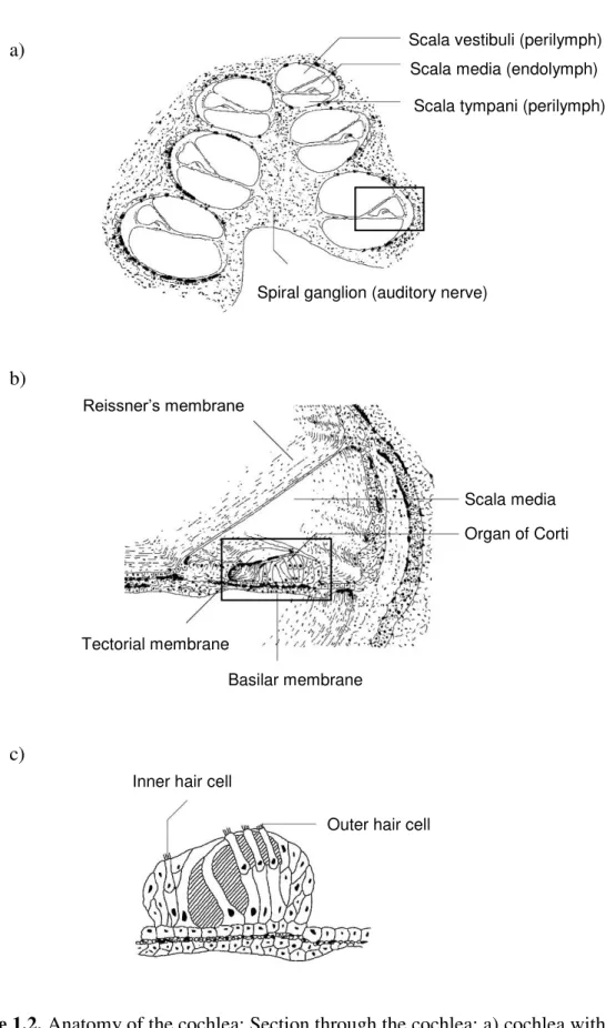
The cochlea has the form of a snail and consists of three fluid filled canals with a length of 31 to 37 mm coiled up in the cochlea (16,17): scala tympani and scala vestibuli are filled with perilymph which has a composition similar to other extracellular fluids whereas the scala media situated between the two other scalae is filled with endolymph (Figure 1.2.a). The latter has an unusual composition with a high concentration of potassium ions of 150 mM leading to a high potential in the endolymphatic fluid. The scalae tympani and vestibuli are connected at the apex of the cochlea via the helicotrema and have a volume of 70 µL in humans and 2.78 µl in gerbils which in both species is nearly ten times higher than the volume of the endolymphatic space (Table 1.1.).
To separate the three scalae from each other there are two membranes in the inner ear: Reissner’s membrane between scala vestibuli and media as well as the basilar membrane between scala media and tympani (Figure 1.2.b). In the middle, the organ of Corti is situated in the scala media. The highly specialized inner and outer hair cells situated on the basilar membrane of the organ of Corti (Figure 1.2.c) are responsible for the translation of mechanical waves into electrical signals leading to the perception of sound in the brain (16).
4 a)
b)
c)
Figure 1.2. Anatomy of the cochlea: Section through the cochlea: a) cochlea with three coiled
up fluid filled spaces: scala vestibuli, media and tympani; b) zoom into the scala media with the Organ of Corti – the organ containing the sensory cells; c) zoom into the Organ of Corti with three rows of outer hair cells and one row of inner hair cells, adapted from (16).
Inner hair cell
Outer hair cell
Scala vestibuli (perilymph) Scala media (endolymph)
Scala tympani (perilymph)
Spiral ganglion (auditory nerve)
Reissner’s membrane
Basilar membrane Tectorial membrane
Organ of Corti Scala media
INTRODUCTION
5
Table 1.1. Characteristics of fluids inside the cochlea: Perilymph and Endolymph in humans,
adapted from (18,19). Perilymph Endolymph Volume, µL 70 8 Volume (gerbil), µL 2.78 0.38 Na+, mM 160 1 K+, mM 4-5 150 Cl-, mM 120 130 H2CO3, mM 20 30 Ca2+, mM 1.2 0.02 Glucose, mM 4 0.5 Proteins, g L-1 1 0.15 pH 7.4 7.4 Osmolality, mOsm kg-1 290 315 Potential, mV 0 +80
The vestibular system consists of the three semicircular canals as well as the vestibule which comprises of the utricle and saccule. It stays in contact with the fluids of the cochlea. That is why the inner ear can also be divided into the bony labyrinth, filled with perilymph, and the membranous labyrinth, filled with endolymph (20,21) (Figure 1.3.). The perilymph of the bony labyrinth stays in contact with the cerebrospinal fluid and surrounds the membranous labyrinth (22). Nevertheless, the flow of the inner ear fluids is very low which means that the local conditions in the vestibular system and the cochlea are maintained locally in each compartment of the two labyrinths (23).
Part of the membranous labyrinth of the vestibular system are the semicircular canals: the superior, the horizontal and the posterior canal. They are arranged at right angles to each other and open out into correspondent ampullae leading to the utricle (Figure 1.3. on the left hand side). The ampullae, the utricle and the saccule contain specialized hair cells detecting movement of the head: the macula is situated in the utricle and saccule whereas the crista ampullaris is situated in the ampullae (21).
6
Figure 1.3. Anatomy of the inner ear: The fluids of the cochlea (right) stay in contact with the
fluids of the vestibular system (left). The bony labyrinth with the Perilymph (light grey) surrounds the membranous labyrinth containing Endolymph (dark grey), adapted from (22).
This system reacts very sensitive to potentially toxic changes and, though, is protected by several barrier systems described in the following chapter.
1.1.1. Barriers of the Inner Ear
The highly sensitive inner ear is protected via three different barriers: The Blood-cochlea barrier, the tympanic membrane as well as the oval and the round window (18).
The blood-cochlea barrier, also called the blood-perilymph barrier, is similar to the blood-brain barrier: Diffusion of drugs from the systemic blood circulation into the inner ear is limited due to the special composition of the capillary endothelium of the blood vessels. It is blocking the entrance of drugs from blood stream into the cochlea via tight junctions without fenestrations (24–26). Furthermore, p-glycoprotein (p-gp) as well as multidrug resistance protein 1 (MRP1) has been detected in the inner ear indicating that it is also protected by efflux pumps (27,28). The impact on clinical results is important, e.g., dexamethasone administered i.v. resulted in significant lower cochlear concentrations compared to drug administered intratympanically (29). Nevertheless, it seems that drugs can enter the inner ear depending on their chemical characteristics. Small lipophilic drugs can
Cochlea Semicircular canals:
Posterior
Utricle Saccule
Vestibule Cochlear nerve
Bony labyrinth Membranous labyrinth Apex Lateral Anterior Lateral Posterior Connection between Cochlea and Vestibular System Ampullae:
INTRODUCTION
7 enter the perilymph more easily than big hydrophilic, charged or protein binding drugs (25). Finally, positively charged drugs are less likely to enter the endolymphatic space from the perilymph because of the electrical gradient (Table 1.1.) (25). Importantly, various conditions can disturb the blood-cochlea barrier, e.g. noise exposure, inflammation, the administration of diuretics or several osmotic agents (18).
The tympanic membrane (Figure 1.1.) protects the middle ear from toxic substances entering through the ear canal of the outer ear and has an area of 85 to 90 mm2. It consists of
an outer epidermal layer, followed by a fibrous layer as well as an inner mucosal layer and has an almost oval and conical shape (15,30). During intratympanic injection this membrane is damaged.
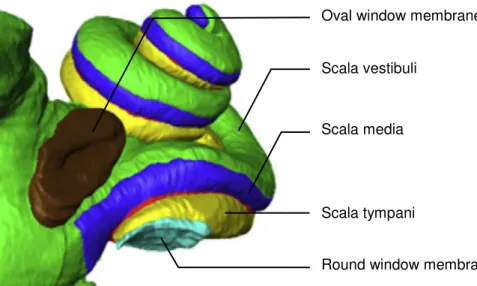
Figure 1.4. Barriers of the inner ear: 3D-reconstruction of a human inner ear. The round
window membrane (RWM) stays in contact with the scala tympani whereas the oval window membrane (OWM) is connected to the scala vestibuli, adapted from (31).
The round and the oval window connect the middle ear with the cochlea which is surrounded by the petrous bone (Figure 1.4.). Unfortunately, drug delivery to the inner ear through the petrous bone – one of the densest bones in the body – seems to be limited in humans. Importantly, drug delivery through this bone seems to be overestimated in animal experiments because the bone in animals is very thin compared to the bone in humans (32).
Both, the round and oval window membrane, are not only barriers but also a potential target for local drug delivery. The round window is connected to the scala tympani at the basal turn of the cochlea. It consists of three layers, an outer epithelium with a single layer of cells, a middle layer of connective tissue containing fibroblasts, blood vessels, collagen and
Scala tympani
Oval window membrane
Scala vestibuli
Scala media
8 elastic fibers as well as an inner layer consisting of squamous epithelium (18). The round window niche has an opening width of about 0.5 to 3 mm, the membrane has a thickness of about 50 - 100 µm in humans compared to 10 to 14 µm in rodents (18,33,34). The ovoid surface of the round window is around 2.2 mm2 in humans compared to 1 mm2 in rodents and
can have various shapes (18,34). Unfortunately, the round window membrane is often plugged by a pseudomembrane, a fat plug or fibrous tissue which makes the quantification of drug delivery quite challenging. From 85 patients, 22 % had obstructions in both ears whereas only 56 % of the examined patients had no obstacle in both ears at the round window niche (35). Additionally, the transport of a drug through the round window membrane depends highly on the size, concentration, solubility, electrical charge and uptake mechanism of the drug (18) which makes the development of an appropriate drug delivery system very challenging and time consuming.
The second membrane connecting the middle with the inner ear is the oval window which stays in contact with the perilymph of the scala vestibuli at the base of the stapes. The stapes’ footplate is attached to the oval window by the annular ligament and has a normal thickness of 0.3 to 0.5 mm in humans (33). The length of the footplate has been measured to be 2.5 to 3.36 mm compared to a width of 0.7 to 1.66 mm (36). It has been calculated that the surface area of the stapes footplate is about 3.97 mm2 (36). In the past, clinicians thought that the drug enters the inner ear mainly through the round window membrane. Recent studies indicate that drugs can also enter the inner ear via the stapes footplate (37): It has been calculated that the ionic marker trimethylphenylammonium (TMPA) enters the inner ear mainly through the round window membrane, but, importantly, one third of the drug enters through the oval window membrane (31).
Those barriers protect the inner ear, more precisely the inner ear hair cells. This mechanoreceptor cells are responsible for the auditory perception that will be described in the following chapter.
1.1.2. Auditory perception
The sound that is processed in the inner ear and detected in the brain depends on the characteristics of the sound waves arriving at the outer ear. Sound waves can be described regarding the amplitude (or intensity), the wavelength, the frequency and the phase (16). Briefly, the sound wave is collected by the auricle, passes the ear canal where it is amplified and, subsequently, causes movement of the tympanic membrane (15). This movement is
INTRODUCTION
9 converted into mechanical vibrations that are – again – amplified and transferred via the ossicles to the oval window membrane. The movement of the stapes is converted at the oval window into a pressure wave which is spread throughout the fluid filled cochlea - from the oval window of the scala vestibuli via the apex of the cochlea to the round window of the scala tympani.
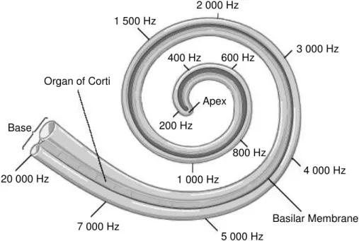
Inside the cochlea, the sound wave causes vibration of the basilar and the tectorial membrane. Depending on the frequency of the sound wave, especially the cells in the corresponding area of the cochlea are stimulated: Human beings can detect low frequencies from approximately 20 Hz at the apex until high frequencies of 20 000 Hz at the base of the cochlea (Figure 1.5.) (15). Mongolian gerbils have a hearing frequency range of 100 to 60 000 Hz (38). In humans, the ability to detect high frequencies is typically decreasing with age.
Figure 1.5. Perception of sound inside the cochlea depending on the frequency of the sound
wave in humans: High frequencies stimulate the hair cells at the base whereas low frequencies vibrate the hair cells at the apex of the cochlea, adapted from (39).
The difference between the vibration of the basilar and the tectorial membrane causes a shearing force. Subsequently, this mechanical signal is translated into an electrical signal in the specialized outer and inner hair cells of the organ of Corti: The stereocilia situated on top of the hair cells vibrates depending on the mechanical wave. This vibration causes the hair cells to depolarize and repolarize by opening of potassium and calcium channels. The sound is amplified by the outer hair cells which leads to vibration and release of transmitters from the
200 Hz 1 500 Hz 2 000 Hz 4 000 Hz 3 000 Hz Apex 400 Hz 600 Hz 800 Hz 1 000 Hz 7 000 Hz 5 000 Hz 20 000 Hz Basilar Membrane Organ of Corti Base
10 inner hair cells that activate receptors in the nerve leading to the brain. Subsequently, this signal is transferred to the brain where the sound is perceived (16,40).
Along with this first perceptional system situated in the cochlea, the second main system in the inner ear, the vestibular system, is responsible for the equilibrioception and will be described in the following chapter.
1.1.3. Sense of balance
The semicircular canals and the vestibule of the inner ear (Figure 1.3.) are part of the system maintaining the balance of the body. Not only the inner ear is involved in this process but also the eyes, muscles, the brainstem, the cerebellum and the cortex (21,41). In this context, the inner ear hair cells play a major role in translating the movement of the head into electrical signals which can be interpreted by superordinate systems.
Therefore, two types of hair cell containing membranes exist in the vestibular system: the macula, also called otolitic organ, and the crista ampullaris. The mechanosensitive hair cells inside those membranes consist of a kinocilium and 70 to 100 stereocilia (21).
Macula membranes exist inside the utricle and the sacculus of the vestibule and are responsible for the detection of linear acceleration and head tilt (41). Those membranes contain not only hair cells but also “heavy” calcium carbonate crystals, so called otoliths. They are embedded in the otolitic membrane which covers the gelatinous layer containing the hair cells. When the head is leaned forwards or moved linearly these crystals are displaced. They cause a shearing force between the otolitic membrane and the macular surface leading to a bending of the hair cells followed by an electrical signal which can be detected in the brain (21).
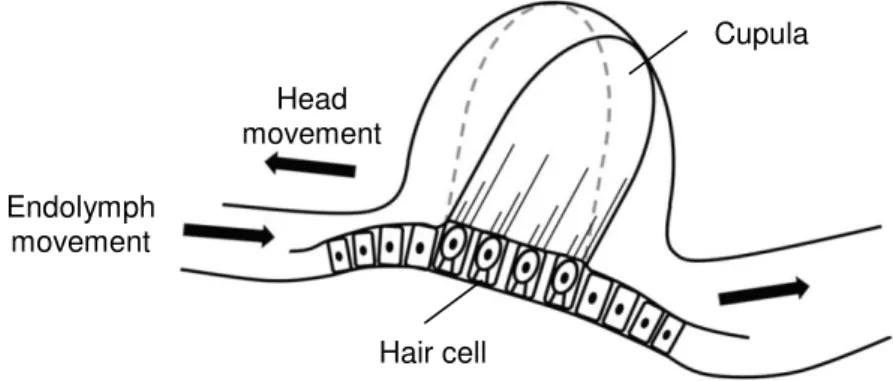
The crista ampullaris inside the ampullae at the end of the semicircular canals detect angular acceleration. Since the three semicircular canals are arranged orthogonal to one another the hair cells in each ampulla can detect movement in the three dimensions (22). Therefore, the hair cells are embedded into a gelatinous structure, the cupula, similar to the macula. In contrast to the otolitic structure of the macula, the hair cells of the crista ampullaris are bent due to the movement of the endolymph of the membranous labyrinth and contain no calcium carbonate crystals. When the head is moved the endolymph inside the semicircular canals flows in the opposite direction of the movement causing the bending of the cupula. In consequence, the hair cells are bent and stimulated (Figure 1.6.). A continued uniform movement of the head results in a return of the cupula to the original position, stopping the
INTRODUCTION
11 motion results in a bending of the cupula in the opposite direction with correspondent hair cell polarization (21). The signals from the vestibular system are transduced via the nerve to the brain where head and eye movement are matched to maintain the balance of the body (22).
Figure 1.6. Function of the crista ampullaris: Rotation of the head causes endolymph flow
inside membranous labyrinth in the opposite direction. The cupula waves depending on the flow leading to a stimulation of the hair cells (21).
Damage in the cascade of auditory perception or the sense of balance in only one step can lead to inner ear disorders that are described in the next chapter.
1.2. Diseases of the inner ear
Current strategies to treat inner ear diseases aim at the treatment of Noise Induced and Sudden Sensorineural Hearing Loss (NIHL and SSHL respectively), of Autoimmune Inner Ear Disease (AIED), Tinnitus or Meniere’s Disease and the protection of the inner ear, e.g., during aminoglycoside or anti-cancer therapy. In this introduction a major focus will be on Hearing Loss. Additionally, a short overview on other illnesses will be given here.
Autoimmune Inner Ear Disease causes bilateral, generally asymmetric, progressive or fluctuating hearing loss that is often combined with a systemic autoimmune disease of the patient as well as vestibular symptoms and responds to immunosuppressive therapy (42,43). Researchers assume that the etiology of the disease includes inflammation, vascular and cochlear tissue damage (e.g., Stria vascularis, Spiral ganglion, Organ of Corti) due to a disproportionate Th1 immune response (42). Therapy includes systemic and intratympanic administration of corticosteroids for a prolonged period. Sometimes other immunosuppressive agents like methotrexate or cyclophosphamide seem to be beneficial for the patient by reducing the dose of steroids. Recent research focuses on fusion proteins and monoclonal
Cupula Head movement Endolymph movement Hair cell
12 antibodies to block the inflammatory reaction. A second promising approach might be the application of stem cell and gene therapy to repair damaged inner ear tissues (43).
Tinnitus is defined as the perception of sound without an external acoustic stimulation (44,45). The cause of the disease is unclear, researchers discuss not only a peripheral but also a central neural origin (46). This disease can occur following to excessive noise exposure or during the normal process of aging and can be associated with additional symptoms like hearing loss, sleep disturbance, hearing loss, anxiety and depression (45). Therapy aims at interrupting or masking the “phantom” sound via sound therapy (47) but also includes an appropriate treatment of the additional symptoms. This treatment might involve supply with hearing aids, education, psychological support, relaxation and cognitive behavioral therapy for the patient (44,45). Research on drugs that might be promising for the treatment of Tinnitus focuses on corticosteroids, e.g., dexamethasone, local anesthetics, e.g., lidocaine, and n-Methyl-d-aspartate receptor antagonists (18).
Patients suffering from Meniere’s disease report intermittently occurring episodes of vertigo, often associated with hearing loss, tinnitus or an aural pressure (48). This illness has an enormous impact on patient’s lives and researchers still discuss about its origin. Autoimmune reactions or viral infections might cause endolymphatic hydrops as well as fibrosis and tissue degeneration leading to the major symptoms of Meniere’s disease (48). The treatment with intratympanic Aminoglycoside antibiotics, e.g., gentamicin, seems to reduce vertigo but increases the risk to suffer from hearing loss (49). Also transtympanic injection of steroids seems to have a beneficial effect on vertigo attacks but further studies should be performed to proof those promising results (50).
Otoprotective actions should be taken to prevent hearing loss due to Cisplatin or Aminoglycoside related toxicity. Both groups of drugs cause similar damage to the inner ear hair cells. Mainly outer hair cells inside the cochlea are degraded, while the damage is increasing from the apex to the base of the cochlea (51). Therefore, an increasing hearing impairment at the correspondent frequencies can be observed. Hearing Loss due to Cisplatin administration during anti-cancer therapy is not only age- (very young and the elder patients are more affected) but also dose-dependent (25): Administration of the ototoxic drug via an osmotic pump with concentrations from 0 to 300 µg/mL respectively resulted in greater and faster hearing loss when a higher concentration is administered (52). Spiral ganglion cells can also be affected. Additionally to this hearing loss, during aminoglycoside administration vestibular toxicity can be observed. The mechanism behind seems to be an excessive level of reactive oxygen species damaging especially outer hair cells (25). Local administration of
INTRODUCTION
13 antioxidants seems to be promising but systemically administered methionine or sodium thiosulfate decreases the effectiveness of the cisplatin therapy (25). Furthermore, the use of cytoprotective agents, e.g., amifostine, has not been proven to prevent hearing loss due to cisplatin therapy in children (53). During aminoglycoside therapy, otoprotection can be achieved by the administration of antioxidants as well as steroids (25).
Hearing loss can be related to all of the inner ear illnesses described above and, thus, will be discussed in detail in the following chapter.
1.2.1. Hearing Loss
Especially when it occurs suddenly, hearing loss is a frightening disorder for the patient. In addition to the hearing loss patients may report tinnitus (“ringing” of the ears), dizziness or fullness of the ear (54).
According to the World Health Organization there are five grades of hearing impairment (Table 1.2.): no, slight, moderate, severe and profound impairment (Grades 0 to 4 respectively). Following this classification, disabling hearing impairment occurs when the patient has at least a hearing loss of Grade 2. This moderate impairment with a loss of 41 dB or more on the better hearing ear means that words can still be understood and repeated at 1 m distance with a raised voice (55).
Table 1.2. Hearing impairment according to the definition of the WHO: with a grade
exceeding grade 1 hearing aides are recommended, adapted from (55).
Grade Threshold shift of the better ear, dB Effect
0 25 or better No/slight problems, even whispers are heard.
1 26 to 40 normal voice can be heard and repeated. Words spoken in 1 m distance with
2 41 to 60 Words spoken in 1 m distance with raised
voice can be heard and repeated.
3 61 to 80 Some words can be heard when shouted.
4 81 or greater No words can be heard and understood even when shouted. The causes of hearing loss are various. In general, they can be classified as congenital or acquired (1). Congenital hearing loss refers to causes occurring during or shortly after birth, e.g., rubella, toxoplasmosis or other infections of the mother as well as treatment with
14 inappropriate drugs during pregnancy, asphyxia and low weight of the newborn (56). Importantly, genetic factors also play a major role in 25 % of the cases, over 400 gene related syndromes have been identified (4,56). Unfortunately, in 57 % of the cases the cause of congenital hearing loss still remains unknown (4).
Acquired hearing loss refers to cases occurring at every age of the patient and can develop suddenly or over a long period. Hearing loss can develop due to infections, e.g. meningitis, measles, mumps or otitis media, as well as traumata of the head or the ear following an accident or surgery (1,55,57). Other causes can be autoimmune diseases, e.g. systemic lupus erythematosus, tumor growth and treatment, neurologic diseases, e.g. Multiple sclerosis, or vascular events (58–61). Additionally, certain drugs can have a toxic effect on the ear, e.g. aminoglycoside antibiotics as well as several chemotherapeutic agents and anti-malaria drugs (51,62,63). Importantly, also acute or long term noise exposure can cause noise-induced hearing loss (NIHL), e.g. recreational noise during a sport event or from a MP3-Player and noise from machines or explosions. Also, a certain degree of hearing loss is age-related (64) and can be considered as a normal process: It was estimated that 30 % of the men and 20 % of the women over 70 suffer from hearing loss (threshold shift of at least 30 dB) in Europe (65). Frequently, patients are also diagnosed with hearing loss due to excessive ear wax stuck in the ear canal (66). Nevertheless, only in 7 to 45 % of patients with Sudden Sensorineural Hearing Loss (SSNHL with a threshold shift of at least 30 dB over three continuous frequencies during 72 h) the cause can be identified, a major part of cases remains idiopathic (57).
Table 1.3. Types of hearing loss with the concerned region, according to (15). Type of hearing loss Concerned region
Conductive Disease of external and/or middle ear
Sensorineural Disease of the cochlea and/or nerve
Mixed Combination of conductive and sensorineural
Central Disease of the auditory pathway higher than the auditory nerve Depending on the region, there are several types of hearing loss (Table 1.3.) (15). Conductive loss occurs when the stiffness of the outer or middle ear is changed, e.g. when the ear canal is stuck with ear wax or in case that the ossicular chain is damaged because of
INTRODUCTION
15 otosclerosis (67). Sensorineural hearing loss (SNHL) occurs when the cochlea or the nerve is damaged, e.g. this is the case when hair cells of the organ of Corti are damaged due to gentamicin administration (37). A combination of conductive and sensorineural is a so called mixed hearing loss. When the auditory system is damaged in higher regions than the auditory nerve a central hearing loss occurs.
Nevertheless, the cause of hearing loss is unknown in most of the cases and therapy still remains challenging. Ongoing research on different strategies to treat hearing loss is discussed in the following chapter.
1.3. Drug delivery to the inner ear
Current strategies used in clinic focus mainly at treating Sudden Sensorineural Hearing Loss and autoimmune diseases as well as at a protection of the inner ear (25). Besides the strategy of providing the patient with appropriate medical devices, e.g., hearing aids or cochlear implants to cure persistent hearing loss, different drug delivery tools are a major topic in research.
Since the rate of spontaneous recovery from Sudden Sensorineural Hearing Loss is relatively high (32 to 65 %) and the etiology of Hearing Loss is not fully understood yet, clinicians discuss about the appropriate treatment of hearing loss. Nevertheless, in case that the cause is known, the patient should be treated accordingly (57,66). In the case of Idiopathic Hearing Loss, current therapeutic strategies often include systemic or local administration of steroids but also antivirals, diuretics, vasodilators, antioxidants, hyperbaric oxygen treatment, middle ear surgery and bedrest are used to treat hearing loss (57,66).
Systemic drug delivery (described in section 1.3.1.) is still used to treat inner ear diseases but is progressively replaced by local drug delivery (described in section 1.3.2.) to avoid adverse events caused by high systemic blood concentrations of the drug.
1.3.1. Systemic drug delivery
Unfortunately, the systemic administration of both, steroids, optionally combined with antivirals, and vasodilators did not show a significant improvement in Cochrane Reviews (54,68,69). This may be partially due to insufficient patient numbers and inconsistent inclusion criteria or study designs.
However, oral steroids may be useful in the treatment of sudden sensorineural hearing loss but their influence on hearing recovery remains uncertain. Only one of three studies
16 included in the Cochrane Review showed a significant effect of oral steroids on hearing recovery with a hearing improvement of 61 % compared to 32 % in the placebo/untreated group (70). In two other studies no improvement of hearing loss can be seen when oral steroids are administered (71,72).
The systemic administration of antivirals to treat idiopathic sudden sensorineural hearing loss neither shows improvement: Two studies included in a Cochrane review showed no improvement when aciclovir was administered additionally to prednisolone (73,74). Accordingly, patients treated with valaciclovir in addition to prednisone, or aciclovir administered additionally to hydrocortisone, showed no hearing improvement (75,76). Nevertheless, animal studies support the assumption that an early treatment of patients with antivirals could be beneficial. Unfortunately, in clinical practice most patients present very late so the impact of the treatment with antivirals may be difficult to prove (54).
The administration of vasodilators or vasoactive substances could be beneficial for the treatment of hearing loss but due to the small number of patients included in the studies the benefit remains unproven (68). A significant hearing improvement has been reported for patients receiving carbogen additionally to several other drugs compared to no inhalation of carbogen (77). In a study where patients received Prostglandin E1 additionally to hydrocortisone only the hearing in higher frequencies was improved (78). The hearing in lower frequencies was improved by the administration of low molecular weight Dextran with additional Naftidrofuryl (79). Those results are promising clinicians should be aware of potential side effects of drugs whose benefit for the patient is not yet approved in clinical practice (66).
In addition to the unknown cause of the disease in most of the cases, during systemic administration of drugs side effects are more likely to occur. The patient often needs an elevated dose to enhance absorption of the drug into the inner ear to reach therapeutic drug concentrations. This is due to the barriers protecting the highly sensitive inner ear as described before (chapter 1.1.1. Barriers of the Inner Ear). Additionally, the small volume of the inner ear fluids and its complicated anatomical access make local drug delivery very difficult.
Nevertheless, local drug delivery seems to be a promising approach to limit adverse events during the treatment of hearing loss and will be discussed in the following chapter.
INTRODUCTION
17
1.3.2. Local drug delivery
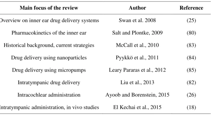
Local inner ear drug delivery has been the topic of several reviews in the last years (Table 1.4.). Most reviews concentrate on either intratympanic or intracochlear administration of drugs: El Kechai et al. recently published an interesting update focusing on intratympanic administration and in vivo studies (18), whereas Ayoob and Borenstein focused on intratympanic drug delivery (26). The review of Salt and Plontke deals with the pharmacokinetics of the inner ear (80). Salt also provides a program to simulate cochlear fluids of several species (81).
Table 1.4. Reviews on inner ear drug delivery, adapted from (82).
Main focus of the review Author Reference
Overview on inner ear drug delivery systems Swan et al. 2008 (25) Pharmacokinetics of the inner ear Salt and Plontke, 2009 (80) Historical background, current strategies McCall et al., 2010 (83)
Drug delivery using nanoparticles Pyykkö et al., 2011 (84)
Drug delivery using micropumps Leary Pararas et al., 2012 (85)
Intratympanic drug delivery Liu et al., 2013 (82)
Intracochlear administration Ayoob and Borenstein, 2015 (26) Intratympanic administration, in vivo studies El Kechai et al., 2015 (18)
The two major strategies to deliver drugs locally to the inner ear are intratympanic and intracochlear drug delivery. Depending on the intended treatment both systems have various benefits and drawbacks.
During intratympanic delivery the drug is placed inside the tympanic cavity where the drug is absorbed mainly via the round but also by the oval window. The advantage of this strategy is the relatively save, usually ambulatory administration, often requiring no general anesthesia, allowing for short and mid-term drug delivery to the middle or inner ear. Unfortunately, the preparation might be washed away through the Eustachian tube or degraded rapidly and, though, often requires repeated application. These repetitions increase the risk of introducing pathogens into the inner ear. Additionally, the anatomy of the ear
18 varies from patient to patient leading to different drug concentrations in the inner ear. Depending on the characteristics of the drug, a gradient along the length of the cochlea can occur (18,25).
Intracochlear delivery allows for the release of drugs directly inside the cochlea and requires a cochleostomy. The main advantage of this administration is the direct access to the inner ear ensuring a defined long term drug delivery during months or years bypassing inter-patient anatomical differences. Importantly, the characteristics of the drug only play a minor role since the drug is not obliged to pass the barriers protecting the inner ear. However, the patient has to stay in hospital during the treatment which is rather invasive and the surgeon risks to introduce pathogens during the operation (18,25).
1.3.2.1.
Intratympanic drug delivery
Today, intratympanic administration of a drug loaded solution is commonly used in clinical practice. Therefore, the tympanic cavity is filled with the solution which is injected via the tympanic membrane using a thin needle. The outcome is promising: Patients suffering from Sudden Sensorineural Hearing Loss whose first line treatment with oral steroids failed could benefit from a treatment with intratympanic steroids which has led to a reduction in hearing thresholds (86). Other diseases of the inner ear might be treated accordingly using aminoglycosides, glutamate receptor antagonists, protease inhibitors, antioxidants or neurotrophins (25).
Unfortunately, the drug solution is often eliminated very fast from the middle ear cavity. To enhance the residence time at the round window membrane, promising devices are the Silverstein MicroWick®, the Round Window µ-CathTM and the Round Window E-CathTM. Another strategy is to place biodegradable polymers loaded with either a drug solution or nanoparticulate systems inside the middle ear, e.g., close to the round window membrane (18,87).
Medical devices
MicroWick®The MicroWick® is a cylinder (dimensions 9 or 19 x 1 mm) consisting of polyvinyl acetate. It stays in contact with the round window membrane and passes through a perforation in the tympanic membrane. A drug solution (that can be administered dropwise into the ear canal of the outer ear by the patient himself) is absorbed by the polymer and, thus, transported
INTRODUCTION
19 to the round window (25). The device is often used to treat vertigo occurring during Meniere’s disease with gentamicin (88), but also patients suffering from Sudden Sensorineural Hearing Loss can profit from a prolonged drug delivery: 26 patients receiving methylprednisolone during 10 days (after failure of the conventional therapy against hearing loss) had improved mean speech discrimination scores. The score recorded at 40, 55 and 70 dB improved by 24.2 ± 8.7 % (89). Despite these promising results, the application of the MicroWick® might result in a permanent perforation of the tympanic membrane (82,83).
Additionally, the risk of infection of the middle ear is increased due to the connection to the outer ear (83). The compliance of the patient is important because the drug solution is administered usually several times per day during weeks.
µ-CathTM and E-CathTM
The microcatheters can be used to deliver drugs intratympanically and via an intracochlear approach. They have two different canals: The first serves to infuse a solution, the other to withdraw fluids. The E-CathTM has a third canal that can be used to insert an electrode to control inner ear function during surgery (25). The tip of the microcatheter is inserted through a tunnel drilled into the temporal bone and fixed near the round window. This device has been used successfully to treat Sudden Sensorineural Hearing Loss (90). To facilitate the removal of plugs blocking the round window niche and the intratympanic injection of drug preparation, an otoendoscope has been developed that can visualize the middle ear during surgery. It has two canals: the first one serves removing mucosal adhesions, via the second one a drug solution can be injected into the middle ear (91). After treatment during several weeks, the catheter can be removed (83). Potential drawbacks are the risk of catheter dislocation or obstruction, the formation of granulation tissue and a potentially permanent perforation of the tympanic membrane (83).
Polymeric matrices
Hydrophilic polymers are widely used in research since the residence time of the formulation at the round window compared to intratympanically injected solutions is increased. These polymers can not only be administered in the form of solid sponges or discs but also as injectable in situ forming gels. The drug is released through degradation of the matrix, diffusion of the drug or a combination of both mechanisms (82). Thus, drug
20 concentration inside the cochlea seems to be more consistent, the concentration gradient along the scala tympani seems to decrease.
Gelfoam®
Gelfoam® is a compressed biodegradable sponge based on purified porcine gelatin which is used because of its hemostatic and fluid absorbing properties. Prior to the use as a drug delivery device, the polymer is soaked in drug solution. Promising results have been reported for the delivery of brain-derived neurotrophic factor (BDNF): Guinea pigs have been deafened and treated with a sponge that was loaded with BDNF and placed onto the round window. This treatment increases spiral ganglion cell survival in the basal turn of the cochlea after 2 and 4 weeks, thus, provides a protection to inner ear cells. Unfortunately, this effect in lower compared to studies working with an intracochlear approach (92). Silverstein et al. treated patients suffering from Meniere’s disease with a Gelfoam® sponge loaded with
gentamicin solution. Vertigo was controlled in 75 % of the patients; hearing was preserved in 90 % of the cases (93).
SeprapackTM
SeprapackTM is a bioresorbable device consisting of carboxymethyl cellulose and hyaluronic acid. Several studies evaluated the capacity of dexamethasone loaded SeprapackTM gels to reduce hearing loss due to trauma, e.g., during cochlear implantation (82). The administration of dexamethasone-loaded SeprapackTM before the implantation resulted in detectable drug concentrations inside the cochlea what was not the case when other types of delivery beads were applied. Dexamethasone protects residual hearing during cochlear implantation (94). In another study, it was confirmed that an administration of the dexamethasone-loaded device before the implantation resulted in increased hearing thresholds from 2 to 32 kHz. Importantly, protection increased with longer application time of the drug loaded device. Also, higher concentrations of dexamethasone applied onto the round window membrane resulted in better hearing protection in the second turn of the cochlea (95).
Hydrogels
Hydrogels can also be administered to the inner ear via intratympanic injection of the gel itself or in the form of an in situ forming gel. Various polymers have been tested to adjust drug delivery from the gels, e.g., gelatin, chitosan glycerophosphate, hyaluronic acid, alginate, siloxane, poloxamer 407 and collagen (25,82,83).
INTRODUCTION
21 Chitosan, a non-toxic cationic polymer, has been used to deliver dexamethasone to the inner ear of mice. In vitro, the chitosan-glycerophosphate hydrogel released 92 % of the drug during 4 days. In vivo, dexamethasone has been detected during 5 days. Reversible hearing loss has been reported after surgery but mice recovered spontaneously after 10 days (96).
Gelatin is not only administered as the solid Gelfoam® but also as a gel: The biodegradable polymer has been used to deliver the recombinant human insulin-like growth factor 1 (rhIGF-1) to prevent damage of the inner ear cells upon excessive noise exposure. Histological evaluation confirmed a higher survival of outer hair cells when the gel is applied onto the round window membrane (97).
An interesting approach to prolong drug delivery is to use temperature-sensitive systems: The formulation can be injected intratympanically at room temperature as a liquid solution and forms a gel at body temperature (sol-gel-transition), e.g., on the round window membrane. Poloxamer has been used to provide prolonged dexamethasone release to the inner ear by forming an in situ forming gel. Concentration gradients along the scala tympani were lower compared to when injecting a solution. This can be partially due to a formation of the gel at the thin bone at the apex of the cochlea (which is more permeable in rodents than in humans) but can possibly related also to an extended release of the drug (98). This promising formulation is currently under clinical evaluation in Phase IIb to treat Meniere’s disease with a sustained release of dexamethasone (OTO-104, Otonomy) (18,99).
Other candidates for clinical practice are two formulations based on hyaluronic acid to cure Noise Induced Hearing Loss and Tinnitus, administering dexamethasone and esketamine (AM-111 and AM-101 respectively, Auris Medical) (18). Additionally, a gelatin-based preparation releasing IGF-1 to cure Sudden Sensorineural Hearing Loss is clinically evaluated (18).
A potential drawback concerning hydrogel-based drug delivery could be that the formulation has to be placed precisely at the round window niche. Another problem might occur when excessive gel in the middle ear cavity causes transient hearing loss by blocking the ossicular chain. Quick elimination of the formulation via the Eustachian tube might limit application of hydrogels to treat chronic diseases (83).
Nanoparticles
Nanoparticles are drug delivery systems with diameters of less than 1000 nm, typically a diameter of 200 nm or smaller is requested for otological use (83). They should be incorporated into formulations or devices that sustain drug release and prevent the elimination
22 via the Eustachian tube, e.g., by using hydrogels or microcatheters. Different drug delivery systems have been investigated to treat inner ear illnesses, e.g., silica nanoparticles, PLGA- or GMO-based systems, liposomes, lipid nanocapsules, hyperbranched polylysine nanoparticles, polymerosomes, as well as dendrimer-based nanoparticles and SPIONs (18,26). Nanoparticles can be used to counteract low drug solubility, problems with degradation, with passage of the round window membrane or short half-life of the drug (18). Functionalization of the nanoparticles’ surface offer interesting possibilities to target single cell types inside the inner ear. An interesting approach to enhance diffusion through the round window membrane might be to combine PLGA-nanocarriers with magnetite to release dexamethasone-acetate. After the administration of the nanoparticles on the round window niche, a permanent magnet was placed on the opposite site of the round window. Drug transport through the membrane has been increased using magnetic nanocarriers with a magnet compared to pure diffusion (100). For further interesting studies on drug release from nanoparticles the author refers to the review published by El Kechai et al. were a vast amount of different strategies are discussed in detail (18).
1.3.2.2.
Intracochlear drug delivery
In contrast to intratympanically administered drugs which have to be absorbed via the round window membrane, intracochlear delivery offers the potential to release drugs directly to the inner ear. Strategies include direct intracochlear injection, drug release using osmotic pumps or microcatheters (described in section 1.3.2.1. Intratympanic drug delivery), as well as reciprocating perfusion systems and cochlear prosthesis-mediated drug delivery. A rather invasive cochleostomy through the round window or the temporal bone is needed to provide access to the inner ear (26).
Intracochlear injection
The intracochlear injection of drugs is mainly used for research, e.g., to conduct pharmacokinetic studies or to study the effect of new drugs on inner ear cells. Therefore, a few microliters of the drug solution are injected via cochleostomy. Potential drawbacks are the short period of drug delivery as well as a possible leakage of cochlear fluids that might wash the drug solution out of the cochlea. Furthermore, high drug concentrations at the application side might damage the highly sensible inner ear cells. In human, intracochlear injection is used only during surgery (18).
INTRODUCTION
23
Osmotic pumps
Osmotic pumps are used similarly to the microcatheters already described above. Both systems can be used to provide intratympanic or intracochlear drug release. The osmotic pump can be implanted subcutaneously providing flow rates from 0.1 to 10 µl/h from a reservoir containing 0.1 to 2 mL during 1 day up to 6 weeks. Osmotic pressure ensures low but permanent drug delivery rates. A drawback is that the flowrate cannot be adjusted in vivo (85). Those systems can be used to evaluate new therapies in animal models: betamethasone has been administered using an osmotic pump during 14 days following to a damage of the right semicircular canal of guinea pigs. Animals treated with the drug showed better recovery from the induced vestibular illness compared to non-treated animals (101).
Reciprocating perfusion systems
Those systems combine microsystems and microfluidics technologies to create new drug delivery devices that are able to provide drugs to the inner ear more precisely (83). A micropump is infusing and withdrawing inner ear fluids in a cyclic manner nearly simultaneously so that the volume inside the cochlea stays constant (26). This device has been studied in guinea pigs administering 6,7-dinitroquinoxaline-2,3-dione (DNQX), a glutamate receptor blocker. DNQX allowed for following of drug release by recording the Compound Action Potential (CAP) (102). A new version of the reciprocating perfusion system has been presented recently (26).
Cochlear prosthesis-mediated drug delivery
Cochlear implants have been used widely since 35 years to cure hearing loss and consist of an electrode array that is inserted via cochleostomy inside the scala tympani of the cochlea. Different insertion depths are used in practice and in research, ranging from 16 to 31.5 mm (26). The usually drug free electrode is coiled up inside the turns of the cochlea providing a relatively large surface for potential drug delivery. Different strategies are discussed: combining a cochlear implant with a micropump or drug-eluting coatings of the electrode as well as introducing the drug directly into the silicone of the electrode (26). The aim is to reduce damage of the inner ear cells due to the insertion force during surgery. Therefore, dexamethasone-eluting electrodes have been developed and evaluated in vitro and in vivo showing promising results (103–105). Another approach is to deliver the drug (or a dye in this case) via tiny delivery ports that are connected via the implant with a micropump. The distribution of the dye along an artificial cochlea was satisfactory when two outlets served to
24 release the dye (106). Furthermore, electrodes have been coated with hydroxyl ethyl cellulose to adjust drug release and have been used to deliver neurotrophic factors, e.g., brain-derived neurotrophic factor (BDNF) or neurotrophin-3 (26).
Since silicones are already widely used inside the inner ear, e.g. in the form of cochlear implants, this polymer seems to be advantageous to deliver drugs to the inner ear in a sustained manner. Therefore, the following chapter will be focused on drug release from silicone matrices.
1.4. Drug release from silicone matrices
The incorporation of drugs within silicone matrices can be very helpful to improve the therapeutic efficacy and safety of a large variety of medical treatments. The basic idea is that the polymeric system accurately controls the resulting drug release rate during pre-programmed periods of time. Examples for promising applications include the local delivery of drugs to sites of action, which are difficult to reach (without causing major side effects in the rest of the human body due to high systemic drug concentrations). This includes for instance the treatment of diseases and disorders of the inner ear (107,108). But also local treatments of the vagina (109), heart (110), eye (111,112) or scars (113) can be very challenging and silicone matrices can be highly beneficial in these cases. Furthermore, silicone matrices offer a great potential for the design of advanced intraperitoneal controlled release implants (114) and central venous catheters (115).
Importantly, the release periods can be very long (e.g. several months or years) and the resulting advantages for the patient long-lasting. For example, Mond and Stokes (110) reported on the benefits of silicone-based, dexamethasone-eluting electrodes in pacemakers, which effectively lower the stimulation threshold at the “electrode-tissue interface”. In a double-blind human study over 10 years the mean stimulation thresholds for the drug-eluting devices remained almost constant (exhibiting a narrow standard deviation), whereas the drug-free systems showed an unpredictable increase in the threshold values and wide standard deviations. Interestingly, 20 % of the dexamethasone is estimated to still remain within the silicone matrices even after 10 years implantation in humans (based on the analysis of explanted devices). The authors state that drug release may well continue at sufficient levels for an additional 10 years. This is a very promising clinical evidence for the benefits of silicone-based controlled drug delivery systems. However, the development of such devices is
INTRODUCTION
25 generally very cumbersome, since often long release periods are targeted and so far very little information is available on the impact of the device design on the resulting system performance (in particular drug release kinetics) in a quantitative way. So, highly time-consuming and cost-intensive series of trial-and-error experiments are mandatory.
In order to adjust a desired drug release profile from a silicone matrix, different formulation parameters can be varied. For instance, the type of silicone (e.g. with a particular type of side chains and contents of amorphous silica) can be altered, different types and amounts of additives can be incorporated and/or the initial drug content can be varied. Also, the geometry and dimensions of the system might be changed. Both determine the pathway lengths, which have to be overcome by the drug to be released. Interesting reports are available in literature on the effects of the composition of silicone matrices on the resulting drug release kinetics (116–119). For example, Di Colo and co-workers studied the impact of adding glycerol, ethylene glycol and poly(ethylene glycol) to silicone disks loaded with prednisolone (120,121). Importantly, the presence of these hydrophilic additives effectively increased the resulting drug release rate. It has to be pointed out that silicones are generally hydrophobic and water penetration into the systems is very limited. Furthermore, commonly used silicones do not degrade in the human body. Craig and co-workers (109) published a very interesting study on the importance of the solubility of the drug in silicone matrices for the resulting release kinetics. Clindamycin, estradiol, estradiol-3-acetate, 17-estradiol diacetate, metronidazole, norethisterone, norethisterone acetate and oxybutynin release was studied from intravaginal rings, prepared by injection molding. Also, Liu et al. (105) investigated the impact of the initial drug loading and of the dimensions of differently shaped silicone matrices on dexamethasone release. Waever et al. (122) used dexamethasone loaded silicone rods and discs for controlled local delivery in order to modulate inflammation in islet transplantation, and varied the initial drug loading.
It has to be highlighted that the underlying mass transport mechanisms in polymeric drug delivery systems can be rather complex (123–125). The basic idea is that the presence of the polymer prevents immediate drug release upon contact with aqueous body fluids. Generally, first water penetrates into the system and dissolves the drug (126). Once dissolved (in the form of individual molecules), the drug can diffuse through the polymeric system into the surrounding environment. Drug diffusion might take place through an intact polymeric network and/or through water filled pores. The amount of water available for drug dissolution in the system and the drug solubility in the matrix can be decisive (127). In the case of substantial polymer swelling and/or dissolution, important time- and position-dependent
26 changes in the system’s composition might occur over time, altering the conditions for drug transport (128). Furthermore, the homogeneity of the initial drug distribution within the silicone matrix can be of importance (129). More or less complex mathematical theories can be used to quantify the involved mass transport processes and describe drug release from polymeric delivery systems (130–133). Also neural networks can be applied (134,135). Ideally, the mathematical theory should be mechanistically realistic and take all decisive phenomena into account, thus, allowing for the quantitative prediction of the effects of the device design on the resulting drug release kinetics (136). Negligible mass transport phenomena should not be considered, to keep the model as simple as possible. However, yet there is a lack of reliable mathematical theories allowing for such in-silico simulations of the impact of formulation parameters on the resulting system performance.
1.5. Objectives
The objective of this thesis was to develop implants capable of releasing the drug in a time controlled manner to the inner ear to treat inner ear diseases. The following steps have been selected to achieve this aim:
Characterization of dexamethasone loaded silicone matrices in vitro to identify easy tools allowing for the adjustment of drug release kinetics from thin films and extrudates. Therefore, different formulation parameters have been varied: the ratio of PEG addition to the silicone, the molecular weight of PEG, the chemical structure of the silicone and the dexamethasone loading. Mathematical modeling helped to elucidate the underlying drug release mechanisms.
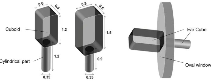
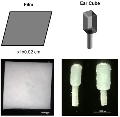
Development of dexamethasone loaded implants using the most promising silicone. Thin films and Ear Cube implants have been prepared and studied in vitro. Additionally, the physicochemical properties of Ear Cube implants have been analyzed.
In vivo study with in situ forming dexamethasone loaded implants to examine the feasibility of an implantation of the implant besides the oval window. Additionally, the drug released from the implant has been detected directly inside the explanted gerbil cochlea using Confocal Laser Scanning Microscopy.