Characterization of Saccharomyces cerevisiae Pseudohyphal Development
by
Carlos Joaquin Gimeno
B.A. University of California at Berkeley (1990)
Submitted in partial fulfillment of the
requirements for the degree of Doctor of Philosophy
at the
Massachusetts Institute of Technology May 1994
© Carlos Joaquin Gimeno, 1994, All rights reserved
The author hereby grants to M I T permission to reproduce and to distribute copies of this thesis document in whole or in part.
Signature of Author
h
Department of Biology May 1994 Certified by Gerald R. Fink Thesis Supervisor I, - -Accepted by K- rank Solnmen Chairperson, Departmental Committee on Graduate Studies MASSACLUSEFTS INSTIT TEOF 'VCHNOLOGY
Characterization of Saccharomyces cerevisiae Pseudohyphal Growth
by Carlos Joaquin Gimeno
Submitted to the Department of Biology in May 1994 in partial fulfillment of the
requirements for the Degree of Doctor of Philosophy
Abstract:
When starved for nitrogen, MATa/a Saccharomyces cerevisiae cells
undergo a dimorphic transition to pseudohyphal growth (PHG).
Pseudohyphae are filaments of elongated incompletely separated cells that grow by budding, often invasively. PHG may be a foraging mechanism:
pseudohyphal cells may be vectors that deliver ellipsoidal cells to new substrates. PHG is a MATa/a-diploid-specific dimorphic transition. Mating type locus regulation may occur because the mating type locus programs the
polar budding pattern of newborn MATa/a cells that is required for PHG.
Mutations in SHR3 cause cells to inappropriately enter the pseudohyphal pathway when growing on media where proline is sole nitrogen source. This probably occurs because shr3/shr3 strains have impaired proline uptake so that when they grow on proline medium they are
nitrogen starved. The constitutively active RAS2val 19 allele inappropriately
activates PHG while overexpression of cAMP phosphodiesterase 2 inhibits PHG. The latter mutation is epistatic to the former suggesting that the RAS-cAMP pathway may directly regulate PHG.
PHD1 overexpression enhances PHG and also induces PHG on rich
medium. PHD1 is a nuclear protein and has a DNA binding motif similar to the DNA binding domains of the SWI4 and MBP1 transcription factors and a
Aspergillus nidulans protein that regulates two PHG-like cell divisions. phdl/phdl strains undergo normal PHG when starved for nitrogen.
Overexpression of PHD1 induces DAL81 and POL2. DAL81 encodes a transcriptional inducer of nitrogen catabolic genes and POL2, a gene
regulated by MBP1, encodes a S phase-specific DNA polymerase. These results raise the possibility that PHD1 may regulate nitrogen metabolism and the cell cycle, two processes with logical connections to PHG.
SOK2 is a PHD1 homolog (80% identity over the DNA binding motif) that may participate in the RAS-cAMP pathway. Mutant sok2/sok2 strains
undergo greatly enhanced PHG. Mutant phdl/phdl sok2/sok2 strains exhibit moderately enhanced PHG revealing a genetic interaction between PHD1 and SOK2. PHD1 and SOK2 may act together to integrate and transduce signals that control PHG.
Thesis supervisor: Dr. Gerald R. Fink, American Cancer Society Professor of Genetics and Director of the Whitehead Institute for Biomedical Research
Acknowledgments
I would like to first thank my advisor and mentor Gerald Fink for making the laboratory portion of my graduate education comprehensive, exciting, and rewarding.
I also would like to express my gratitude to the members of my thesis
committee, Phillips Robbins, Terry Orr-Weaver, Jun Liu, and John Chant, for their time and valuable input into my thesis.
I would also like to thank Cora Styles and my mentor and collaborator Per L.jungdahl for sharing their knowledge and enthusiasm with me.
I also feel indebted to Judy Bender, Chip Celenza, Jan-Huib Franssen, Julia Koehler, Steve Kron, Hans-Ulrich MOsch, and David Pellman for their help and encouragement.
I would also like to thank those people with whom I collaborated, interacted, or both and all of the people with whom I coincided in the Fink lab for helping to make my graduate career a success.
Finally, I would like to thank Ruth Hammer and my parents, Joaquin and Rosalie Gimeno, for their support and encouragement.
Table of Contents
Chapter 1: Genetic Regulation of Saccharomyces cerevisiae Pseudohyphal Growth
Introduction ... 14
Genes and Proteins that Directly or Indirectly Regulate Pseudohyphal G row th ... ... ... ... ...17
SHR3 and Amino Acid Permeases ... 17
The Mating Type Locus and BUDGenes ... ...18
The RAS-cAMP Signaling Pathway ... 20
Putative Transcription Factors PHD1 and SOK2 ... 25
Pheromone ResponseGenes ... ... 28
Protein Kinases, Protein Phosphatases, and Interacting Proteins ... 29
Conclusions ... ... ... ...32
LiteratureC ited ... ... ... ...33
Chapter 2: Initial Characterization of Saccharomyces cerevisiae Pseudohyphal Growth Summary ... ... 43
Introduction
...
45
Results Pseudohyphal Cells Originate by Budding from Ellipsoidal C ells ... ... ... ...53
As Pseudohyphae Elongate They Become Covered with Ellipsoidal Cells ... 53
Discussion
...
57
Materials and Methods Saccharomyces cerevisiaeStrains ... 58
Media, Microbiological Techniques, and Light Microscopy ...58
LiteratureC ited ... ...60
Appendix to Chapter 2: SHR3: A Novel Component of the Secretory Pathway Specifically Required for Localization of Amino Acid Perm easesin Yeast ... 6...3...63
Chapter 3: Unipolar Cell Divisions in the Yeast S. cerevisiae Lead to Filamentous Growth: Regulation by Starvation and RAS Summary ... 81
Introduction . ... ... ...81
Results The Dimorphic Switch to Pseudohyphal Growth Is Induced by Nitrogen Starvation ... 82
Mutations in the SHR3 Gene Enhance Pseudohyphal Growth..82
Activation of the RAS2 Protein Enhances Pseudohyphal
Growth
...
83
Pseudohyphal Growth Is a Diploid-Specific Pathway ... 84
The Cells of the Pseudohypha Are a Morphologically Distinct
CellType ... ...85
The Daughter of a Pseudohyphal Cell Can Be a Pseudohyphal Cell or a Blastospore-Like Cell ... 85
Pseudohyphal Cells Invade the Semisolid Agar Growth M edium ... 86
A Mutation in RSR1/BUD1 Causing Random Bud Site Selection Suppresses Pseudohyphal Growth ... ...8686... Discussion ... ... ... ... 88
Conclusions ... ... ... ... 90
Experimental Procedures Strains, Media, and Microbiological Techniques ... 91
Yeast Strain Construction ... ... ...91... 91
Bud Site Selection Assay ... ... ... ...92
SEM Methods ... 92
Light Microscopic Techniques ... 92
Quantitation of Yeast Cell Dimensions ... 92
Acknowledgments ... 92
References
...
...
93
Chapter 4: Induction of Pseudohyphal Growth by Overexpression of PHD1, a Saccharomyces cerevisiae Gene Related to Transcriptional Regulators of Fungal Development A bstract ... ...96
Intro duction
...
96
Materials and Methods Yeast Strains, Media, and Microbiological Techniques ...97
Yeast Strain Construction ... 97
Physical Mapping of PHD1 to PHD7. ... 97
Genetic Mapping of PHD1 ...98
Qualitative Pseudohyphal Growth Assay ... 98
Agar Invasion Assay ... ... 98
Light Microscopic Techniques ... ... ... 98
Plasmid Construction ... 98
DNA Sequence Analysis of PHD1 ...99
Epitope Tagging of PHD1 ... 99
Immunofluorescence of PHD1 ::FLU1 ...99
Construction of a Precise PHD1 Deletion ... ... 99
Deletion of PH D 1 ... ...99
Results A Gene Overexpression Screen Identifies Multicopy Enhancers of Pseudohyphal Growth ... ... 99
Mapping PHD1 to PHD7to the S. cerevisiaeGenome ... 101
DNA and Predicted Amino Acid Sequences of PHD1 ...101
PHD1 Is Related to Transcriptional Regulators of Fungal D evelopm ent ... 101
Immunolocalization of Functionally Epitope-Tagged PHD1 Protein to the Nucleus ... 103
PHD1 Overexpression Induces Pseudohyphal Growth on Rich
M edium ... 103
Analysis of Pseudohyphal Growth in Haploids Overexpressing PHD 1 ... .. ... ... ... 1... 104
Deletion of PHD1 . ... ... ...104
Discussion ... 104
Acknow ledgm ents ... 106
References ... ... ... 1...06
Chapter 5: Isolation and Characterization of Genes Induced by PHD1 Overexpression Introduction ... 110
Materials and Methods Yeast Strains, Media, and Microbiological Techniques ... 112
Yeast Strain Construction ... ... ... 1 12 Qualitative Pseudohyphal Growth Assay ... 116
Light MicroscopicTechniques ... 116
Plasmid Construction ... 1 16 DNA Sequencing ... 119
Construction of a Plasmid Containing PHD1 Under the Control of the GAL1-10Promoter .... ... ... ... ...120
13-Galactosidase Assays ... ... 120
Results A Genetic Screen Identifies Genes Induced by PHD1 O verexpression ... 122
Sequence Analysis of the 5 PHD1-lnducible Fusion Genes ....126
Analysis of Strains Carrying Disruptions of PHD1-lnducible G enes ... ... 129
Analysis of Pseudohyphal Growth in Strains that Have Disruption Alleles of IP01 and DAL81 ... 131
Analysis of DAL81 Regulation ... 131
D iscussion ... 133
Literature Cited ... 136
Chapter 6: Investigation of the Role of PHD1 Homolog SOK2 in the Regulation of Saccharomyces cerevisiae Pseudohyphal Growth
Intro duction
...
142
Materials and Methods Yeast Strains, Plasmids, Media, and Microbiological
Techniques
.
...
145
Yeast Strain Construction ... ... 145
Qualitative Pseudohyphal Growth Assay ... 145
Light Microscopic Techniques ... 149
Disruption of SOK2...149
Construction of phdl/phdl sok2/sok2 Double Mutant
Strains
.
...
149
Results100 Amino Acids of SOK2 Are 80% Identical to the DNA Binding
Motif Present in PHD1 ... ... ... 151151... Analysis of Pseudohyphal Growth in Strains that Have Mutant
Alleles of SOK2 ... ... ... ... 151 Overexpression of cAMP Phosphodiesterase 2 Inhibits
Pseudohyphal Growth and Is Epistatic to Activated RAS2 ... 159
Discussion
...
163
LiteratureCited
...
166
Chapter 7:
C onclusions ... 171 Prospectus
Characterizing PHD1 and SOK2 and Identifying Pseudohyphal
Growth Genes ... ... . ... 1... 176
Discovering a Pseudohyphal Growth-Specific Gene ... 178
A Quantitative Assay for Cell Elongation ... 1...79 Materials and Methods
Quantitation of Pseudohyphal Cell Dimensions ... 182
Chapter 1:
Genetic Regulation of
Saccharomyces
cerevisiae
Introduction
This chapter reviews and integrates recently published information
about Saccharomyces cerevisiae pseudohyphal growth. For a discussion of
older work on pseudohyphal growth and related issues, readers are referred to several excellent articles and books (Barnett, 1992; Barnett et al., 1979; Guilliermond, 1920; Kendrick, 1985; Mortimer and Johnston, 1986; Scherr and Weaver, 1953; van der Walt, 1970; von Wettstein, 1983).
The dimorphic transition to pseudohyphal growth is MATa/a
diploid-specific and is induced by nitrogen starvation (Gimeno et al., 1992).
Pseudohyphal growth can be divided into at least three distinct phases. In the first phase, an ellipsoidal yeast cell buds an elongated pseudohyphal cell (Gimeno et al., 1993). In the second phase, the elongated daughters bud identical elongated cells that compose the backbone of the pseudohypha. In the third phase, the elongated cells of the pseudohypha, with the exception of those very near the growing tip of the filament, bud vegetative ellipsoidal yeast cells. The net result is that pseudohyphal growth disperses asexually
produced vegetative ellipsoidal yeast cells to new and otherwise inaccessible growth substrates. This has been interpreted to mean that pseudohyphal growth is a foraging mechanism.
Component processes of pseudohyphal growth include the diploid-specific polar budding pattern of newborn cells, cell elongation, invasive growth into substrates, and the incomplete separation of pseudohyphal
mother and daughter cells (Gimeno et al., 1992). With the exception of the
polar budding pattern, a property of all wild-type MATa/a diploid newborn
RSR1/BUD1 genes affect pseudohyphal growth because they regulate one of
these processes, polar budding (Bender and Pringle, 1989; Chant and Herskowitz, 1991). MATa/a diploid cells have the polar budding pattern that permits pseudohyphal growth, whereas MATa and MATa haploid cells have the axial pattern that precludes pseudohyphal growth (Chant and Pringle,
1991; Freifelder, 1960). The RSRl/BUD1 gene is also required for the polar budding pattern; a Bud1- diploid has a random budding pattern (Chant and Herskowitz, 1991; Ruggieri et al., 1989) that impairs pseudohyphal growth (Gimeno et al., 1992).
The SHR3 gene indirectly regulates pseudohyphal growth by a mechanism understood in some molecular detail (Gimeno et al., 1993; Ljungdahl et al., 1992). Shr3- strains inappropriately induce pseudohyphal growth when grown on proline as the sole nitrogen source. SHR3, a
component of the endoplasmic reticulum (ER), is required for amino acid permeases, including the proline permease PUT4, to exit the endoplasmic reticulum and reach the plasma membrane. Thus, Shr3- strains have
impaired proline uptake that results in nitrogen starvation, which consequently leads to induction of pseudohyphal growth when proline is the sole nitrogen source.
Several other genes have been reported to affect pseudohyphal growth directly or indirectly. These include protein phosphatase 2A, ELM1, PAM1,
PDE2, PHD1, RAS2, SOK2, STE7, STE11, STE12, and STE20. Mutations in
genes encoding any of the three subunits of protein phosphatase 2A can cause filamentous growth (Blacketer et al., 1993; Healy et al., 1991; Ronne et al., 1991; van Zyl et al., 1992) and some of these mutations interact with mutant alleles of the PAM1 or ELM1 genes (Blacketer et al., 1993; Hu and Ronne, 1994). The PAM1 protein is unrelated to proteins in databases while
ELM1 is related to protein kinases. PHD1 and SOK2 encode putative transcription factors that strongly enhance pseudohyphal growth when
overexpressed and disrupted, respectively (Gimeno, 1994; Gimeno and Fink, 1994; Ward and Garrett, 1994). The PHD1 and SOK2 proteins are
homologous to Aspergillus nidulans StuA, a regulatory protein that controls two pseudohyphal growth-like cell divisions during conidiogenesis
(Timberlake, 1991). The RAS2 and PDE2 genes encode components of the RAS-cAMP signal transduction pathway (Broach and Deschennes, 1990). Ectopic activation of RAS2 induces pseudohyphal growth under inappropriate
conditions while PDE2 overexpression inhibits pseudohyphal growth (Gimeno
et al., 1992; Gimeno et al., 1993). STE7, STE11, STE12, and STE20 encode
components of a signal transduction pathway that regulates mating-specific genes and some genes linked to either transposon Ty or the repeated element
sigma (Sprague and Thorner, 1992). Loss of function of any of these four genes causes impaired pseudohyphal growth (Liu et al., 1993). The
mechanisms by which these varied genes regulate pseudohyphal growth are not yet clear and their elucidation is the next frontier in the pseudohyphal growth field.
Genes and Proteins that Directly or Indirectly Regulate
Pseudohyphal Growth
SHR3 and Amino Acid Permeases
SHR3 is the gene whose mechanism of pseudohyphal growth
regulation is understood in most detail. SHR3 was identified as a gene that when mutated confers upon yeast cells resistance to 30 mM histidine
(Ljungdahl et al., 1992). Mutant shr3 strains have pleiotropically impaired amino acid uptake (Gimeno et al., 1993; Ljungdahl et al., 1992) and the shr3 mutation is synthetically lethal with several mutations that cause amino acid auxotrophies. In addition, shr3 strains constitutively induce the translation of
GCN4, a gene encoding a positive regulator of amino acid biosynthetic genes
that is only translated under conditions of amino acid starvation (Hinnebusch, 1988).
Cloning and sequencing of SHR3 revealed that its predicted protein product is not homologous to known proteins and is probably an integral membrane protein and other experiments showed that it is a component of the ER membrane. The impaired amino acid transport observed in shr3 strains occurs because many amino acid permeases fail to exit the ER and reach the plasma membrane, where they are functionally expressed. The permeases are found to be at least partially inserted into the ER membrane but do not assume their normal topology. This phenomenon appears to be specific to amino acid permeases because several other secretory proteins including the plasma membrane [H+]ATPase, a factor, a factor, invertase, and the vacuolar enzyme carboxypeptidase Y are secreted normally in shr3 strains.
Haploid shr3 strains grow slowly when they utilize proline as sole nitrogen source because of their proline uptake defect. Diploid shr3/shr3 strains not only grow slowly on proline, but also ectopically enter the
pseudohyphal growth pathway (Gimeno et al., 1992). It seems likely that the nitrogen starvation experienced by shr3 strains utilizing proline as sole
nitrogen source induces them to undergo pseudohyphal growth. Thus, SHR3 affects pseudohyphal growth indirectly by controlling the secretion of amino acid permeases.
The Mating TyvDe Locus and BUD Genes
The polar budding pattern of newborn diploid cells is regulated by a
cell's ploidy as reflected by its genotype at the mating type (MAT) locus and is
the only component process of pseudohyphal growth that is not induced by
nitrogen starvation. Only MATa/a diploids and not MATa or MATa haploids or
MATa/a or MATa/a diploids show pseudohyphal growth, indicating that the
MAT locus controls this dimorphic transition (Gimeno et al., 1992). The MAT
locus also controls the budding pattern of yeast strains (Chant and Pringle,
1991; Herskowitz, 1989). Cells expressing only MATa or MATa bud in the
axial pattern whereas cells expressing both MATa and MATa bud in a polar
manner (see Table 1 in Chapter 2). The axial pattern leads to budding at the junction of two cells and cannot extend the filament. Only the polar budding
pattern of wild-type diploid cells in which new buds emerge from the daughter on the side opposite the junction to her mother is consistent with
pseudohyphal growth. In fact, it has been clearly shown by time lapse
photomicroscopy that pseudohyphae elongate by polar budding by newborn cells (Gimeno et al., 1992).
Budding pattern genes represented by BUD 1-BUD5 are required for selection of the proper bud site and consequently for establishing the proper axis of cell division (Bender and Pringle, 1989; Chant et al., 1991; Chant and Herskowitz, 1991; Park et al., 1993). BUD 1, BUD2, and BUD5 convert the
default random budding pattern to bipolar and subsequent action of BUD3 and BUD4 converts bipolar to axial (Chant and Herskowitz, 1991). To explain the observed cell type specificity (diploids are bipolar, haploids axial) an elegant model was proposed that either or both BUD3 and BUD4 are repressed by the repressor al a2 found only in MATa/a cells (Chant and Herskowitz, 1991). That a MATa/a diploid strain with a random budding pattern caused by a mutant allele of BUD1 has impaired pseudohyphal
growth demonstrated that BUD genes are important for pseudohyphal growth. This observation was recently expanded with the discovery that a bud4
haploid strain that has the polar budding pattern undergoes pseudohyphal growth when starved for nitrogen (Sanders and Herskowitz, 1994).
The cell type specificity of pseudohyphal growth is best understood by
considering the different roles of diploid and haploid cells in the S. cerevisiae
life cycle (Gimeno and Fink, 1992). The diploid is the assimilative phase of S.
cerevisiae and the predominant cell type found growing in nature (Lodder, 1970). Thus, the diploid phase must be specialized to deal with a variety of nutritional conditions and stresses. Diploids have two cell type-specific
nutrient stress responses, sporulation and pseudohyphal growth. It has been proposed that the diploid polar budding pattern exists at least in part to allow one of these processes, pseudohyphal growth (Gimeno et al., 1992). In this model, diploid cells are specifically able to undergo pseudohyphal growth and foraging because they are the cell type that is specialized for assimilating nutrients.
The haploid phase is transient in the life cycle of S. cerevisiae and functions as a gamete. Haploid cells are specialized to rapidly reform the diploid phase immediately after ascospore germination. As I explain below, it is thought that haploids have the axial budding pattern, which precludes pseudohyphal growth, to enable them to perform mating efficiently (Nasmyth,
1982). The diploid phase is usually reconstituted when the two MATa spores
in the ascus mate with their two MATa sister spores (Hicks et al., 1977). If an
ascospore becomes separated from its mate, it uses its mating type
interconversion system and axial budding pattern to efficiently reconstitute the diploid phase. The action of the mating type interconversion system results in
two cells of ascospore-derived four celled microcolonies being MATa and two
cells being MATa. The axial budding pattern causes the cells of opposite
mating types to be juxtaposed allowing efficient mating. Thus haploid cells are specialized for mating and specifically have the axial budding pattern, which prevents pseudohyphal growth, to make mating more efficient. In this model, haploids do not need to be able to undergo pseudohyphal growth because the goal of their transient existence is to reform the pseudohyphal growth-competent diploid phase.
The RAS-cAMP Signaling Pathway
The RAS-cAMP signaling pathway regulates mitotic growth, sporulation, stationary phase, and pseudohyphal growth (Broach, 1991; Broach and Deschennes, 1990; Gimeno, 1994; Gimeno et al., 1992). The
RAS1 and RAS2 proteins are low molecular weight GTP-binding proteins that are associated with the plasma membrane (Fig. 1.1). When they are bound to GTP they are active as signal transduction molecules, and when they are
Filure 1.1 The RAS-cAMP pathway. The RAS1 and RAS2 proteins are
inactive as signal transducers when bound to GDP. CDC25 promotes the RAS proteins to undergo GDP-GTP exchange. When bound to GTP, RAS1 and RAS2 stimulate adenylate cyclase (CYR1) to convert ATP to cAMP. cAMP binds to the regulatory subunit of cAMP-dependent protein kinase (BCY1) releasing the catalytic subunits (TPK1, TPK2, TPK3) which are active protein kinases. IRA1 and IRA2 turn off RAS1 and RAS2 by promoting hydrolysis of RAS-bound GTP to GDP. The cAMP phosphodiesterases PDE1 and PDE2 turn off cAMP-dependent protein kinase by hydrolyzing cAMP to AMP. When
intracellular cAMP levels decrease BCY1 releases cAMP and associates with the TPK proteins, inactivating them.
CDC25
RAS
1
-GDP
RAS2-GDP
RA~~s2-RAS 1 -GTP
RAS2-GTP
IRA1
IRA2
CYR1
ATP
cAMP
PDE1
PDE2
BCY1
-cAMP
TPK1
TPK2
TPK3
I
bound to GDP they are inactive. Regulation of the activity of the RAS proteins involves the CDC25 protein which promotes the exchange of GDP for GTP, and the functionally redundant IRA1 and IRA2 proteins which stimulate the intrinsic GTPase activity of the RAS proteins, causing their bound GTP to be converted to GDP. GTP-bound RAS proteins stimulate CYR1-encoded adenylate cyclase to convert ATP to cAMP, a second messenger molecule. cAMP acts by controlling the activity of cAMP-dependent protein kinase, an enzyme that, when inactive, exists in the cell as a heterotetramer composed of two regulatory subunits encoded by BCY1 and two catalytic subunits encoded by three functionally redundant genes, TPK1, TPK2, and TPK3. When high levels of cAMP are generated in the cell, cAMP molecules bind to BCY1 proteins, and these complexes disassociate from the TPK proteins. Free TPK proteins are catalytically active serine-threonine protein kinases that regulate the activity of their substrates by phosphorylating them. Substrates are
thought to include several enzymes involved in carbohydrate metabolism and some RAS-cAMP pathway components (Broach and Deschennes, 1990). At the level of cAMP, the RAS-cAMP pathway is downregulated by two genes,
PDE1 and PDE2, encoding low and high affinity phosphodiesterases,
respectively. Other genes that may participate in the RAS-cAMP signaling pathway are known, but how they fit into the pathway is not well understood (Broach, 1991).
Strains that have an inactive RAS-cAMP pathway are inviable (Broach and Deschennes, 1990). Such strains include cdc25 strains, rasl ras2 strains, cyrl strains, and tpkl tpk2 tpk3 strains. In general, strains that
experience a sudden inactivation of the RAS-cAMP pathway arrest growth as unbudded cells if they are haploid and sporulate if they are diploid. Strains with a hypoactive RAS-cAMP pathway cannot grow on nonfermentable carbon
sources and accumulate carbohydrate reserves. MATa/a strains that have a
hypoactive RAS-cAMP pathway because they overexpress PDE2 have
severely inhibited pseudohyphal growth (Gimeno, 1994, Chapter 6). Strains that have a hyperactive RAS-cAMP pathway are hypersensitive to heat shock and nitrogen starvation, lose their carbohydrate reserves, and cannot
sporulate efficiently. A MATa/a yeast strain that has a hyperactive RAS-cAMP
pathway because it carries the dominant constitutively active RAS2val 9
allele (Kataoka et al., 1984) exhibits enhanced pseudohyphal growth. Inhibition of pseudohyphal growth by PDE2 overexpression is epistatic to enhancement of pseudohyphal growth by RAS2val19 suggesting that
RAS2val 9 enhances pseudohyphal growth by raising intracellular cAMP levels which probably then cause activation of cAMP-dependent protein kinase. Taken together, these results suggest that cells consider information transmitted by the RAS-cAMP signaling pathway when deciding whether or
not to undergo pseudohyphal growth.
The only well characterized environmental signal that activates the RAS-cAMP pathway is the addition of glucose or a related fermentable sugar to yeast cells that are starved for glucose, have been growing on a
nonfermentable carbon source, or are in stationary phase (Broach and
Deschennes, 1990). Within two minutes of this treament, intracellular levels of cAMP rise 10 to 50 fold. Soon after this cAMP spike, cAMP levels fall to a basal level that is about two fold higher than the level of cAMP that existed in the cell when it was starved for carbon. Thus, upregulation of the RAS-cAMP pathway is associated with growth on a good carbon source and
downregulation of the RAS-cAMP pathway correlates with carbon starvation. These relationships are consistent with the effects of perturbations in the RAS-cAMP pathway on carbohydrate reserves: upregulation of the RAS-RAS-cAMP
pathway causes depletion of carbohydrate reserves, whereas downregulation of the RAS-cAMP pathway causes accumulation of these reserves. These effects result because active cAMP-dependent protein kinase directly or
indirectly activates enzymes that break down storage carbohydrates and inactivates enzymes that synthesize them. These facts suggest that one role of the RAS-cAMP pathway is to communicate information to the cell about the
availability of carbon sources. As discussed in detail in Chapter 6, information about carbon status is relevant for a cell's decision to undergo pseudohyphal growth and this may explain why the RAS-cAMP pathway regulates
pseudohyphal growth.
Putative Transcription Factors PHD1 and SOK2
PHD 1 and SOK2 are two related genes that have interesting
pseudohyphal growth regulatory properties. PHD1 was identified as a high copy number enhancer of pseudohyphal growth (Gimeno and Fink, 1994).
PHD1 overexpression causes MATa/a strains to manifest prolific
pseudohyphal growth earlier than wild-type controls. Also, the pseudohyphal cells of strains overexpressing PHD1 are more elongated than wild-type
pseudohyphal cells. Significantly, PHD1 overexpression also induces pseudohyphal growth in MATa/a cells growing on rich medium. As
determined by indirect immunofluorescence, PHD1 protein localizes to the nucleus. The 366 amino acid PHD1 protein has a 100 amino acid DNA
binding motif related to that of the S. cerevisiae transcription factors SWI4 and
MBP1. These facts suggest that PHD1 may be a transcription factor. The DNA binding motif in PHD1 is about 70% identical to a region of A. nidulans StuA. This homology is interesting because StuA regulates the two pseudohyphal
growth-like cell divisions that occur during conidiophore morphogenesis (Miller et al., 1991; Miller et al., 1992; Timberlake, 1990; Timberlake, 1991). A
MATa/a phdl/phdl strain undergoes normal pseudohyphal growth in a
qualitative pseudohyphal growth assay.
Insights into the possible cellular role of PHD1 were gained by the identification of genes that are induced when PHD1 is overexpressed
(Gimeno, 1994, Chapter 5). Three essential and two probably nonessential genes that are induced by PHD1 overexpression were identified. Two of these genes are POL2 and DAL81 and the three others have not been
previously characterized. POL2 is a DNA polymerase involved in DNA repair and chromosomal DNA replication (Budd and Campbell, 1993; Morrison et al., 1990; Wang et al., 1993). POL2is regulated by MBP1 (Johnston and
Lowndes, 1992), a transcription factor whose DNA binding domain is related to the DNA binding motif in PHD1, which induces POL2 expression during the
G1 to S transition. This raises the interesting possibility that PHD1 may control
the MBP1 regulon which includes genes that regulate cell elongation and cell separation, two component processes of pseudohyphal growth (Gimeno and Fink, 1994).
DAL81 is a transcriptional regulator required for the induced expression
of several genes encoding enzymes that catabolize poor sources of nitrogen (Bricmont and Cooper, 1989; Bricmont et al., 1991; Coornaert et al., 1991).
DAL81 is induced by nitrogen starvation and RAS2val19 (Gimeno, 1994), two
conditions that activate the pseudohyphal growth pathway (Gimeno et al., 1992). It is possible that PHD1 is involved in the transduction of the nitrogen
starvation signal, the RAS2val19 signal, or both to DAL81. DAL81 appears
not to be required for the pseudohyphal growth component processes of cell elongation, agar invasion, and incomplete cell separation. The absense of a
pseudohyphal growth phenotype in dal81/da81 mutants raises the possibility that PHD1 may induce DAL81 so that cells in the pseudohyphal growth mode have the catabolic enzymes they need to utilize the poor nitrogen sources that they are likely to encounter while foraging.
SOK2 was isolated by Mary Ward and Stephen Garrett as a high copy
number suppressor of the temperature-sensitive growth defect of a
tpkl-tpk2(ts) tpk3- mutant (Ward and Garrett, 1994). Subsequently, deletion of SOK2 was found to exacerbate the temperature sensitivity of the tpk2(ts)
mutation. Together, these results suggest that SOK2 may function in the
RAS-cAMP pathway downstream of RAS-cAMP-dependent protein kinase. SOK2 is predicted to encode a 470 amino acid protein that contains a DNA binding motif that is 81.7% identical to the DNA binding motif found in PHD1. Because PHD1 and the RAS-cAMP pathway have been implicated in pseudohyphal
growth control, the possible role of SOK2 in pseudohyphal growth was
studied.
Overexpression of SOK2 from a 2pm plasmid weakly enhances pseudohyphal growth and disruption of SOK2 causes strongly enhanced pseudohyphal growth. MATa/a phdl/phdl sok2/sok2 double mutants exhibit enhanced pseudohyphal growth that is less vigorous than that of MATa/a
sok2/sok2 strains. Thus, the enhanced pseudohyphal growth manifested by MATa/a sok2/sok2 strains is partially PHD1-dependent. The bulk of these
observations could be explained if one or more pseudohyphal growth genes
were positively regulated by PHD1 and negatively regulated by SOK2 and if
these two proteins competed for the same regulatory binding sites in the promoters of these pseudohyphal growth genes. Because phdl/phdl strains exhibit normal pseudohyphal growth and sok2/sok2 phdl/phdl strains exhibit moderately enhanced pseudohyphal growth, this model predicts that yeast
has at least one gene whose function is redundant with the pseudohyphal growth regulatory function of PHD1.
It is interesting that the RAS-cAMP pathway regulates pseudohyphal
growth and genetically interacts with SOK2 and that SOK2 genetically
interacts with PHD 1. It is possible that SOK2 and PHD1 are members of a
group of structurally related transcription factors that respond to and integrate the signals that influence a cell's decision to enter the pseudohyphal growth
mode.
Pheromone Response Genes
Haploid S. cerevisiae cells have a signal transduction pathway that
transmits the signal that a mating partner is nearby (Sprague and Thorner, 1992). At the apex of this pathway is a plasma membrane receptor (STE2 or STE3) that binds to a peptide pheromone (MFA1, MFA2, MFal, and MFa2)
secreted by a cells of the opposite mating type. The pheromone bound receptor interacts with a heterotrimeric G protein (GPA1, STE4, and STE18) that is thought to directly or indirectly activate a protein kinase (STE20) and a protein whose function is unknown (STE5). These proteins are thought to stimulate various protein kinases (STE11, STE7, KSS1, and FUS3) that directly or indirectly activate a transcription factor (STE12).
STE12 has several functions including the activation of mating-specific genes and some genes linked to transposon Ty (Errede and Ammerer, 1989). Additionally, STE12 probably regulates some genes linked to the repeated element sigma (Van Arsdell et al., 1987). STE12 protein activates mating-specific genes by binding to a mating-specific DNA sequence present in their promoters (Errede and Ammerer, 1989). Ty contains this specific STE12
binding site so that it often puts genes into whose promoters it has transposed under the control of STE12 and upstream pheromone signaling pathway components (Ciriacy et al., 1991; Dubois et al., 1982; Errede et al., 1980; Taguchi et al., 1984). Interestingly, STE12 and other components of the
pheromone signaling pathway are required for carbon source regulation of some Ty-linked genes (Taguchi et al., 1984).
Of the genes that encode the pheromone response pathway components mentioned in the preceding paragraph, only KSS1, STE7,
STE11, STE12, and STE20 are known to be expressed in MATa/a cells (Liu
et al., 1993; Sprague and Thorner, 1992). Mutations in STE7, STE11, STE12,
and STE20 but not mutations in KSS1 impair pseudohyphal growth (Liu et al., 1993). Activated alleles of STE11 and STE12 enhance pseudohyphal growth and enhancement by active STE 1 requires STE12. Mutations in STE2,
STE3, STE4, STE18, and FUS3 do not affect pseudohyphal growth. It is not
known whether mutations in MFA 1, MFA2, MFa 1, MFa2, or GPA 1 affect
pseudohyphal growth. These results show that some of the pheromone signaling pathway components that are expressed in MATa/a cells directly or
indirectly regulate pseudohyphal growth. It would be interesting to know if these genes regulate pseudohyphal growth by activating mating-specific
genes, Ty-linked genes, or by some other mechanism.
Protein Kinases, Protein PhosDhatases, and Interacting Proteins
Recently a genetic screen was reported that identified haploid yeast mutants that are constitutively elongated (Blacketer et al., 1993). The elm 1,
elm2, and elm3 mutants were the focus of this study. Mutations in ELM1,
pseudohyphal growth. Interestingly, strains that are heterozygous at the ELM2 or ELM3 loci have enhanced pseudohyphal growth on nitrogen starvation medium. ELM1 was cloned and encodes a putative protein kinase. Deletion of ELM1 in haploid strains caused cell elongation and incomplete cell
separation while elm 1/ielm li diploids exhibited constitutive pseudohyphal
growth.
Protein phosphatase 2A (PP2A) is one of four superfamilies of protein phosphatases (Cohen, 1989). In both yeast and higher organisms this enzyme is comprised of at least two regulatory subunits (A and B ) and a catalytic subunit (C) (Cohen, 1989; Peng et al., 1991). The product of TPD3 is thought to encode the A regulatory subunit of PP2A (van Zyl et al., 1992) while the product of the CDC55 gene probably encodes the B regulatory subunit (Healy et al., 1991). Deletion of TPD3 or CDC55 causes a filamentous
phenotype (Blacketer et al., 1993; Healy et al., 1991; van Zyl et al., 1992). The filamentous structures formed by Tpd3- or Cdc55- strains are elongated, multinucleate, and have constrictions and diffuse nuclei. Three redundant genes, PPH3, PPH21, and PPH22, have been reported to encode putative catalytic subunits (C subunits) of protein phosphatase 2A (Ronne et al., 1991). Interestingly, mutant alleles of these gene cause morphological phenotypes (Ronne et al., 1991) including cell elongation, which is caused by PPH22 overexpression. There is evidence that PP2A is involved in carbohydrate
metabolism. PP2A activates glycogen synthase in vitro by dephosphorylating
it (Peng et al., 1991). This is particularly interesting because glycogen
synthase is thought to be activated directly or indirectly by phosphorylation by cAMP-dependent protein kinase (Broach and Deschennes, 1990). Thus, PP2A and the RAS-cAMP pathway, which has been implicated in
pseudohyphal growth control (Gimeno, 1994, Chapter 6; Gimeno et al., 1992),
may have a common target.
A gene called PAM1 was isolated as a high copy number suppressor of the inviability of pph3 pph21 pph22 triple mutants (Hu and Ronne, 1994). The
PAM1 product is predicted to be a hydrophilic 93 kD protein that contains two
coiled coil motifs and has a basic carboxyl-terminal region. PAM1 is not significantly related to known proteins. Interestingly, overexpression of PAM1 induces pseudohyphal growth (Hu and Ronne, 1994). The pseudohyphal cells in the PAM1 overexpression-induced pseudohyphae are well separated and mononucleate.
Interestingly, the ELM1 gene interacts with CDC55 (Blacketer et al., 1993). Double cdc55 elm mutants have a synthetic growth defect, have highly abnormal morphologies, have a defect in either or both cytokinesis and cell separation. This result suggests that ELMI and CDC55 may be involved in the same cellular function. Other evidence that CDC55 may be involved in pseudohyphal growth is that strains heterozygous for CDC55 have enhanced pseudohyphal growth on nitrogen starvation medium and no pseudohyphal growth phenotypes on rich medium (Blacketer et al., 1993).
It should also be mentioned that the redundant PPZ1 and PPZ2 putative protein phosphatases have also been implicated in pseudohyphal growth control. PPZ1 and PPZ2 are thought to be involved in the PKC signaling pathway and ppzl ppz2 strains undergo pseudohyphal growth when grown in osmotically supported medium (Lee et al., 1993). Taken together, these
results suggest that protein phosphorylation is important in regulating pseudohyphal growth. It is now important to systematically study the role of the genes discussed in this section in pseudohyphal growth regulation.
Conclusions
The genetic regulation of S. cerevisiae pseudohyphal growth promises
to be complicated (Table 1. 1). To date, research into this area has focused on the identification of genes whose mutation affects pseudohyphal growth. Only in the case of SHR3 is the molecular mechanism of pseudohyphal growth control understood in some detail. The mating type locus and BUD genes control pseudohyphal growth most likely by programming the polar budding pattern of newborn cells. The molecular mechanism of this programming as well as how the BUD gene products determine budding pattern are active research areas. Genes directly involved in the well characterized RAS-cAMP and pheromone signal transduction pathways have been implicated in
pseudohyphal growth control. Also, PHD1 and SOK2, new genes whose products have interesting homologies to well characterized transcription factors and developmental regulators have also been implicated in
pseudohyphal growth control. ELM1, a protein kinase of unknown function, protein phosphatases, and PAM1, a protein phosphatase interacting protein may also play direct roles in pseudohyphal growth control.
Now that a long list of possible direct regulators of pseudohyphal growth exists, it is possible to systematically determine each one's function in pseudohyphal growth control. In Chapter 7, possible systematic approaches to determine whether a gene whose mutation affects pseudohyphal growth directly affects this process are presented. Reagents and technical advances that would help the pseudohyphal growth field make rapid progress are also presented.
Literature Cited
Barnett, J. A. (1992). The taxonomy of the genus Saccharomyces Meyen ex
Reess: a short review for non-taxonomists. Yeast 8, 1-23.
Barnett, J. A., Payne, R. W., and Yarrow, D. (1979). A Guide to Identifying and
Classifying Yeasts. (Cambridge: Cambridge University Press).
Bender, A., and Pringle, J. R. (1989). Multicopy suppression of the cdc24 budding defect in yeast by CDC42 and three newly identified genes including the ras-related gene RSR1. Proc. Natl. Acad. Sci. USA 86, 9976-9980.
Blacketer, M. J., Koehler, C. M., Coats, S. G., Myers, A. M., and Madaule, P.
(1993). Regulation of dimorphism in Saccharomyces cerevisiae: involvement of the novel protein kinase homolog Elm p and protein phosphatase 2A. Mol. Cell. Biol. 13, 5567-5581.
Bricmont, P. A., and Cooper, T. G. (1989). A gene product needed for induction
of allantoin system genes in Saccharomyces cerevisiae but not for their transcriptional activation. Mol. Cell. Biol. 9, 3869-3877.
Bricmont, P. A., Daugherty, J. R., and Cooper, T. G. (1991). The DAL81 gene
product is required for induced expression of two differently regulated nitrogen catabolic genes in Saccharomyces cerevisiae. Mol. Cell. Biol. 11, 1161-1166.
Broach, J. R. (1991). RAS genes in Saccharomyces cerevisiae: signal transduction in search of a pathway. Trends Genet. 7, 28-33.
Broach, J. R., and Deschennes, R. J. (1990). The function of RAS genes in
Saccharomyces cerevisiae. Adv. Cancer Res. 54, 79-139.
Budd, M. E., and Campbell, J. L. (1993). DNA polymerases a and are
required for chromosomal replication in Saccharomyces cerevisiae. Mol. Cell. Biol. 13, 496-505.
Chant, J., Corrado, K., Pringle, J. R., and Herskowitz, I. (1991). Yeast BUD5,
encoding a putative GDP-GTP exchange factor, is necessary for bud site selection and interacts with bud formation gene BEM1. Cell 65, 1213-1224.
Chant, J., and Herskowitz, I. (1991). Genetic control of bud site selection in yeast by a set of gene products that constitute a morphogenetic pathway. Cell
65, 1203-1212.
Chant, J., and Pringle, J. R. (1991). Budding and cell polarity in
Saccharomyces cerevisiae. Curr. Opin. Genet. Dev. 1, 342-350.
Ciriacy, M., Freidel, K., and Lohning, C. (1991). Characterization of trans-acting mutations affecting Ty and Ty-mediated transcription in Saccharomyces
cerevisiae. Curr. Genet. 20, 441-448.
Cohen, P. (1989). The structure and regulation of protein phosphatases. Annu. Rev. Biochem. 58, 453-508.
Coornaert, D., Vissers, S., and Andr6, B. (1991). The pleiotropic UGA35
(DURL) regulatory gene of Saccharomyces cerevisiae: cloning, sequence,
and identity with the DAL81 gene. Gene 97, 163-171.
Dubois, E., Jacobs, E., and Jauniaux, J.-C. (1982). Expression of the ROAM mutations in Saccharomyces cerevisiae: involvement of trans-acting
regulatory elements and relation with the Tyl transcription. EMBO J. 1,
1133-1139.
Errede, B., and Ammerer, G. (1989). STE12, a protein involved in
cell-type-specific transcription and signal transduction in yeast, is part of protein-DNA complexes. Genes Dev. 3, 1349-1361.
Errede, B., Cardillo, T. S., Wever, G., and Sherman, F. (1980). ROAM
mutations causing increased expression of yeast genes: their activation by signals directed toward conjugation functions and their formation by insertion of Tyl repetitive elements. Cold Spring Harbor Symp. Quant. Biol. 45, 593-602.
Freifelder, D. (1960). Bud position in Saccharomyces cerevisiae. J. Bacteriol.
80, 567-568.
Gimeno, C. J. (1994). Characterization of Saccharomyces cerevisiae Pseudohyphal Development. (Ph.D. Thesis: Massachusetts Institute of Technology).
Gimeno, C. J., and Fink, G. R. (1992). The logic of cell division in the life cycle of yeast. Science 257, 626.
Gimeno, C. J., and Fink, G. R. (1994). Induction of pseudohyphal growth by
overexpression of PHD1, a Saccharomyces cerevisiae gene related to transcriptional regulators of fungal development. Mol. Cell. Biol. 14, 2100-2112.
Gimeno, C. J., Ljungdahl, P. O., Styles, C. A., and Fink, G. R. (1992). Unipolar cell divisions in the yeast S. cerevisiae lead to filamentous growth: regulation by starvation and RAS. Cell 68, 1077-1090.
Gimeno, C. J., Ljungdahl, P. O., Styles, C. A., and Fink, G. R. (1993).
Characterization of Saccharomyces cerevisiae pseudohyphal growth. In Dimorphic Fungi in Biology and Medicine, H. Vanden Bossche, Odds, F. C., and Kerridge, D., eds. (New York, New York: Plenum).
Guilliermond, A. (1920). The Yeasts. (New York: John Wiley and Sons, Inc.).
Healy, A. M., Zolnierowicz, S., Stapleton, A. E., Goebl, M., DePaoli-Roach, A.
A., and Pringle, J. R. (1991). CDC55, a Saccharomyces cerevisiae gene
involved in cellular morphogenesis: identification, characterization, and homology to the B subunit of mammalian type 2A protein phosphatase. Mol. Cell. Biol. 11, 5767-5780.
Herskowitz, I. (1989). A regulatory hierarchy for cell specialization in yeast. Nature 342, 749-757.
Hicks, J. B., Strathern, J. N., and Herskowitz, . (1977). Interconversion of yeast mating types ill. Action of the homothallism (HO) gene in cells homozygous for the mating type locus. Genetics 85, 395-405.
Hinnebusch, A. G. (1988). Mechanisms of gene regulation in the general control of amino acid biosynthesis in Saccharomyces cerevisiae. Microbiol. Rev. 52, 248-273.
Hu, G.-Z., and Ronne, H. (1994). Overexpression of yeast PAM1 gene permits
survival without protein phosphatase 2A and induces a filamentous phenotype. J. Biol. Chem. 269, 3429-3435.
Johnston, L. H., and Lowndes, N. F. (1992). Cell cycle control of DNA synthesis in budding yeast. Nucleic Acids Res 20, 2403-2410.
Kataoka, T., Powers, S., McGill, C., Fasano, O., Strathern, J., Broach, J., and
Wigler, M. (1984). Genetic analysis of yeast RAS1 and RAS2 genes. Cell 37, 437-445.
Kendrick, B. (1985). The Fifth Kingdom. (Waterloo: Mycologue Publications).
Lee, K. S., Hines, L. K., and Levin, D. E. (1993). A pair of functionally
redundant yeast genes (PPZ1 and PPZ2) encoding type 1-related protein phosphatases function within the PKC1-mediated pathway. Mol. Cell. Biol. 13, 5843-5853.
Liu, H., Styles, C. A., and Fink, G. R. (1993). Elements of the yeast pheromone
response pathway required for filamentous growth of diploids. Science 262, '1741-1744.
Ljungdahl, P. O., Gimeno, C. J., Styles, C. A., and Fink, G. R. (1992). SHR3: a
novel component of the secretory pathway specifically required for localization of amino acid permeases in yeast. Cell 71, 463-478.
Lodder, J. (1970). The Yeasts: a Taxonomic Study. (Amsterdam: North-Holland Publishing Co.).
Miller, K. Y., Toennis, T. M., Adams, T. H., and Miller, B. L. (1991). Isolation and
transcriptional characterization of a morphological modifier: the Aspergillus
nidulans stunted (stuA) gene. Mol. Gen. Genet. 227, 285-292.
Miller, K. Y., Wu, J., and Miller, B. L. (1992). StuA is required for cell pattern
formation in Aspergillus. Genes Dev. 6, 1770-1782.
Morrison, A., Araki, H., Clark, A. B., Hamatake, R. K., and Sugino, A. (1990). A
third essential DNA polymerase in S. cerevisiae. Cell 62, 1143-1151.
Mortimer, R. K., and Johnston, J. R. (1986). Genealogy of principal strains of
the yeast genetic stock center. Genetics 113, 35-43.
Nasmyth, K. A. (1982). Molecular genetics of yeast mating type. Annu. Rev. Genet. 16, 439-500.
Park, H.-O., Chant, J., and Herskowitz, I. (1993). BUD2 encodes a
GTPase-activating protein for Budl/Rsrl necessary for proper bud-site selection in yeast. Nature 365, 269-274.
Peng, Z. Y., Wang, W., Wilson, S. E., Schlender, K. K., Trumbly, R. J., and Reimann, E. M. (1991). Identification of a glycogen synthase phosphatase
from yeast Saccharomyces cerevisiae as protein phosphatase 2A. J. Biol.
Chem. 266, 10925-10932.
Ronne, H., Carlberg, M., Hu, G. Z., and Nehlin, J. 0. (1991). Protein
phosphatase 2A in Saccharomyces cerevisiae: effects on cell growth and bud
morphogenesis. Mol. Cell. Biol. 11, 4876-4884.
Ruggieri, R., Bender, A., Matsui, Y., Powers, S., Takai, Y., Pringle, J. R., and
Matsumoto, K. (1989). RSR1, a ras-like gene homologous to
Krev-11smg21AIrapA: role in the development of cell polarity and interactions with
the Ras pathway in Saccharomyces cerevisiae. Mol. Cell. Biol. 12, 758-766.
Sanders, S., and Herskowitz, I. (1994). Personal communication.
Scherr, G. H., and Weaver, R. H. (1953). The dimorphism phenomenon in
yeasts. Bacteriol. Rev. 17, 51-92.
Sprague, G. F., and Thorner, J. W. (1992). Pheromone response and signal
transduction during the mating process of Saccharomyces cerevisiae. In The
Expression, E. W. Jones, Pringle, J. R., and Broach, J. R., eds. (Cold Spring Harbor, New York: Cold Spring Harbor Laboratory Press).
Taguchi, A. K. W., Ciriacy, M., and Young, E. T. (1984). Carbon source
dependence of transposable element-associated gene activation in
Saccharomyces cerevisiae. Mol. Cell. Biol. 4, 61-68.
Timberlake, W. E. (1990). Molecular genetics of Aspergillus development. Annu. Rev. Genet. 24, 5-36.
Timberlake, W. E. (1991). Temporal and spatial controls of Aspergillus development. Curr. Opin. Genet. Dev. 1, 351-357.
Van Arsdell, S. W., Stetler, G. L., and Thorner, J. (1987). The yeast repeated element sigma contains a hormone-inducible promoter. Mol. Cell. Biol. 7, 749-759.
van der Walt, J. P. (1970). Genus 16. Saccharomyces Meyen emend. Reess. In The Yeasts: a Taxonomic Study, J. Lodder, eds. (Amsterdam: North-Holland Publishing Co.).
van Zyl, W., Huang, W., Sneddon, A. A., Stark, M., Camier, S., Werner, M., Marck, C., Sentenac, A., and Broach, J. R. (1992). Inactivation of the protein
phophatase 2A regulatory subunit A results in morphological and
transcriptional defects in Saccharomyces cerevisiae. Mol. Cell. Biol. 12, 4946-4959.
von Wettstein, D. (1983). Emil Christian Hansen Centennial Lecture: from pure yeast culture to genetic engineering of brewers yeast. (European Brewery Convention Congress: London). 97-119.
Wang, Z., Wu, X., and Friedberg, E. C. (1993). DNA repair synthesis during base excision repair in vitro is catalyzed by DNA polymerase and is
influenced by DNA polymerases a and a in Saccharomyces cerevisiae. Mol.
Cell. Biol. 13, 1051-1058.
Chapter 2:
Initial Characterization of
Saccharomyces
cerevisiae
Summary
This chapter will introduce the logic and results of the initial
experiments of my pseudohyphal growth project. An appendix that contains some of these experiments is included as an aid to the reader. Results pertaining to the development of pseudohyphae are presented.
Microscopic analysis of pseudohyphal growth in diploid MATa/a and
MATa/a shr3/shr3 Saccharomyces cerevisiae strains revealed that the first
pseudohyphal cell in a pseudohypha originates by budding from an ellipsoidal mother. Mature pseudohyphae have a backbone of elongated pseudohyphal cells that typically invades the agar medium. Pseudohyphal cells that are located a few cells away from the growing tip of the filament often
bud mitotic ellipsoidal cells. These vegetative ellipsoidal cells colonize the new substrates to which they have been delivered by the filament of
pseudohyphal cells.
These observations have been used to formulate a model of how pseudohyphae forage for nitrogen. In this model, the elongated
pseudohyphal cells and the ellipsoidal cells that together compose the pseudohypha have distinct roles in foraging. Pseudohyphal cells have the role of penetrating and exploring substrates. Their elongated shape aids in exploration because it increases the linear growth rate of the pseudohypha. This occurs because pseudohyphal cells are about 1.5 times longer than ellipsoidal cells and have a similar doubling time to them. Once the
pseudohypha has penetrated a new substrate it begins to generate vegetative ellipsoidal cells. In the model it is the role of the ellipsoidal cells to colonize new substrates and assimilate the nutrients that are present in them. Thus,
pseudohyphal cells can be viewed as vectors that deliver mitotic ellipsoidal cells to new and otherwise inaccessible growth substrates.
Introduction
The logic and results of the initial experiments of my pseudohyphal growth project are important for the reader to put in perspective the body of work described in this thesis. For the reader's convenience, an appendix that contains most of the research on the SHR3 gene described in this introduction is included at the end of the chapter. Research not presented in the Appendix is presented in Chapter 3. This introduction also describes my specific
contributions to the Appendix to Chapter 2 and Chapter 3.
When I began my graduate studies, I started to work on SHR3, a previously uncharacterized gene. SHR3 had been cloned and was being characterized by my mentor Per Ljungdahl and its product was unrelated to known proteins. SHR3 was interesting because its mutation resulted in the impairment of the uptake of several different amino acids. Although several mutations that resulted in this phenotype had been described (Grenson and Hennaut, 1971; Lasko and Brandriss, 1981; McCusker and Haber, 1990; Sorsoli et al., 1964; Surdin et al., 1965), none of them had been molecularly characterized. Thus, the mechanism by which the mutation of a single gene pleiotropically affected amino acid uptake remained a mystery.
Histidine is a noncatabolizable nitrogen source that is toxic at media concentrations greater than 1 mM (Ljungdahl et al., 1992). SHR3 had been identified by Per in a genetic selection called SHR (super high
istidine-resistant) for genes whose mutation permitted a histidine auxotrophic yeast
strain to grow on a synthetic medium that contained 30 mM histidine and had proline as sole nitrogen source (SPD plus 30 mM histidine). Proline was used as sole nitrogen source because at least three different permeases that take up histidine are expressed by yeast cells growing on this amino acid
(Ljungdahl et al., 1992). Thus, only mutations that affected the cellular target of histidine or that diminished histidine transport by all three histidine transport systems were expected to be isolated in the SHR screen. shr3 mutations fit into the second class described above and caused an additional phenotype, slow growth on SPD medium that contained 0.2 mM histidine.
To learn more about the phenomenon of super high
histidine-resistance and the SHR3 gene, I studied whether some already characterized amino acid transport mutants were resistant to 30 mM histidine. I was
especially interested in the apf mutant described by Grenson because it had pleiotropically impaired amino acid uptake and was biochemically well
characterized (Grenson and Hennaut, 1971). Grenson had sent a MATa apf
strain derived from the X1278b genetic background called RA68 (Grenson and Hennaut, 1971) to Dr. Fink in 1971 and it had been entered into the Fink laboratory strain collection as F35. I obtained F35 from the strain collection and determined that it grew well on SPD plus 30 mM histidine medium and that it grew poorly on SPD plus 0.2 mM histidine medium. In these respects
apf and shr3 mutants behaved similarly. Thus, it seemed possible that apf and shr3 were alleles of the same gene.
The fact that apf strains have impaired proline uptake (Grenson and Hennaut, 1971) suggested that F35 grew slowly on SPD plus 0.2 mM histidine, which has proline as sole nitrogen source, because it could not efficiently import its nitrogen source and consequently was nitrogen starved. When I examined the small colonies made by F35 on the SPD plus 1 mM histidine medium under the microscope I noticed that they had a fuzzy
appearance. Closer examination of the colonies revealed that they had many filaments of elongated cells emanating away from them. Furthermore, these filaments actually invaded the agar medium on which they were growing. F35
was observed to make these filaments when grown on SPD plus 0.2 mM histidine medium but not on rich media that were tested, including SPD plus 0.2 mM histidine medium that had been supplemented with other nitrogen sources (Gimeno et al., 1992). Because of this result and because apf strains are known to have impaired proline uptake (Grenson and Hennaut, 1971), it seemed possible that the filamentous growth was caused by the nitrogen starvation that F35 probably experiences when it utilizes proline as sole nitrogen source.
These filaments of cells resembled the hyphae or filaments produced by filamentous fungi (Kendrick, 1985). After some library research, I found several yeast taxonomy books that reported that Saccharomyces cerevisiae makes filaments called pseudohyphae which are defined as chains of
elongated yeast cells that have not completely separated and that elongate by budding growth (Guilliermond, 1920; Lodder, 1970). This definition perfectly described the filaments produced by F35 that I had observed. I realized that I was potentially working on a part of the S. cerevisiae life cycle that had never been studied by molecular geneticists.
Next, I wanted to demonstrate that F35 was a S. cerevisiae strain and not a contaminant and to do a complementation experiment to determine whether apf and shr3 were allelic. I decided to do these experiments
simultaneously by setting up a cross between F35 (reported to be MATa apf) and a MATa shr3 strain. The result of this and subsequent crosses was that F35 did not mate with either MATa or MATa strains. This meant that F35 was either not a S. cerevisiae strain or that F35 had become diploid. I tested the latter possibility by subjecting F35 to a sporulation experiment. F35
sporulated and tetrad analysis showed that the four ascospores derived from F35 asci gave rise to nonmating strains that behaved just like F35. The
simplest explanation for this observation was that the ho allele of RA68 (MATa apf ho) had reverted to HO permitting a mitotic RA68 segregant to switch
mating types and subsequently mate to its mother, producing a strain with the genotype MATa/a apf/apf HO/HO (for a review of mating type switching see (Herskowitz, 1989)). Such a strain would sporulate and generate four MATa/a
apf/apf HO/HO meiotic segregant strains per ascus, just as F35 did.
To test this hypothesis, I sporulated F35 and mixed the resulting asci
containing haploid ascospores with a haploid ho -S. cerevisiae strain that had
an auxotrophic marker (strain MB758-5B, MATa ho ura3-52) on rich media. I
incubated the mixture of cells and ascospores at 30° C in the hope that after
germination some of the ascospores would manage to mate with MB758-5B
and not with their sister ascospores. After the incubation at 30° C, I replica
plated the mating mix to a SPD plus 0.2 mM histidine plate to select for the diploid strains that resulted from the mating. This double selection works because F35 derived apf strains grow poorly on SPD plus 0.2 mM histidine and MB758-5B cells require uracil for growth. After several days, a few
colonies had grown out of the patch of cells on the SPD plus 0.2 mM histidine plate. Tetrad analysis of a strain derived from one of these colonies
(CGDY53) revealed that its genotype was MATa/a apf/APF ho/HO
ura3-52/URA3. This result proved that F35 was a S. cerevisiae strain with the
genotype MATa/a apf/apf HO/HO.
I went on to show that apf was allelic to shr3 (Ljungdahl et al., 1992). One of the ways that I did this utilized a MATa ho apf ura3-52 strain (CGAS53-2E) that was an ascospore segregant of the CGDY53 strain described above. I transformed CGAS53-2E with a control plasmid and also with a plasmid from Per that contained the cloned SHR3 gene. The CGAS53-2E control plasmid transformant grew on SPD plus 30 mM histidine medium while the
CGAS53-2E SHR3 plasmid transformant did not: Thus the SHR3 gene on a plasmid complemented the apf mutation.
This information proved useful because Per's molecular analysis of
SHR3 provided important information about how SHR3 might regulate
pseudohyphal growth. Per had shown that SHR3 protein resides in the ER (Ljungdahl et al., 1992). This result immediately suggested that SHR3 might be involved in the secretion of amino acid permeases. In this model, shr3 mutants fail to secrete normal levels of amino acid permeases to the plasma membrane resulting in a pleiotropic defect in amino acid transport. Per's elegant experiments subsequently proved this model and contributed significantly to our understanding of nitrogen starvation regulation of
pseudohyphal growth (Ljungdahl et al., 1992). These results suggested that
MATa/a shr3/shr3 strains undergo vigorous pseudohyphal growth when
utilizing proline as sole nitrogen source probably because abnormally low levels of functional proline permeases in these strains cause impaired proline uptake that results in nitrogen starvation.
In the course of these classical genetic experiments, three important
facts about pseudohyphal growth became apparent to me. First, MATa/a
shr3/shr3 diploid strains but not MATa shr3 or MATa shr3 haploid strains
undergo pseudohyphal growth when grown on proline medium (Gimeno et al., 1992). This result means that pseudohyphal growth in S. cerevisiae is a
diploid-specific developmental pathway. This observation makes sense because in S. cerevisiae the diploid phase is the assimilative phase found in nature while the haploid phase has a transient existence in the life cycle (Gimeno and Fink, 1992).
The second fact was that strains of only some genetic backgrounds undergo pseudohyphal growth. I found that MATa/a shr3/shr3 1278b
(Grenson et al., 1966) strains undergo vigorous pseudohyphal growth when
grown on proline medium while the MATa/a shr3/shr3 strains that Per had
been working with that were related to the S288c (Mortimer and Johnston,
1986) genetic background did not. Interestingly, a MATa/a shr3/shr3 hybrid
strain that had one '1 278b parent and one S288c parent did undergo
vigorous pseudohyphal growth when grown on proline medium suggesting that S288c strains have cryptic recessive mutations in genes required for pseudohyphal growth (Gimeno et al., 1992).
The third and most important fact was that pseudohyphal growth is a
latent developmental pathway of wild-type 1278b MATa/a diploid S.
cerevisiae strains. I first showed this by demonstrating that a wild-type MATa/a diploid strain undergoes modest pseudohyphal growth when it is grown on proline medium (Gimeno et al., 1992). Proline is a well documented poor nitrogen source (Cooper, 1982) and thus it seemed likely that the yeast strain was moderately starved for nitrogen when growing on proline and thus induced modest pseudohyphal growth. I confirmed this result by developing SLAHD medium, a medium identical to SPD plus 0.2 mM histidine that instead of proline contains 0.05 mM ammonium sulfate as sole nitrogen source (as a comparison, standard synthetic complete SC medium contains 37.8 mM ammonium sulfate). This nitrogen starvation medium induces prolific and
vigorous pseudohyphal growth in MATa/a wild-type strains (Gimeno et al.,
1992).
During this time I also performed many observational experiments with the technical help of Cora Styles. This work showed that pseudohyphal cells are morphologically distinct from ellipsoidal cells, that pseudohyphae
elongate by budding, that a pseudohyphal cell can bud to produce another pseudohyphal cell or a mitotic ellipsoidal cell, and that newborn

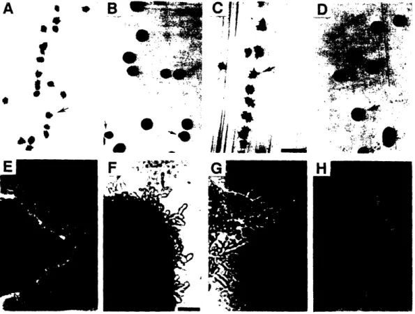
![Figure 6. Localization of Plasma Membrane IH+]ATPase (PMA1) in SHR3 and shr3 Strains](https://thumb-eu.123doks.com/thumbv2/123doknet/14246475.487593/72.933.138.640.185.760/figure-localization-plasma-membrane-atpase-pma-shr-strains.webp)