Abstract.
Breast cancer is the leading cause of
cancer-related death in women worldwide and a critical public
health concern. Here we investigated the anticancer
potential and effects of low-molecular-weight bridgehead
oxygen and nitrogen-containing spiro-bisheterocycles on
proliferation and apoptosis of the human breast cancer cell
lines MCF-7 and MDA-MB-231. The compounds feature a
hydantoin moiety attached to either diazole, isoxazole,
diazepine, oxazepine or benzodiazepine via the privileged
tetrahedral spiro-linkage. Treatment with compounds spiro
[hydantoin-isoxazole] and spiro [hydantoin-oxazepine]
resulted in a dose-dependent decrease of cell proliferation
and induction of apoptosis in both breast cancer cell lines,
whereas spiro [hydantoin-diazepine] was only active against
MDA-MB 231. Quantitative reverse transcription polymerase
chain reaction analysis showed up-regulation of murine
double minute 2 (MDM2), strictly p53-dependent, and
detected an increase in expression of pro-apoptotic caspase
3 and BCL2-associated X (BAX) genes in both breast cancer
cell lines expressing wild-type and mutant p53. In summary,
the results suggest that our compounds promote apoptosis of
breast cancer cell lines via p53-dependent and -independent
pathways.
Breast cancer is the most common invasive cancer in women
and the leading cause of cancer-related death in women
worldwide, making it an urgent public health issue (1).
Despite intensive research into new chemotherapeutic agents,
treatment is still characterized by unwanted side-effects and
the spread of drug resistance, and thus falls short of
expectations (2). Consequently, it is essential to pursue the
drug development effort to treat breast cancer cells.
Many spiro compounds play fundamental roles in
biological processes and have demonstrated therapeutic
properties. (3), especially the spiro-heterocyclic compounds,
which have shown promising results in chemotherapy of
various cancer types (4). Spiro compounds are usually a
structural system of two rings positioned orthogonally one
to the other due to the sp
3hybridization of the central spiro
carbon, which can be chemically adjusted by introducing
various heterocyclic motifs mimicking those found in
biomolecules, such as DNA and proteins, in order to increase
their ability to interact with biological systems. This very
rare spatial arrangement of spiro-bicyclic scaffolds is
characterized by high rigidity, preventing freely-rotating
bonds, which can also be configured with several essential
functional groups.
Spiro functionality also remains a primary structural tool
for creating powerful molecular antitumor and anticancer
agents used in latest-generation chemotherapy. Nature
provides outstanding spiro structures, as evidenced by the
discovery of new spiro-bicyclic triterpenoid models were
found to be cytotoxic against breast human cancer cell lines
(5). However, the low natural availability of spiro
compounds has prompted researchers to build on these
molecular templates to optimize novel spiro-based chemical
structures capable of significant bioactivity.
Correspondence to: Lamia Hamdan Ramdani, Scientific and Technical Research Center in Physico-Chemical Analysis CRAPC, BP 384, Industrial zone of Bou-Ismail, RP 42004, Tipaza, Algeria. E-mail: lamia_pharm@yahoo.fr
Key Words: Spiro-bisheterocycles, oxygen and nitrogen heterocycles, breast cancer, cell proliferation, apoptosis, p53, MDM2.
Effects of Spiro-bisheterocycles on Proliferation and
Apoptosis in Human Breast Cancer Cell Lines
LAMIA HAMDAN RAMDANI
1,2,3, OUALID TALHI
1,5, NADIA TAIBI
1,
LAETITIA DELORT
2,3, CAROLINE DECOMBAT
2,3, ARTUR SILVA
5, KHALDOUN BACHARI
1,
MARIE-PAULE VASSON
2,3,4and FLORENCE CALDEFIE-CHEZET
2,31
Scientific and Technical Research Center in Physico-Chemical Analysis (CRAPC), Tipaza, Algeria;
2Clermont University, University of Auvergne, UMR 1019, UNH CellulaR Micro-Environment,
Immunomodulation and Nutrition ECREIN, Clermont-Ferrand, France;
3
Research Center in Human Nutrition CRNH Auvergne, Clermont-Ferrand, France;
4Anticancer Center Jean-Perrin, Clermont-Ferrand, France;
5
Organic Chemistry, Natural Products and Agrifood (QOPNA),
main mediators regulating the cell cycle and induction of
apoptosis in response to cellular damage (9). p53 regulation
in the cell is essentially based on its negative regulator
murine double minute 2 (MDM2). Targeting p53–MDM2
interaction by using appropriate molecules offers an
attractive strategy for p53 activation (8) and a promising
approach for new anticancer therapies (10).
Previous studies have reported that spiro compounds are
potent antagonists of the p53–MDM2 interaction (6). Given
these biorelevant properties, spiro compounds make very
attractive targets for cancer treatment screening. Here we
provide an account of the evaluation of our previously
reported small molecules containing oxygen and nitrogen
spiro-bisheterocycles in terms of effects on in vitro
proliferation and apoptosis of human breast cancer cell lines
(MCF-7 and MDA-MB231). The wider aim is to identify
novel low-molecular-weight, easily-accessible synthetic spiro
compounds for cancer chemotherapy.
Materials and Methods
Chemistry. Spiro-bisheterocyclic compounds 2-7 (Table I; Figure 2) were chemically synthesized following our previously described procedure (11). Their preparation is based on the action of bisnucleophiles such as methylhydrazine, hydroxylamine, ethylenediamine, hydroxyethyleneamine and ortho-phenylenediamine on the key spiro[chromanone-hydantoin] (1) dyad, which results in a spiro-to-spiro ring transformation of the chromanone residue into new substituted spiro[hydantoin-diazole] (2), spiro[hydantoin-isoxazole] (3), spiro[hydantoin-diazepine] (4-5), spiro[hydantoin-oxazepine] (6) and spiro[hydantoin-benzodiazepine] (7) (Figure 2). During these chemical reactions, the hydantoin cycle is preserved, as confirmed by relevant 2D nuclear magnetic resonance and single-crystal X-ray diffraction studies. Our spiro-bisheterocycles (2-7) were produced in optimal 42-67% yields after chromatographic purification and recrystallization, thus affording high purity as checked by TLC showing a single spot.
Cell culture and treatment. MCF-7 and MDA/MB 231 human breast adenocarcinoma cell lines were purchased from the American Type Culture Collection (Molsheim, France). MCF-7 and MDA-MB-231 were grown in RPMI-1640 and L-15 medium (GIBCO, Invitrogen, Saint-Aubin, France), respectively. RPMI-1640 medium was supplemented with 10% heat-inactivated fetal bovine serum (FBS) (Eurobio, Courtaboeuf, France), 1% L-glutamine (2 mM) (GIBCO,
Invitrogen, Saint-Aubin, France), 0.5% gentamycin (50 mg/ml) (Fisher Scientific, Strasbourg, France) and 0.05% insulin (100 UI/ml). Fluoresceinisothiocyanate (FITC) conjugated to annexin V was purchased from BioLegend (San Diego, CA, USA) and propidium iodide (PI) was purchased from Biotium (Hayward, CA, USA). A 10 mM stock solution of compounds 2-7 was prepared in dimethylsulfoxide (DMSO) and stored at 4˚C. All compounds were added to cells at different concentrations. The DMSO concentration of controls was <0.1% (v/v) and 1 μM doxorubicin was used as a positive control.
Cell proliferation assay. Cell proliferation was assessed by resazurin assay. Human breast adenocarcinoma cell lines (1×10⁴/well) were
cultured in 96-well plates with complete media for 24 h. At the end of incubation, cells were treated with compounds 2-7 at concentrations of 0, 10, 25, 50 and 100 μM for 72 h at 37˚C under a 5% CO2atmosphere without replacing the medium. Control cells and
positive control cells were treated with 0.1% DMSO and 1 μM doxorubicin, respectively. After incubation, 200 μl of a 25 μg/ml solution of resazurin in medium was added to each well. Plates were incubated for 2 h at 37˚C in a humidified atmosphere containing 5% CO2. Fluorescence was then measured on an automated 96-well plate
reader (Fluorskan Ascent FL; Thermo Fisher Scientific, Wilmington, NC, USA) at 530 nm excitation wavelength and 590 nm emission wavelength. Under these conditions, fluorescence (OD value) was proportional to the number of living cells in the well. Cell
proliferation assays were performed at least six times for each cell line (in replicates of six wells per concentration tested) (12). DNA quantification using Hoechst 33342. 96-Well plates containing treated and control cells were taken out of freezer storage at −20˚C and left to defrost for 1 h at room temperature. Then, 100 μl of 0.01% sodium dodecyl sulfate was added to enable the DNA to come into contact with the Hoechst solution. After 1 h of incubation, plates were put back in the freezer at −80˚C for 1 h to trigger thermal shock. After defrosting for 2 h, 100 μl of Hoechst solution 33342 (30 μg ml–1) was added to each well. Plates were
placed under agitation for 1 h shielded from light. The reading of the fluorescence was made by Fluorskan Ascent FL (Thermo Fisher Scientific). Hoechst 33342 dye is a fluorescent nucleic acid which exhibits a maximum emission at 460 nm when bound specifically to double-stranded DNA. Data represent viable cells.
Annexin V–FITC/PI apopstosis assay. MCF-7 and MDA-MB 231 tumor cells (105cells/well) were treated with 100 μM of compounds
3, 4 and 6 for 72 h at 37˚C andwith 5% CO2, then washed with phosphate-buffered saline, recovered by centrifugation at 1,000 × g for 5 min at room temperature (RT; 25˚C), and resuspended in 40 μl Figure 1. Synthetic bicyclic compounds as anticancer agents: A: 5.5 oxindoles; B: 6.5.5 bicyclic lactams; and C: spiro-isoxazolidine.
Figure 2. Reaction pathway for the synthesis of spiro-bisheterocycles 2-7 (11). RT: Room temperature; DBU: 1,8-diazabicyclo[5.4.0]undec-7-ene; THF: tetrahydrofuran.
of annexin V binding buffer (140 mm NaCl, 10 mm HEPES/NaOH, 2.5 mm CaCl2). The suspension was stained with 5 μl of annexin
V-FITC and 5 μl of PI, then incubated for 15 min at RT in the dark. To the sample was added 250 μl of 1×phosphate-buffered saline, this was then centrifuged at 1,000 × g for 5 min at RT, and cells were resuspended in 50 μl of annexin V binding buffer. Stained cells were then analyzed on a Cellometer K2 image cytometer (Nexcelom Bioscience, Lawrence, MA, USA).
Isolation of RNA and real-time polymerase chain reaction. Total RNA was extracted from untreated and treated breast cancer cell lines, MCF-7 and MDA-MB-231, using Trizol and quantified on a NanoDrop spectrophotometer (Nanodrop 2000; Thermo Scientific, Waltham, MA, USA). Purity was estimated by 260/280 nm absorbance ratio. cDNA was synthesized using the High-Capacity cDNA Reverse Transcription kit (Applied Biosystems, Courtaboeuf, France). Quantitative real-time PCR analysis was performed using a SYBR Green PCR Master Mix (Applied Biosystems, Courtaboeuf, France) as follows: first step at 50˚C for 2 min, denaturation step at 95˚C for 10 min, then 40 cycles at 95˚C for 15 s and 60˚C for 1 min. In total, 10 ng of cDNA was added to 18 μl of reaction containing primers. Relative expression levels of p53, MDM2 and BCL2-associated X (BAX) were calculated using the 2−ΔΔCt method. Experiments were
repeated in triplicate. The primer sequences used are listed in Table II. Data analysis. Data are expressed as the mean±SEM of multiple experiments. Statistical significance was determined by Student’s t-test using SPSS software (ver. 18.0; SPSS Inc., Chicago, IL, USA).
Results
Effects of spiro-bisheterocycles 2-7 on MCF-7 and MDA-MB231
cell proliferation. In order to determine the impact of
spiro-bisheterocycles 2-7 on cell proliferation of human breast cancer
cell lines, MCF-7 and MDA-MB 231 cells were treated with
compounds 2-7 at different concentrations from 10 to
100 μM for 72 h. Cell proliferation was assessed after 2 h of
incubation with resazurin solution. Proliferation of MCF-7
human breast cancer cell line was significantly reduced with
compounds 3 and 6, with The half-maximal inhibitory
concentration (IC
50) corresponding to 42.3 μM (17.4 μg/ml) and
66.3 μM (30.8 μg/ml) (Figure 3), respectively. We also observed
significant inhibition of MDA-MB-231 cells (Figure 3) up to
67.9 μM (28.5 μg/ml) for 3 and 97.1 μM (42.7 μg/ml) for 6.
Proliferation of MDA-MB 231 cells was also significantly
reduced with compound 4, with IC
50of 44.6 μM (19.6 μg/ml),
but not of MCF-7 cells. On the other hand, treatment with
compounds 2, 5 and 7 did not induce a significant decrease in
cell proliferation in the two breast cancer cell lines. At a 100 μM,
compound 3 was the most effective, inducing a major
suppressive effect of 98.15% and 97.02% against MCF-7 and
MDA-MB 231 cells, respectively.
Hoechst staining confirmed results of the resazurin assay
and showed significant impact of compounds 3 and 6 on
MCF-7 and MDA-MB-231 tumor cell proliferation (Figure
4), with similar IC
50. Compound 4 induced tumor inhibition
at an IC
50of 42 μM inMDA-MB-231 cells.
Effects of spiro-bisheterocycles 3, 4 and 6 on apoptosis. In
order to determine the apoptotic efficacy of the
spiro-bisheterocycles, cells were treated with 100 μM of 3, 4 and
6 for 72 h (Figure 5), then collected and subjected to
Cellometer annexin FITC/PI apoptosis assay. Annexin
V-FITC
−/PI
−flagged viable cells, annexin V-FITC
+/PI
−flagged
early-stage apoptotic cells, and annexin V-FITC
+/PI
+flagged
late-stage apoptotic cells.
Analysis showed a sharp induction of apoptosis by
compounds 3 and 6 in MCF-7 cells and by 3, 4 and 6 in
MDA-MB-231 cells (Figure 5A). The rates of early and late
apoptosis were 9.5%, 85.4% and 71.3% in MCF-7 for
control, and compounds 3 and 6, respectively, and 5.9%,
88.1%, 79.4% and 66.4% in MDA-MB-231 cells for control,
and compounds 3, 4 and 6, respectively (Figure 5B).
Effects of spiro-bisheterocycles 3, 4 and 6 on p53, MDM2,
BAX and caspase 3 expression. In an effort to investigate
whether spiro compounds 3, 4 and 6 induce apoptosis of
breast cancer cell lines by targeting p53–MDM2 interaction,
we used quantitative reverse transcription-polymerase chain
reaction to analyze the wild-type p53, MDM2, BAX and
R: TGAGGGTCTCTCTCTTCCTCTTGTcaspase 3 gene expression levels. Results for MCF-7 breast
cancer cells show that compound 3 more clearly increased the
expression of wild-type p53 (6.707-fold vs. control) than did
compound 6 (3.275-fold vs. control) (Figure 6). We also found
that compounds 3 and 6 induced up-regulation of MDM2 in
MCF-7 cells at levels of 4.282 and 2.658, respectively.
However, in MDA-MB-231 p53-mutant breast cancer cells,
no significant changes in the levels of p53 and MDM2 were
Figure 3. Effect of compounds 2-7 on the in vitro growth and proliferation of human breast cancer cells. MCF-7 and MDA-MB231 cell line were either untreated, treated with 1 μM doxorubicin (Dox) or treated with 10, 25, 50 and 100 μM of compounds 2-7 for 72 h as described in the Materials and Methods section. Proliferation was assessed using resazurin on an automated 96-well Fluoroskan Ascent FL. Columns show means±SD, n=6. *Significantly different at p<0.05.observed with compounds 3, 4 and 6, indicating that
overexpression of MDM2 is under control of p53. Compared
to levels in untreated control MCF-7 cells, compounds 3 and
6 led to 6.738- and 3.532-fold higher BAX and 6.498- and
2.703-fold higher caspase 3 gene expression, respectively.
Interestingly, similar results were obtained in MDA-MB-231
cells, where an increase of the expression of caspase 3 and
BAX genes was also noted with compounds 3, 6 and, even
with compound 4. This demonstrates the capacity of our
compounds to induce apoptosis independently of p53 status.
Discussion
Tumor suppressor p53 plays a key role in the regulation of
cell cycle and apoptosis (13-15). We previously reported that
p53 inhibition by its negative regulator MDM2 is involved
in the onset of breast cancer (16, 17). An approach based on
restoring p53 activity by inhibiting p53–MDM2 interaction
could lead to tumor suppression and thus be a promising
strategy for future control of breast cancer (18, 19). Many
spiro compounds possess very promising biological and
pharmacological activities, especially antitumor and
antimicrobial properties (4, 20, 21).
These specific, potent, non-peptide small-molecule inhibitors
are reported to mimic the α-helix recognition motif of the p53–
MDM2 complex (22), thereby efficiently reactivating p53
tumor-suppressor activity and consequently inducing a
p53-mediated signaling pathway that will culminate in cell death
by apoptosis (23). Our results clearly showed that the
spiro-heterocyclic compounds substituted by isoxazole (3), diazepine
(4) and oxazepine (6) significantly reduced the proliferation of
MCF-7 and MDA-MB-231 cells in a dose-dependent manner.
These results were consistent with other studies demonstrating
the cytotoxicity of spiro-bicyclic compounds in human cancer
cell lines (5, 24). Our data are further supported by another
report which demonstrated similar in vivo anticancer potential
of a spiro-heterocyclic compound on the development on
MCF-7 human breast cancer cells (25). The antiproliferative
effect was more pronounced with spiro-isoxazole (3).
Structurally, the presence of the isoxazole heterocyclic unit is
likely tightly related to these clear antiproliferative effects, as
already reported for other spiro-models containing the
isoxazole motif (7). It was also observed that the presence of
oxazepine substituent demonstrated good activity against breast
cancer cells (26), thus explaining the antiproliferative effect
observed with compound 6. Furthermore, the significant
antiproliferative activity of compound 4 could stem from the
presence of a diazepine molecule in the structure, since
diazepine inhibits the adenosine A2B receptor involved in the
development of tumors and proliferation (27). In addition, a
Figure 4. Inhibitive effect of compounds 3, 4 and 6 on growth of cancer cell lines MCF-7 and MDA-MB-231. *Significantly different at p<0.05.previous study reported that adenosine A
2Breceptors are
expressed at high levels in the estrogen-negative
MDA-MB-231 cell line but not expressed in the estrogen-positive
MCF-7 cell line, which could explain why the antiproliferative effect
of compound 4 was observed here only in MDA-MB-231 cells
(28). Moreover, the presence of cyclohexyl fragments in these
three compounds might also contribute to their antiproliferative
activity (29), allowing better understanding of why compounds
5 and 7 had no effect.
Apoptosis plays a key role in the maintenance of
homeostasis (30) and is considered a primary form of cancer
cell death (31). This pathway results in DNA fragmentation,
degradation of cytoskeletal and nuclear proteins, formation of
apoptotic bodies, and finally uptake by phagocytic cells (32).
Recent reports indicate that most spiro compounds induce
apoptosis of cancer cells (33, 34), but especially
spiro-isoxazolidine derivatives (35). Here, we performed annexin
V-FITC/PI apoptosis assays and demonstrated that our
spiro-bisheterocyclic compounds induced apoptosis of breast cancer
cell lines. Annexin V is a protein that interacts strongly and
specifically with phosphatidylserine. During early apoptosis,
phosphatidylserine translocates to the external leaflet and can
be used for the detection of apoptosis (36). Analysis using
Cellometer K2 image cytometry indicated that compounds 3,
4 and 6 induced a large proportion of breast cancer cell death
by apoptosis in vitro. The strong effect was observed with
compound 3 in both breast cancer cell lines.
As previously demonstrated (18), the most likely
mechanism by which spiro compounds initiate apoptosis of
wild-type p53 breast cancer cells is by inhibition and
disruption of the p53–MDM2 interaction, leading to
accumulation of p53 and up-regulation of MDM2. The tumor
suppressor p53 binds to DNA and mediates transcriptional
activation which ultimately overexpress BAX and promote
Figure 5. Drug-induced apoptosis in breast cancer cell lines MCF-7 and MDA-MB-231 at 72 h of incubation with spiro-bisheterocyclic compounds. A: Bright-field (top row), and fluorescent images of annexin V-fluorescein isothiocyanate (FITC) (middle row) and propidium iodide (PI) (bottom row) staining. The images show significant induction of apoptosis with compounds 3 and 6 in MCF-7cells, while strong apoptosis was observed with compounds 3, 4 and 6 in MDA-MB-231 cells. B: Quantitative results of percentage of viable, and early apoptotic and late apoptotic cells after treatment with compounds. *Significantly different at p<0.05.p53-dependent cell apoptosis (37, 38). The pro-apoptotic
BAX protein is one of the most important regulators of the
intrinsic pathway of apoptosis (39). Excess of BAX protein
leads to formation of BAX–BAX homodimers, that stimulate
release of cytochrome c from mitochondria and activate
caspase-3, then inducing apoptosis (40).
Quantitative reverse transcription-polymerase chain
reaction data support these findings and showed an increase
of wild-type p53 expression in MCF-7 cells, accompanied
by up-regulation of MDM2, with treatment of compound 3
and 6. We extended this study by demonstrating that spiro
compounds 3 and 6 enhance expression of caspase-3 and
Figure 6. Results of polymerase chain reaction detection. p53, murine double minute 2 (MDM2), BCL2–associated X (BAX) and caspase-3 genes expression in MCF-7 and MDA-MB-231 cells following treatment with 100 μM of compounds 3, 4 and 6 for 72 h. *Significantly different at p<0.05.BAX in MCF-7 wild-type p53 breast cancer cells, and
consequently induce apoptosis. At the same time, in order to
determine whether our spiro compounds induce apoptosis
regardless of cell p53 status, we also employed
MDA-MB-231 cells which harbor mutant p53. Treatment with
compounds 3, 4 and 6 had no significant effect on expression
of p53 or MDM2 in cancer cells lacking wild-type p53 but
increased caspase-3 and BAX expression. These results
correlate with a previous study revealing induction of
apoptosis through p53-independent pathways (41).
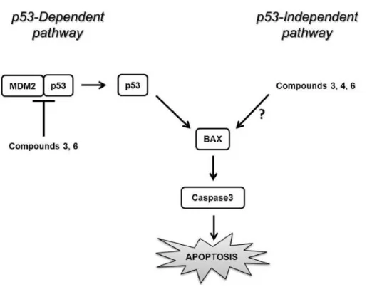
In summary (Figure 7), this study brings the first
demonstration that new small spiro-bisheterocyclic
molecules can sensitize breast cancer cells to apoptosis by
targeting p53–MDM2 interaction. We also highlighted that
these spiro compounds promote apoptosis via
p53-independent pathway(s), suggesting that these compounds
represent interesting candidates as therapeutic targets for the
treatment of breast cancer.
Acknowledgements
The Authors thank the Unité de Nutition Humaine UMR 1019 INRA-UdA – Equipe ECREIN (France) and the University of Aveiro, Fundação para a Ciência e a Tecnologia (Portugal), EU, QREN, FEDER, COMPETE, for funding this biological and organic chemistry research. They also thank Algeria’s General Directorate for Scientific Research and Technological Development (DGRSDT) for financial support.
References
1 Ferlay J, Shin HR, Bray F, Forman D, Mathers C and Parkin DM: Estimates of worldwide burden of cancer in 2008. Int J Cancer 127: 2893-2917, 2010.
2 Rokhlin OW, Gudkov AV, Kwek S, Glover RA, Gewies AS and Cohen MB: p53 is involved in tumor necrosis factor-alpha-induced apoptosis in the human prostatic carcinoma cell line LNCaP. Oncogene 19: 1959-1968, 2000.
3 El-Hashash MA, Rizk SA and Atta-Allah SR: Synthesis and regioselective reaction of some unsymmetrical heterocyclic chalcone derivatives and spiro heterocyclic compounds as antibacterial agents. Molecules 20: 22069-22083, 2015. 4 Miao Y, Juan X, Jing-Jing Z, Jun-Lin H, Yun Z, Hui F, Yang GX,
Xia G and Hu JF: Leonurusoleanolides E−J, minor spirocyclic triterpenoids from Leonurus japonicus fruits. J Nat Prod 77: 178-182, 2014.
5 Moujir LM, Seca AM, Araujo L, Silva AM and Barreto MC: A new natural spiro heterocyclic compound and the cytotoxic activity of the secondary metabolites from Juniperus brevifolia leaves.Fitoterapia 82: 225-229, 2011.
6 Ding K, Lu Y, Nikolovska-Coleska Z, Qiu S, Ding Y, Gao W, Stuckey J, Krajewsk,i K, Roller PP, Tomita Y, Parrish DA, Deschamps JR and Wang S: Structure-based design of potent non-peptide MDM2 inhibitors. J Am Chem Soc 127: 10130-10131, 2005.
7 Das P, Omollo AO, Sitole LJ, McClendon E, Valente EJ and Raucher D: Synthesis and investigation of novel spiro-isoxazolines as anti-cancer agents. Tetrahedron Lett 56: 1794-1797, 2015. 8 Vassilev LT: MDM2 inhibitors for cancer therapy. Trends Mol
Med 13: 23-31, 2007.
13 El-Deiry WS: Regulation of p53 downstream genes. Semin Cancer Biol 8: 345-357, 1998.
14 Levine AJ: p53, the cellular gate keeper for growth and division. Cell 88: 323-331, 1997.
15 Vogelstein B, Lane D and Levine AJ: Surfing the p53 network. Nature 408: 307-310, 2000.
16 Rayburn E, Zhang R, He J and Wang H: MDM2 and human malignancies: expression, clinical pathology, prognostic markers, and implications for chemotherapy. Curr Cancer Drug Targets 5: 27-41, 2005.
17 Vassilev LT, Vu BT, Graves B, Carvajal D, Podlaski F, Filipovic Z, Kong N, Kammlott U, Lukacs C, Klein C, Fotouhi N and Liu EA: In vivo activation of the p53 pathway by small-molecule antagonists of MDM2. Science 303: 844-848, 2004.
18 Shangary S, Qin D, McEachern D, Liu M, Miller RS, Qiu S, Nikolovska-Coleska Z, Ding K, Wang G, Chen J, Bernard D, Zhang J, Lu Y, Gu Q, Shah RB, Pienta KJ, Ling X, Kang S, Guo M, Sun Y, Yang D and Wang S: Temporal activation of p53 by a specific MDM2 inhibitor is selectively toxic to tumors and leads to complete tumor growth inhibition. Proc Natl Acad Sci USA 105: 3933-3938, 2008.
19 Shangary, S and Wang S: Targeting the MDM2–p53 interaction for cancer therapy. Clin Cancer Res 14: 5318-5324, 2008. 20 Rompaey KV, Ballet S, Tömböly C, Wachter RD,
Vanommeslaeghe K, Biesemans M, Willem R and Tourwé DA: Synthesis and evaluation of the β-turn properties of 4-amino-1,2,4,5-tetrahydro-2-benzazepin-3-ones and of their spirocyclic derivative.Eur J Org Chem 13: 2899-2911, 2006.
21 Rosy P, Manabendra P, Behera, AK, Mishra BK and Behera RK: A synthon approach to spiro compounds. Tetrahedron 62: 779-828, 2006.
22 Somu RV and Johnson RL: Synthesis of pipecolic acid-based spiro bicyclic lactam scaffolds as β-turn mimics. J Org Chem 70: 5954-5963, 2005.
23 Shangary S and Wang S: Small-molecule inhibitors of the MDM2–p53 protein–protein interaction to reactivate p53 function: a novel approach for cancer therapy. Annu Rev Pharmacol Toxicol 49: 223-241, 2009.
24 Manner S, Oltner VT, Oredsson S, Ellervik U and Frejd T: Spiro-bicyclo[2.2.2]octane derivatives as paclitaxel mimetics. Synthesis and toxicity evaluation in breast cancer cell lines. Org Biomol Chem 7: 7134-44, 2013.
25 Chin YW, Salim AA, Su BN, Mi Q, Chai, HB, Riswan S, Kardono LB, Ruskandi A, Farnsworth NR, Swanson SM and Kinghorn AD: Potential anticancer activity of naturally occurring and semisynthetic derivatives of aculeatins A and B from Amomum aculeatum. J Nat Prod 71: 390-395, 2008.
Lead optimization of 2-cyclohexyl-N-[(Z)-(3-methoxyphenyl/3-hydroxyphenyl) methylidene]hydrazine-carbothioamides for targeting the HER-2 overexpressing breast cancer cell line SKBr-3. Molecules 20: 18246-18263, 2015.
30 Norbury CJ and Hickson ID: Cellular responses to DNA damage. Annu Rev Pharmacol Toxicol 41: 367-401, 2001. 31 Elmore S: Apoptosis: A review of programmed cell death.
Toxicol Pathol 35: 495-516, 2007.
32 Martinvalet D, Zhu P and Lieberman J: Granzyme A induces caspase-independent mitochondrial damage, a required first step for apoptosis. Immunity 22: 355-370, 2005.
33 Molvi KI, Haque N, Awen BZS and Zameeruddin M: Synthesis of spiro compounds as medicinal agents; new opportunities for drug design and discovery. World J Pharm Sci 3: 536-563, 2014. 34 Yu B, Shi XJ, Qi PP, Yu DQ and Liu HM: Design, synthesis and biological evaluation of novel steroidal spiro-oxindoles as potent antiproliferative agents. J Steroid Biochem Mol Biol 141: 121-134, 2014.
35 Khazir J, Singh PP, Reddy DM, Hyder I, Shafi S, Sawant SD, Chashoo G, Mahajan A, Alam MS, Saxena AK, Arvinda S, Gupta BD and Kumar HM: Synthesis and anticancer activity of novel spiro-isoxazoline and spiro-isoxazolidine derivatives of α-santonin. Eur J Med Chem 63: 279-289, 2013.
36 Sean RS, Arthur CM, Ju T, Rodrigues LC, Riul TB, Dias-Baruffi M, Miner J, McEver RP and Cummings RD: Galectin-1 induces reversible phosphatidylserine exposure at the plasma membrane. Mol Biol Cell 20: 1408-1418, 2009.
37 Li B and Dou QP: BAX degradation by the ubiquitin/ proteasome-dependent pathway: involvement in tumor survival and progression. Proc Natl Acad Sci USA 97: 3850-3855, 2000. 38 Scheffner M, Werness BA, Huibregtse JM, Levine AJ and
Howley PM: The E6 oncoprotein encoded by human papillomavirus types 16 and 18 promotes the degradation of p53. Cell 63: 1129-1136, 1990.
39 Williams GT and Smith CA: Molecular regulation of apoptosis: genetic controls on cell death. Cell 74: 777-779, 1993. 40 Kurabayashi A1, Furihata M, Matsumoto M, Ohtsuki Y, Sasaguri
S and Ogoshi S. Expression of BAX and apoptosis-related proteins in human esophageal squamous cell carcinoma including dysplasia. Mod Pathol 14: 741-747, 2001.
41 Lanni JS, Lowe SW, Licitra EJ, Liu JO and Jacks T: p53-independent apoptosis induced by paclitaxel through an indirect mechanism. Proc Natl Acad Sci USA 94: 9679-9683, 1997.
![Figure 2. Reaction pathway for the synthesis of spiro-bisheterocycles 2-7 (11). RT: Room temperature; DBU: 1,8-diazabicyclo[5.4.0]undec-7-ene;](https://thumb-eu.123doks.com/thumbv2/123doknet/14117874.467204/3.892.125.786.115.318/figure-reaction-pathway-synthesis-spiro-bisheterocycles-temperature-diazabicyclo.webp)