Blood-brain barrier model on a microfluidic chip for
the study of tumor cell extravasation
By
Cynthia Hajal
B.S., Mechanical Engineering
B.A., Economics
Columbia University, 2016
Submitted to the department of Mechanical Engineering in Partial
Fulfillment of the Requirements for the Degree of Master of Science in
Mechanical Engineering
At the
Massachusetts Institute of Technology
June 2018
0 2018 Massachusetts Institute of Technology. All rights reserved.
Signature of Author:
Certified by:
Accepted by:
MASSACH SL I OF TECHNOL(JUN 2
5 2
LIBRARI
Signature redacted
Department of Mechanical Engineering
May 9, 2018
Signature redacted
Roger D. Kamm
Professor of Mechanical and Biological Engineering
Thesis Supervisor
____Signature
redacted
NSTITUTE
-ohan Abeyaratne
)GY
Professor of Mechanical Engineering
018
Chairman, Committee for Graduate Students
Blood-brain barrier model on a microfluidic chip for the study of
tumor cell extravasation
By Cynthia Hajal
Submitted to the Department of Mechanical Engineering on May 9, 2018 in Partial Fulfillment of the Requirements for the Degree of Master of Science in Mechanical Engineering
Abstract
With up to 40% of cancer patients showing metastatic lesions to the brain and a 30% five-year survival rate post-diagnosis, secondary tumors to the brain are a leading cause of cancer-related deaths'. Understanding the mechanisms of tumor cell extravasation at the brain is therefore crucial to the development of therapeutic agents targeting this step in cancer metastasis, and to the overall improvement of cancer survival rates2. Investigating the interactions between tumor cells and brain stroma is of particular interest due to the site's unique microenvironment. In fact, the interface between brain and blood, known as the blood-brain barrier (BBB), is the tightest endothelial barrier in humans'4. The presence of tight junctions between brain endothelial cells, coupled with the spatial organization of pericytes and astrocytes around the vasculature, restrict the entry of most solutes and cells into the brain',6. Yet, the brain constitutes a common metastatic site to many primary cancers originating from the lung, breast and skin'. This suggests that tumor cells must employ specific mechanisms to cross the blood-brain barrier'. While in vitro models aimed at replicating the human blood-brain barrier exist, most are limited in their physiological relevance. In fact, the majority of these platforms rely on a monolayer of human brain endothelial cells in contact with pencytes, astrocytes and neurons9-". While this approach focuses on incorporating the relevant cell types of the brain microenvironment, it fails to accurately replicate the geometry of brain capillaries, the barrier tightness of the BBB, and the juxtacrine and paracrine signaling events occurring between brain endothelial cells and stromal cells during vasculogenesis5. To integrate these features into a physiologically relevant blood-brain barrier model, we designed an in vitro microvascular network platform formed via vasculogenesis, using endothelial cells derived from human induced pluripotent stem cells, primary human brain pericytes, and primary human brain astrocytes. The vasculatures formed with brain pericytes and astrocytes exhibit decreased cross-section areas, increased endothelial cell-cell tight junction expression and basement membrane deposition, as well as reduced and more physiologically relevant values of vessel permeability, compared to the vasculatures formed with endothelial cells alone. The addition of pericytes and astrocytes in the vascular system was also coupled with increased extravasation efficiencies of different tumor cell subpopulations, despite the lower permeability values measured in this BBB model. Moreover, an increase in the extravasation potential of metastasized breast tumor cells collected from the brain was recorded with the addition of pericytes and astrocytes, with respect to the parental breast tumor cell line. These results were not observed in metastasized breast tumor cells collected from the lung, thus validating our BBB model and providing useful insight into the role of pericytes and astrocytes in extravasation. Our microfluidic platform certainly provides advantages over the current state-of-the-art in vitro blood-brain barrier models. While being more physiologically relevant than most in vitro platforms when it comes to geometry, barrier function and juxtacrine/paracrine signaling between the relevant cell types, our model provides a robust platform to understand tumor cell-brain stromal cell interactions during extravasation. Thesis supervisor: Roger D. Kamm
Title: Professor of Mechanical and Biological Engineering
Acknowledgements
I would like to thank my advisor, Professor Roger Kamm, for his continuous support and valuable scientific
insight throughout my time at MIT. I am very grateful for all the opportunities he has given me, to pursue my own research and scientific interests and to collaborate with other groups. Thanks to his mentorship, I was able to explore new avenues of research, learn valuable skills in the lab, and start to contribute to the scientific world.
I would also like to thank my labmates in the Kamm group for helping me throughout my learning phase
in the lab and providing me with useful advice throughout my two years as a Masters student. I would especially like to thank Dr. Michelle Chen and Dr. Alexandra Boussommier-Calleja for sharing their valuable expertise in tumor cell extravasation assays using microvascular networks, Dr. Yoojin Shin for sharing her knowledge and insight on microfluidics and blood-brain barrier modelling, Dr. Kristina Haase, Dr. Giovanni Offeddu, Dr. Tatsuya Osaki, Jean-Carlos Serrano-Flores, Lina Ibrahim, and Anya Roberts for asking me challenging yet constructive bioengineering questions that have guided me throughout my project. I am also grateful for my mentors and collaborators from other labs, Dr. Leanne Li and Dr. Fred Lam, who have provided me with useful insight on the biological aspect of my work.
I would like to thank my family for their never-ending love, support, and advice. They have helped me
navigate countless challenges I have encountered in the past 7 years I spent living abroad. My parents have always been my idols and remind me every day that hard-work and an honest work ethic will always allow me to achieve my goals. My brother has continuously supported me in stressful times with his thoughtful words of encouragement and advice. I would also like to thank my fianc6, Christian, for always standing
by my side even during intense periods of work in the lab and for bringing happiness into my life every
day.
Finally, I would like to dedicate this thesis to my two grandfathers, Salim Haddad and Assaad Hajal, who left us early. I am thankful that progress is being made in this community to further the general understanding of diseases and push forward novel treatments where they are needed.
This work is supported by the National Cancer Institute (UOI CA202177).
Table of Contents
CH APTER 1: INTRODUCTION... 7
1.1 THE BLOOD-BRAIN BARRIER ... 7
1.2 METASTATIC CASCADE AND EXTRAVASATION AT THE BRAIN... 8
1.3 STATE-OF-THE-ART BLOOD-BRAIN BARRIER MODELS AND EXTRAVASATION ASSAYS AT THE BRAIN... 11
1.4 OBJECTIVE AND THESIS OVERV IEW ... 14
CHAPTER 2: IN VITRO BLOOD-BRAIN BARRIER MICROVASCULAR NETWORK FO RM ATION ... 17
2.1 INTRODU CTION ... ... 17
2.1.1 Bridging the gap: from in vivo brain models to in vitro BBB microvascular networks... 17
2.1.2 Blood-brain barrier formation and stabilization in vivo and in vitro... 18
2.2 M ETHODS ... 19
2 .2 .1 A ssay d esign ... 19
2.2.2 Cells and device preparation...21
Microfluidic devicefabrication ... 21
C e ll cu ltu re ... 2 1 Gel fabrication and cell seeding ... 22
2.2.3 Fluorescent imaging and analysis ... 23
2.2.4 Calculation of vessel permeability...23
2.2.5 Microvascular network metrics ... 24
2.3 RESULTS ... 26
2.3.1 Vessel formation and stability ... 26
2.3.2 M icrovascular network metrics and morphology ... 29
2.3.3 Vessel permeability measurements...32
2.3.4 Tight junction protein expression and basement membrane deposition ... 33
2.4 DISCU SSION ... 36
CHAPTER 3: TUMOR EXTRAVASATION AT THE BRAIN FROM IN VITRO BLOOD-BRAIN BARRIER MICROVASCULAR NETWORKS ... 37
3.1 INTRODUCTION ... 37
3.2 M ETHODS ... 38 4
3.2.1 Blood-brain barrier microvascular network formation and maintenance ... 38
3.2.2 Tumor cell perfusion in the microvascular networks... 38
3.2.3 Tumor cell extravasation quantification ... 39
3.2.4 Barrier permeability measurements following tumor cell extravasation ... 39
3.3 RESULTS ... 40
3.3.1 Tumor cell perfusion and extravasation in the BBB microvascular networks... 40
3.3.2 Extravasation potential of metastasized tumor cells collected from different organs ... 43
3.3.3 Effect of tumor cell extravasation on barrier permeability ... 45
3.4 DTSCUSSION ... 47
CHAPTER 4: CONCLUSIONS ... 49
REFERENCES... 50
List of Figures and Tables
Figure 1. The Blood-Brain Barrier... 7
Table 1. Localization and role of most notable TJ and AJ proteins at the BBB...8
Figure 2. The m etastatic cascade... 9
Figure 3. Outlook on brain metastases ... 10
Figure 4. State-of-the-art blood-brain barrier models ... 12
Figure 5. Current extravasation assays at the brain employing endothelial monolayers .... 13
Figure 6. Extravasation assays employing 3D self-organized HUVEC vasculatures ... 14
Figure 7. Device schematic and platform conditions... 21
Figure 8. Microvascular network quantification metrics ... 25
Figure 9. Microvascular networks formed in the three culture conditions... 27
Figure 10. Vessel stability over time... 29
Figure 11. Microvascular network metrics at day 7 in culture ... 31
Figure 12. Morphological changes with the addition of PCs and ACs ... 32
Figure 13. Permeability measurements in the three culture conditions ... 33
Figure 14. Tight junction protein expression ... 34
Figure 15. Basement membrane deposition in three culture conditions... 35
Figure 16. Visualization of MDA-MB-231 and A549 extravasation events... 40
Figure 17. Extravasation efficiencies of MDA-MB-231 over time ... 41
Figure 18. Extravasation efficiencies of A549 over time ... 42
Figure 19. Extravasation potential of breast parental tumor cells (MDA-Parental) and metastasized tumor cells collected from the brain (MDA-BrM) and lung (MDA-LM)... 45
Figure 20. Effect of tumor cell extravasation on barrier permeability... 46
Chapter 1: Introduction
1.1 The Blood-Brain Barrier
The BBB is a selective interface between microvasculature and brain parenchyma formed by the endothelial cells that line the cerebral microvessels, brain pericytes enclosed within the basal lamina, and astrocytes touching their end-feet to the abluminal side of the vessels". It is described as the tightest endothelial barrier in humans, thanks to the presence of tight junctions (TJs) and adherens junctions (AJs) between adjacent endothelial cells5. Intercellular gaps measuring 3 to 6 nm were recorded between endothelial cells of the brain in mice, significantly smaller in size than the gaps measured in other endothelial cells (-10 nm)13
. The TJs and AJs characteristic of the gaps between neighboring brain
endothelial cells are known to be the main obstacles to paracellular transport across the BBB, forcing most solutes (of size larger than -6 nm) to travel via transcellular routes by melding into cell membrane "holes", via receptor-mediated or adsorptive-mediated transcytosis, or with the help of carriers, such as GLUT 1 for glucose transport and LATI for amino acid transport.
General capiay Interoellular cleft Fenestra KEnnothebaM A Brain capoillary
ffncran T~t Juncnn illono
En ce & eal En UBlooda
Figure 1. The Blood-Brain Barrier. Brain capillaries are constituted of endothelial cells lining the microvessels,
brain pericytes enclosed in a basement membrane, and astrocytes touching their glial feet on the abluminal side of the vessels. The presence of tight junctions and adherens junctions closes the intercellular clefts between adjacent endothelial cells at the brain14'15.
Most notable components of TJ and AJ proteins at the BBB, as well as their localization and role, are listed below:
Type of Junction protein Localization in Role
junction brain endothelial
cells
Occludin Membrane Promote redox-sensitive processes of
TJ assembly
Claudins Membrane Homodimerize with claudins on
adjacent endothelial cells
Tight junctions JAMs Membrane Facilitate assembly of TJ components and recruit polarity complex
Zonula Cytoplasm Bind to claudins and JAMs via PDZ
Occludens (ZOs) domain and to occluding via other
regions to sustain TJ integrity
Adherens VE-cadherin Membrane Promote actin bundling
junction N-cadherin Membrane Regulate angiogenesis Table 1. Localization and role of most notable TJ and AJ proteins at the BBB"-"
In addition to forming an obstacle to solutes, the BBB also prevents the passage of most cells into the brain parenchyma. However, certain circulating mononuclear cells at the brain, such as leukocytes, monocytes and macrophages, have been shown to undergo diapedesis and cross the barrier via both paracellular and transcellular routes20. During inflammatory pathology, trafficking of leukocytes across the BBB is highly upregulated. In these cases, several studies suggest that transcellular diapedesis seems to be the preferential route employed by leukocytes to cross the BBB, leaving TJs and barrier function undisrupted-1 7 2 1 2 2
. Brain pericytes seem to play a role in neutrophil chemoattraction and transmigration, by secreting interleukin-8 (IL-8) and matrix metalloprotease-9 (MMP-9) in response to inflammation characterized by tumor necrosis factor-alpha (TNF-a), IL-l
IP,
orlipopolysaccharide
(LPS) expression.Although solute transport and cell migration across the BBB are highly limited, several tumor cells originating from specific primary sites metastasize preferentially to the brain2
1. The supporting role of the
BBB in metastasis formation is still not fully elucidated but several findings suggest a potential role for pericytes and astrocytes in promoting tumor cell extravasation and homing at the brain2 .
1.2 Metastatic cascade and extravasation at the brain
As cancer cells multiply uncontrollably at a specific site, a primary tumor mass forms and self-vascularizes as tumor cells start secreting pro-angiogenic factors29. These primary tumors are, however, only responsible for 10% of cancer-related deaths. The remaining 90% of deaths is attributed to metastasis, the spread of cancer cells from primary tumors to surrounding tissues and distant organs30
The initial phase of the metastatic cascade involves the loss of adhesion molecules, such as E-cadherin, and cytokeratins, which leads to the detachment of carcinoma cells from the primary tumor mass and their acquisition of a motile phenotype. In the vascularized tumor microenvironment, MMP-expressing cancer cells are able to digest the basement membrane surrounding the primary tumor site to come in contact and intravasate into the circulation". The circulating tumor cells (CTCs) must then travel to distant sites or tissues via the bloodstream, while escaping immune surveillance. CTCs are able to arrest in the microvasculature of the secondary site via mechanical entrapment in the capillary beds due to size restriction or via adhesion to the vascular endothelium32. A fraction of these adhered or entrapped tumor cells may extravasate through the endothelial walls and colonize the secondary site. The overall metastatic process from intravasation to colonization is highly inefficient - only about 0.01% of CTCs actually lead to the formation of tumors at the secondary site, mainly due to the failure of these single cells to escape immune surveillance and reach the distant site to initiate growth, and to the failure of early micrometastases to continue growth into potentially fatal macrometastases 29,33.
Primary tumour Vascularization Detachment Intravasation
0 0 0 0
Circulating Adhesion to Growth of
tumour cet blood vessel wall Extravasation secondary tumour
Figure 2. The metastatic cascade. Cancer cell progression from the primary tumor site to the secondary site for colonization.
While the general mechanisms underlying tumor cell extravasation have been examined both in vivo and in physiologically relevant in vitro platforms, the precise mechanisms governing extravasation at the brain, on the other hand, remain poorly understood2 4 263 4. It has been observed that melanoma cells undergo
brain, on the other hand, remain poorly understood242634. It has been observed that melanoma cells undergo paracellular transendothelial migration of tumor cells across the BBB, where TJs were disrupted and barrier integrity was damaged35. Transcellular migration of tumor cells at the brain is possible, given the fact that TJs seal the intercellular route, and several mononuclear cells have been shown to cross the BBB via the transcellular route20. However, this phenomenon has yet to be detected either in in vivo animal models or in in vitro systems36.
Gathering data from several hundreds of patients showing metastatic lesions at the brain, a few primary cancers have been identified to preferentially metastasize to the brain. Around 40-50% of non-small cell lung cancer patients have been shown to develop brain metastases, followed by breast cancer patients at -20% and melanoma patients (-10%)'. These findings have raised questions regarding the specific genes and underlying mechanisms governing the behavior of cancer cells from different primary sites. Although certain genes in breast cancer have been identified to play a role in preferential brain metastatic colonization, additional studies are necessary to fully elucidate the physical mechanisms and signaling pathways activated in different primary cancer cells crossing the BBB37.
Table 1 Pcmxntafcs of brain metastascs fmm diffirmnt primay cancers
Primary MSKCC [II* Nussbawum t al. Counscl t a. [64 Lageoaard ct al, Stark ci a!. [II R Fabi et al. [12-1
cancer 1994; s=210 [19 Q996;. n=729 1996; n=214 [23]. 1999; a-1292 2011; n=309 2011; - =29) Lung 41% 39% 53% 56% 50% NSCLC 35% 24% 42% 44% SCLC 6% 15% ... 8% Breast 19% 17% 13% 16% 15% 30% Melanoma 10% 11% 8% 7% 6% Renal 3% 6% 2% 4% -- -GI 7% 6% -. - 9% Coloreal 4% 3% - 8% -Unknown 2% 5% 14% 8%
-Figure 3. Outlook on brain metastases. Lung primary cancer cells preferentially metastasize to the brain, followed
by breast primary cancer cells and melanoma cells'.
The specific BBB microenvironment allows for distinct cellular and molecular interactions. It is not surprising that extravasating tumor cells interact closely with the endothelium, but also with brain pericytes and astrocytes located on the abluminal side of the vasculature. It has been observed that extravasated tumor cells at the brain maintain close contact with the abluminal side of the endothelium, where they can benefit from the protection of the BBB against anti-tumoral immune cells circulating in the brain, and the support of the basement membrane lining the vasculature20 3 . In the context of brain metastasis, two subpopulations
of pericytes have been identified in connection with tumor cell extravasation. CD 13-positive pericytes have been linked with normal BBB function and barrier integrity, while desmin-positive pericytes were found in brain metastases where BBB permeability is significantly increased. CD13+ pericytes could transform into desmin+ pericytes in the presence of tumor cells, thus impairing barrier function and promoting extravasation, however, desmin+ pericytes might have originated from their own expansion, the bone marrow or potentially endothelial cells during endothelial-mesenchymal transion2 7
. In addition, several studies suggest that astrocytes might promote cancer invasion and survival at the brain. It has been observed that astrocytes are activated in the vicinity of tumor cells, even prior to their extravasation. These reactive astrocytes, characterized by up-regulated glial fibrillary acidic protein (GFAP) expression, secrete
MMP-9, promoting tumor cell extravasation and invasion into the matrix26,39. Tumor cells have also been shown
to activate the production of TNF and interferon (IFN)-a in astrocytes, which in turns supports brain metastasis via the STAT1 and NF-K1 pathways25. While the exact mechanisms and cellular interactions between tumor cells and brain stromal cells have not been fully elucidated, evidence seems to suggest that brain pericytes and astrocytes play a role in promoting tumor cell extravasation. Recapitulation of tumor cell arrest and extravasation in a highly controlled and physiologically relevant brain microenvironment would then provide useful insight into the precise mechanism of action of pericytes and astrocytes, and the molecular alterations associated with tumor cell extravasation across the BBB. This assay would therefore be crucial to the development of therapeutic agents targeting this step in cancer metastasis, and to the overall improvement of cancer survival rates2.
1.3 State-of-the-art blood-brain barrier models and extravasation assays at the
brain
Most extravasation studies at the brain are performed in animal models. These in vivo studies allow for the investigation of specific genes and proteins involved in tumor cell extravasation at the brain25,37,40,41
Although these models offer a highly physiologically relevant platform of study where most in vivo responses can take place, significant challenges associated with the translation of experimental results in animal brains to conclusive outcomes in human brains persist. While most in vivo models are limited to time-point data collection, real-time imaging of tumor cell extravasation at the brain has been performed in mice; however, the study required the use of immunodeficient mice, which may have perturbed the tumor cell response, and success rates were low (-50%), which resulted in low throughput experiments 42 (Fig.
4A). Controlling for specific parameters in animal studies, as well as observing the underlying mechanisms
In an attempt to circumvent these limitations, several groups have focused their efforts on developing
in vitro platforms replicating the human BBB physiology. The most commonly used in vitro model relies
on the use of a Transwell chamber, where human brain cells can be cultured in 2D in a simple high-throughput platform4 3-47
(Fig 4B). While these platforms offer an additional degree of physiological relevance with the incorporation of human cells from the brain, they do not allow real time imaging of the entire extravasation process and fail to fully replicate the geometry and spatial cellular organization of the BBB microvasculature.
Moving away from 2D models, assembled 3D monolayer systems have been adopted to more closely recapitulate the geometry of brain capillaries. In these systems, 3D channels are fabricated in microfluidic devices where cells can be cultured as a monolayer coating the inner cavity layer. Several groups have adopted this technique to recreate geometrically relevant platforms where brain endothelial cells coat the inner channel walls and brain stromal cells can be cultured in adjacent fluidic channels or in a gel on the abluminal side of the assembled endothelial "vessels"'1'4849 (Fig. 4C). Assembled 3D monolayer systems allow for greater control of the vessel geometry as well as the spatial organization of brain stromal cells and neurons around the vasculature. However, they fail to fully replicate 3D self-assembled vasculatures in terms of branching, diameter sizes, barrier tightness and activated signaling pathways during vasculogenesis between the relevant brain cell types".
AB
C
E
EndoheW CsSAstrocytes SNeurons/Pedcytes
10,11,2510,11,24
Figure 4. State-of-the-art blood-brain barrier models. (A) In vivo models of mice brains employed for the study of tumor cell extravasationl,42. (B) Transwell assay model of the BBB: brain endothelial cells are cultured on a 2D membrane with astrocytes on the opposite side of the membrane, and pericytes and neurons at the bottom of the well'0.
(C) Assembled 3D monolayer model of the BBB: a liquid flow is used to create a cavity inside a collagen gel where
brain endothelial cells are subsequently plated. Pericytes and astrocytes can be cultured prior to the addition of brain endothelial cells in order to replicate the cellular spatial organization of the BBB".
-1
Only a few studies of extravasation at the brain have been performed in microfluidic in vitro platforms, as opposed to in vivo animal models. Most of these assays rely on the use of a 2D monolayer of brain endothelial cells, adjacent to a layer of brain astrocytes or pericytes. These platforms validated the trends of Fig. 4, suggesting that lung (A549), breast (MDA-MB-231) and melanoma (M624) cancer cells preferentially migrated across the brain endothelium, as opposed to liver (BEL-7042) cancer cells5 1'5 2.
Endowelialcalls derived
*om cord blood
hent7oei dern
cals
ECU ... >Brainn pefttes Blood * brain tum ce *,ls metastac cels nlow
Figure 5. Current extravasation assays at the brain employing endothelial monolayers. Lung, breast and melanoma tumor cells have been shown to preferentially cross the 2D brain endothelium adjacent to astrocytes or
pericytes5 ,5 2.
Recently, microfluidic technologies employing 3D microvascular "networks" have been used to model tumor cell extravasation across a physiologically relevant endothelial barrier". Along with others, our group has grown these microvascular networks from the self-organization of human umbilical vein endothelial cells (HUVECs) in a hydrogel, along with several stromal cell types, such as normal human lung fibroblasts (NHLFs), placental pericytes, osteoblasts and mesenchymal stem cells (MSCs), in the context of tumor migration assays3 453-5 . These platforms have provided tremendous insight into the specific mechanisms of tumor cell extravasation, as well as the different signaling pathways activated and molecular alterations occurring during transmigration and colonization of cancer cells. For instance, Chen
et al. examined the roles of tumor integrin
P1
in extravasation using an in vitro platform with HUVEC microvascular networks and NHLFs in adjacent channels. While in vivo models do not allow to fully elucidate the role ofP1
in the different steps of metastatic dissemination, the in vitro platform employed demonstrated that tumor cells send activated 1-rich protrusions past the endothelium to engage with subendothelial matrix, particularly laminin, via a3pl and a6P1 integrins5 6. Similarly, Jeon et al. demonstrated that tumor cell extravasation rates were significantly higher in the presence of human bone marrow-derived MSCs and osteo-differentiated MSCs. By investigating the effects of adenosine, known to be secreted in the skeletal muscle microenvironment, on metastasizing tumor cells in this controlledplatform, it was observed that extravasation rates were reduced, confirming the anti-metastatic and protective role of skeletal muscle cells54.
MDA-MB-231 HUVEC Intrava.cular
A B
Transmigratng
.2~
-
U..._-
hrhFuly franwmnrat-2 h
Figure 6. Extravasation assays employing 3D self-organized HUVEC vasculatures. (A) 3D microvascular networks formed with HUVECs and perfused with breast MDA-MB-23 1. Cross-sectional views of the transmigration steps of a single tumor cell are shown over time'. (B) 3D microvascular networks formed with HUVECs, with osteo-differentiated MSCs in an adjacent channel, and perfused with bone seeking breast MDA-MB-23 1, extravasating over
54
time
While these 3D self-organized HUVEC vasculatures offer a general, more physiologically relevant platform for the study of tumor cell extravasation than 2D assembled monolayer models, they do not fully recapitulate different organ specificities, in terms of cell types incorporated, organ-specific barrier tightness, vessel morphology, and paracrine/juxtacrine signaling5 4'57'58. In the context of modelling the BBB on a chip for tumor cell extravasation studies, there remains a need for the development of 3D vasculatures in vitro, incorporating the advantages offered by microfluidic technologies, as well as the physiological relevance pertaining to incorporated cell types, cellular interactions and localizations, barrier function and protein
expression.
1.4 Objective and thesis overview
The objective of this project is to incorporate the unique features of the BBB in current 3D in vitro microfluidic platforms, in an attempt to design a physiologically relevant model that can be employed for the study of tumor cell extravasation at the brain. While current brain planar models and 2D assembled monolayer systems aim to incorporate most relevant brain cell types, they fail to replicate the appropriate geometry of brain vasculature, and exhibit suboptimal barrier function. On the other hand, state-of-the-art
3D vasculature systems do not fully encompass organ-specific characteristics, in terms of vascular
monolayer systems aim to incorporate most relevant brain cell types, they fail to replicate the appropriate geometry of brain vasculature, and exhibit suboptimal barrier function. On the other hand, state-of-the-art
3D vasculature systems do not fully encompass organ-specific characteristics, in terms of vascular
architecture, cellular interactions and protein expression. Most tumor cell extravasation studies are currently performed in in vivo animal models. However, these are restricted in their physiological relevance to humans, and in their ability to perform controlled, high-throughput studies. The lack of physiologically relevant 3D vascularized BBB models for the study of tumor cell extravasation may limit our understanding of the mechanisms, cellular interactions and signaling pathways involved in this step of brain metastasis formation. Our goal is then to build an in vitro BBB-specific vascularized extravasation assay that allows for high-throughput capture of extravasation events across physiologically relevant BBB vasculatures, exhibiting low permeabilities when compared to those of animal brains, and human in vivo-like vessel morphology and cellular interactions.
The first aim of this project, discussed in Chapter 2, is to establish in vitro perfusable BBB-like vasculatures that can be used for the study of tumor cell extravasation at the brain, across this barrier. This can be achieved by developing 3D microvascular networks (MVNs) composed of physiologically relevant endothelial cells and stromal cells to recapitulate the brain microenvironment. The MVNs should be stable in terms of morphology and barrier function, and should exhibit permeabilities similar to those measured in animal brains. Tumor cells must be able to flow through the vascular networks and their arrest, transmigration and colonization should be observed in the in vitro platform. The MVNs must be able to incorporate relevant brain cell types successively, in order to examine the underlying effect of each cell type on tumor cell extravasation. The platform should allow for a high level of throughput in order to produce several devices per condition (at least 15 devices per condition per experimental repeat).
Different conditions pertaining to the incorporated cell types are examined in terms of morphology, barrier tightness, and overall physiological relevance. By progressively incorporating different cell types specific to the brain, this assay determines the roles of each one of them when it comes to vascular parameters and barrier function.
In Chapter 3, the platform employed for tumor cell extravasation studies across the BBB is described. It consists of endothelial cells derived from human induced pluripotent stem cells (iPS-ECs) in juxtacrine co-culture with brain primary pericytes (PCs) and brain primary astrocytes (ACs). The platform is validated previously by tracking vessel stability over time with and without the different brain stromal cells, as well as vessel morphology, barrier tightness, EC tight junction protein expression, and basement membrane deposition after vascular stability is achieved. This assay is then used to understand the underlying mechanisms of tumor cell extravasation, including the differences in extravasation efficiencies over time
in different vasculatures progressively exhibiting BBB features, the disparity in extravasation patterns of distinct tumor cell lines originating from various primary sites, and the correlation of preferential homing site with extravasation ability. With this platform, we aim to elucidate the mechanisms underlying extravasation across a 3D BBB vasculature in vitro, as well as shed the light on potential cellular interactions and factors that may favor extravasation at the brain.
Chapter 2: In vitro blood-brain barrier microvascular network formation
2.1 Introduction
2.1.1 Bridging the gap: from in vivo brain models to in vitro BBB microvascular networks
The unique microenvironment of the brain, its central role in vital functions, as well as the severity of brain disorders, have been drivers in the use and development of brain assays59 .60 In order to fully
understand the complexities of the brain microenvironment and to isolate specific features, cellular interactions and signaling pathways characteristic of certain brain disorders, there is a tremendous need for engineered vascularized brain tissue models.
Currently, the majority of in vitro 3D vascularized assays rely on the self-organization of endothelial cells (ECs) into microvascular networks in a hydrogel. These assays can be achieved in two ways - ECs seeded into a matrix of collagen or fibrin can come in contact and self-organize into an in vivo-like vasculature via a process called vasculogenesis, or ECs sprout into a gel, following angiogenic stimuli, to form vessels which split into new ones via angiogenesis 61. The vasculatures formed through either process are mature, interconnected tubular networks with patent lumens, allowing for the perfusion of drugs, dyes and other cells in order to replicate physiological responses and perform in vitro studies.
In the context of tissue engineering models, most assays employ generic HUVECs for vascularization, owing to their robustness, homogenous phenotype, as well as ease of access and comparison with state-of-the-art models6 2. To generate organ-mimicking microenvironment and investigate the role of organ-specific stromal cells in biological responses, several platforms also incorporate stromal cells in the matrix, in addition to the ECs. For instance, bone- or muscle-specific microenvironment were recapitulated by co-culturing HUVECs with osteo-differentiated MSCs or myoblasts, respectively54
. While these organ-mimicking microfluidic models aim to replicate closely the in vivo tissue, there remain limitations in the use of HUVECs for vascularization. While stromal cells play an important role in several biological events, such as the metastatic cascade, immune cell infiltration or the development of certain diseases, organ-specific endothelial cells still remain the principal barrier that cells and drugs come in contact with when circulating in the vasculature63'6.
3D vascularized networks formed with organ-specific ECs are extremely rare in current models. Instead,
a few in vitro platforms rely on human induced pluripotent stem cells (iPSCs) and embryonic stem cells (ESCs), which can be differentiated into cells possessing endothelial properties, to develop vasculatures in
microfluidic devices65-67
. These iPS-ECs or ESC-ECs are non-organ specific, and thus provide the advantage of being further differentiated into ECs possessing the biological characteristics and phenotype of organ-specific ECs. For instance, Lippmann et al. have generated human iPS- and ESC-derived brain
microvascular endothelial cells (BMECs) by co-differentiation of neural and endothelial progenitors67
.
Various differentiation protocols have been developed to obtain generic ECs from iPS and ESC, or organ-specific ECs from iPS-ECs and ESC-ECs. Most differentiation procedures into generic ECs rely on treatments with specific growth factors or metabolites 68. In order to obtain organ-specific ECs from stem cells or stem cell-derived ECs, current protocols rely on the use of growth factors and on the co-culture of iPS-ECs or ESC-ECs with stromal cells found in the microenvironment of the organ of interest6
9
71. Several studies have determined a prominent role for brain pericytes, astrocytes and neurons in differentiating iPSCsand ESCs into ECs with BBB characteristics 68,72
-74
.
2.1.2 Blood-brain barrier formation and stabilization in vivo and in vitro
In addition to promoting iPSCs and ESCs differentiation into brain-like ECs, primary brain pericytes and astrocytes also play a significant role in BBB formation and stabilization, both in vivo and in vitro75'76. The development of the BBB in vivo begins with angiogenesis, where pre-existing vessels sprout into the neuroectoderm producing vascular endothelial growth factor (VEGF). These sprouts already exhibit certain BBB properties, such as the expression of TJs and transporters. These barrier properties mature further as the endothelial sprouts come in contact with pericytes and astrocytes77
.
Specifically, pericyte-endothelial cell interactions are mediated by bidirectional signaling between transforming growth factor-P (TGF-3) and its receptor. This dictates the upregulation of endothelial cadherin-2 which in turn leads to firm adhesion between endothelial cells and pericytes. The pericytes then produce angiopoietin I (Ang-1) which promotes vascular development, and deposit extracellular matrix
(ECM) components, contributing to basement membrane formation. Astrocytes, on the other hand, release
Sonic Hedgehog (Shh) involved in development and cell division. These interactions between brain stromal cells and endothelial cells lead to the development of more advanced TJs, loss of leukocyte adhesion molecules and inhibition of transcytosis, all characteristics of a mature BBB78.
The role of pericytes and astrocytes has also been investigated in vitro. In the case of 2D monolayer models, the addition of astrocytes has been shown to decrease barrier permeability and increase transendothelial electrical endothelial resistance (TEER) in the presence of flow47 '1. Furthermore, Hatherell et al. detccted increased TEER values in Transwell assays where BMECs were co-cultured with astrocytes
or pericytes on the abluminal side of the membrane. However, their tri-culture systems of BMECs with
both pericytes and astrocytes, culturing one of the stromal cell on the bottom of the dish and the other on the abluminal side of the membrane, yielded lower TEER values than the co-culture platforms. They concluded that juxtacrine cellular contacts between BMECs, pericytes and astrocytes are needed to induce proper barrier function in brain endothelial cells76. In addition, Wang et al. were able to identify in vivo-like permeability values of
10-7-10-8
cm/s for FITC-dextrans ranging from 4 to 70kDa in their 2D monolayer model constituted of human iPS-ECs (IMR90-4) differentiated to brain microvascular ECs, and rat primary astrocytes. In the case of 3D assembled monolayer models, both Adriani et al. and Herland et al. demonstrated improved barrier function when BMECs lining hollow 3D channels were co-cultured with pericytes, astrocytes, or neurons1'49.While these results provide useful insight into the different cellular interactions, activated signaling pathways and proteins involved in BBB development, there remain limitations in the platforms used for these studies. The aim of this project is to engineer a tightly controlled BBB model, incorporating self-organized 3D vasculatures with relevant brain cell types, and displaying physiological barrier function and morphology, for the study of tumor cell extravasation at the brain.
2.2
Methods
2.2.1 Assay design
In order to develop an in vitro vascularized BBB extravasation assay meeting the requirements listed in Chapter 1, we employ microfluidic tools to recapitulate the brain microenvironment50. Using a microfluidic
device for cell culture applications, we generate perfusable 3D human iPS-ECs microvascular networks assembled via vasculogenesis, surrounded by brain stromal cells (pericytes and astrocytes). The procedure for HUVEC vascular network formation via vasculogenesis has been previously established and employed in our group for various assays pertaining to tumor cell extravasation with and without immune cells, interstitial flow applications, and ECM-cellular interactions455'80.
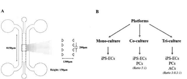
In this study, we employ a vasculogenesis device based on previous designs from our group. The polydimethylsiloxane (PDMS) device consists of a single gel region for cell culture, lined by media channels which can all be independently filled by either gels or media. The interface between the single gel region and the media channels consists of micro-posts facing towards the central channel, allowing for surface tension-assisted filling. All the cells employed in the model are cultured in the single, central gel region where they can interact with soluble factors in the media via diffusion. A schematic of the device
used is described in Fig. 7. The single central channel is
1300[tm
wide and 150pm high, and the length is 8150pm. The gaps between micro-posts are 200ptm.To develop 3D vascularized networks, we use a similar vasculogenesis approach based on earlier models in our lab, in which HUVECs were seeded as a suspension in a central gel channel and normal human lung fibroblasts (NHLFs) were seeded in adjacent channels 34
,
81
. In this project, however, the assay described employs iPS-ECs instead of HUVECs, co-cultured with brain primary pericytes and astrocytes in the same gel region instead of NHLFs in side channels. This different approach carries several advantages:
a. the contact culture of non-organ specific iPS-ECs with brain stromal cells allows for a certain degree of additional differentiation of the iPS-ECs into brain-specific ECs, as mentioned in differentiation protocols relying on co-cultures of iPS-ECs and ESC-ECs with organ-specific stromal cells69-7
b. the use of iPS-ECs with brain pericytes and astrocytes renders the vascular platform more
physiologically relevant than generic HUVEC models when it comes to re-creating the BBB on-a-chip
c. the platform is more suitable for brain-specific assays, such as the study of brain disorders or drug delivery, where the unique features of the BBB are required to shed light on certain mechanisms of interest
When seeded into the central gel channel of the device, iPS-ECs spontaneously form into 3D interconnected lumens, open to the side channels in the inter-post regions. When co-cultured with brain pericytes and astrocytes, iPS-ECs form interconnected vascular networks, pericytes wrap around the vasculature and astrocytes extend their end-feet to touch the abluminal side of the vessels. The different cells cultured in fibrin gel spontaneously adopt the appropriate spatial organization to re-create the morphology and characteristic features of BBB vasculatures. The networks formed are perfusable with cells, beads and dyes from the media channels on either side of the vasculature region.
Platforms
D 1 Mono-culture Co-culture Tri-culture
SI~Opm~
1
~0
ITII
iPS-ECs iPS-ECs iPS-ECs
Im PCs PCs
(
H,: (Ratio 5:1) ACsH h (Ratio 5:0.5:1)
Figure 7. Device schematic and platform conditions. (A) The microfluidic device used consists of a single central gel region for cell seeding. The micro-posts separating the gel region from the adjacent media channels allow for surface tension-assisted filling. When iPS-ECs (with or without brain stromal cells) are seeded into the central channel, they spontaneously re-organize into 3D microvascular networks, perfusable with tumor cells from the media channels. (B) Three different platforms were considered: a mono-culture of iPS-ECs alone used as a baseline for comparisons, a co-culture of iPS-ECs and brain pericytes, and a tri-culture of iPS-ECs, brain pericytes and brain astrocytes to fully recapitulate the BBB microenvironment. Ratios of cell densities for the different cell types included are indicated for each platform.
2.2.2 Cells and device preparation
Microfluidic device fabrication
Microfluidic devices were fabricated using soft lithography, as described in a previous protocol 2 Polydimethylsiloxane (PDMS, Ellsworth Adhesives, MA) was mixed at a 10:1 ratio of base to curing agent and poured over a silicon master. The PDMS poured in the mold was incubated for at least 2 hours at 70C before being cut from the master, punched, trimmed and autoclaved in water first and then dry autoclaved. The PDMS slabs were then incubated overnight in a 70C oven. Once cooled, the PDMS slabs and No. I glass coverslips were plasma treated for
1
minutes and 30 seconds (Harrick Plasma, Ithaca, NY) and bonded together. The devices were each coated with 40pL of 1mg/mL poly-D-lysine in water (Sigma-Aldrich, Burlington, MA) and incubated at 37C, 5% CO2 for at least 2 hours. The devices were then washed three times with sterile water and left at 70C for at least 24 hours before use.Cell culture
Induced pluripotent stem cell-derived endothelial cells (iPS-ECs, Cellular Dynamics International, Madison, WI) were cultured on fibronectin coated flasks (30mg/mL fibronectin in water for 2 hours) in Vasculife (Lifeline Cell Technology, Frederick, MD) growth media, supplemented with 50mL of fetal
21
bovine serum (FBS) and I0mL of L-glutamine, according to the protocol developed by Cellular Dynamics International (CDI). Primary brain penicytes (PCs, ScienCell, Carlsbad, CA) and astrocytes (ACs, ScienCell) were cultured on poly-L-lysine (PLL) coated flasks (0.15ptg/mL PLL in water for 2 hours). PCs were grown in pericyte media (ScienCell) and ACs in astrocyte media (ScienCell). All cells were used at passage 5.
Gel fabrication and cell seeding
Fibrinogen solution was prepared by dissolving 30mg of bovine fibrinogen (Sigma-Aldrich) in 5mL of PBS in a 37C water bath for 2-3 hours. The solution was sterile filtered using a 0.2im filter and stored at 4C until use. iPS-ECs (CDI, WI) grown in Vasculife (Lifeline Cell Technology) growth media were lifted from the flasks using TrypLE IX (Thermo Fisher Scientific, Waltham, MA). PCs and ACs were trypsinized for lifting. For the mono-culture model, iPS-ECs were resuspended at 10 million cells per mL in a solution of EGM-2 (Lonza, Basel, Switzerland) with thrombin (4 U/mL, Sigma-Aldrich). The cell suspension was then mixed in a 1:1 ratio with IOptL of 6mg/mL of fibrinogen to achieve a final seeding density of 5 million cells per mL when injected directly into the central gel channel of the device. In the co-culture model, iPS-ECs were resuspended at 20 million cells per mL, and PCs at 4 million cells per mL, in the EGM-2-thrombin solution, before being seeded directly into the central channel. The final concentrations for the co-culture model are 5 million cells per mL of iPS-ECs and
1
million cell per mL of PCs providing a 5-to-l cell seeding ratio of ECs to PCs, similar to cell ratios quantified in freshly isolated mice brain capillaries". Finally, for the tri-culture model (BBB model with ECs, PCs and ACs), iPS-ECs were resuspended at 30 million cells per mL, PCs at 3 million cells per mL, and ACs at 6 million cells per mL for final seeding densities of 5 million cells per mL for the iPS-ECs, 0.5 million cells per mL for the PCs, and1
million cells per mL for the ACs (Fig. 7B). The final seeding density of PCs was reduced in the tni-culture system (0.5 million cells per rnL) when compared to the co-culture system (1 million cells per mL) to account for their increased propensity to proliferate in the fibrin gel in the presence of ACs. The seeded devices were placed in a Petri dish (which includes 5mL of water in a smaller dish for humidity) and left in the 37C, 5% CO2incubator for 30 minutes until the fibrinogen gel cured completely. The different devices were filled with the same iPS-EC media, supplemented with 50ng/mL of VEGF-A for the first four days of culture (Peprotech, Rocky Hill, NJ), and placed back in the incubator at 37C and 5% CO2. VEGF-A was
supplemented to promote vasculogenesis in the early stages of cell seeding in the microfluidic channel. From day 4 onward, Vasculife media (Lifeline Cell Technology) without any additional growth factors was used.
To perform permeability studies, a monolayer of iPS-ECs was seeded in the media channels of the device on Day 2 of the culture to prevent dye diffusion into the matrix. Briefly, after seeding the devices, the media channels were coated with 60ptg/mL of human plasma fibronectin (EMDmillipore, Burlington, MA) and incubated at 37C and 5% CO2 for 30min. The fibronectin was then removed and iPS-ECs were
perfused in one media channel side at 1.5 million cells per mL of EGM-2 (Lonza). The device was tilted to the side for 10 minutes to allow the cells to settle at the media-gel channel interface. This procedure was repeated with the other media channel side to allow for the formation a homogenous monolayer of ECs on both sides of the gel. The devices were incubated for 2 hours at 37C and 5% CO2 prior to changing the media and adding Vasculife (Lifeline Cell Technology) with 50ng/mL of VEGF-A (Peprotech).
2.2.3 Fluorescent imaging and analysis
To visualize microvascular network formation throughout the 7 days of culture, live devices were
imaged using phase contrast microscopy (Nikon, Japan). Confocal microscopy (Olympus, Japan) was used to study and visualize end-point network morphology on day 7, at which point the devices were fixed with 4% PFA for 15 minutes and permeabilized with 0.01% Triton X-100 for 5 minutes. Devices were then blocked with 4% w/v BSA (Sigma-Aldrich) and 0.5% v/v goat serum (Gibco) in PBS overnight at 4C. Following washing, primary antibodies were added at 1:200 in PBS and devices were incubated overnight at 4C on a shaker. Following another wash, devices were incubated with secondary antibodies (Invitrogen, Carlsbad, CA) at 1:200 in PBS overnight at 4C on a shaker. If needed, devices were also counterstained with Phalloidin (1:200, Invitrogen) for actin and DAPI (1:1000, Invitrogen) for nuclei. All fluorescent images were obtained via confocal microscopy and processed using the IMARIS imaging software (Bitplane, Belfast, Ireland).
2.2.4 Calculation of vessel permeability
To assess the permeability of the vasculatures formed, fluorescent tracers were perfused in the networks,
based on a protocol described previousl 484
The media in all reservoirs was aspirated and the top reservoir
on one side was injected with 5.7pL of PBS. The top reservoir on the other side was subsequently injected with 6piL of 0.4mg/mL I OkDA MW or 40kDa MW FITC-dextran (Sigma) in PBS. The volume difference on either side of the gel channel allowed for dextran convection and diffusion through the microvascular networks. The devices were then placed in the confocal microscope environmental chamber for imaging. Once equilibrium was established and the intensity inside the vessels (Iv) remained constant, fluorescent
images were captured every 3 minutes for 15 minutes. The permeability coefficient was calculated by obtained the average intensity in a measured z-stack containing both vasculature and surrounding gel at the initial and final timepoints. .
One ROI was imaged per device and n=6 devices per vasculature condition (mono-culture, co-culture or tri-culture) were selected for analysis. Prior to performing permeability studies, a monolayer of
iPS-ECs
was seeded in the media channels of the device, as mentioned above, to prevent diffusion of dextran into the gel matrix. Care was taken to select vessel segments in the central region of the gel channel, farthest away from the media-gel channel boundaries. Vessel permeability coefficients P, (cm/s) were computed as:
1 (t2 - I) V (It' - I)
At
Asurfacewhere
I'
and I2 are the tissue intensities at initial (ti) and final (t2) timepoints, If' is the initial vesselintensity, At is the time interval between initial and final timepoints (seconds), V is the vasculature volume in the region considered (cm3
), and Asurface is the 3D surface area of the vessels (cm2
). All imaging was performed using a confocal microscope (Olympus) and timepoint images were analyzed using ImageJ
(NIH, Bethesda, MD).
2.2.5 Microvascular network metrics
Several parameters were employed to quantify vascular network formation and stabilization. During the first seven days of culture until stabilization, the average vessel branch length, the number of branches per device and the average lateral diameter of the microvascular networks were computed in each culture condition (mono-, co-, and tri-culture). At day 7, once the networks had formed and were stable, the number of branches per vascularized area, the average transverse diameter, and the average cross-section area of the vessels were computed, in addition to the parameters listed above. For each metric quantified, n=15 devices were considered per culture condition.
A branch is defined as the portion of a vessel between two intersecting points (Fig. 8A depicts 7
branches). Since most vessels are oriented in a plane parallel to the glass substrate, lateral (parallel to the glass) and transverse (perpendicular to the glass) diameters can be computed. Lateral diameters are obtained as the ratio of the projected lateral vessel area to the total branch length per ROT. Transverse diameters are computed using the 3D vessel volume and the surface area of the vessels in 3D (Fig. 8B describes the difference between lateral and transverse diameters for each culture condition). During stabilization, images
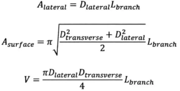
were captured using phase contrast microscopy (Nikon, Japan) to compute the parameters listed above. At day 7, devices were fixed and stained, and z-stack images obtained via confocal microscopy (Olympus, Japan) were used to quantify the different vascular parameters with ImageJ (NIH, MD). The equations used to compute the diameters and average cross-section area of the vessels are listed below:
Alaterai = DIateralLbranch
Aslrf ace - LDtransverse + D12ateral
Asr c
r2
LbranchS= lDiateraDtransverse Lbranch
Using the three equations above for 2D lateral and 3D surface areas, as well as 3D volume, the lateral and transverse diameters, as well as the cross-section area can be computed as described below:
Diaterai =Aaterai Lbranch DIateral Dtransverse = A2 2 AsurfaceDlateral Across-section =tDtransverseDiaterai 4 A A 2 B /, B Schematic 0077of an elliptical vessel 1 4 Transverse diameter
S (perpendicular to device glass)
Lateral diameter
6 7 (parallel to device glass)
Figure 8. Microvascular network quantification metrics. (A) Depiction of a vessel skeleton containing 7 branches. (B) Schematic of an elliptical vessel cross-section where the major axis (horizontal) refers to the lateral diameter and the minor axis (vertical) refers to the transverse diameter.
2.3 Results
2.3.1 Vessel formation and stability
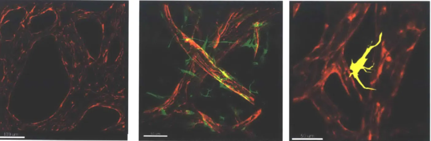
Three different platforms were employed for the study of BBB-like microvascular network formation. In the mono-culture model, iPS-ECs alone reorganized into microvascular networks as early as 5 days after seeding, and remained stable until day 7. Networks were fixed and stained on day 7 with anti-CD31 for iPS-ECs, anti-platelet derived growth factor receptor (PDFR)-o for PCs, and anti-glial fibrillary acidic protein (GFAP) for ACs (Abcam, MA). Image analysis reveals networks spanning over the entire gel region with wide lumens and elliptical vessel cross-sections (Fig. 9A). In the co-culture model, iPS-ECs formed stable and perfusable microvascular networks on day 7, while PCs spontaneously wrapped around the vessels on their abluminal side, as they would in the brain in vivo. Confocal imaging shows a high degree of branching and narrower lumens when compared to the mono-culture case (Fig. 9B). Via image analysis, it can be observed that the microvascular networks formed in the tri-culture platform possess a BBB-like morphology, similar to the one shown in Fig. 1. iPS-ECs spontaneously reorganize into highly interconnected vasculatures with the narrowest lumens of all three culture conditions. PCs coat the abluminal side of the vessels, while ACs are observed to stretch their end-feet to touch several branches of the networks (Fig. 9C).
Figure 9. Microvascular networks formed in the three culture conditions. (A) Mono-culture of iPS-ECs after 7
days in culture. The vasculature covers most of the gel area, with elliptically-shaped lumen cross-sections. iPS-ECs are stained with anti-CD31 (red) and DAPI (blue). (B) Co-culture of iPS-ECs and PCs after 7 days in culture. PCs wrap around the narrow vessels, on their abluminal side, as they would in the brain in vivo. iPS-ECs are stained with anti-CD31 (red) and PCs are stained with anti-PDGFR-s (green), indicative of the recruitment of PCs to the vessel wall. (C) Tri-culture of iPS-ECs with PCs and ACs after 7 days in culture (PCs not shown in this image). ACs extend their end-feet to touch the abluminal side of the vasculature. Cellular organization is similar to the one observed in the
in vivo BBB. iPS-ECs are stained with anti-CD31 (red) and ACs are stained with GFAP (yellow), characteristic of
ACs.
Live, daily tracking of the same devices for each culture condition was performed using phase contrast microscopy (Nikon, n= 15 devices for each culture condition). In the first 6 days of culture, the average branch length steadily increased in all three conditions from 80pm on day 2 to -160-170pm on day 6. However, on day 7 in the case of the co-culture and tri-culture conditions, the average branch length remained constant at -160-170pm, while that of the mono-culture condition kept increasing to 210 m. At the same time, the average number of branches per device decreased over time. At day 6, this number of branches stabilized for the co-culture and tri-culture conditions and remained equal to an average of -60 branches per device at day 7. This was not observed in the mono-culture condition, where the average number of branches per device kept decreasing from day 6 to day 7, reaching a final average value of 30 branches per device. These observations suggest that PCs and ACs, when co-cultured with iPS-ECs, can promote increased vascular branching, while reducing branch length, to achieve a more stable and physiologically relevant vasculature. Finally, the average lateral diameter was computed using live devices as well. The tri-culture condition exhibited the lowest average lateral diameter of all conditions (-35pm), followed by the co-culture condition with an average lateral diameter of 60pm. The mono-culture platforms exhibited very large lumens with an average lateral diameter of 125pm. This confirms that both PCs and ACs are required to achieve capillary-like vasculatures with narrower lumens (Fig. 10).
Mono-culture Co-culture Tr-culture 2 3 4 5 6 Day B 7 180- 160-E 140 -- 120-CID loo-80. E z 60- 40- 20-A -a- Mono-culture - Co-culture Tri-culture -- --
~-
- --2 3 4 5 6 7 Day Day 3 Day 5 Mono-culture Mono-culture Day 7 Mono-culture - Mono-culture - Co-culture - Tri-culture 2 3 4 5 6 7 Day 240-a 220200 - 180- 160- 140-C 120 - 100-oC
140- 120-S100 -15 so- S60- 40- 20-D OwE
Day3
Day5
Co-culture
Co-culture
Day 7
Co-culture
F Day 3 Tri-culture Day 5 Ii-culture Day 7 7T.-cultureFigure 10. Vessel stability over time. (A) Average branch length increases in all three culture conditions and
stabilizes at day 6 for the co- and tri-culture systems. It increase further in the mono-culture condition until day 7. (B) The average number of branches per device decreases in all conditions and stabilizes at day 6 for the co- and tri-culture models. It decreases further in the mono-culture model until day 7. (C) The tri-culture condition exhibits the lowest average lateral diameter, followed by the co-culture condition and mono-culture condition with the highest lateral diameter. (D-F) Phase contrast microscopy images of networks in the mono-, co-, and tri-culture systems respectively, at days 3, 5 and 7. Lateral diameters are shown to increase over time, at a faster and higher rate for the mono-culure system compared to the tri-culture one. Average number of branches decreases over time. These observations all suggest higher interconnectivity and branching, as well as narrower vessels in the tri-culture model. The vasculatures formed with PCs and ACs are more physiological in terms of morphology.
2.3.2 Microvascular network metrics and morphology
At day 7, once vessel stability was reached in most culture conditions, devices were fixed and stained, and z-stack images were obtained using confocal microscopy (Olympus). The stacks were analyzed using ImageJ (NIH) to compute the average branch length, the number of branches per vascularized area, the
3
IM
job,