The heart and other organs
The heart and the gut
Gerhard Rogler
1*
and Giuseppe Rosano
21
Division of Gastroenterology and Hepatology, Department of Internal Medicine, University Hospital Zurich, Raemistrasse 100, Zurich 8091, Switzerland; and2
Department of Medical Sciences, IRCCS San Raffaele, Via della Pisana 235, Roma 00161, Italy
Received 12 November 2012; revised 13 June 2013; accepted 18 June 2013; online publish-ahead-of-print 17 July 2013
This paper was guest edited by Roberto Ferrari, Department of Cardiology and LTTA Centre, University Hospital of Ferrara and Salvatore Maugeri Foundation, IRCCS, Lumezzane, Italy.
The heart and the gut seem to be two organs that do not have much in common. However, there is an obvious and clinically relevant impact of gut functions on the absorption of drugs and oral therapies on the one hand. On the other hand, the gut determines the quantity of nutrient uptake and plays a central role in metabolic diseases. Patients with inflammatory bowel diseases appear to have a higher risk for coronary heart disease despite a lower prevalence of ‘classical’ risk factors, indicating additional links between the gut and the heart. However, they certainly have a ‘leaky’ intestinal barrier associated with increased permeability for bacterial wall products. An impaired intestinal barrier function will be followed by bacterial translocation and presence of bacterial products in the circulation, which can contribute to atherosclerosis and chronic heart failure (CHF) as recent data indicate. Impaired cardiac function in CHF vice versa impacts intestinal microcirculation leading to a barrier defect of the intestinal mucosa and increased bacterial translocation. These pathways and the most recent insights into the impact of the gut on acute and chronic heart disease will be discussed in this review.
-Keywords Intestinal microbiome † Intestinal barrier † Bacterial translocation † Atherosclerosis † Chronic heart failure
The gut and its impact on heart
diseases
The gut certainly is not the first organ we would think about when we consider the pathophysiology of heart diseases. However, its basic functions, digestion, and absorption are obviously clinically relevant for almost all oral drug treatments of diseases.
The absorption of drugs from the small intestine is altered in its kinetics in patients with Crohn’s disease or celiac disease.1Patients with undetected celiac disease or with inconsequent diet have a decreased expression of some cytochrome P450 (CYP) isoenzymes such as CYP3A.2CYP3A is constitutively expressed in small intestinal villi and contributes to an important pre-hepatic metabolism of a number of drugs. Already in the intestine, CYP3A mediates the oxidative biotransformation of various clinically important drugs.3 Macrolide antibiotics (which will be discussed in another role further below) are important inhibitors of CYP3A.3 Statins have been reported to increase CYP3A isoenzymes expression4and, on the other hand, are metabolized by them.4CYP3A4 and CYP3A5 metabolize statins and thus have been demonstrated to influence the pharmacokinetics, efficacy, and safety of statins,4indicating that
small intestinal disease such as Crohn’s disease and celiac disease may well have a profound impact on the medical therapy of heart diseases.
It is not surprising that diarrhoea, associated with the mentioned diseases but also with other gut pathologies such as infectious enteritis, ulcerative colitis, radiation colitis, alters the absorption of drugs,5,6which has to be kept in mind when treating patients with heart diseases.
Gut and heart disease: is there
a link?
Several intestinal diseases have been reported to be associated with an increased risk for coronary heart disease (CHD). In a recent study from Finland, it was found that CHD occurred significantly more frequently in inflammatory bowel disease (IBD) patients compared with an age- and sex-matched control group (P ¼ 0.004).7Patients with IBD, however, usually do not have the ‘classical’ risk factors. In a respective analysis, only hypertension was confirmed as risk factor.8In addition, Crohn’s disease patients seem to have lower levels of high-density lipoprotein (HDL).9This could be due to the
*Corresponding author. Tel:+41 44 255 9477; fax: +41 44 255 9497, ext 9477, Email:[email protected]
chronic inflammation, as it was mainly associated with flares of the disease.9As most patients with IBD are in remission, the question arises whether there could be additional clinically relevant connec-tions between the gut and the heart. A lower absorption of drugs during active flares of the disease as indicated above might certainly be relevant; however, the increased risk for CHD was also observed in IBD patients without cardiological medication.
The gut, the intestinal bacteria,
and general health/metabolic
syndrome
Recent years have brought interesting insights into the interaction of the gut microbes (the so-called microbiome) with the intestinal mucosa. Those interactions may impact the function of other organs such as the lung, the heart, or the lymphatic system. It is obvious that learning more about these interactions will become clin-ically relevant in the near future. Signals sent out from the intestinal microbiome, factors released by microbes and then absorbed, com-ponents of microbes (such as endotoxin or DNA) or factors induced in and secreted by intestinal epithelial cells or intestinal dendritic cells appear to have important physiological and pathophysiological functions.
It is estimated that there are 1000 – 1500 bacterial species that colonize the human gut, and that the gene content of microbes in the human gut may exceed that of the host by a factor of 100 or more.10,11Recent analyses of the human microbiome have revealed that even healthy individuals differ remarkably in their gut microbes.12 It is clear that diet, bacterial composition of the environment, and host genetics play an important role for the individual composition of the microbiome.12
Many acute and chronic disorders affecting the heart, such as obesity13–18or metabolic syndrome,19have been linked to inadequate or disturbed post-natal microbiome acquisition or environmental micro-organism exposure during early childhood.20Obese patients seem to harbour different bacterial species compared with the lean population, especially Firmicutes.13–18Further, chronic inflammatory diseases such as atopic dermatitis,21asthma,22allergy,23and IBD24–27 also have been linked with disturbances of the intestinal microbiome. The commensals are important components of the digestive system and provide a number of micronutrients and small molecules further shaping the metabolome of the gut28 and the overall metabolism of the organism. The commensal flora takes part in orchestrating immune responses in physiological and pathophysio-logical situations.29
As mentioned, obesity and metabolic syndrome, well-known risk factors for hypertension or heart disease, have been linked to the presence of specific bacteria or families of bacteria in the intestinal microbiome.13–18,30,31 Especially, the landmark studies by Turn-baugh and Gordon have raised important insights into the role of gut bacteria for the metabolic syndrome.14,17,18,32,33Lean mice trans-planted with the microbiome of obese mice showed a significant weight gain despite no change in food intake. Similar microbiome patterns as in obese mice were observed in obese patients or indivi-duals with a metabolic syndrome. Unfortunately, the transplantation of the microbiome of lean mice into obese mice did not induce a
weight loss in the latter. Therefore, these findings have no impact on clinical practice so far. However, the findings indicate that there are indeed patients who may have more weight gain and higher blood glucose levels with the same amount of daily caloric intake depending on the type of bacteria they host in their gut. This may at least change our attitude to patients with metabolic syndrome to some extent.
The gut and atherosclerosis
A recent study by Wang et al.34using a metabolomics approach identified a novel pathway linking dietary lipid intake, gut microflora, and atherosclerosis. The investigators identified the metabolism of phosphatidylcholine by the gut flora to be important for the develop-ment of cardiovascular disease.34Three metabolites of phosphatidyl-choline (phosphatidyl-choline, trimethylamine N-oxide and betaine) were shown to predict risk for cardiovascular disease in a large clinical cohort. This was not observed in germ-free animals, confirming a crucial role for the gut flora in phosphatidylcholine metabolism. Additional prospective studies will be needed to evaluate whether these parameters are useful in clinical practice.
The above results have raised a number of speculations that pro-biotic interventions may be beneficial and prevent the development of atherosclerosis and heart disease. Such conclusions should be handled with care. Health claims for food products are now more restricted and supervised by the European Food Safety Authority.
Several studies have shown an association between both viral and bacterial infections and degree of atherosclerosis. The mechanisms through which viral infections may favour the development of athero-sclerosis are not obvious, although there is plausibility for the influence of intestinal bacterial infections and atherosclerosis.35 Bacterial lipopolysaccharides (LPS) may interact with low-density lipoprotein (LDL) and influence lipoprotein metabolism, thereby contributing to the development of atherosclerosis.36–40 Further-more, LPS induces endothelial cell damage41–43 and stimulates the production and release of superoxide anions (O2−)44,45 and
the oxidation of LDL.46Oxidized LDL in turn favours the develop-ment of atherosclerosis and the release of cytokines, such as interleukin-1 and tumour necrosis factor alpha (TNFa), from macro-phages, stimulating their transformation into foam cells (Figure1).47,48 Whether the progression of atherosclerosis is supported or accel-erated by bacterial infection or by LPS is still a matter of speculation. Although the results of antibiotic intervention studies have been somewhat discouraging, mechanistic evidence suggests a shift of focus from bacteria to endotoxins. Patients with highest serum LPS levels have an increased incidence of carotid atherosclerosis.49This might be clinically relevant in patients with an impairment of the intestinal barrier function, such as IBD patients or patients with liver cirrhosis. Those patients frequently have largely increased serum LPS levels. Since the ability of endotoxin to promote athero-sclerosis may depend on its ability to initiate an inflammatory response, additional regulatory factors have been investigated. Polymorphisms of the Toll receptor 4, which is the receptor for endotoxin of Gram-negative bacteria, have been implicated in the development of coronary artery disease.50The Toll receptor 4 is expressed among other tissues on cardiomyocytes and foam cells.47,51–54 Kiechl et al.50 have shown that the presence of a
common polymorphism of TLR4 predicted low levels of circulating inflammatory molecules and conferred a reduced risk of atheroscler-osis. Thus, some evidence supports a link between gut-originated endotoxins and progression of atherosclerosis; however, further studies are needed to confirm this link, to understand better the mechanisms and develop clinical consequences.
The gut and coronary artery disease
The link between enteric bacterial translocation and coronary artery disease is more elusive. Lam et al.55treated rats orally with the broad-spectrum antibiotic vancomycin to reduce total microbiota numbers and change the composition of the gut microbiome in an ischaemia/ reperfusion model of myocardial infarction. Orally administrated vancomycin is absorbed only to a very low amount, thus excluding a direct effect on the myocardium. The addition of the antibiotic to the drinking water was associated with a reduction of infarction size, and cardioprotection already was achieved after 2 days of antibiotic treatment.55 The protection, however, was lost again after vancomycin supplementation was stopped for .3 days. It remains unclear whether the association between CHD and bacterial pathogens, such as Helicobacter pylori and Chlamydia pneumonia, may play a role here.56–62It is generally believed that a chronic infection with these bacteria and the subsequent immune responses area pre-requisite for a slow development of atherosclerosis.63–65 Subsequently, those mechanisms are not likely to play a role in an ischaemia/reperfusion model of myocardial infarction. Nevertheless, a direct anti-inflammatory effect of the drug in this artificial setting cannot be excluded.
As a clinical attempt to improve the outcome of acute myocardial ischaemia in patients, the administration of various antibiotics was studied in well-designed randomized trials. In the STAMINA trial, 325 patients with acute myocardial infarction or unstable angina (acute coronary syndromes) were randomized to receive either a 1-week course of placebo or two different classical Helicobacter eradication antibiotic therapies [either amoxicillin (500 mg twice daily), metronidazole (400 mg twice daily), and omeprazole (20 mg twice daily) or azithromycin (500 mg once daily), metronidazole (400 mg twice daily), and omeprazole (20 mg twice daily)].66Patients were followed for 1 year; the endpoint was cardiac death or re-admission with acute coronary syndrome. The authors report 17 cardiac deaths and 71 re-admissions with acute coronary syn-drome in their study group. No difference was observed between the two antibiotic treatments; however, at 12 weeks and during the 1-year follow-up, there was a 36% reduction in all endpoints in patients receiving antibiotics compared with placebo (P ¼ 0.02).66
In the ROXIS study, the effect of roxithromycin on the outcome of 202 patients with unstable angina or non-Q-wave myocardial
Figure 1 Potential pathways of gut involvement in the pathogenesis of atherosclerosis and coronary heart disease. The intestinal microbiota has a profound influence on mucosa barrier functions and on the nutritional/metabolic status of its ‘host’. Certain bacterial families such as Firmicutes con-tribute to a higher uptake, for example, of short-chain fatty acids. In addition, a leaky barrier or impaired intestinal epithelial barrier function allows bacterial products such as lipopolysaccharide, bacterial DNA (CpG motifs), or peptidoglycans to enter the circulation. Furthermore, the microbiota can directly influence the cytokine production of epithelial cells and innate immune cells. Those mediators also enter the circulation. Lipopolysac-charide itself but also the metabolic situation can induce the production of oxidized low-density lipoprotein. These mediators are recognized by specific receptors (such as Toll-like receptors for bacterial wall products, cytokines receptors, or scavenger receptors) on (or in) endothelial cells, macrophages, or smooth muscle cells (SMCs) of the arterial wall. They are able to induce endothelial damage, foam cell formation, and SMC proliferations, which are features of atherosclerosis and coronary heart disease.
infarction was assessed in a double-blind, randomized, prospective, multicentre, parallel-group, placebo-controlled study.67 Patients either received the macrolide roxithromycin 150 mg orally twice a day or placebo orally twice a day for 30 days.67The primary clinical endpoints (cardiac ischaemic death, myocardial infarction, and severe recurrent ischaemia) were assessed at day 31 in 202 patients on an intention-to-treat basis, and a statistically significant reduction in the primary composite triple endpoint rates was observed in the roxithromycin group.67As reported in the publication, the rates of severe recurrent ischaemia, myocardial infarction, and ischaemic death were 5.4, 2.2, and 2.2% in the placebo group and 1.1, 0, and 0%, in the roxithromycin group.67
In contrast to the two described studies in the WIZARD trial, no positive effect was reported—7747 adults with previous myocardial infarction that had occurred at least 6 weeks previously were rando-mized to placebo treatment or azithromycin (600 mg/day for 3 days during week 1, then 600 mg/week during weeks 2 – 12; n ¼ 3879).68 After a median of 14 months of follow-up, no significant risk reduc-tion in the likelihood of occurrence of death, nonfatal re-infarcreduc-tion, coronary revascularization, or hospitalization for angina was found comparing azithromycin with placebo [RRR: 7% (95% confidence interval: 25 to 17%), P ¼ 0.23].68
For the interpretation of the results, it appears to be important that in the large WIZARD study, patients were included with an AMI at least 6 months previously (median 2.6 years), thus lacking those cases with early cardiac events after AMI. This is in contrast with STAMINA and ROXIS studies, which evaluated patients with ACS treated with antibiotics shortly after the initial event.
It has been discussed that the positive effects of the clinical interventions may be attributed to the anti-Chlamydia activity of the antibiotics. However, as the impact of Chlamydia on atherosclerosis has been suggested to be mediated by a chronic inflammatory response, the positive effect to the antibiotic treatment in acute myo-cardial infarction especially with respect to short-term (and not long-term) outcome is surprising. A direct anti-inflammatory effect of the antibiotics also might be relevant. Further studies are needed to finally answer these questions as an RRR between 37 and 80% would be clinically very important.
The gut and heart failure
An involvement of the gut in the progression and clinical evolution of heart failure has been discussed for years. Although the pathogenetic role of the gut microbiome and function have only recently started to be investigated in more detail in patients with chronic heart failure (CHF), data are accumulating to suggest that the gut plays an important pathophysiological role in both chronic inflammation and malnutrition in CHF.
In patients with CHF, disturbed intestinal microcirculation and barrier function may trigger cytokine production that in turn contri-butes to impaired cardiac function.69On the other hand, the circula-tory adaptations that occur in patients with CHF as consequence of myocardial dysfunction may favour microcirculatory injuries leading to a disruption in the intestinal barrier, thereby amplifying inflamma-tion.69–71
Patients with CHF have morphological and functional alterations of the gut.69–71In these patients, all parts of the large bowel display
a thickened wall compared with control subjects of similar age.70 This is associated with a functionally altered gut mucosa with increased permeability for lactulose/mannitol and sucralose in both the small and large intestine as well as with a reduced passive carrier-mediated transport forD-xylose. Furthermore, in patients with CHF,
the concentration of bacteria in the sigmoidal mucosal biofilm and the extent of their adherence are higher than those in control subjects.72 The translocation of bacteria across the intestinal barrier and the systemic presence of endotoxin such as LPS or other bacterial wall compounds such as peptidoglycans (e.g. muramyl dipeptide) may also play a pathophysiological role in CHF.73The hypothesis is sup-ported by increased levels of soluble CD14 in patients with CHF.74 CD14 is a part of the LPS receptor, and soluble CD14 (a form of CD14 that is shed from the cell membrane) is believed to have im-portant regulatory functions in the sensing of LPS. As mentioned above, another component of the LPS receptor, the Toll-like recep-tor 4 (TLR-4), is expressed on cardiomyocytes.75Binding of endo-toxin to TLR-4 on cardiomyocytes is associated with impaired function,76decreased contractility,52–54induction of an inflamma-tory response,52,54and structural tissue damage.
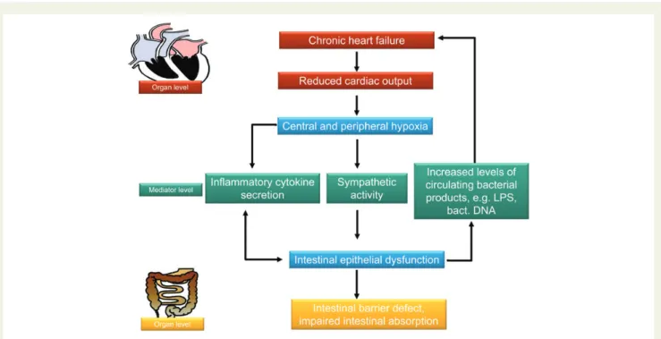
It is well known that CHF is a state of chronic inflammation with elevated circulating levels of pro-inflammatory cytokines, such as TNFa. In patients with CHF, increased circulating levels of pro-inflammatory cytokines have been shown to be closely related to predict poor short- and long-term survival.77,78Circulating cyto-kines have cardiosuppressor effects via different pathways that include alterations in myocardial intracellular calcium homeostasis, reduction in mitochondrial activity, alterations in matrix metallopro-teinase expression, cardiomyocyte hypertrophy, and apoptosis.79–83 Although the origin of inflammation in patients with CHF with ele-vated concentrations of pro-inflammatory cytokines is still a matter of debate, it has been shown that very small, but pathophysiologically relevant amounts of LPS may induce TNFa release.84,85 Further-more, growing evidence suggests that increased amounts of LPS enter the systemic circulation because of an altered intestinal micro-circulation in CHF, with LPS levels being 35% higher in the hepatic venous blood than in the left ventricle.86An important point in gut-derived inflammation in patients with CHF is the altered gut circula-tion as a consequence of reduced cardiac output and venous conges-tion (Figure2).
In patients with CHF, increased sympathetic tone and peripheral vasoconstriction contribute to a redistribution of blood flow away from the splanchnic circulation. The reduced intestinal perfusion may lead to an increase in intramucosal carbon dioxide pressure. Intramucosal acidosis may occur in nearly 50% of patients with circu-latory failure, suggesting the presence of inadequate oxygen supply and intestinal ischaemia.87,88The altered mucosal perfusion increases intestinal mucosal permeability with the disruption of the epithelial barrier function that favours the bacterial colonization and the pene-tration of LPS. Besides its effect on the release of cytokines that further aggravates CHF, LPS is able to trigger catecholamine release by granulocytes and phagocytes.89This increased release of catecholamines exerts additional unfavourable effects on gut perfu-sion and further increases the already hyperactive sympathetic tone. Another mechanism through which CHF may favour bacterial translocation is related to intestinal mucosa congestion as a conse-quence of raised right atrial pressure. As CHF is associated with
mucosal oedema in the intestine, which will impair the intestinal barrier function, this again may be followed by increased bacterial translocation (across the impaired barrier), increased amounts of endotoxin in the circulation,90 and aggravated heart disease—a typical vicious circle. Niebauer et al.90found that intensified diuretic
treatment normalized circulating endotoxin concentrations in patients with acute exacerbation of chronic heart disease.
A number of alterations in gastrointestinal function have been described in patients with CHF (Table1). It remains a matter of discus-sion whether these alterations are primary to the heart disease or caused by it.
In summary, recent data on potential interaction between the gut and the heart are intriguing. However, the evidence we have so far is preliminary. In large cohort studies, it needs to be evaluated whether, indeed, increased levels of bacterial products can be found in patients with atherosclerosis or CHF. The interesting and innovative field of heart – gut interaction still waits for more cardiologists and gastroen-terologists to collaborate on these important topics.
Authors’ contribution
Both authors wrote the manuscript together.Funding
The writing of this review was supported by grant SNF 310030-120312 to G.R. and by Grant Ricerca Corrente Ministero della Salute 2012 to G.R. Conflict of interest: none declared.
Figure 2 The heart and the gut in the pathophysiology of chronic heart failure. Chronic heart failure will cause a reduction in cardiac output which in turn will cause central and peripheral hypoxia. Among the organs that are affected by peripheral hypoxia is the small and large intestine. Hypoxia will cause an increase in inflammatory cytokine production, sympathetic activity, and production of other mediators (such as leucotrienes, prosta-glandins, and others that are not depicted in this graph). These mediators and the sympathetic activity may cause a malfunction of the gut. A further contributor will be a venous stasis increasing mucosal hypoxia. The mentioned factors have been shown to impair epithelial barrier function leading to a penetration of bacterial products across the intestinal barrier. Preliminary data indicate that the presence of those products in the circulation further aggravate chronic heart failure. Further studies with modern technologies such as mass spectroscopy and pyro sequencing of bacterial DNA will be necessary to confirm this. On the other hand, a dysfunction of the intestinal barrier will also cause impaired absorption negatively influencing the nutritional status of patients with end-stage heart disease.
Table 1 Alterations of gastrointestinal function in
patients with chronic heart failure (according to Sandek et al.70)
Increased small intestinal and large intestinal paracellular permeability in stable compensated chronic heart failure patients
Diminished carrier-mediated transport forD-xylose
Excessive enteric protein loss in infants with severe congenital heart disease
Decreased absorption of fat and protein
Thickened bowel wall of the terminal ileum and the colon Elevated collagen content in small intestinal biopsies
Increased distance between the capillary wall and the basal membrane of the enterocyte
References
1. Holt S, Heading RC, Clements JA, Tothill P, Prescott LF. Acetaminophen absorption and metabolism in celiac disease and Crohn’s disease. Clin Pharmacol Ther 1981;30: 232 – 238.
2. Lang CC, Brown RM, Kinirons MT, Deathridge MA, Guengerich FP, Kelleher D, O’Briain DS, Ghishan FK, Wood AJ. Decreased intestinal CYP3A in celiac disease: reversal after successful gluten-free diet: a potential source of interindividual vari-ability in first-pass drug metabolism. Clin Pharmacol Ther 1996;59:41 – 46. 3. Wilkinson GR. Cytochrome P4503A (CYP3A) metabolism: prediction of in vivo
ac-tivity in humans. J Pharmacokinet Biopharm 1996;24:475 – 490.
4. Willrich MA, Hirata MH, Hirata RD. Statin regulation of CYP3A4 and CYP3A5 ex-pression. Pharmacogenomics 2009;10:1017 – 1024.
5. Sakurai E, Hikichi N, Niwa H. Alteration of histamine, serotonin and primary prosta-glandin in case of diarrhea induced by endotoxin and gastrointestinal absorption of drug. J Pharmacobiodyn 1985;8:186 – 192.
6. Melichar B, Dvorak J, Krcmova L, Hyspler R, Urbanek L, Solichova D. Intestinal per-meability and vitamin A absorption in patients with chemotherapy-induced diarrhea. Am J Clin Oncol 2008;31:580 – 584.
7. Haapamaki J, Roine RP, Turunen U, Farkkila MA, Arkkila PE. Increased risk for cor-onary heart disease, asthma, and connective tissue diseases in inflammatory bowel disease. J Crohns Colitis 2011;5:41 – 47.
8. Gandhi S, Narula N, Marshall JK, Farkouh M. Are patients with inflammatory bowel disease at increased risk of coronary artery disease? Am J Med 2012;125:956 – 962. 9. van Leuven SI, Hezemans R, Levels JH, Snoek S, Stokkers PC, Hovingh GK, Kastelein JJ, Stroes ES, de Groot E, Hommes DW. Enhanced atherogenesis and altered high density lipoprotein in patients with Crohn’s disease. J Lipid Res 2007; 48:2640 – 2646.
10. Qin J, Li R, Raes J, Arumugam M, Burgdorf KS, Manichanh C, Nielsen T, Pons N, Levenez F, Yamada T, Mende DR, Li J, Xu J, Li S, Li D, Cao J, Wang B, Liang H, Zheng H, Xie Y, Tap J, Lepage P, Bertalan M, Batto JM, Hansen T, Le Paslier D, Linneberg A, Nielsen HB, Pelletier E, Renault P, Sicheritz-Ponten T, Turner K, Zhu H, Yu C, Jian M, Zhou Y, Li Y, Zhang X, Qin N, Yang H, Wang J, Brunak S, Dore J, Guarner F, Kristiansen K, Pedersen O, Parkhill J, Weissenbach J, Bork P, Ehrlich SD. A human gut microbial gene catalogue established by metagenomic se-quencing. Nature 2010;464:59 – 65.
11. Markowitz VM, Chen IM, Chu K, Szeto E, Palaniappan K, Jacob B, Ratner A, Liolios K, Pagani I, Huntemann M, Mavromatis K, Ivanova NN, Kyrpides NC. IMG/M-HMP: A metagenome comparative analysis system for the Human Microbiome Project. PLoS One 2012;7:e40151.
12. Human Microbiome Project Consortium. Structure, function and diversity of the healthy human microbiome. Nature 2012;486:207 – 214.
13. Thompson AL. Developmental origins of obesity: early feeding environments, infant growth, and the intestinal microbiome. Am J Hum Biol 2012;24:350 – 360. 14. Greenblum S, Turnbaugh PJ, Borenstein E. Metagenomic systems biology of the
human gut microbiome reveals topological shifts associated with obesity and inflam-matory bowel disease. Proc Natl Acad Sci USA 2012;109:594 – 599.
15. Tilg H, Kaser A. Gut microbiome, obesity, and metabolic dysfunction. J Clin Invest 2011;121:2126 – 2132.
16. Ley RE. Obesity and the human microbiome. Curr Opin Gastroenterol 2010;26:5 – 11. 17. Turnbaugh PJ, Gordon JI. The core gut microbiome, energy balance and obesity.
J Physiol 2009;587(Pt 17):4153 – 4158.
18. Turnbaugh PJ, Ley RE, Mahowald MA, Magrini V, Mardis ER, Gordon JI. An obesity-associated gut microbiome with increased capacity for energy harvest. Nature 2006;444:1027 – 1031.
19. Fava F, Gitau R, Griffin BA, Gibson GR, Tuohy KM, Lovegrove JA. The type and quan-tity of dietary fat and carbohydrate alter faecal microbiome and short-chain fatty acid excretion in a metabolic syndrome ‘at-risk’ population. Int J Obes (Lond) 2013;37: 216 – 223.
20. Murgas Torrazza R, Neu J. The developing intestinal microbiome and its relationship to health and disease in the neonate. J Perinatol 2011;31(Suppl. 1):S29 – S34. 21. Kong HH, Oh J, Deming C, Conlan S, Grice EA, Beatson MA, Nomicos E, Polley EC,
Komarow HD, Murray PR, Turner ML, Segre JA. Temporal shifts in the skin micro-biome associated with disease flares and treatment in children with atopic derma-titis. Genome Res 2012;22:850 – 859.
22. Ege MJ, Mayer M, Normand AC, Genuneit J, Cookson WO, Braun-Fahrlander C, Heederik D, Piarroux R, von Mutius E. Exposure to environmental microorganisms and childhood asthma. N Engl J Med 2011;364:701 – 709.
23. Cernadas M. It takes a microbiome: commensals, immune regulation, and allergy. Am J Respir Crit Care Med 2011;184:149 – 150.
24. Michail S, Durbin M, Turner D, Griffiths AM, Mack DR, Hyams J, Leleiko N, Kenche H, Stolfi A, Wine E. Alterations in the gut microbiome of children with severe ulcerative colitis. Inflamm Bowel Dis 2012;18:1799 – 1808.
25. Zella GC, Hait EJ, Glavan T, Gevers D, Ward DV, Kitts CL, Korzenik JR. Distinct microbiome in pouchitis compared to healthy pouches in ulcerative colitis and famil-ial adenomatous polyposis. Inflamm Bowel Dis 2011;17:1092 – 1100.
26. Docktor MJ, Paster BJ, Abramowicz S, Ingram J, Wang YE, Correll M, Jiang H, Cotton SL, Kokaras AS, Bousvaros A. Alterations in diversity of the oral microbiome in pediatric inflammatory bowel disease. Inflamm Bowel Dis 2012;18:935 – 942. 27. Sartor RB. Genetics and environmental interactions shape the intestinal microbiome
to promote inflammatory bowel disease versus mucosal homeostasis. Gastroenter-ology 2010;139:1816 – 1819.
28. Li M, Wang B, Zhang M, Rantalainen M, Wang S, Zhou H, Zhang Y, Shen J, Pang X, Wei H, Chen Y, Lu H, Zuo J, Su M, Qiu Y, Jia W, Xiao C, Smith LM, Yang S, Holmes E, Tang H, Zhao G, Nicholson JK, Li L, Zhao L. Symbiotic gut microbes modulate human metabolic phenotypes. Proc Natl Acad Sci USA 2008;105: 2117 – 2122.
29. Round JL, Mazmanian SK. The gut microbiota shapes intestinal immune responses during health and disease. Nat Rev Immunol 2009;9:313 – 323.
30. Flier JS, Mekalanos JJ. Gut check: testing a role for the intestinal microbiome in human obesity. Sci Transl Med 2009;1:6ps7.
31. Tsai F, Coyle WJ. The microbiome and obesity: is obesity linked to our gut flora? Curr Gastroenterol Rep 2009;11:307 – 313.
32. Turnbaugh PJ, Backhed F, Fulton L, Gordon JI. Diet-induced obesity is linked to marked but reversible alterations in the mouse distal gut microbiome. Cell Host Microbe 2008;3:213 – 223.
33. Turnbaugh PJ, Ridaura VK, Faith JJ, Rey FE, Knight R, Gordon JI. The effect of diet on the human gut microbiome: a metagenomic analysis in humanized gnotobiotic mice. Sci Transl Med 2009;1:6ra14.
34. Wang Z, Klipfell E, Bennett BJ, Koeth R, Levison BS, Dugar B, Feldstein AE, Britt EB, Fu X, Chung YM, Wu Y, Schauer P, Smith JD, Allayee H, Tang WH, DiDonato JA, Lusis AJ, Hazen SL. Gut flora metabolism of phosphatidylcholine promotes cardio-vascular disease. Nature 2011;472:57 – 63.
35. Valtonen VV. Infection as a risk factor for infarction and atherosclerosis. Ann Med 1991;23:539 – 543.
36. Costales P, Castellano J, Revuelta-Lopez E, Cal R, Aledo R, Llampayas O, Nasarre L, Juarez C, Badimon L, Llorente-Cortes V. Lipopolysaccharide downregulates CD91/ low-density lipoprotein receptor-related protein 1 expression through SREBP-1 overexpression in human macrophages. Atherosclerosis 2013;227:79 – 88. 37. Wiesner P, Choi SH, Almazan F, Benner C, Huang W, Diehl CJ, Gonen A, Butler S,
Witztum JL, Glass CK, Miller YI. Low doses of lipopolysaccharide and minimally oxi-dized low-density lipoprotein cooperatively activate macrophages via nuclear factor kappa B and activator protein-1: possible mechanism for acceleration of atheroscler-osis by subclinical endotoxemia. Circ Res 2010;107:56 – 65.
38. Feng X, Zhang Y, Xu R, Xie X, Tao L, Gao H, Gao Y, He Z, Wang H. Lipopolysacchar-ide up-regulates the expression of Fcalpha/mu receptor and promotes the binding of oxidized low-density lipoprotein and its IgM antibody complex to activated human macrophages. Atherosclerosis 2010;208:396 – 405.
39. Maziere C, Conte MA, Dantin F, Maziere JC. Lipopolysaccharide enhances oxidative modification of low density lipoprotein by copper ions, endothelial and smooth muscle cells. Atherosclerosis 1999;143:75 – 80.
40. Brand K, Banka CL, Mackman N, Terkeltaub RA, Fan ST, Curtiss LK. Oxidized LDL enhances lipopolysaccharide-induced tissue factor expression in human adherent monocytes. Arterioscler Thromb 1994;14:790 – 797.
41. Zhao Y, Cui G, Zhang N, Liu Z, Sun W, Peng Q. Lipopolysaccharide induces endo-thelial cell apoptosis via activation of Na(+)/H(+) exchanger 1 and calpain-dependent degradation of Bcl-2. Biochem Biophys Res Commun 2012;427:125 – 132. 42. Yang Y, Li Q, Deng Z, Zhang Z, Xu J, Qian G, Wang G. Protection from lipopolysaccharide-induced pulmonary microvascular endothelial cell injury by acti-vation of hedgehog signaling pathway. Mol Biol Rep 2011;38:3615 – 3622. 43. Koide N, Morikawa A, Tumurkhuu G, Dagvadorj J, Hassan F, Islam S, Naiki Y, Mori I,
Yoshida T, Yokochi T. Lipopolysaccharide and interferon-gamma enhance Fas-mediated cell death in mouse vascular endothelial cells via augmentation of Fas ex-pression. Clin Exp Immunol 2007;150:553 – 560.
44. Konter JM, Parker JL, Baez E, Li SZ, Ranscht B, Denzel M, Little FF, Nakamura K, Ouchi N, Fine A, Walsh K, Summer RS. Adiponectin attenuates lipopolysaccharide-induced acute lung injury through suppression of endothelial cell activation. J Immunol 2012;188:854 – 863.
45. Dayoub JC, Ortiz F, Lopez LC, Venegas C, Del Pino-Zumaquero A, Roda O, Sanchez-Montesinos I, Acuna-Castroviejo D, Escames G. Synergism between mela-tonin and atorvastatin against endothelial cell damage induced by lipopolysaccharide. J Pineal Res 2011;51:324 – 330.
46. Morel DW, DiCorleto PE, Chisolm GM. Modulation of endotoxin-induced endo-thelial cell toxicity by low density lipoprotein. Lab Invest 1986;55:419 – 426. 47. Howell KW, Meng X, Fullerton DA, Jin C, Reece TB, Cleveland JC Jr. Toll-like
recep-tor 4 mediates oxidized LDL-induced macrophage differentiation to foam cells. J Surg Res 2011;171:e27 – e31.
48. Pataki M, Lusztig G, Robenek H. Endocytosis of oxidized LDL and reversibility of mi-gration inhibition in macrophage-derived foam cells in vitro. A mechanism for athero-sclerosis regression? Arterioscler Thromb 1992;12:936 – 944.
49. Wiedermann CJ, Kiechl S, Dunzendorfer S, Schratzberger P, Egger G, Oberhollenzer F, Willeit J. Association of endotoxemia with carotid atherosclerosis and cardiovascular disease: prospective results from the Bruneck Study. J Am Coll Cardiol 1999;34:1975 – 1981.
50. Kiechl S, Lorenz E, Reindl M, Wiedermann CJ, Oberhollenzer F, Bonora E, Willeit J, Schwartz DA. Toll-like receptor 4 polymorphisms and atherogenesis. N Engl J Med 2002;347:185 – 192.
51. Schilling J, Lai L, Sambandam N, Dey CE, Leone TC, Kelly DP. Toll-like receptor-mediated inflammatory signaling reprograms cardiac energy metabolism by repres-sing peroxisome proliferator-activated receptor gamma coactivator-1 signaling. Circ Heart Fail 2011;4:474 – 482.
52. Avlas O, Fallach R, Shainberg A, Porat E, Hochhauser E. Toll-like receptor 4 stimu-lation initiates an inflammatory response that decreases cardiomyocyte contractility. Antioxid Redox Signal 2011;15:1895 – 1909.
53. Fallach R, Shainberg A, Avlas O, Fainblut M, Chepurko Y, Porat E, Hochhauser E. Car-diomyocyte Toll-like receptor 4 is involved in heart dysfunction following septic shock or myocardial ischemia. J Mol Cell Cardiol 2010;48:1236 – 1244.
54. Boyd JH, Mathur S, Wang Y, Bateman RM, Walley KR. Toll-like receptor stimulation in cardiomyoctes decreases contractility and initiates an NF-kappaB dependent in-flammatory response. Cardiovasc Res 2006;72:384 – 393.
55. Lam V, Su J, Koprowski S, Hsu A, Tweddell JS, Rafiee P, Gross GJ, Salzman NH, Baker JE. Intestinal microbiota determine severity of myocardial infarction in rats. FASEB J 2012;26:1727 – 1735.
56. Danesh J, Collins R, Peto R. Chronic infections and coronary heart disease: is there a link? Lancet 1997;350:430 – 436.
57. Epstein SE, Speir E, Zhou YF, Guetta E, Leon M, Finkel T. The role of infection in re-stenosis and atherosclerosis: focus on cytomegalovirus. Lancet 1996;348(Suppl. 1): s13 – s17.
58. Patel P, Mendall MA, Carrington D, Strachan DP, Leatham E, Molineaux N, Levy J, Blakeston C, Seymour CA, Camm AJ. Association of Helicobacter pylori and Chlamydia pneumoniae infections with coronary heart disease and cardiovascular risk factors. BMJ 1995;311:711 – 714.
59. Saikku P, Leinonen M, Mattila K, Ekman MR, Nieminen MS, Makela PH, Huttunen JK, Valtonen V. Serological evidence of an association of a novel Chlamydia, TWAR, with chronic coronary heart disease and acute myocardial infarction. Lancet 1988;2: 983 – 986.
60. Khan S, Okamoto T, Enomoto K, Sakashita N, Oyama K, Fujii S, Sawa T, Takeya M, Ogawa H, Yamabe H, Akaike T. Potential association of Helicobacter cinaedi with atrial arrhythmias and atherosclerosis. Microbiol Immunol 2012;56:145 – 154. 61. Dore MP, Sepulveda AR, Bacciu PP, Blasi F, Simula L, Marras L, Piccolo D, Cherchi GB,
Graham DY, Realdi G. Detection of Chlamydiae pneumoniae but not Helicobacter pylori DNA in atherosclerosis plaques. Dig Dis Sci 2003;48:945 – 951.
62. Mayr M, Kiechl S, Willeit J, Wick G, Xu Q. Infections, immunity, and atherosclerosis: associations of antibodies to Chlamydia pneumoniae, Helicobacter pylori, and cyto-megalovirus with immune reactions to heat-shock protein 60 and carotid or femoral atherosclerosis. Circulation 2000;102:833 – 839.
63. Sessa R, Nicoletti M, Di Pietro M, Schiavoni G, Santino I, Zagaglia C, Del Piano M, Cipriani P. Chlamydia pneumoniae and atherosclerosis: current state and future pro-spectives. Int J Immunopathol Pharmacol 2009;22:9 – 14.
64. Fazio G, Giovino M, Gullotti A, Bacarella D, Novo G, Novo S. Atherosclerosis, in-flammation and Chlamydia pneumoniae. World J Cardiol 2009;1:31 – 40.
65. Campbell LA, Kuo CC. Chlamydia pneumonia—an infectious risk factor for athero-sclerosis? Nat Rev Microbiol 2004;2:23 – 32.
66. Stone AF, Mendall MA, Kaski JC, Edger TM, Risley P, Poloniecki J, Camm AJ, Northfield TC. Effect of treatment for Chlamydia pneumoniae and Helicobacter pylori on markers of inflammation and cardiac events in patients with acute coronary syndromes: South Thames Trial of Antibiotics in Myocardial Infarction and Unstable Angina (STAMINA). Circulation 2002;106:1219 – 1223.
67. Gurfinkel E, Bozovich G, Daroca A, Beck E, Mautner B. Randomised trial of roxithro-mycin in non-Q-wave coronary syndromes: ROXIS Pilot Study. ROXIS Study Group. Lancet 1997;350:404 – 407.
68. O’Connor CM, Dunne MW, Pfeffer MA, Muhlestein JB, Yao L, Gupta S, Benner RJ, Fisher MR, Cook TD. Azithromycin for the secondary prevention of coronary heart disease events: the WIZARD study: a randomized controlled trial. JAMA 2003;290:1459 – 1466.
69. Arutyunov GP, Kostyukevich OI, Serov RA, Rylova NV, Bylova NA. Collagen accu-mulation and dysfunctional mucosal barrier of the small intestine in patients with chronic heart failure. Int J Cardiol 2008;125:240 – 245.
70. Sandek A, Anker SD, von Haehling S. The gut and intestinal bacteria in chronic heart failure. Curr Drug Metab 2009;10:22 – 28.
71. Sandek A, Rauchhaus M, Anker SD, von Haehling S. The emerging role of the gut in chronic heart failure. Curr Opin Clin Nutr Metab Care 2008;11:632 – 639. 72. Sandek A, Bauditz J, Swidsinski A, Buhner S, Weber-Eibel J, von Haehling S,
Schroedl W, Karhausen T, Doehner W, Rauchhaus M, Poole-Wilson P, Volk HD, Lochs H, Anker SD. Altered intestinal function in patients with chronic heart failure. J Am Coll Cardiol 2007;50:1561 – 1569.
73. Krack A, Sharma R, Figulla HR, Anker SD. The importance of the gastrointestinal system in the pathogenesis of heart failure. Eur Heart J 2005;26:2368 – 2374. 74. Anker SD, Egerer KR, Volk HD, Kox WJ, Poole-Wilson PA, Coats AJ. Elevated
soluble CD14 receptors and altered cytokines in chronic heart failure. Am J Cardiol 1997;79:1426 – 1430.
75. Chong AJ, Shimamoto A, Hampton CR, Takayama H, Spring DJ, Rothnie CL, Yada M, Pohlman TH, Verrier ED. Toll-like receptor 4 mediates ischemia/reperfusion injury of the heart. J Thorac Cardiovasc Surg 2004;128:170 – 179.
76. Tavener SA, Long EM, Robbins SM, McRae KM, Van Remmen H, Kubes P. Immune cell Toll-like receptor 4 is required for cardiac myocyte impairment during endotox-emia. Circ Res 2004;95:700 – 707.
77. Ferrari R, Bachetti T, Confortini R, Opasich C, Febo O, Corti A, Cassani G, Visioli O. Tumor necrosis factor soluble receptors in patients with various degrees of congest-ive heart failure. Circulation 1995;92:1479 – 1486.
78. Rauchhaus M, Doehner W, Francis DP, Davos C, Kemp M, Liebenthal C, Niebauer J, Hooper J, Volk HD, Coats AJ, Anker SD. Plasma cytokine parameters and mortality in patients with chronic heart failure. Circulation 2000;102:3060 – 3067. 79. Kumar A, Krieger A, Symeoneides S, Parrillo JE. Myocardial dysfunction in septic
shock. Part II: Role of cytokines and nitric oxide. J Cardiothorac Vasc Anesth 2001; 15:485 – 511.
80. Kumar A, Haery C, Parrillo JE. Myocardial dysfunction in septic shock. Part I: Clinical manifestation of cardiovascular dysfunction. J Cardiothorac Vasc Anesth 2001;15: 364 – 376.
81. Muller-Werdan U, Engelmann H, Werdan K. Cardiodepression by tumor necrosis factor-alpha. Eur Cytokine Netw 1998;9:689 – 691.
82. Anker SD, von Haehling S. Inflammatory mediators in chronic heart failure: an over-view. Heart 2004;90:464 – 470.
83. Gao CQ, Sawicki G, Suarez-Pinzon WL, Csont T, Wozniak M, Ferdinandy P, Schulz R. Matrix metalloproteinase-2 mediates cytokine-induced myocardial con-tractile dysfunction. Cardiovasc Res 2003;57:426 – 433.
84. Sharma R, Bolger AP, Rauchhaus M, von Haehling S, Doehner W, Adcock IM, Barnes PJ, Poole-Wilson PA, Volk HD, Coats AJ, Lim S, Anker SD. Cellular endotoxin desensitization in patients with severe chronic heart failure. Eur J Heart Fail 2005;7: 865 – 868.
85. Genth-Zotz S, von Haehling S, Bolger AP, Kalra PR, Wensel R, Coats AJ, Anker SD. Pathophysiologic quantities of endotoxin-induced tumor necrosis factor-alpha release in whole blood from patients with chronic heart failure. Am J Cardiol 2002; 90:1226 – 1230.
86. Peschel T, Schonauer M, Thiele H, Anker SD, Schuler G, Niebauer J. Invasive assess-ment of bacterial endotoxin and inflammatory cytokines in patients with acute heart failure. Eur J Heart Fail 2003;5:609 – 614.
87. Gutierrez G, Palizas F, Doglio G, Pusajo J, Wainsztein N, Klein F, Gallesio A, San Roman E, Pacin J, Dorfman B, Dubin A, Schiavi E, Shottender J, Jorge M, Giniger R. Gastric intramucosal pH as a therapeutic index of tissue oxygenation in critically ill patients. Lancet 1992;339:195 – 199.
88. Maynard N, Bihari D, Beale R, Smithies M, Baldock G, Mason R, McColl I. Assessment of splanchnic oxygenation by gastric tonometry in patients with acute circulatory failure. JAMA 1993;270:1203 – 1210.
89. Flierl MA, Rittirsch D, Nadeau BA, Chen AJ, Sarma JV, Zetoune FS, McGuire SR, List RP, Day DE, Hoesel LM, Gao H, Van Rooijen N, Huber-Lang MS, Neubig RR, Ward PA. Phagocyte-derived catecholamines enhance acute inflammatory injury. Nature 2007;449:721 – 725.
90. Niebauer J, Volk HD, Kemp M, Dominguez M, Schumann RR, Rauchhaus M, Poole-Wilson PA, Coats AJ, Anker SD. Endotoxin and immune activation in chronic heart failure: a prospective cohort study. Lancet 1999;353:1838 – 1842.