SUPPLEMENTARY MATERIAL
Supplementary methods
List of Drosophila stocks
The following strains were used in this work: w1118 (courtesy of R. Stocker); Canton-S (courtesy of R. Stocker); salm-Gal4, UAS-H2B::YFP (courtesy of C. Desplan; Mollereau et al., 2000); UAS-syt::eGFP, UAS-mCD8::Cherry (courtesy of O. Urwyler; Zhang et al., 2002);
FRT42D, so3(courtesy of F. Pignoni; Pignoni et al., 1997), UAS-hazy (courtesy of J. Bischof;
Bischof et al., 2013), otd (courtesy of H. Reichert; Blanco et al., 2011),
UAS-mCD8::GFP and FRT82B, atow (courtesy of B. Hassan; Choi et al., 2009). Stocks containing
glass mutant alleles were previously characterised in G. Rubin's lab (Moses et al., 1989), and
obtained from the Bloomington stock center: gl60j (No. 509); FRT82B, gl60j (No. 6333); gl2, e4 (No. 507) and gl3 (No. 508). We used the following stocks as drivers for analysing mutant clones: eyFLP;; FRT82B, ubiGFP (courtesy of B. Hassan), eyFLP; FRT42D, arm-lacZ (courtesy of J. Curtiss) and hsFLP; tub-Gal4, UAS-mCD8::GFP; FRT82B, tub-Gal80 (courtesy of B. Bello). For flip-out misexpression experiments we used hsFLP; tub(FRT
cassette)Gal4, UAS-lacZ.nls (courtesy of E. Piddini).
We took advantage of commonly used balancers and phenotypic markers for performing crosses and selecting flies of the desired genotype, in particular nocSco, Sp, CyO, TM2,
MKRS, TM6b (Lindsley and Zimm, 1992) and CyO dfd-eYFP (Le et al., 2006).
Antibodies and fluorescent dyes
We used the following primary antibodies: rabbit anti-ȕGal (1:1000, Cappel, No. 55976), chicken anti-ȕGal (1:1000, Abcam, ab9361), rabbit anti-GFP (1:1000, Molecular probes,
A-1
Published in Development 143(8): 1413–1423, 2016
which should be cited to refer to this work
6455), chicken anti-GFP (1:1000, Abcam, ab13970), rabbit anti-DsRed (1:1000, Clontech, No. 632496), rabbit anti-HRP (1:200, Sigma, P7899), guinea pig anti-Otd (1:750, courtesy of T. Cook; Ranade et al., 2008), rabbit anti-Hazy (1:500, courtesy of A. Zelhof; Zelhof et al., 2003), rabbit anti-Rh2 (1:40, Mishra et al., 2016), rabbit anti-Rh6 (1:10000, courtesy of C. Desplan; Tahayato et al., 2003), rabbit anti-histamine (1:1000, ImmunoStar, No. 22939) and rabbit anti-Ato (1:5000, courtesy of B. Hassan). Mouse monoclonal antibodies anti-Rh4, Rh5, and Rh6 were obtained from S. Britt and used 1:40 (Chou et al., 1999). A number of rabbit polyclonal antibodies against proteins of the phototransduction cascade were produced in C. Zuker's lab and kindly provided by N. Colley: anti-Arr1 (1:100), anti-GĮq (1:100), anti-NorpA (1:100), anti-Trpl (1:100) and anti-InaD (1:100). We obtained the following antibodies from Developmental Studies Hybridoma Bank (DSHB) at The University of Iowa: mouse anti-ȕGal (1:20, 40-1a), rat anti-Elav (1:30, No. 7E8A10), mouse anti-Chp (1:20, No. 24B10), mouse anti-Fas2 (1:20, ID4), mouse anti-Fas3 (1:20, 70G10), mouse anti-Futsch (1:20, 22C10), mouse anti-Glass (1:10, 9B2.1), mouse anti-Rh1 (1:20, 4C5) and mouse anti-Trp (1:20, MAb83F6).
Secondary antibodies were conjugated with Alexa Fluor fluorescent proteins (405, 488, 546, 568 or 647) and we used them in a 1:200 dilution (Molecular Probes; No. A-11029, A11001, 11031, 21235, 31553, 11006, 11077, 21247, 11008, 11011, 21244, 11039, 11041, 11073, 11074, 21450, 10037, 31571, 21206, 10042 and A-31573). We also used Hoechst 33258 (1:100, Sigma, No. 94403) and DAPI (in Vectashield mounting medium, Vector, H-1200) as fluorescent markers of cell nuclei, and phalloidin conjugated with ATTO 647N to label actin microfilaments (No. 65906).
Generation and analysis of clones
MARCM analysis of glass mutant clones was performed in hsFLP; tub-Gal4,
days after the flies had laid the eggs with a 20 minute long heat shock at 37°C. We could identify gl60jclones in the pupal retina positively labelled with mCD8::GFP.
Rescue of the glass mutant retina was tested by inducing Hazy and Otd-expressing clones. For this, we crossed hsFLP; tub(FRT cassette)Gal4, UAS-lacZ.nls; gl60j flies with others carrying combinations of the UAS-hazy and UAS-Otd constructs with the gl60jmutation. Hazy expression was induced alone and together with Otd in 6 day old animals (mainly pupae, at about 24 hours after pupation) with a 5 minute long heat shock at 37°C. We induced the expression of Otd alone in 5 day old animals (mainly late third instar larvae) with a 5 minute long heat shock at 37°C. For both Hazy and Otd we aimed to express them at the time point in which they should be expressed during development in wild-type PRs (Vandendries et al., 1996; Zelhof et al., 2003). We were able to identify Hazy and Otd-expressing cells in the adult glass mutant retina because of the co-expression of nuclear ȕGal.
To test the potency of Glass to ectopically induce PR markers we generated clones in which combinations of Glass with Hazy and Otd were ectopically expressed. For this we crossed
hsFLP; tub(FRT cassette)Gal4, lacZ.nls flies with others carrying the glass, UAS-hazy and UAS-otd constructs. Clones were induced in 4-6 hour old embryos by a 6 minute
heat shock at 37°C. Expression of the UAS promoters was driven by tub(FRT cassette)Gal4, which we activated by removing the FRT cassette through hsFLP mediated recombination (Blair, 2003; Struhl and Basler, 1993). Gal4-expressing cells in the CNS of 4 day old larvae were labelled with nuclear ȕGal.
so3 mutant clones were obtained in the eye discs of eyFLP; FRT42D, arm-lacZ/FRT42D, so3 larvae, and could be identified as groups of cells negatively labelled for ȕGal.
atow mutant clones were generated in the eye discs of eyFLP;; FRT82B, ubiGFP/FRT82B,
atow larvae, and we could recognise them as groups of cells negatively labelled for GFP.
3
Generation of transgenic flies
In order to generate the hazy(wt)-GFP reporter construct, a 1085 bp fragment upstream of the hazy start codon was amplified by PCR from wild-type flies and cloned into pBluescript using KpnI and NotI sites attached to the primers. The two Glass binding motifs were then mutated individually and in combination using site directed mutagenesis (Stratagene), to produce the hazy(gl1mut)-GFP, hazy(gl2mut)-GFP and hazy(gl1,2mut)-GFP reporter constructs. The wild-type and mutant sequences were then transferred into an attB-GFP-hsp70 3'UTR reporter vector (modified from a plasmid provided by J. Rister). All constructs were injected into nos-PhiC31; attP40 flies. For primer sequences see table S1.
To generate the otd(wt)-GFP reporter construct, the 1.5 kb PR enhancer element (Vandendries et al., 1996) was amplified by PCR from wild-type flies and cloned into pBluescript using the endogenous KpnI and BamHI sites flanking this element. For making
otd(glmut)-GFP, the Glass binding motif was mutated by PCR amplification from wild-type
flies of two fragments of the enhancer with the 5' and 3' flanking primers combined with primers extending to and from the Glass binding motif with a XhoI restriction site replacing the Glass binding motif. The wild-type and mutant constructs were then transferred into the attB-GFP-hsp70 3'UTR reporter vector. Both constructs were injected into nos-PhiC31;
attP40 flies. For primer sequences see table S1.
For generating the UAS-glass flies we used the Glass PA isoform (REFSEQ:NP_476854, FBpp0083005), containing all five zinc fingers, which has been reported to be functional (O'Neill et al., 1995). To obtain this isoform we had to remove the last intron from the only fully sequenced BDGP DGC glass cDNA clone (GH20219) available encoding the Glass PB isoform. This isoform lacks the last 47 amino acids including half of the last zinc-finger due to the presence of a stop codon within the last intron. We removed this intron by PCR
that match the coding sequence at the other side of the intron, ligating the two fragments together and PCR amplifying the entire Glass PA coding region and 5'UTR. We cloned this PCR product into the BamHI and XhoI sites of pBluescript using restriction sites added to the flanking primers. We next PCR amplified the Glass PA coding region with primers for gateway cloning and inserted it into a pUASg.attB plasmid (courtesy of J. Bischof; Bischof et al., 2013). The construct was injected into nos-PhiC31; attP40 flies. We tested the ability of the UAS-glass flies to ectopically express the protein by antibody staining against Glass, and found that this construct rescues the glass mutant phenotype when expressed in the eye during development (data not shown). For primer sequences see table S1.
For the glass-GFP reporter constructs a 293 bp BamHI-EcoRI fragment from the middle of the 5.2 kb upstream genomic region of glass was cloned in front of a minimal hsp70 promoter + GFP reporter gene using the endogenous BamHI and EcoRI sites. The BamHI site present in our genomic sequence is missing in the Flybase sequence due to a single nucleotide polymorphism. The putative So binding sites were mutated by PCR amplifications using primers with overhangs replacing the So sites with restriction sites for SpeI (so1) and NcoI (so2 and so3). Since so2 and so3 are very close to each other (within 25 bp) they were mutated together. The PCR fragments were ligated and cloned in front of the minimal hsp70 promoter + GFP reporter gene. Both transgenes were injected into nos-PhiC31;; attP2 flies. For primer sequences see table S1.
5
Table S1: primer sequences
hazy -1.1 Kpn fw ctggtaccACATGTGTGCAGAGGCAAAGGG
hazy noStart Not re aagcggccgcGCGAATCCTGAGCTTCCTGTTGG hazy gl1 site mut Sph fw GGGCGACTTCTACgcatgcTGTCGACGGACAGCACG hazy gl1 site mut Sph re CGTGCTGTCCGTCGACAgcatgcGTAGAAGTCGCCC hazy gl2 site mut RV fw GAAGAAGCAGCGACGCgatatcCTCGAAGTGTCGACG hazy gl2 site mut RV re CGTCGACACTTCGAGgatatcGCGTCGCTGCTTCTTC otd PRenh Kpn fw cggagcgttGGTACCtcgtc
otd PRenh BamHI re ggccagaccatcGGATCCcc otd PRenh gl mut Xho fw agCTCGAGcctgcagtggtcggctcc otd PRenh glmut Xho re ggCTCGAGtccttaatcgctgttgctttttacggc glass 5'UTR BamH fw gaggatCCTCGCCAAAAGTCGCTTCTTG
glass exon 4 re ccccgactgcgaaaatCTGAGCAGGCAGAGCTTGCAC
glass exon 5 fw gctctgcctgctcagATTTTCGCAGTCGGGGAACTTG
glass Stop Xho re ggctcgaGTCATGTGAGCAGGCTGTTGCC
glass Start+Kozak attB1 fw ggggacaagtttgtacaaaaaagcaggcttcaaCATGGGATTGTTATATAAGG GTTCCAAACT
glass Stop attB2 re ggggaccactttgtacaagaaagctgggtcgTCATGTGAGCAGGCTGTTGCC
glass BRenh so1 mut Spe fw acACTAGTttgaagcgaagtaaaaaaaaaaagaaatataaaaattgaaaactgg
glass BRenh so1 mut Spe re ttTGATCAgtttcatgtcaacaacttggctaaggac
glass BRenh so2+3 mut Nco fw ggGACGCtgggggatatagctCCATGGgtatgcgatcactgcaagcc
glass BRenh so2+3 mut Nco re acCCATGGagctatatcccccaGCGTCccttaccttatcgatgggaatttcagg
Immunohistochemistry
In the case of adult heads, we incubated them in cryoprotectant solution (sucrose 25% in PB) at 4°C overnight. Next we embedded them in OCT and cut 14 m cryosections in the transverse plane, after which we proceeded to stain them.
For staining both cryosections and whole mounted samples, we first washed them at room temperature with PBT (Triton X-100 0.3% in PB) at least three times for a minimum of 20 minutes each: this procedure was repeated in all washing steps that follow. Incubation in primary antibody solution was done overnight at 4°C and was followed by PBT washes. Next, we incubated our samples in secondary antibody solution overnight at 4°C, after which we washed them. We mounted our samples either in 50% glycerol or Vectashield.
7
Supplementary figures
Fig. S1. glass mutant PR precursors are not correctly recruited into the developing ommatidia in the third instar eye disc. It has been reported that, at this stage, glass mutant PR precursors fail to acquire a correct subtype identity, based on the expression of subtype specific PR markers (Hayashi et al., 2008; Jarman et al., 1995; Lim and Choi, 2004; Treisman and Rubin, 1996). To analyse in detail the order in which PR precursors are recruited in glass mutant, we counted the number of Elav-positive cells in the third instar eye disc of wild-type and gl60jlarvae. Each ommatidium was pseudo-coloured according to the number of PR precursors that it contains. This image illustrates how PR precursors in the the wild-type eye disc are orderly recruited into the developing ommatidia (A). By comparison, in the glass mutant eye disc PR precursors are recruited slower and disorderly (B). Scale bars represent 30 ȝm.
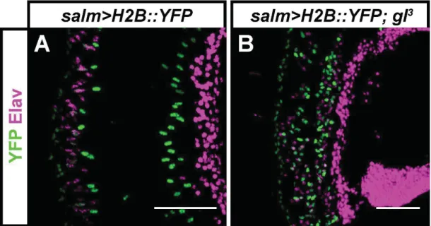
Fig. S2. glass mutant PR precursors survive metamorphosis and are still present in the adult retina. Expression of YFP (green) and Elav (magenta) in the salm>H2B::YFP reporter line in the adult retina of control (A) and gl3 mutant flies (B). In both cases, a subset of presumptive PRs can be identified by the co-expression of YFP and Elav, while cone cells express YFP but not Elav. Scale bars represent 40 ȝm.
9
Fig. S3. glass mutant PR precursors are irregularly distributed in the retina. (A-C''') We used MARCM analysis to induce the formation of gl60j mutant clones, labeled with the expression of UAS-mCD8::GFP. Retinas were dissected ~50 hours after pupation, and stained against
GFP (green), Elav (used to label the nuclei of neurons, red) and Fas3 (used to label the membranes of interommatidial cells, blue). For each image, these three channels are shown below in greyscale. All images belong to the same confocal stack (Movie S1), in which those cells that are wild-type for glass are located in the upper half of the area that is shown, while a big homozygous gl60j clone crosses the lower half. Distally in the retina, cones strongly express Fas3 and can be seen as groups of 4 cells in the wild-type (GFP-negative) region of the image (arrowheads; A, A'''). This kind of organization is not present in the GFP-labeled
glass mutant clone (A, A'''). More proximally, PR precursors are abundant in the wild-type
area, where they distribute in rosettes of 8 Elav-positive cells (arrows; B, B''). Rosettes are separated from each other by pigment and bristle cells, which form the hexagonal lattice of the ommatidia, and are strongly stained for Fas3 (arrowheads; B, B'''). By contrast, in the
glass mutant region there are fewer Elav-positive cells, and cells do not group in any
structure resembling an ommatidium (B, B'', B'''). This is different from earlier developmental stages, since ommatidial clusters can still be seen in the glass mutant eye disc (Figs. 1E, S1; Moses et al., 1989; Treisman and Rubin, 1996). The most proximal region of the wild-type retina contains the nuclei of bristle neurons, which are orderly arranged between the
ommatidia (arrowheads; C, C''). At this level, the glass mutant clone contains densely packed groups of neurons (C, C''), including the PR precursors missing in the medial section. Scale bar represents 30 ȝm.
11
Fig. S4. Glass is required for the acquisition of the phototransduction machinery. (A-T) Expression of different proteins involved in the phototransduction cascade in the adult retina of control (salm>H2B::YFP) and gl2 mutant flies. Samples were stained against phototransduction proteins (green) and counterstained with DAPI (magenta). Rhodopsins Rh1 (A), Rh4 (B), Rh5 (C), and Rh6 (D) are expressed in different subsets of PRs in control retinas. In the gl2 mutant retina there is no expression of Rh1 (F), Rh4 (G), Rh5 (H) or Rh6 (I). Proteins downstream in the phototransduction cascade are expressed in all PRs in the retina of control flies: Arr1 (E), GĮq (K), NorpA (L), Trp (M), Trpl (N) and InaD (O). These proteins are not expressed in the retina of gl2 mutant flies: Arr1 (J), GĮq (P), NorpA (Q), Trp (R), Trpl (S), or InaD (T). Scale bars represent 40 ȝm.
Fig. S5. Glass is required for Rh2 expression. Both control and glass mutant flies were stained against Rh2 (green) and counterstained with Elav (magenta). Rh2 is expressed in ocellar PRs in control flies (A). In glass mutant, there is no expression of Rh2 in the presumptive ocelli PRs (arrowheads, B). Scale bars represent 40 ȝm.
13
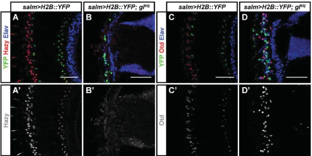
Fig. S6. Glass is required for the correct expression of Hazy and Otd. (A-D') We used the
salm>H2B::YFP reporter to label the retina of adult flies, and stained against GFP (green),
either Hazy or Otd (red), and the neuronal marker Elav (Blue). For each image, the red channel is shown below in greyscale. There is expression of Hazy in the nuclei of PRs in control flies (A, A') but not in the presumptive PRs of glass mutant flies (B, B'). Otd is expressed in the PRs of control flies (C, C') but only a fraction of presumptive glass mutant PRs express Otd (D, D'). Scale bars represent 50 m.
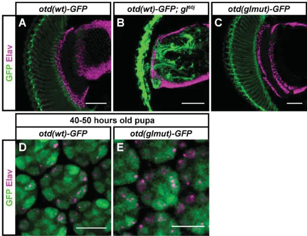
Fig. S7. Expression of the otd(wt)-GFP reporter is independent of Glass. Samples were stained against GFP (green) and against the neuronal marker Elav (magenta). (A-C) In adult flies, otd(wt)-GFP is expressed both in control (A) and glass mutant background (B). This reporter is also expressed when the Glass binding motif is mutated (C). (D, E) At 40-50 hours after pupation, all PR precursors express the otd(wt)-GFP reporter in control animals (D). After mutation of the Glass binding motif the reporter is still expressed in all PRs (E). Scale bars represent 10 m in panels D-E, and 50 m in panels A-C.
15
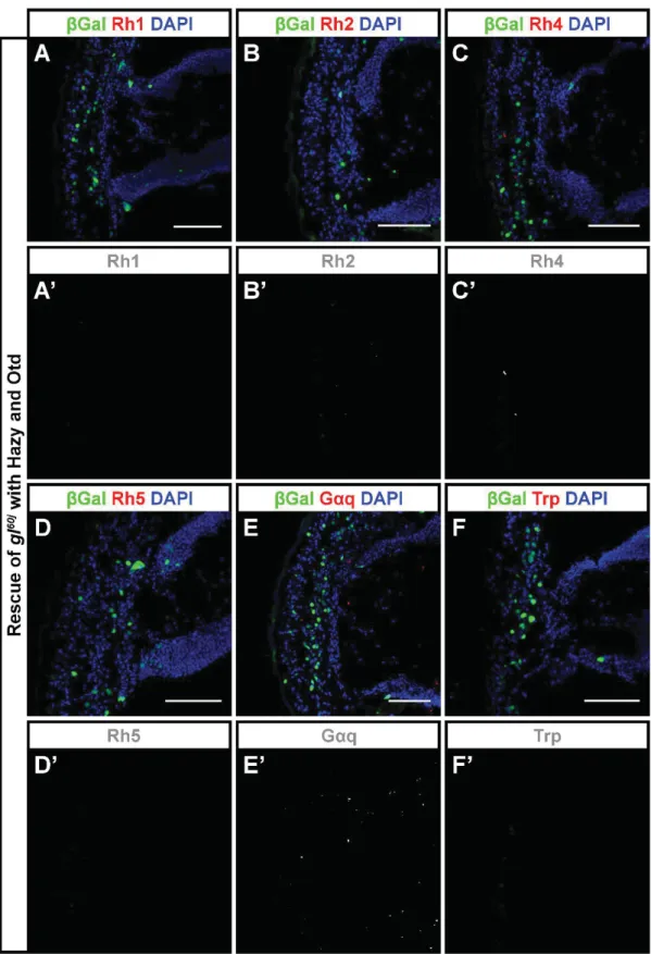
Fig. S8. Induced co-expression of Hazy and Otd does not rescue the glass mutant phenotype better than Hazy alone. (A-F') Hazy and Otd were expressed in the glass mutant retina in clones labelled with nuclear ȕGal. Samples were stained against ȕGal (green),
different proteins involved in the phototransduction cascade (red) and with DAPI (used to label cell nuclei, blue). For each image, the red channel is shown below in greyscale. We did not observe the rescue of any of those proteins that were not rescued by Hazy alone (the rescue of glass mutant with Hazy is shown in Fig. 5), namely the expression of Rh1 (A, A'), Rh2 (B, B'), Rh4 (C, C'), Rh5 (D, D'), GĮq (E, E') and Trp (F, F') was not induced in Hazy-Otd-expressing clones. Scale bars represent 40 ȝm.
17
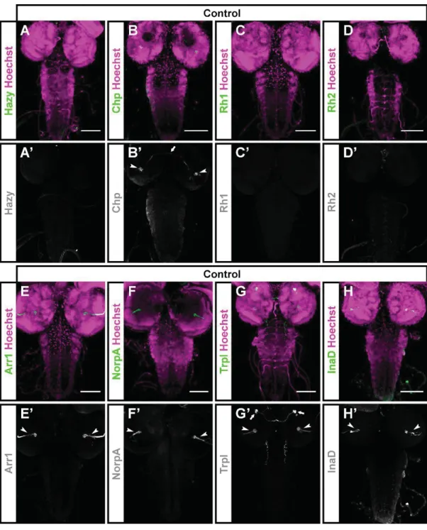
Fig. S9. Expression of PR markers in the CNS of third instar larvae. (A-H') The CNS of control (Canton-S) animals was stained with antibodies against different PR proteins (green) and counterstained with Hoechst 33258 (used to label cell nuclei, magenta). For each image, the green channel is shown below in greyscale. Hazy is expressed in the nuclei of PRs in the Bolwig organ (Zelhof et al., 2003), and cannot be seen in the CNS (A, A'). Chp is primarily expressed in the axons of PRs in the Bolwig organ, which project into the optic lobe (arrowheads; B, B'). In addition,a small number of cells in the brain are stained (arrow; B, B').
Neither Rh1 nor Rh2 are expressed in the CNS of the larvae (C, D). Both Arr1 and NorpA are expressed in the axon projections of the Bolwig organ PRs (arrowheads; E, E', F, F'). Trpl is expressed in the axons of PRs in the Bolwig organ (arrowheads; G, G') and in 3-4 cells located rostrally in each of the brain lobes (arrows; G, G'). InaD is expressed in the axon projections of the Bolwig organ PRs (arrowheads; H, H'). Scale bars represent 80 m.
19
Fig. S10. Co-misexpression of Glass and Hazy is not sufficient to ectopically induce all the phototransduction proteins that we have tested, and co-misexpression of Glass and Otd does not ectopically induce more phototransduction proteins than Glass alone (for a comparison, see Fig. 6). Misexpression of these transcription factors was induced during embryonic development in clones, which were labelled by the presence of nuclear ȕGal. We dissected and stained the CNS of third instar larvae with antibodies against ȕGal (green), different phototransduction proteins (red/magenta) and with Hoechst 33258 (used to label cell nuclei, blue). Close-ups are shown below each sample. (A-E') Co-misexpressing Glass and Hazy is not sufficient to ectopically induce Rh4 (A, A'), Rh5 (B, B'), Rh6 (C, C'), GĮq (D, D') nor Trp (E, E'). (F-N') Co-misexpressing Glass and Otd is not sufficient to ectopically induce Rh4 (F, F'), Rh5 (G, G'), Rh6 (H, H'), GĮq (I, I'), Trp (J, J'), Rh1 (K, K'), Arr1 (L, L'), NorpA(M, M') nor InaD (N, N'). Scale bars represent 10 ȝm in A'-N', and 80 ȝm in A-N.
Fig. S11. The proneural transcription factor Ato is not required for glass expression. (A) During the development of the third instar eye disc, Ato expression (red) precedes that of Glass (green). Counterstaining with phalloiding (blue) serves to locate the position of the morphogenetic furrow (arrow), where both transcription factors overlap in a narrow band of cells. The three channels are shown in greyscale on the right (A'-A'''). (B) To test whether Ato is required for the expression of glass we induced the formation of atow mutant clones in the third instar eye disc, which were labelled by the absense of GFP staining. Samples were stained against Glass (red), GFP (green) and with Hoechst 33258 (used to label cell nuclei, blue). A close-up on the right shows that Glass (magenta) is expressed in atonal mutant clones, which lack GFP (green) (B''). Scale bars represent 10 ȝm in B', and 40 ȝm in A and B.
21
Movie S1. Confocal stack showing the structure of a retina that contains a GFP-labeled gl60j clone, induced by MARCM. Images from this stack were used for Fig. S1. Z-stack frames are ordered from distal to proximal, and show staining against GFP (green, A'), Elav (used to label the nuclei of neurons, red, A'') and Fas3 (used to label the membranes of interommatidial cells, blue, A'''). The typical structure of the ommatidia can be recognized in the upper half of the stack, where cells possess the wild-type version of glass, but not in the lower half of the stack, where cells are homozygous for gl60j and are disarrayed. Taking the expression of Elav as a guide, it seems that most PR precursors relocate basally in the gl60j clone. Scale bar represents 30 ȝm.
Supplementary references
Bischof, J., Bjorklund, M., Furger, E., Schertel, C., Taipale, J. and Basler, K. (2013). A versatile platform for creating a comprehensive UAS-ORFeome library in Drosophila.
Development 140, 2434-2442.
Blair, S. S. (2003). Genetic mosaic techniques for studying Drosophila development.
Development 130, 5065-5072.
Blanco, J., Pandey, R., Wasser, M. and Udolph, G. (2011). Orthodenticle is necessary for survival of a cluster of clonally related dopaminergic neurons in the Drosophila larval and adult brain. Neural development 6, 34.
Choi, C. M., Vilain, S., Langen, M., Van Kelst, S., De Geest, N., Yan, J., Verstreken, P. and Hassan, B. A. (2009). Conditional mutagenesis in Drosophila. Science 324, 54. Chou, W. H., Huber, A., Bentrop, J., Schulz, S., Schwab, K., Chadwell, L. V., Paulsen, R.
and Britt, S. G. (1999). Patterning of the R7 and R8 photoreceptor cells of Drosophila: evidence for induced and default cell-fate specification. Development 126, 607-616.
Hayashi, T., Xu, C. and Carthew, R. W. (2008). Cell-type-specific transcription of prospero is controlled by combinatorial signaling in the Drosophila eye. Development 135, 2787-2796.
Jarman, A. P., Sun, Y., Jan, L. Y. and Jan, Y. N. (1995). Role of the proneural gene, atonal, in formation of Drosophila chordotonal organs and photoreceptors. Development 121, 2019-2030.
Le, T., Liang, Z., Patel, H., Yu, M. H., Sivasubramaniam, G., Slovitt, M., Tanentzapf, G., Mohanty, N., Paul, S. M., Wu, V. M., et al. (2006). A new family of Drosophila balancer chromosomes with a w- dfd-GMR yellow fluorescent protein marker.
Genetics 174, 2255-2257.
Lim, J. and Choi, K. W. (2004). Induction and autoregulation of the anti-proneural gene Bar during retinal neurogenesis in Drosophila. Development 131, 5573-5580.
23
Lindsley, D. L. and Zimm, G. G. (1992). The Genome of Drosophila melanogaster. San Diego, California: Academic Press, Inc.
Mishra, A. K., Bargmann, B. O., Tsachaki, M., Fritsch, C. and Sprecher, S. G. (2016). Functional genomics identifies regulators of the phototransduction machinery in the Drosophila larval eye and adult ocelli. Developmental biology 410, 164-177.
Mollereau, B., Wernet, M. F., Beaufils, P., Killian, D., Pichaud, F., Kuhnlein, R. and Desplan, C. (2000). A green fluorescent protein enhancer trap screen in Drosophila photoreceptor cells. Mechanisms of development 93, 151-160.
Moses, K., Ellis, M. C. and Rubin, G. M. (1989). The glass gene encodes a zinc-finger protein required by Drosophila photoreceptor cells. Nature 340, 531-536.
O'Neill, E. M., Ellis, M. C., Rubin, G. M. and Tjian, R. (1995). Functional domain analysis of glass, a zinc-finger-containing transcription factor in Drosophila. Proceedings of the
National Academy of Sciences of the United States of America 92, 6557-6561.
Pignoni, F., Hu, B., Zavitz, K. H., Xiao, J., Garrity, P. A. and Zipursky, S. L. (1997). The eye-specification proteins So and Eya form a complex and regulate multiple steps in Drosophila eye development. Cell 91, 881-891.
Ranade, S. S., Yang-Zhou, D., Kong, S. W., McDonald, E. C., Cook, T. A. and Pignoni, F. (2008). Analysis of the Otd-dependent transcriptome supports the evolutionary
conservation of CRX/OTX/OTD functions in flies and vertebrates. Developmental
biology 315, 521-534.
Struhl, G. and Basler, K. (1993). Organizing activity of wingless protein in Drosophila. Cell 72, 527-540.
Tahayato, A., Sonneville, R., Pichaud, F., Wernet, M. F., Papatsenko, D., Beaufils, P., Cook, T. and Desplan, C. (2003). Otd/Crx, a dual regulator for the specification of ommatidia subtypes in the Drosophila retina. Developmental cell 5, 391-402.
Treisman, J. E. and Rubin, G. M. (1996). Targets of glass regulation in the Drosophila eye disc. Mechanisms of development 56, 17-24.
Vandendries, E. R., Johnson, D. and Reinke, R. (1996). orthodenticle is required for photoreceptor cell development in the Drosophila eye. Developmental biology 173, 243-255.
Zelhof, A. C., Koundakjian, E., Scully, A. L., Hardy, R. W. and Pounds, L. (2003).
Mutation of the photoreceptor specific homeodomain gene Pph13 results in defects in phototransduction and rhabdomere morphogenesis. Development 130, 4383-4392. Zhang, Y. Q., Rodesch, C. K. and Broadie, K. (2002). Living synaptic vesicle marker:
synaptotagmin-GFP. Genesis 34, 142-145.
25