HAL Id: hal-02679864
https://hal.inrae.fr/hal-02679864
Submitted on 31 May 2020
HAL is a multi-disciplinary open access archive for the deposit and dissemination of sci-entific research documents, whether they are pub-lished or not. The documents may come from teaching and research institutions in France or abroad, or from public or private research centers.
L’archive ouverte pluridisciplinaire HAL, est destinée au dépôt et à la diffusion de documents scientifiques de niveau recherche, publiés ou non, émanant des établissements d’enseignement et de recherche français ou étrangers, des laboratoires publics ou privés.
Profiling of oxygen-modulated gene expression in early
human placenta by systematic sequencing of suppressive
subtractive hybridization products
Françoise Mondon, Thérèse-Marie Mignot, Régis Rebourcet, Hélène Jammes,
Jean-Louis Danan, Françoise Ferré, Daniel Vaiman
To cite this version:
Françoise Mondon, Thérèse-Marie Mignot, Régis Rebourcet, Hélène Jammes, Jean-Louis Danan, et al.. Profiling of oxygen-modulated gene expression in early human placenta by systematic sequencing of suppressive subtractive hybridization products. Physiological Genomics, American Physiological Society, 2005, 22 (1), pp.99-107. �10.1152/physiolgenomics.00276.2004�. �hal-02679864�
doi:10.1152/physiolgenomics.00276.2004
22:99-107, 2005. First published Mar 29, 2005; Physiol Genomics
Jean-Louis Danan, Françoise Ferré and Daniel Vaiman
Françoise Mondon, Thérèse-Marie Mignot, Régis Rebourcet, Hélène Jammes,
You might find this additional information useful...
for this article can be found at:
Supplemental material
http://physiolgenomics.physiology.org/cgi/content/full/00276.2004/DC1
33 articles, 15 of which you can access free at:
This article cites
http://physiolgenomics.physiology.org/cgi/content/full/22/1/99#BIBL
1 other HighWire hosted article:
This article has been cited by
[PDF] [Full Text] [Abstract] , January 1, 2007; 49 (1): 76-83. Hypertension Vaiman
Rebourcet, P. Laissue, V. Tsatsaris, F. Goffinet, V. Rigourd, B. Carbonne, F. Ferre and D. S. T. Chelbi, F. Mondon, H. Jammes, C. Buffat, T.-M. Mignot, J. Tost, F. Busato, I. Gut, R.
SERPINA3 Is a Potential Marker of Preeclampsia
Expressional and Epigenetic Alterations of Placental Serine Protease Inhibitors:
including high-resolution figures, can be found at:
Updated information and services
http://physiolgenomics.physiology.org/cgi/content/full/22/1/99
can be found at:
Physiological Genomics
about
Additional material and information
http://www.the-aps.org/publications/pg
This information is current as of September 9, 2010 .
http://www.the-aps.org/. the American Physiological Society. ISSN: 1094-8341, ESSN: 1531-2267. Visit our website at
July, and October by the American Physiological Society, 9650 Rockville Pike, Bethesda MD 20814-3991. Copyright © 2005 by techniques linking genes and pathways to physiology, from prokaryotes to eukaryotes. It is published quarterly in January, April,
publishes results of a wide variety of studies from human and from informative model systems with Physiological Genomics
on September 9, 2010
physiolgenomics.physiology.org
Profiling of oxygen-modulated gene expression in early human placenta by
systematic sequencing of suppressive subtractive hybridization products
Franc¸oise Mondon,1The´re`se-Marie Mignot,1Re´gis Rebourcet,1He´le`ne Jammes,1,3
Jean-Louis Danan,2Franc¸oise Ferre´,1and Daniel Vaiman1,4
1Ge´ne´tique et Epige´ne´tique des Pathologies Placentaires, GEPP, U709 Institut National de la Sante´ et de la
Recherche Me´dicale-Universite´ Rene´ Descartes-Institut Alfred Jost, Pavillon Baudelocque, Hoˆpital Cochin, Paris;2Centre National de la Recherche Scientifique UPR 9078-Universite´ Paris V Rene´ Descartes,
Hoˆpital Necker-Enfants Malades, Paris; and3De´partement de Physiologie Animale and4De´partement de
Ge´ne´tique Animale, Institut National de la Recherche Agronomique, Domaine de Vilvert, Jouy-en-Josas, France
Submitted 22 November 2004; accepted in final form 24 March 2005 Mondon, Franc¸oise, The´re`se-Marie Mignot, Re´gis Rebourcet, He´le`ne Jammes, Jean-Louis Danan, Franc¸oise Ferre´, and Daniel Vaiman. Profiling of oxygen-modulated gene expression in early
human placenta by systematic sequencing of suppressive subtractive hybridization products. Physiol Genomics 22: 99 –107, 2005. First published March 29, 2005; 10.1152/physiolgenomics.00276.2004.— Villi from first-trimester human placenta were exposed to oxygen concentrations of either 2 or 20% during 3 h to construct two reciprocally subtracted libraries using the suppressive subtractive hybridization (SSH) methodology. After cloning, sequencing, and gene identification, the genes (1,071 clones corresponding to 822 different sequences) were classified according to 1) the subtracted library from which they originated and 2) within 58 groups of gene functions. We then developed a logarithm of the odds (LOD) test to identify a possible excess of genes in each group. We show that genes involved in angiogenesis are significantly overrepresented in the “hypoxic” condition (2% O2), whereas apoptotic genes are overrep-resented in the “normoxic” condition (20% O2). Furthermore, we observed an excess of kinases relative to phosphatases and an excess of genes involved in proliferation over genes involved in cell growth in the hypoxic condition. To validate our results, we used quantitative RT-PCR to analyze the set of eight genes involved in angiogenesis on six independent placentas. Finally, we studied the distribution of gene clusters on human chromosomes to check whether their chromosomal distribution was random or not. We observed on human chromosome 11 a clear clustering of genes regulated similarly by O2tension, and we also discovered indications that such clustering exists on chromo-somes 6 and 12.
hypoxia; genomic clustering; suppressive subtractive hybridization
THE PLACENTAis a gestation-specific organ that plays an
essen-tial role in the feto-maternal relationship. In humans, the placental barrier separating the maternal and fetal blood circu-lations is made of three different cell layers of fetal origin: the vascular endothelium, the connective tissue, and the tropho-blast, comprising cytotrophoblast cells and the syncytiotropho-blast, this latter covering the villous surface and lining the maternal intervillous blood space.
It is now well established that trophoblast plugging of the maternal uterine spiral arteries and low perfusion of the
inter-villous space until 8 –12 wk of gestation protect the conceptus from the effects of high O2levels during the crucial steps of
early differentiation, when free radicals could easily damage DNA molecules and thus trigger mutations (26, 36). The end of the first trimester is a decisive period in which there are drastic modifications of the physicochemical conditions at the feto-maternal interface, modifications that culminate in the estab-lishment of a fully functional hemochorial placenta, a neces-sary condition for the rapid growth of the human fetus during the last trimester of gestation. The specific needs of the human feto-placental unit in terms of O2 requirements, and
particu-larly of the fetal brain (estimated to consume 50% of the O2
available for the fetus, compared with about 20% for most other mammals) have been associated with the existence of human pathologies, such as preeclampsia (29, 30).
In this study, we have systematically evaluated the effects of an acute and short-term lowering of O2 pressure on gene
expression inside placental villi at the end of the first trimester of pregnancy. Thus we carried out a suppressive subtractive hybridization (SSH) experiment starting from cDNA material obtained from purified villi from a first-trimester placenta maintained either at 20% (O⫹ condition) or at 2% PO2(O⫺
condition). The SSH methodology is based on a kinetic (PCR-based) subtraction of cDNAs between two physiological situ-ations. In addition to the subtractive effect, the SSH induces a strong normalization of the cDNA libraries under scrutiny. After cloning and systematic sequencing, we could evaluate the impact of variations in PO2on various cellular functions.
We demonstrate that significant effects of hypoxia are visible on the expression of genes involved in angiogenesis and cell proliferation. Variations in the relative expression of genes coding for kinases and phosphatases were also observed, as well as an increased expression level of apoptotic genes in the 20% O2condition. These results were confirmed by
quantita-tive RT-PCR analysis for 8 genes involved in angiogenesis. In addition, we have statistically studied the chromosomal distri-bution of O2-inhibited and O2-induced genes. We could show
that large clusters of genes that share a common regulation by O2concentration, are present on human chromosome 11. This
observation suggests specific long-distance regulation mecha-nisms of gene expression.
MATERIALS AND METHODS
Human placental samples. All the early-gestation placentas used in
this study were a gift from Cochin Hospital Maternity (Paris, France).
Article published online before print. See web site for date of publication (http://physiolgenomics.physiology.org).
Address for reprint requests and other correspondence: D. Vaiman, Ge´ne´t-ique et Epige´ne´tGe´ne´t-ique des Pathologies Placentaires, GEPP, U709 INSERM, Hoˆpital Cochin, 123 Boulevard de Port-Royal, 75014, Paris, France (e-mail: [email protected]).
Physiol Genomics 22: 99 –107, 2005.
First published March 29, 2005; 10.1152/physiolgenomics.00276.2004.
1094-8341/05 $8.00 Copyright©2005 the American Physiological Society 99
on September 9, 2010
physiolgenomics.physiology.org
Placentas were obtained from healthy women undergoing legal abor-tion by vacuum curettage. Six placentas were used: two at 10 wk, two at 11 wk, one at 12 wk, and one at 13 wk of amenorrhea.
Villi preparation and hypoxia. Floating villi isolated by fine
me-chanical dissection were cleaned to remove fetal membranes, large vessels, and tissue of maternal origin. They were then rinsed and minced in Ca2⫹- and Mg2⫹-free Hanks’ balanced salt solution (HBSS). Villi were plated on 60-mm-diameter glass dishes (0.4 g villi/dish) in 3 ml of RPMI-1640 medium supplemented with 10% (vol/vol) fetal bovine serum, 25 mM HEPES, 2 mM glutamine, and antibiotics (100 IU/ml penicillin and 100g/ml streptomycin), which results in a medium height of only 1 mm, ensuring a very rapid exchange between the atmosphere and the culture medium. They were made hypoxic by placing them at 37°C in a Lwoff chamber exposed to low O2 atmosphere (2% O2, 5 %CO2, 93% N2) for 3 h after introduction of the gases into the Lwoff chamber at a flow rate of 10 l/min for 15 min. For the controls, villi were maintained at 37°C under normal conditions in humidified 5% CO2-95% air for 3 h. The atmospheric O2 pressure was checked at the end of the experiment using an ABL725 gas analyzer (Radiometer, Copenhagen, Denmark). Previous experiments in our laboratory, using an Oxi 315i oxymeter (wtw; Fisher Bioblock Scientific, Illkirch, France), demonstrated that, after 3 h, the O2 tension in the dish medium had equilibrated and stabilized at 15–20 Torr (unpublished observations). In addition the pH was checked and was not changed by the 3-h exposure to hypoxia (pH 7.4). After incubation, villi were frozen in TRIzol reagent (Life Technologies, Cergy, France) for RNA isolation and stored at⫺80°C until processing.
Total RNA, poly(A)⫹preparation, and SSH. For the SSH
experi-ment (10), the starting material was constituted by villi from an 11-wk placenta. After dissection, two batches of 2 g of villi from the placenta were put under either 2 or 20% O2 during 3 h. Total RNA was extracted from villous tissue using TRIzol reagent according to the method of Chomczynski (6). After total RNA isolation, poly(A)⫹was fractionated from total RNA on oligo(dT) latex beads using Mach-erey-Nagel columns. cDNAs were synthesized from 1.5–2 g of poly(A)⫹using the RT of the cDNA-select kit (PCR-Select cDNA Subtraction Kit, CLONTECH). Then, after Rsa1 enzymatic restric-tion, each cDNA was divided into two batches; one was ligated to specific adaptors (“tester” DNA) and the other was left intact (“driver” DNA). Then two successive hybridizations were carried out between each tester and an excess of driver according to the manufacturer’s recommendations. The first hybridization was carried out during 8 h and the second overnight (14 h). The resulting hybridized products were subjected to two PCR amplifications using oligonucleotides complementary to the adaptors, the last being a nested PCR of 15 cycles. The possible controls were performed at each step (control of the adaptor ligations, control of the GAPDH normalization). The quality of the subtraction was evaluated by PCR using primers for vascular endothelial growth factor (VEGF) (sense: 5 ⬘-ATGAACTT-TCTGCTGTCTTGGGTG-3⬘ and antisense: 5⬘-CTCACCGCCTCG-GCTTGTCAC-3⬘). This gene was chosen because it is known to be highly induced in hypoxia. While the PCR product was clearly detectable by electrophoresis on an ethidium bromide-stained 2% agarose gel, after 25 PCR cycles in the hypoxia vs. normoxia SSH product (2-20 library), no signal was seen after 40 cycles in the normoxia vs. hypoxia SSH product (20-2 library, not shown).
Cloning and sequencing of SSH products. The final SSH products
(originating from the nested PCR at the end of the subtraction) were cloned in the pGEMT vector (Promega) by incubation of 300 ng of each of the PCR products with 50 ng of vector with 1l of T4 DNA ligase (Biolabs) in the appropriate buffer for 48 h at 4°C, in a total volume of 20l. The ligation mixtures were diluted to the fifth in sterile water and used to transform lab-prepared DH10B electrocom-petent Escherichia coli bacteria with a Biolabs electroporator. The transformation titer was evaluated on 8-cm LB-agar plates prepared with 100g/ml ampicillin and isopropylthiogalactoside/X-Gal (21),
and the next day⬃1,500 colonies from each of the two subtractions were plated on 22⫻ 22-cm LB-agar trays. After an overnight growth, colonies were manually picked and grown individually in 96-well megaplates containing 1 ml LB䡠100 g⫺1䡠ml ampicillin⫺1and 10% glycerol. The plates were covered with porous covers and grown overnight at 37°C under rocking at 200 rpm. Two hundred microliters were then kept in 96-well microtiter plates at⫺80°C. Sequencing was carried out by transferring a frozen aliquot of the bacteria to 3 ml LB䡠100 g⫺1䡠ml ampicillin⫺1. After an overnight growth, the plas-mid DNA was isolated by classical miniprep and sequenced using an ABI 16 capillaries sequencer. The sequences obtained were blasted against the National Center for Biotechnology Information (NCBI) library (http://www.ncbi.nlm.nih.gov/). The known gene function [Gene Ontology (GO) annotation information] was then systemati-cally recorded, as well as the chromosomal location in megabases.
Quantitative RT-PCR analysis. Quantitative RT-PCR was carried
out by use of the Roche LightCycler Fast Start Syber Green I kit in 15-l reaction volumes, using the InvitroGen quantitative PCR kit with primers designed for each gene with the Primer3 software (available at
http://frodo.wi.mit.edu/cgi-bin/primer3/primer3_www.cgi). The list of
primers and PCR conditions are presented in Table 1. PCR amplification was followed by a melting curve rising from 65 to 99°C in 10 min with a continuous measure of the fluorescence intensity, enabling analysis of the purity of the PCR product for each couple of primers. This purity and the molecular weight and length of the amplified DNA were further assessed by electrophoresing the PCR products on 3% ethidium bromide-stained agarose gels.
Statistical analysis of differences between expected and observed gene categories. Genes were classified according to their functions
and were grouped into three categories: genes found once or more than once only in the 2-20 library (“hypoxic” genes), genes found once or more than once only in the 20-2 library (“normoxic” genes), and genes found in both libraries (“insensitive” genes). After classi-fication of the genes according to their functions, the total number of hits per function was recorded. To assess the significance of these observed totals, we estimated the expected number of genes in each of the three categories (hypoxic, normoxic, insensitive) by multiplying the sum of genes in each group by the percentage of specific hypoxic, normoxic, and insensitive genes in the whole set of sequences. Two separate analyses were then carried out: either considering that the genes belong to one of the three possible categories or considering only the two categories hypoxic and normoxic. The probabilities of obtaining a given number of genes were thus calculated using either a multinomial (three categories) or binomial (two categories) law.
Table 1. Primer sequences for quantitative RT-PCR
Primer Name Primer Sequence
THBS.1 ACTCAGACCGCATTGGAGAT THBS.2 GTCATCATCGTGGTCACAGG ANGPTL4.1 GGGTCTGGAGAAGGTGCATA ANGPTL4.2 GTCCCAAGTGGAGAAGGGTA IL8.1 GTGCAGTTTTGCCAAGGAGT IL8.2 AATTTCTGTGTTGGCGCAGT QKI.1 TCAGGGAAGATTTGGTCACA QKI.2 AAATCAAAGCCCAGAAGGTG AMOTL2.1 GCCAGACACTCATTCCCATT AMOTL2.2 GGCTGAATCCTCATTCTCCA FLT1.1 TGTGGAAGAAATGGCAAACA FLT1.2 ACGAGCTCCCTTCCTTCAGT IGF2.1 TCCTCCCTGGACAATCAGAC IGF2.2 AGAAGCACCAGCATCGACTT IL24.1 GAACCTTCCACCCACAGCTA IL24.2 CCAAGCAGCCTCAATTCTTC Melting temperature⫽ 58°C. PCR conditions were as follows: 50°C for 2 min, initial denaturation at 94°C for 2 min, cycling at 58°C, denaturation at 94°C for 5 s, 58°C for 15 s, and 72°C for 20 s.
100 INFLUENCE OF OXYGEN ON THE TRANSCRIPTOME OF HUMAN EARLY PLACENTAL VILLI
Physiol Genomics•VOL 22 •www.physiolgenomics.org
on September 9, 2010
physiolgenomics.physiology.org
These values were compared by a LOD score test considering that above LOG(20/1) ⬃ 1.301; the values were significant at the 5% threshold.
Statistical analysis of gene distribution in the hypoxic or normoxic clusters. This analysis was achieved by counting in each chromosome
the number of consecutive hypoxic (H) or normoxic (N) genes, therefore enabling us to count the number of singletons (i.e., genes that are framed by genes or group of genes that are not regulated in the same way, for instance NNHNN or HNHH) and groups of two, three, four, and so forth, contiguous genes identically regulated. The distri-bution of the groups was then compared with a random distridistri-bution obtained by a Monte Carlo simulation where 1,000 random orders were worked out by the computer, each of these orders being evalu-ated for the number of singletons and groups of two or more consec-utive genes regulated in the same fashion.
RESULTS
Characterization of the two SSH libraries. The two SSH libraries were constructed from mRNA originating from the same placenta maintained during 3 h either at 2 or 20% O2(see MATERIALS AND METHODS). After SSH and PCR amplification,
the two products were ligated and cloned into two distinct sublibraries. The two sublibraries will be referred to as “2-20” and “20-2,” as stated in MATERIALS AND METHODS, without
reference to the normal O2status of placental villi at the end of
the first trimester, since many studies indicate that the normal O2pressure is low in the maternal intervillous space, in front of
the placental villi at early stages of pregnancy (seeDISCUSSION).
One thousand seven hundred fifty clones were sequenced, 1,071 of which resulted in exploitable gene sequences. These sequences were aligned systematically against the NCBI data-base. The other sequences were empty clones, poly(A) stretches, or unreadable sequences. Eight hundred twenty-two different genes could be identified, 454 in the 2-20 library and 319 in the 20-2 library, while 49 were present in both libraries. These sequences were classified according to their redun-dancy (Fig. 1). This clearly demonstrated that a large majority of the genes (80.4%) were present only once in the complete set of sequenced clones, while 14.7% were present twice. These proportions reflect the normalizing effect of the SSH procedure. This was also confirmed by the RT-PCR analysis of GAPDH transcripts, which exhibited a 3,000-fold reduction in the subtracted products. To test the efficiency of the two O2
conditions in modulating the expression of specific genes in terms of RNA levels, VEGF was used as a well-known marker
induced by hypoxia (14). While a band was clearly detectable after 25 PCR cycles in the “2-20” SSH product, no signal could be seen after 40 cycles in the “20-2” SSH product (data not shown). The effects of the O2concentration on gene expression
were also evaluated by comparing the consistency of the presence of clones occurring more than once in the two libraries. Forty-nine sequences were found more than once in both libraries, while 112 clones were found more than once specifically in one library only, which corresponds to a highly significant excess (P ⫽ 6.8 ⫻ 10⫺7). This confirms the effect of the subtraction between the two normoxic and hypoxic states of the villi. Among the most frequent sequences found in both libraries, and therefore encoding genes affected slightly, if at all, by the O2pressure, the ␣-chain of the chorionic
gona-dotrophins (CGA) was found in 15 occurrences, the ubiquitous eukaryotic translation elongation factor-1␣1 (EE1F1A1) in 9 occurrences, and the extracellular matrix component fibronec-tin-1 (FN1) in 9 occurrences.
Transcripts found consistently in one library only encode proteins involved in various cellular functions. Seven tran-scripts were found more than three times in the 20-2 library, while thirteen were found more than three times in the 2-20 library. Among those, three encode mitochondrial proteins: ATP5B, COX1, and the gene encoding the VAL tRNA (Table 2). Other mitochondrial genes were also found induced in the 2-20 library, supporting the idea that mitochondria react very early to O2pressure changes in human trophoblasts, as well as for
other cell types (9). By contrast, two genes involved in hemo-globin activity [ferritin, heavy polypeptide-1 (FTH1), an im-portant iron transporter, and fem1 homolog c (FEM1C), known as a transcriptional inducer of fetal globin synthesis] were found twice and four times, respectively, in both libraries, however. This suggests that genes involved in globin function are somehow independent from O2pressure conditions, at least
in early placentas. This also suggests that the role of O2sensors
is mainly assigned to mitochondria in the placental villi. The other genes from the 2-20 library found in three occurrences or more were involved in other functions, such as angiogenesis (quaking homolog; QKI), lipid transport and adipocyte differ-entiation (adipophilin; ADFP), inhibition of protein degrada-tion (serine protease inhibitor E2; SERPINE2), and immuno-modulation in the placenta (pregnancy-specific glycoprotein-4; PSG4). Intriguingly, the WBSCR1 gene involved in the Wil-liams-Beuren syndrome occurred four times in the 2-20 library.
Fig. 1. Histogram of gene representation after the suppressive subtractive hybridization (SSH) procedure. Most genes are present only once in the library (80.4%); genes present once or twice represent 95.1% of the clones, emphasizing the normaliz-ing effect of the method.
101
INFLUENCE OF OXYGEN ON THE TRANSCRIPTOME OF HUMAN EARLY PLACENTAL VILLI
Physiol Genomics•VOL 22 •www.physiolgenomics.org
on September 9, 2010
physiolgenomics.physiology.org
The Williams-Beuren syndrome leads to a mild mental retar-dation in children sometimes endowed with a specific musical talent (21). Interestingly, BAZ1B, also called WBSCR9, also belongs to the minimal chromosomal region deleted in this disease (35) and was found once in the 2-20 library. Altogether these observations indicate the existence of a placental partic-ipation in this syndrome.
Systematic analysis of the function of the sequenced genes. To further analyze our raw sequencing results, the genes were categorized in 58 groups or subgroups of functions according to their known biological role(s), according to the GO catego-ries (GO database). This made it possible to distribute the genes from the 20-2 and the 2-20 libraries into functional groups as well as those present in both libraries (insensitive, called “mixed;” see Supplemental Table S1, available at the Physiological Genomics web site).1
When analyzing the three gene categories, significant dis-crepancies appeared between the observed and expected oc-currences for genes involved in numerous functions (22 func-tions among 58 had a significant LOD score; see Table 3).
The high number of statistically significant categories ob-tained when analyzing together the three categories prompted us to develop a more stringent analysis by focusing only on the 2-20 and 20-2 categories and comparing them by a LOD score test, enabling us to compare the expected and the observed frequency of the genes found in each functional category (Table 3). In this case, the number of categories exhibiting a significant discrepancy from what was expected dropped to two. They encompass genes involved in angiogenesis (LOD⫽ 2.19) and genes involved in apoptosis regulation (LOD ⫽ 1.45). Close to significance were genes involved in favoring apoptosis (LOD⫽ 1.19), genes involved in protein synthesis and ribosome function (LOD⫽ 1.00), and genes of unknown function (LOD⫽ 1.22).
These significant LOD values result from an excess of “hypoxic genes” in the angiogenesis and “unknown function” categories. They also reflect an excess of “normoxic genes” for genes involved in apoptosis and for genes specifically favoring apoptosis, as well as for genes involved in protein biosynthesis and ribosomal function.
To further analyze the sequencing data, we contrasted pairs of gene groups acting in opposite biological functions (for instance, genes involved in amino acid phosphorylation vs. amino acid dephosphorylation, genes favoring apoptosis vs. genes protecting against apoptosis, etc.). The analysis was performed using2-tests between the actual number of genes
and the expected number calculated from a 2⫻ 2 contingency table (Table 4). A significant 2-test value (P ⫽ 0.03) was
obtained while contrasting genes involved in phosphorylation vs. genes involved in dephosphorylation, with an excess of kinases in the 2-20 library (hypoxic condition). Another sig-nificant2-test value (P⫽ 0.02) was observed while
compar-ing genes involved in cell growth and differentiation with genes involved in cell proliferation, the latter appearing in higher numbers than expected statistically in the hypoxic condition.
Quantitative RT-PCR analysis of the expression of genes involved in angiogenesis in the early placenta. To quantify more accurately the increase in transcription of the genes belonging to the categories harboring a significant discrepancy between the two libraries, we decided to focus our interest on angiogenic genes that were found exclusively in the hypoxic part of the SSH clones (2-20 library). We analyzed the expres-sion of ANGPTL4, AMOTL2, IGF2, IL8, IL24, QKI, THBS1, and FLT1 (which is one of VEGF receptors) by quantitative RT-PCR. We used cDNA prepared from the villi of six early placentas (before 13 wk of amenorrhea), and we analyzed the effect of hypoxia vs. normoxia for every individual placental sample maintained during 3 h in 2 or 20% O2.
An important issue was the selection of a normalizing gene that was not affected by hypoxia in the early placenta. We decided to use CGA as a reporter gene because it is the most commonly found in the libraries, with a relatively equilibrated number of occurrences. Therefore, the number of cycles cor-responding to the initiation of the linear portion of the ampli-fication curves (C0) of each sample was systematically sub-tracted by the C0 value obtained for CGA of the same sample. We compared the “normoxia” situation against the “hypoxia” situation by a paired Student’s t-test. Significant values were obtained for THBS1, FLT1, IGF2, QKI, IL8, AMOTL2, and ANGPTL4. Altogether, all the genes studied were induced by exposure to 2% O2for 3 h. The ratios of induction were 2.2,
2.7, 4.6, 2.9, 2.7, 1.3, 6.3, and 4.8, respectively, for THBS1, 1The Supplemental Material for this article (Supplemental Table S1) is
available online at http://physiolgenomics.physiology.org/cgi/content/full/
00276.2004/DC1.)
Table 2. Genes found consistently in more than three occurrences
3 Occurrences 4 Occurrences 5 Occurrences 6 Occurrences
Library 20-2 EIF4G2 FLJ21918 FXR1 HSPC142 IL6ST LOC284738 TUBB (OK/SW-0156)
Library 2–20 MME PSG4 LAMR1 SERPINE2
ATP5B GNB2L1 tRNA Val
QKI WBSCR1
RPS11 HIPK3
ADFP
COX1
Boldface text indicates genes encoding mitochondrial proteins.
102 INFLUENCE OF OXYGEN ON THE TRANSCRIPTOME OF HUMAN EARLY PLACENTAL VILLI
Physiol Genomics•VOL 22 •www.physiolgenomics.org
on September 9, 2010
physiolgenomics.physiology.org
FLT1, IGF2, QKI, IL8, IL24, ANGPTL4, and AMOTL2 (Fig. 2).
Clustering of genes similarly regulated by O2pressure. We used the complete set of sequences to locate on the
chromo-somes the genes found during the experiment. We tested whether the genes were dispersed at random or clustered, according to their induction or repression by O2 for every
human chromosome. This was performed by a simulation Table 3. Comparison between observed and expected gene nos. In each functional category
Observed Occurrences Computed from Proportions of Different Groups
Expected Occurrences Computed from
Proportions of Different Groups ScoresLOD on the 3 Categories LOD Scores for Comparing Hypoxic vs. Normoxic Categories Mixed Normoxic Hypoxic Total Mixed Normoxic Hypoxic
Amino acid phosphorylation/dephosphorylation 4 24 30 58 9.63 20.8 27.6 1.07 0.01 Dephosphorylation/phosphatase 0 8 5 13 2.16 4.65 6.19 1.42 0.39 Protein kinase/kinase associated 4 14 26 44 7.31 15.8 20.9 0.68 0.23
Angiogenesis 2 0 9 11 1.83 3.94 5.23 2.19 2.19 Apoptosis regulation 7 21 11 39 6.48 14 18.6 1.46 1.45 Apoptosis⫹ 5 16 8 29 4.82 10.4 13.8 1.19 1.19 Apoptosis⫺ 2 6 5 13 2.16 4.65 6.19 0.13 0.13 Blood coagulation 4 4 2 10 1.66 3.58 4.76 0.97 0.30 Carbohydrate metabolism 6 5 13 24 3.99 8.59 11.4 0.62 0.38 Cell adhesion/cell-cell signaling 18 11 26 55 9.13 19.7 26.2 2.45 0.59
Cell cycle 6 18 15 39 6.48 14 18.6 0.40 0.39 Cell cycle⫹ 2 4 5 11 1.83 3.94 5.23 0.01 0.00 Cell cycle⫺ 4 11 6 21 3.49 7.52 9.99 0.73 0.71 Cell growth/maintenance/proliferation 14 21 36 71 11.8 25.4 33.8 0.29 0.19 Cell growth/maintenance⫹ 3 15 11 29 4.82 10.4 13.8 0.70 0.50 Cell growth/maintenance⫺ 0 4 5 9 1.49 3.22 4.28 0.71 0.00 Cell proliferation 11 7 20 38 6.31 13.6 18.1 1.51 0.73 Chemotaxis 0 1 2 3 0.5 1.07 1.43 0.26 0.03
Chromatin organization and modification 2 5 13 20 3.32 7.16 9.52 0.54 0.38 Cytoskeleton organization and biogenesis 15 27 26 68 11.3 24.3 32.4 0.59 0.30 DNA engineering 0 21 20 41 6.81 14.7 19.5 3.48 0.25 Ethanol oxidation 0 0 1 1 0.17 0.36 0.48 0.32 0.24 Extracellular matrix 4 2 9 15 2.49 5.37 7.14 0.87 0.66 Fatty acid and lipid metabolism 0 7 20 27 4.48 9.67 12.8 2.86 0.73 General metabolism and energy 25 18 35 78 13 27.9 37.1 2.81 0.39
Glycolysis 6 2 4 12 1.99 4.3 5.71 1.59 0.05
Hematopoiesis 3 0 3 6 1 2.15 2.86 1.50 0.73
Hormone metabolism 15 11 9 35 5.81 12.5 16.7 3.15 0.25 Androgen receptor metabolism 0 1 1 2 0.33 0.72 0.95 0.16 0.01 Immune response 22 19 29 70 11.6 25.1 33.3 2.06 0.05 Inflammatory response 2 2 2 6 1 2.15 2.86 0.23 0.02 Ion/electron transport/binding 18 48 64 130 21.6 46.5 61.9 0.16 0.00 Calcium ion 6 7 13 26 4.32 9.31 12.4 0.27 0.11 Zinc ion 4 18 30 52 8.64 18.6 24.7 0.91 0.13 Iron metabolism 6 4 3 13 2.16 4.65 6.19 1.46 0.12 Learning and memory 0 1 0 1 0.17 0.36 0.48 0.44 0.36
Mitosis 0 4 2 6 1 2.15 2.86 0.77 0.30
Mobility/motility 18 12 19 49 8.14 17.5 23.3 2.54 0.05
Myoblast fusion 2 0 0 2 0.33 0.72 0.95 1.55 0.00
Oxydoreductase/NO synthesis 3 7 5 15 2.49 5.37 7.14 0.27 0.25 Proteolysis, proteasome, ubiquitination 2 23 47 72 12 25.8 34.3 3.76 0.65 Proteasome activity 0 18 29 47 7.81 16.8 22.4 3.79 0.09
Peptidase 2 4 10 16 2.66 5.73 7.61 0.31 0.27
Peptidase protection 0 2 8 10 1.66 3.58 4.76 1.30 0.51 Protein biosynthesis and ribosome 31 25 55 111 18.4 39.7 52.8 2.93 1.00
Pregnancy 0 0 0 0 0 0 0 0.00 0.00
Protein binding/modification 13 42 61 116 19.3 41.5 55.2 0.63 0.04 Response to oxidative stress (and others) 2 12 13 27 4.48 9.67 12.8 0.49 0.06 Nonoxidative stress 2 6 6 14 2.33 5.01 6.66 0.06 0.05
Oxidative stress 0 4 2 6 1 2.15 2.86 0.77 0.30
RNA processing/splicing and binding 20 37 44 10 16.8 36.2 48.1 101.64 0.05 Signal transduction 27 55 70 152 25.2 54.4 72.3 0.04 0.01 Skeletal/cartilage development 3 2 7 12 1.99 4.3 5.71 0.49 0.37 Transcription regulation 11 40 53 10 17.3 37.2 49.5 106.43 0.00 Transcription⫹ 0 2 0 2 0.33 0.72 0.95 0.89 0.73 Transcription⫺ 2 6 6 14 2.33 5.01 6.66 0.06 0.05 Transport 4 42 44 90 14.9 32.2 42.8 3.06 0.26 Unknown 10 76 68 154 25.6 55.1 73.3 4.30 1.22
Boldface text indicates totals and significant LOD score values.
103
INFLUENCE OF OXYGEN ON THE TRANSCRIPTOME OF HUMAN EARLY PLACENTAL VILLI
Physiol Genomics•VOL 22 •www.physiolgenomics.org
on September 9, 2010
physiolgenomics.physiology.org
approach involving the generation of 1,000 random gene or-ders, as described in MATERIAL AND METHODS. Using this
ap-proach, we could evaluate the global clustering of hypoxic and normoxic genes, and we could also compare each class accord-ing to the number of clusters of a given number of genes. When the classes were compared at the chromosome level by a 2-analysis (grouping classes if necessary to achieve a number
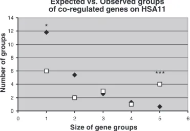
of at least 5 genes in the theoretical class), we obtained a significant test only for human chromosome 11 (HSA11) (P⫽ 0.037). In addition to this global test, we performed an intra-class test by comparing the obtained value with the 1,000 simulations. By this method, we could confirm that HSA11 was significantly depleted in singletons (P ⫽ 0.036) and significantly in excess for groups of five consecutive genes homogenously regulated, i.e., found in the same SSH library of clones (P⫽ 0.001). These results for HSA11 are summarized
in Fig. 3. When applying the same strategy, we could also detect significant values for HSA6 (depleted in singletons, P⫽ 0.04) and for HSA12. This last chromosome has a trend toward a depletion in singletons (P⫽ 0.07) and contains a group of seven consecutive hypoxia-induced genes (P⫽ 0.027).
DISCUSSION
In this paper, we studied the alterations in gene expression that occur in human early placental villi exposed to a short period (3 h) of hypoxia (2% oxygen) by the SSH method. The dissected placental villi were kept for 3 h only in hypoxia to identify mainly genes involved in an acute response to hyp-oxia. The biological significance of such a short exposure is relevant at the placental level, since in early pregnancies, acute and local variations of oxygen pressure are supposed to occur Table 4. Contingency tables and significant2values for matched gene categories
Observed Expected
Phosphatases Kinases Phosphatases Kinases
N 8 14 22 N 6.65 20.47 P⫽ 0.03
H 5 26 31 H 9.37 28.84
13 40 43
Observed Expected
Growth Proliferation Growth Proliferation
N 19 7 26 N 14.68 11.32 P⫽ 0.02
H 16 20 36 H 20.32 15.68
35 27 62
Boldface text indicates the observed no. of genes in each category. N, normoxic; H, hypoxic.
Fig. 2. Real-time PCR analysis of genes found by the SSH procedure to be hypoxia induced and specific for angiogenesis. Fig. 2 has been divided into 2 parts, to take into account the fact that the basal and induced levels of expres-sion are very different for the genes analyzed. The results are given in folds of induction deduced from the C0 values (for explanation of C0 values, seeRESULTS) of the real-time PCR, subtracted by the␣-chain of the chorionic go-nadotrophins (CGA) C0 values, CGA being taken as a reporter gene. Statistical compari-sons were performed by a paired t-test to com-pare for each of the 6 placentas studied the levels of expression in normoxia and in hyp-oxia. Significant values obtained for 7 genes among 8 are presented.
104 INFLUENCE OF OXYGEN ON THE TRANSCRIPTOME OF HUMAN EARLY PLACENTAL VILLI
Physiol Genomics•VOL 22 •www.physiolgenomics.org
on September 9, 2010
physiolgenomics.physiology.org
naturally at the feto-maternal interface. Most of these events will not be prolonged toward a genuine ischemic accident but could in some cases be a starting point of the installation of a long-standing oxygen depletion leading ultimately to a placen-tal vascular disease such as preeclampsia. Therefore, identify-ing early modifications of gene expression due to an acute but short oxygen depletion could point out relevant early markers of placental disease. Three hours correspond to a short but sufficient duration for allowing the oxygen tension to equili-brate with the atmospheric pressure in the medium, as we and others have demonstrated (1). An additional interest in using such short exposure resides in the fact that, after a 3-h lap, factors such as cell death and necrosis will not be relevant.
Several studies indicate that the normal oxygen pressure of the intervillous space in front of placental villi at early stages of pregnancy is low [estimated at 20 mmHg (2.6%) around 8 wk of gestation, but rising to 50 mmHg (6.6%) at 12 wk and to 60 mmHg (7.9%) at the middle of the second trimester; Refs. 16, 22, 34]. At the anatomic level, this first-trimester hypoxia is due to a plug of cytotrophoblasts present in the distal part of the anchoring villi that forms several columns of cells. This proliferation is associated with physiological hypoxic condi-tions (19). Aside from the fetal oxygen consumption, the placental consumption has been evaluated at⬃50% of the total input, one-half of which is involved in oxidative phosphoryla-tions in mitochondria (5, 23, 32).
In our experiment, the placenta used for the SSH was 11 wk old, suggesting that the villi were exposed to a genuine but moderate hypoxia compared with physiological conditions. This is illustrated by the fact that the VEGF transcript was differentially enriched in the two reciprocal SSH libraries. The signal was visible after 25 cycles of PCR, which shows that even in the 2-20 library, VEGF is not a frequent transcript. This substantiates the observation of Kumazaki et al. (20), who could barely detect the VEGF mRNA in hypoxic/ischemic placentas while the protein was clearly detectable by immuno-histochemistry.
We based our analysis on placental villi, a native tissue composed of several cell types (cytotrophoblasts, syncytiotro-hoblast, endothelial cells, lymphocytes, macrophages, vascular smooth muscle cells, etc.). This approach is common in the field (15, 27) and enables one to spot differences that are representative of the complexity of the physiological situation, although the use of complex tissue mainly reveals large dif-ferences affecting relevant biological markers, as a specific cell type may adapt to ameliorate the overall increase or decrease of expression. Complementary studies analyzing the transcrip-tome of specific cell types can be performed in the future to help specify which cell types are involved in the activation of a given gene. The recent study of Ning et al. (25) was based on the serial analysis of gene expression (SAGE) of endothelial cell transcriptome alterations under hypoxia. Although the groups of genes represented in this study are similar to ours, the size of the groups differs. The authors discovered specific alterations of heat shock factors, glycolytic enzymes, extracel-lular matrix factors, cytoskeletal factors, apoptotic factors, cell cycle regulators, and angiogenic factors.
We used an approach based on the systematic sequencing of the SSH products and gene analysis by functional categories. The choice of using SSH was based on the assumption that we would be able to discover factors expressed at a lower level than genes classically detected (33). This approach has been successful, since we could demonstrate that a short exposure to hypoxia is able to modify a group of important cellular func-tions known to be of particular relevance in early placenta when oxygen pressure varies. In a first step, we based our analysis on the genes divided into three categories: insensitive, hypoxic, and normoxic. However, in this analysis, the high number of significant differences in various biological func-tions was attributable to a marked discrepancy between the expected and observed number of genes in the insensitive category. For instance, for genes involved in proteasome ac-tivity, eight genes were theoretically expected but none could be found. This means that the genes implicated in this function were never found simultaneously in both libraries. Similarly, genes involved in transcriptional regulation or genes of un-known function were much less abundant than expected in the mixed category, suggesting that they can be found in hypoxia as well as in normoxia. However, each of them clearly appears to be determined by the oxygen status. Among transcriptional factors that were found more than once in the 2-20 library (hypoxia induced), we find ZNF83, QKI, SQSTM1, and HIP2. The fact that transcription factors appear clearly determined by the oxygen concentration implies that both conditions (hypoxic and normoxic) could be induced by two specific families of transcription factors. Therefore, a new analysis was performed, considering only the genes that were found exclusively in one physiological state. This allowed us to drastically reduce the number of significantly different cellular functions. One of the classes most clearly identified as “hypoxia induced” was an-giogenesis. There is abundant literature about the effects of oxygen on placental vasculogenesis and angiogenesis (see, for instance, Refs. 9, 11, 14, 18, 20, 28, 38). However, most of these studies have focused on factors known to be involved in hypoxia sensing and vasculo/angiogenesis, such as VEGF, VEGFR1 (FLT-1), VEGFR2 (KDR), ANG-1, ANG-2, TIE, and PlGF. In our clones, only FLT-1 belongs to this category, but we could also discover lesser-known actors, such as AMOTL2,
Fig. 3. Analysis of the clustering of “hypoxic/normoxic” genes on human chromosome 11 (HSA11). Squares represent the actual number of clusters observed on the chromosome and are classified according to their size (no. of “normoxic” or “hypoxic” consecutive genes). Diamonds represent the ex-pected no. of clusters, according to 1,000 random repetitions of a Monte Carlo simulation (seeMATERIALS AND METHODS). The extreme classes [singletons and 5 consecutive genes] appear significantly under- or overrepresented in the actual data set, respectively. SeeRESULTSfor statistical thresholds.
105
INFLUENCE OF OXYGEN ON THE TRANSCRIPTOME OF HUMAN EARLY PLACENTAL VILLI
Physiol Genomics•VOL 22 •www.physiolgenomics.org
on September 9, 2010
physiolgenomics.physiology.org
ANGPTL4, and THBS1. Therefore, our SSH approach suggests interesting new targets for future studies. Similarly, it was not at all unexpected to identify genes involved in cell prolifera-tion, as the normal hypoxic conditions pertaining at the tip of first-trimester placental villi are triggering the proliferation of cytotrophoblasts in cell columns, previous to their transforma-tion in invading cytotrophoblasts. In parallel, cytotrophoblasts of the floating villi also have the potential of becoming prolif-erative at the end of the first trimester, showing that the low oxygen concentration at the intervillous space affects all these cells (2). The molecular basis of this proliferation and changes of phenotype is not as well elucidated as those of the angio-genesis pathways in the early placenta. Therefore, the genes we have identified constitute interesting candidates for further studies. The discovery of an excess of apoptotic genes in the 20-2 library is also indicative of the general status of the tissue, where a relative hypoxia may appear as the normal situation. Contrary to the other significant functions that were uncovered by our approach, the excess of kinases over phosphatases in the 2-20 library has not been previously described in the literature. The significance of this excess remains to be explored at the individual gene level.
In addition to our results on gene expression, we have shown that there seems to be a high-level organization of gene function in wide chromosome regions. This is particularly the case for HSA11. One of the four hypoxia-inducible regions of more than five consecutive genes found on this chromosome corresponds to the well-known cluster of imprinted genes found at 11p15.5. This substantiates the importance of this gene cluster for placental physiology, and particularly the importance of IGF2 and H19 as major actors of correct placental development (8, 31). Our findings add another di-mension showing that external factors may contribute to the regulation of gene expression. This is further strengthened by the sequence analysis of the promoters of H19 and IGF2, which displays an extreme richness in CpG islands and puta-tive HIF1␣-binding sites. The existence of coregulated gene clusters in eukaryotic genomes is becoming substantiated by an increasing number of reports (4, 7). The explanation of such a long-range regulation remains to be clarified but will probably be accessible in the near future by techniques of chromatin analysis, which recently have been applied successfully to the Igf2/H19 region in mice (24).
In conclusion, our approach made it possible to identify the cellular functions in the first-trimester placental villi that are strongly modified by a short exposure to particular concentra-tions of oxygen, as well as the funcconcentra-tions that remain un-changed. The relevance of studying the effects of oxygen at the end of the first trimester is clear, since oxygen is recognized at this stage as critically important for a successful colonization of maternal uterine arteries by invasive trophoblasts (17). This is consistent with the fact that, during the first trimester, the low level of endovascular trophoblastic invasion leads to a minimal maternal blood flow to the placenta and therefore to the creation of a hypoxic environment, concomitant with a very high level of placental development, compared with that of the embryo (12, 13). During the invasion, the plugs of cytotropho-blasts are expelled and the cells encounter a positive oxygen pressure gradient that may play an important part in the differentiation of invasive cytotrophoblast cells and their relo-calization in the media of maternal uterine blood vessels. This
colonization of the mother circulation is thought to be the key pathway leading to the loss of the contractile properties of maternal uterine spiraled arteries and hence toward the input of a considerable amount of blood and nutrients in direction of the feto-placental unit (17). Indeed, many experimental results substantiate the major role of oxygen pressure in the normal physiology of cytotrophoblast cells. It is possible to hypothe-size that chronic hypoxia at this stage may constitute an early indication of risk for the major placental pathologies pre-eclampsia and intrauterine growth retardation. Surprisingly, only a few studies have been performed that analyze the effects of oxygen on gene expression in placental villi. A recent study by Tsui et al. (37) makes use of commercially available microarrays and identifies several genes that appear as markers of preeclampsia. However, commercial arrays do not make it possible to easily identify rare transcripts such as transcription factors that may be relevant for explaining the physiology of complex diseases. The complementary roles of SSH and mi-croarrays have recently been thoroughly analyzed in a study (3) involving the in vitro differentiation of human monocytes to myeloid dendritic cells. In this study, SSH products were used as probes for hybridizations on Affymetrix DNA chips (3). It clearly demonstrates that both techniques are complementary and make it possible to discover different sets of genes. We hope the present study will contribute to the identification of new factors that may become powerful tools for deciphering the complexities of placental physiopathology.
REFERENCES
1. Allen CB, Schneider BK, and White CW. Limitations to oxygen diffusion and equilibration in in vitro cell exposure systems in hyperoxia and hypoxia. Am J Physiol Lung Cell Mol Physiol 281: L1021–L1027, 2001.
2. Aplin JD, Haigh T, Jones CJ, Church HJ, and Vicovac L. Develop-ment of cytotrophoblast columns from explanted first-trimester human placental villi: role of fibronectin and integrin-␣51. Biol Reprod 60: 828 – 838, 1999.
3. Cao W, Epstein C, Liu H, DeLoughery C, Ge N, Lin J, Diao R, Cao
H, Long F, Zhang X, Chen Y, Wright PS, Busch S, Wenck M, Wong K, Saltzman AG, Tang Z, Liu L, and Zilberstein A. Comparing gene
discovery from Affymetrix GeneChip microarrays and Clontech PCR-select cDNA subtraction: a case study. BMC Genomics 5: 26, 2004. 4. Caron H, van Schaik B, van der Mee M, Baas F, Riggins G, van Sluis
P, Hermus MC, van Asperen R, Boon K, Voute PA, Heisterkamp S, van Kampen A, and Versteeg R. The human transcriptome map:
clus-tering of highly expressed genes in chromosomal domains. Science 291: 1289 –1292, 2001.
5. Carter AM. Placental oxygen consumption. Part I: in vivo studies—a review. Placenta 21, Suppl A: S31–S37, 2000.
6. Chomczynski P. A reagent for the single-step simultaneous isolation of RNA, DNA and proteins from cell and tissue samples. Biotechniques 15: 532–534, 536 –537, 1993.
7. Cosseddu GM, Perez-Enciso M, Fellous M, and Vaiman D. Interspe-cific chromosome-wide transcription profiles reveal the existence of mam-malian-specific and species-specific chromosome domains. J Mol Evol 59: 317–328, 2004.
8. DeChiara TM, Robertson EJ, and Efstratiadis A. Parental imprinting of the mouse insulin-like growth factor II gene. Cell 64: 849 – 859, 1991. 9. De Marco CS and Caniggia I. Mechanisms of oxygen sensing in human
trophoblast cells. Placenta 23, Suppl A: S58 –S68, 2002.
10. Diatchenko L, Lau YF, Campbell AP, Chenchik A, Moqadam F,
Huang B, Lukyanov S, Lukyanov K, Gurskaya N, Sverdlov ED, and Siebert PD. Suppression subtractive hybridization: a method for
generat-ing differentially regulated or tissue-specific cDNA probes and libraries.
Proc Natl Acad Sci USA 93: 6025– 6030, 1996.
11. Dunk C, Shams M, Nijjar S, Rhaman M, Qiu Y, Bussolati B, and
Ahmed A. Angiopoietin-1 and angiopoietin-2 activate trophoblast Tie-2
106 INFLUENCE OF OXYGEN ON THE TRANSCRIPTOME OF HUMAN EARLY PLACENTAL VILLI
Physiol Genomics•VOL 22 •www.physiolgenomics.org
on September 9, 2010
physiolgenomics.physiology.org
to promote growth and migration during placental development. Am J
Pathol 156: 2185–2199, 2000.
12. Genbacev O. To proliferate or to divide—to be or not to be. Early
Pregnancy 5: 63– 64, 2001.
13. Genbacev O and Miller RK. Post-implantation differentiation and pro-liferation of cytotrophoblast cells: in vitro models—a review. Placenta 21,
Suppl A: S45–S49, 2000.
14. Geva E, Ginzinger DG, Zaloudek CJ, Moore DH, Byrne A, and Jaffe
RB. Human placental vascular development: vasculogenic and angiogenic
(branching and nonbranching) transformation is regulated by vascular endothelial growth factor-A, angiopoietin-1, and angiopoietin-2. J Clin
Endocrinol Metab 87: 4213– 4224, 2002.
15. Gratton RJ, Gluszynski M, Mazzuca DM, Nygard K, and Han VK. Adrenomedullin messenger ribonucleic acid expression in the placentae of normal and preeclamptic pregnancies. J Clin Endocrinol Metab 88: 6048 – 6055, 2003.
16. Jauniaux E, Watson A, and Burton G. Evaluation of respiratory gases and acid-base gradients in human fetal fluids and uteroplacental tissue between 7 and 16 weeks’ gestation. Am J Obstet Gynecol 184: 998 –1003, 2001.
17. Kaufmann P, Black S, and Huppertz B. Endovascular trophoblast invasion: implications for the pathogenesis of intrauterine growth retar-dation and preeclampsia. Biol Reprod 69: 1–7, 2003.
18. Kaufmann P, Mayhew TM, and Charnock-Jones DS. Aspects of human fetoplacental vasculogenesis and angiogenesis. II. Changes during normal pregnancy. Placenta 25: 114 –126, 2004.
19. Kingdom JC and Kaufmann P. Oxygen and placental villous develop-ment: origins of fetal hypoxia. Placenta 18: 613– 621; discussion 623– 616, 1997.
20. Kumazaki K, Nakayama M, Suehara N, and Wada Y. Expression of vascular endothelial growth factor, placental growth factor, and their receptors Flt-1 and KDR in human placenta under pathologic conditions.
Hum Pathol 33: 1069 –1077, 2002.
21. Lashkari A, Smith AK, and Graham JM Jr. Williams-Beuren syn-drome: an update and review for the primary physician. Clin Pediatr
(Phila) 38: 189 –208, 1999.
22. Maltepe E and Simon MC. Oxygen, genes, and development: an analysis of the role of hypoxic gene regulation during murine vascular develop-ment. J Mol Med 76: 391– 401, 1998.
23. Mover-Lev H, Dreval D, Zakut H, and Ar A. O2consumption in the
in-vitro fetal side human placenta. Respir Physiol 106: 199 –208, 1996. 24. Murrell A, Heeson S, and Reik W. Interaction between differentially
methylated regions partitions the imprinted genes Igf2 and H19 into parent-specific chromatin loops. Nat Genet 36: 889 – 893, 2004.
25. Ning W, Chu TJ, Li CJ, Choi AM, and Peters DG. Genome-wide analysis of the endothelial transcriptome under short-term chronic hyp-oxia. Physiol Genomics 18: 70 –78, 2004.
26. Potter CF, Kuo NT, Farver CF, McMahon JT, Chang CH, Agani FH,
Haxhiu MA, and Martin RJ. Effects of hyperoxia on nitric oxide
synthase expression, nitric oxide activity, and lung injury in rat pups.
Pediatr Res 45: 8 –13, 1999.
27. Rajakumar A, Doty K, Daftary A, Harger G, and Conrad KP. Impaired oxygen-dependent reduction of HIF-1␣ and -2␣ proteins in pre-eclamptic placentae. Placenta 24: 199 –208, 2003.
28. Regnault TR, de Vrijer B, Galan HL, Davidsen ML, Trembler KA,
Battaglia FC, Wilkening RB, and Anthony RV. The relationship
be-tween transplacental O2 diffusion and placental expression of PlGF,
VEGF and their receptors in a placental insufficiency model of fetal growth restriction. J Physiol 550: 641– 656, 2003.
29. Robillard PY, Dekker GA, and Hulsey TC. Evolutionary adaptations to pre-eclampsia/eclampsia in humans: low fecundability rate, loss of oes-trus, prohibitions of incest and systematic polyandry. Am J Reprod
Immunol 47: 104 –111, 2002.
30. Robillard PY, Hulsey TC, Dekker GA, and Chaouat G. Preeclampsia and human reproduction. An essay of a long term reflection. J Reprod
Immunol 59: 93–100, 2003.
31. Sasaki H, Ferguson-Smith AC, Shum AS, Barton SC, and Surani MA. Temporal and spatial regulation of H19 imprinting in normal and unipa-rental mouse embryos. Development 121: 4195– 4202, 1995.
32. Schneider H. Placental oxygen consumption. Part II: in vitro studies—a review. Placenta 21, Suppl A: S38 –S44, 2000.
33. Seta KA and Millhorn DE. Functional genomics approach to hypoxia signaling. J Appl Physiol 96: 765–773, 2004.
34. Soothill PW, Nicolaides KH, Rodeck CH, and Campbell S. Effect of gestational age on fetal and intervillous blood gas and acid-base values in human pregnancy. Fetal Ther 1: 168 –175, 1986.
35. Tassabehji M. Williams-Beuren syndrome: a challenge for genotype-phenotype correlations. Hum Mol Genet 12: R229 –R237, 2003. 36. Tolle A, Kolleck I, Schlame M, Wauer R, Stevens PA, and Rustow B.
Effect of hyperoxia on the composition of the alveolar surfactant and the turnover of surfactant phospholipids, cholesterol, plasmalogens and vita-min E. Biochim Biophys Acta 1346: 198 –204, 1997.
37. Tsui NB, Chim SS, Chiu RW, Lau TK, Ng EK, Leung TN, Tong YK,
Chan KC, and Lo YM. Systematic micro-array based identification of
placental mRNA in maternal plasma: towards non-invasive prenatal gene expression profiling. J Med Genet 41: 461– 467, 2004.
38. Zygmunt M, Herr F, Munstedt K, Lang U, and Liang OD. Angiogen-esis and vasculogenAngiogen-esis in pregnancy. Eur J Obstet Gynecol Reprod Biol 110, Suppl 1: S10 –S18, 2003.
107
INFLUENCE OF OXYGEN ON THE TRANSCRIPTOME OF HUMAN EARLY PLACENTAL VILLI
Physiol Genomics•VOL 22 •www.physiolgenomics.org
on September 9, 2010
physiolgenomics.physiology.org