HAL Id: hal-01635553
https://hal.archives-ouvertes.fr/hal-01635553
Submitted on 15 Nov 2017
HAL is a multi-disciplinary open access
archive for the deposit and dissemination of sci-entific research documents, whether they are pub-lished or not. The documents may come from teaching and research institutions in France or abroad, or from public or private research centers.
L’archive ouverte pluridisciplinaire HAL, est destinée au dépôt et à la diffusion de documents scientifiques de niveau recherche, publiés ou non, émanant des établissements d’enseignement et de recherche français ou étrangers, des laboratoires publics ou privés.
Cu(II) binding to various forms of amyloid-β peptides.
Are they friends or foes ?
Valentina Borghesani, Bruno Alies, Christelle Hureau
To cite this version:
Valentina Borghesani, Bruno Alies, Christelle Hureau. Cu(II) binding to various forms of amyloid-β peptides. Are they friends or foes ?. European Journal of Inorganic Chemistry, Wiley-VCH Verlag, 2018, pp.7-15. �10.1002/ejic.201700776�. �hal-01635553�
1
Cu(II) binding to various forms of amyloid-β peptides. Are they
friends or foes ?
Valentina Borghesani,a,b Bruno Aliesc and Christelle Hureau a,b,*
a
CNRS, LCC (Laboratoire de Chimie de Coordination), 205 route de Narbonne, BP 44099 31077 Toulouse Cedex 4, France.
christelle.hureau@cc-toulouse.fr https://www.lcc-toulouse.fr/auteur59.html
b
University of Toulouse, UPS, INPT, 31077 Toulouse Cedex 4, France.
c
Current address: Université de Bordeaux, ChemBioPharm INSERM U1212 CNRS UMR 5320, Bordeaux, France
Abbreviations:
A: Amyloid- Peptides ; AD: Alzheimer Disease ; AFM: Atomic Force Microscopy; APP: Amyloid
Precursor Protein ; ATCUN: Amino Terminal Cu(II) and Ni(II) binding motif (H2N-Xxx-Yyy-His) ;
CCA: Coumarin Carboxylic Acid ; CD: Circular Dichroism ; CSF : Cerebro-Spinal Fluid ; EPR: Electron Paramagnetic Resonance ; EXAFS: Extended X-Ray Absorption Fine Structure ; FRET: Fluorescence Resonance Energy Transfer; ITC: Isothermal Titration Calorimetry ; H2-DCF :
di-chlordihydrofluorescein diacetate ; ROS: Reactive Oxygen Species ; SCE: Saturated Calomel Electrode ; TBARS : 2-Thiobarbituric Acid Reactive Substances; TEM : Transmission Electron Microscopy ; ThT: Thioflavine T ; XANES: X-Ray Absorption Near Edge Structure.
Abstract:
In the present micro-review, we describe the Cu(II) binding to several forms of amyloid- peptides, the peptides involved in Alzheimer’s disease. It has indeed been shown that in addition to the “full-length” peptide originating from the precursor protein after cleavage at position 1, several other shorter peptides do exist in large proportion and may be involved in the disease as well. Cu(II) binding to amyloid- peptides is one of the key interactions that impact both the aggregating properties of the amyloid peptides and the Reactive Oxygen Species (ROS) production, two events linked to the etiology of the disease. Binding sites and affinity are described in correlation with Cu(II) induced ROS formation and Cu(II) altered aggregation, for amyloid peptides starting at position 1, 3, 4, 11 and for the corresponding pyroglutamate forms when they could be obtained (i.e. for peptides cleaved at positions 3 and 11). It appears that the current paradigm which points out a toxic role of the Cu(II) – amyloid- interaction might well be shifted towards a possible protective role when the peptides considered are the N-terminally truncated ones.
2
1. Introduction
1.1 Alzheimer’s disease (AD)
Alzheimer’s disease (AD) is a neurodegenerative disorder characterized by progressive cognitive and memory impairments.[1, 2] AD is the most common type of dementia in the elderly population representing 60-80% of all cases. There are two neurohistological hallmarks of AD: the amyloid (or senile) plaques, which are extracellular deposits of insoluble amyloid- (A) peptides under aggregated forms and the intracellular tau tangles.[3-5] The mechanisms underlying the triggering and progression of the disease are still unclear. Nevertheless, according to the amyloid cascade hypothesis first proposed in the 90’s,[6] the imbalance between production and clearance of the A peptides is at the origin of the accumulation of peptides that further leads to the aggregation of the soluble monomeric peptide into fibrils and amorphous aggregates detected in the senile plaque.[3-9] The amyloid cascade is an early event and plays a causal role in AD (Scheme 1). The complete aggregation pathway from the monomeric peptide to the fibrils contains several steps including oligomers, protofibrils and fibrils. Oligomeric species have been proposed to be the most toxic species, instigating further pathological events, including the formation of intracellular neurofibrillary tangles, destruction of synaptic connections, which would lead ultimately to neuronal cell death and dementia.[10-13] Metal ions have been involved in the amyloid cascade process based on the detection of high levels of copper and zinc in the senile plaques and on in vitro studies probing their abilities to modify the aggregation process.[14-18] In addition, the redox activity of copper is responsible for the production of Reactive Oxygen Species (ROS) that contribute to the oxidative stress observed in AD.[16, 19-23]
3
Scheme 1. Toxic events in the framework of the amyloid cascade hypothesis: (over)-production of the
amyloidogenic A peptide, formation of oligomeric species and ROS production associated to the formation of metal-A complexes.
1.2 The amyloid- peptides
Several A peptides of various lengths are found in vivo and while the A1-40/42 i
have been the focus of most of the reported studies, N-terminally truncated forms are also of interest since they represent a significant amount of the A content in the brain (> 60 %). In addition, the N-terminal modifications do alter dramatically the Cu(II) binding abilities of the peptides. This will be the focus of the next paragraphs.
a. APP cleavage
The A peptides originate from the cleavage of the transmembrane Amyloid Precursor Protein (APP)
by the and γ-secretases, acting at the N- and C-terminal sites, respectively (Scheme 1). Because the C-terminal part of the A sequence is buried in the membrane, proteolysis of the APP by the γ-secretase occurs at several sites and A1-38 to A1-43 peptides can be processed.[2]
b. The full-length peptides
Among the “full-length” peptides (corresponding in the present review to A1-38 - A1-43) the 40 and
42 amino-acid residues forms are the most abundant in the Cerebro-Spinal Fluid (CSF), with the A
i The peptides are noted A
n-m/p/q. This corresponds to a peptide starting at position n and ending at position m,
4
42 representing 5-10% of theA1-40 amount. [2]
The longer form is the most toxic in line with a higher propensity to aggregate. The A peptides encompasses a N-terminal binding site (spanning from residues 1 to 16) and a C-terminal hydrophobic sequence involved in the aggregation process. Hence, it is considered that the A1-16 peptide is an appropriate model with regard to metal ion binding. This
sequence is classically used in spectroscopic studies due to its higher solubility that eases its handling. It has also been extensively used to study the coordination of Cu(II), Cu(I), Zn(II) and Fe(II) to A.[16]
c. N-terminally truncated peptides
As for the C-terminal site, the N-terminal site undergoes some cleavage heterogeneity with peptides starting at positions between 1 and 5.[9, 24-26] The reason for such heterogeneity is unclear. In seminal reports by Masters and coworkers,[27, 28] the quantity of A4-n was found to be predominant in the
amyloid deposits of AD patients. This was further confirmed by mass-spectrometry analyses revealing that the dominating A species in AD brains are A4-42, A1-40 and A1-42,
[25]
and suggesting that N-terminally truncated peptides are abundant in aged human brains.[24] A11-n peptides are also detected
in plaques from sporadic AD brains representing up to 20% of the total content of A peptides.[29]
When the N-terminal deletion occur at position 3 or 11 corresponding to Glu residues, the formation of pyroglutamate groups can occur, leading to Ap3-n and Ap11-n, respectively. The pyroglutamate is
generated by the action of the enzyme glutaminyl cyclase[30] and/or spontaneously but slowly over time.[29]
The various forms of the A peptides discussed in the text are reminded in Scheme 2.
5 10 15 20 25 30 35 40
Aβ1-16/28/40/42 DAEFR HDSGY EVHHQ KLVFF AEDVG SNKGA IIGLM VGGVV IA
Aβ3-16/28/40/42 EFR HDSGY EVHHQ KLVFF AEDVG SNKGA IIGLM VGGVV IA
Aβp3-16/28/40/42 pEFR HDSGY EVHHQ KLVFF AEDVG SNKGA IIGLM VGGVV IA
Aβ4-16/28/40/42 FR HDSGY EVHHQ KLVFF AEDVG SNKGA IIGLM VGGVV IA
Aβ11-16/28/40/42 EVHHQ KLVFF AEDVG SNKGA IIGLM VGGVV IA
Aβp11-16/28/40/42 pEVHHQ KLVFF AEDVG SNKGA IIGLM VGGVV IA
Scheme 2. Sequence of N-terminally truncated Aβ peptides discussed in the present review.
d. Cu(II) binding to the A peptides
In the present micro-review, we focus on Cu(II) binding to the various A peptides. It is indeed anticipated that the N-terminal modifications impact significantly the Cu(II) site and affinity that will further induce changes in ROS production and aggregation of the Cu-peptide complexes, two key events linked to the amyloid cascade.
5
2. Techniques to study Cu(II) coordination, affinity, ROS production
and peptide aggregation
2.1 Coordination sites
There are several techniques to probe the Cu(II) site in a peptide, Electron Paramagnetic Resonance (EPR) being the most appropriate due to its easy access and the large quantity of information that can be extracted from a spectrum. Indeed the values of g//, g, and of the hyperfine and super-hyperfine
couplings give access to the geometry and the nature of the binding atoms.[31-34] Other techniques
include UV-Vis and CD, X-ray absorption spectroscopy (both EXAFS and XANES),[35] and NMR.[32]
The redox properties of the Cu-peptide complexes can be evaluated by cyclic or square-wave voltammetries.[32, 36, 37]
2.2 Affinity
There are mainly two ways of evaluating the affinity of Cu(II) for a peptide: direct titration or indirect competition experiments. In general, for direct titration experiment, quenching of the fluorescence of Tyr10 (in case of A, but could also be Trp residues) or calorimetric (ITC) measurements are used.[38,
39]
For competition experiments, any competitor having an affinity close to the one of the peptide and leading to a Cu(II) complex with a spectroscopic signature (in EPR, UV-Vis or CD) different than that the Cu(II) peptidic species can be used (see for instance, ref. [40]). Potentiometry is another accurate and direct method of establishing affinity constants. Protonation and stability constants for the Cu(II) complexes of short Aβ peptides, such Aβx-16, can be calculated from pH-metric acid-base titrations.
[39]
2.3 ROS production
ROS production that corresponds to the incomplete (Cu-catalyzed) reduction of O2 can be monitored
by several indirect methods and as a function of time (Scheme 3). Consumption of ascorbate, the most classically used reductant, can be followed by UV-Vis spectroscopy at 265 nm.[22] It is almost impossible to probe the formation of superoxide due to its high rate of reaction with ascorbate (and reduced ascorbate). Superoxide was detected via the reduction of CytC only when the Cu(A) was reduced by electrolysis.[41] There are several tools to detect the formation of hydrogen peroxide,[22]
such as the amplex red assay (formation of the resorufin luminophore) and H2DCF assay (formation of
2,7-dichlorofluorescein) and the formation of the hydroxyl radical, such as the CCA assay (formation
6
Scheme 3. Catalytic production of ROS by Cu(Aβ), from dioxygen and ascorbate as reductant and
some methods to detect the production of the various ROS by spectrophotometry.
2.4 Aggregation
The aggregation of amyloid peptides is driven by the auto-catalytic self-assembly of monomeric peptides leading to the formation of -sheet rich fibrils. While the kinetic of fibrils formation is
mathematically described[42] by a sigmoid-like curve (see equation 1 and Scheme 4), the mechanism of
amorphous aggregates formation is not well-understood.
The fibril formation displays three distinct phases. The initial phase is the lag phase, as related to the nucleation process, followed by a steep growing phase corresponding to the elongation process up to a stationary phase when the system have reached the thermodynamic equilibrium (Scheme 4). The first stage (lag phase linked to the nucleation process) is the kinetically limiting step where monomeric peptides dimerize and/or oligomerize. When a critical amount of -sheets is reached, fast auto-assembly occurs (elongation step) finally leading to fibrils. As the system is now at its equilibrium, no more change is observed, this is the stationary phase. In order to describe the fibrils formation, two parameters are generally considered: the quantity of fibrils formed for a given starting concentration of monomeric peptides and the half-time (t1/2), which is the time needed to reach half of the maximum of
fibrils content.[43-46]
7
Where k is the elongation rate, A the amplitude, t1/2 the time point when half of the maximal intensity
is reached, and F0 the baseline before aggregation.
In vitro, the Aβ aggregation process can monitored as a function of time by several spectroscopic
techniques:[47] (i) CD by probing -sheets content,[48] (ii) raman and infra-red spectrosocopy, which provide information on secondary and tertiary structures changes at all stages of fibrillation,[49] (iii) nuclear magnetic resonance (NMR); liquid and solid state,[50-52] (iv) turbidity and light scattering,[53, 54] (v) native gel electrophoresis[55] and (vi) by addition of chromophores either changing their UV-visible absorption upon interactions with -sheets (like Congo Red for instance)[56] or their fluorescent properties.[57, 58] A very classical assay relies on the use of Thioflavin-T (ThT), a dye that shows a characteristic red shift in the excitation spectrum and strong enhancement of fluorescence quantum yield upon binding to fibrillary structures.[59-62]
Amyloid fibrils are characterized by laminated β-sheets whose strands run perpendicular to the long-axis of the fibril.[59, 62, 63] This amyloid composition produces the characteristic ~5 Å and ~10 Å reflections observed in X-ray diffraction experiments, which are attributed to the strand spacing within and between β-sheet layers, respectively.[64]
The detection of intermediates species, especially oligomers, is more challenging. Nevertheless, various techniques can be used.[65] These methods include recognition by specific antibodies,[66] mass spectrometry,[45] FRET,[58] and electrophoresis on native gel.[67]
Furthermore, morphology of the aggregates formed can be evaluated by microscopy (Transmission Electron or Atomic Force Microscopy).[49, 68]
Each of these techniques offers advantages but also drawbacks in terms of technique-related artifacts,[45] which can anyway be overcome by using multiple independent analytical techniques in parallel.
8
Scheme 4. Scheme of the Aβ peptide self-assembly process. Applicability range for the techniques
used to monitor the different phases of the aggregation process.
3. Cu(II) and “full-length” A
peptides
The human Aβ binds preferentially one Cu(II) ion in the N-terminal domain. The rest of the sequence can modify the affinity maybe via second sphere interactions. The Aβ1-16 has been recognized as a
trustworthy model for the study of Cu(II) binding to the full-length peptide. Binding of additional equivalents of Cu(II) ion has been reported[69] but is likely not biologically relevant and thus will not be commented on in the present micro-review.
Coordination site. The Cu(II) binding sites to human Aβ peptides have been extensively studied with
various complementary techniques (e.g. EPR and advanced EPR, NMR, UV-Vis and CD, Raman and potentiometry) and has been recently reviewed.[16, 33, 34] Briefly, around neutral pH, the Cu(II)-(Aβ) complex exists in two main forms, noted component I (predominant at lower pH) and II (predominant at higher pH) (Scheme 5.A). Component II is obtained from component I by the deprotonation of an amide bond of the peptide backbone and subsequent binding to the Cu(II). The pKa corresponding to the transition between I and II is near pH 7.8. Both forms bind the Cu(II) in a distorted square planar geometry. In component I, Cu(II) is equatorially bound to Asp1 via the terminal amine, the adjacent CO from the Asp1-Ala2 peptide bond, a N atom from imidazole ring of His6 and another one from His13 (component Ia) or from His14 (component Ib) or two N atoms from the His13 and 14 (component Ic). The apical position may be occupied by an oxygen atom, coming either from a water molecule,[70] or from carboxylate groups from side chains of Asp or Glu.[71] In component II, the main equatorial Cu(II) ligands are the N-terminal amine, the amidyl function from the Asp1-Ala2 peptide
9 bond, the adjacent CO from the Ala2-Glu3 peptide bond, and one N atom from the imidazole ring of
one of the three His. The carboxylate group from Asp1 has been proposed as apical ligand,[70] likely
via a water molecule as bridge.
Scheme 5. Proposed coordination sites of Cu(II)-hAβ (A), Cu(II)-Aβ3-16 (B), Cu(II)-Aβp3-16 (C),
Cu(II)-Aβ4-16 (D) and Cu(II)-Aβ11-16 (E).
Affinity. As stated above, the high affinity Cu(II) binding site is located in the first 16 N-terminal
residues. There are many reports on the evaluation of the Cu(II) affinity for the A peptide. The divergence in the affinity value between studies originated from the use of different evaluation methods.[39] This has recently been sorted out,[38, 40] and a conditional value of 1.6 109 M-1 at pH 7.1 and of 1.1 1010 M-1 at pH 7.4 for the A1-16 make consensus (the conditional value is the value at a
10
given pH with no competing buffer).[72] The Cu(II) affinity seems to weakly increase with the length
of peptide.[38, 40, 72, 73]
ROS production. The ability of Cu(Aβ) complexes to generate ROS is in line with the hypothesis that
they participate in the oxidative stress observed in AD brains post mortem.[74] Such an ability relies on the possibility to have a reaction between the reductant (the physiological one is ascorbate) and the CuII(Aβ) and between the CuI(Aβ) and dioxygen, superoxide and/or hydrogen peroxide. Those reactions were recently probed by several methods (see § 2.3) and it was shown that (i) the Cu(Aβ) do produce ROS, (ii) the rate of ROS production is weaker than for “loosely bound” Cu, corresponding to Cu in buffer and (iii) the rate is significant if compared to other biologically relevant peptides, such as GHK and DAHK.[74-79] A direct comparison between ROS production by “loosely bound” Cu and Cu(Aβ) species is difficult since it will depend on the experimental conditions (concentrations in Cu, ascorbate, dioxygen and peptides). Hence, the ascorbate consumption rate of Cu(Aβ) is about half that of “loosely bound” Cu. The length of the peptide has almost no impact on the ROS production rate, but because the peptide can itself be oxidized by the ROS produced, the quantity of ROS escaping the system is weaker when the peptide is longer. [74]
Aggregation. The effect of the Cu(II) on Aβ1-40/42 aggregation has been discussed widely,
[17, 43, 80]
and consensus takes long time to be reached. Indeed, a major controversial point is due to the intrinsic complex nature of Aβ aggregates. Amorphous or/and fibrillar aggregates could be formed from monomeric Aβ. Depending of analytical techniques used, researchers were able (or not) to discriminate the nature of the aggregates. Also, experimental parameters such Cu(II) / Aβ stoichiometry or pH seem to have a dramatic impact on the outcome of aggregation assays. Nowadays, studies seem to converge on different aspects. On one hand, the presence of sub-stoichiometric Cu(II) seems to accelerate the formation of fibrillar aggregates[81] likely due to a metal-mediated dimerization,[82, 83] and a stabilization of oligomers.[84, 85] On the other hand, in excess, Cu(II) generates more amorphous and less fibrillar aggregates.[86-88] It has also been shown that removing Cu(II) from Aβ amorphous aggregates using an external chelator restore fibrillary-type aggregates.[89]
Another key parameter in the Aβ aggregation is the pH. It has been shown that Aβ aggregation occurs faster at acidic pH,[90, 91] and Cu(II) is able to interact with Aβ aggregation even at mildly acid pH,[92] in a rapid time frame.[93] Nevertheless, below pH=5, Cu(II) do not impact Aβ aggregation,[82] likely due to its inability to strongly interact with Aβ.[73]
11
4. N-truncated peptides
4.1 ATCUN type motifs
For a long time the “full-length” Aβ1-40/42 peptides (or the corresponding C-terminally truncated forms
Aβ1-16/28) have been the primary focus of Cu(II) binding studies.
[16]
However, it has been reported that the N-truncated Aβ4-42 peptide is one of the major form of Aβ peptides detected in healthy and
diseased brains,[24-28] representing more than 60% of the total brain amyloid pool. N-truncation also lead to the formation of the Aβ11-40/42.
[94-96]
The Aβ4-40/42 and Aβ11-40/42 peptide share a common feature
with respect to Cu(II) binding site, since they present a N-terminal ATCUN (amino-terminal copper
and nickel) motif characterized by the sequence H2N-Xxx-Yyy-His.
[97, 98]
It is worth noting that a main difference between the Aβ4-x and Aβ11-x is the possibility for the latter to form pyroglutamate
counterparts. Hence, while the Aβ11-40/42 have been detected in large quantityin the CSF and amyloid
plaques,[29, 94, 99-101] they can degrade rapidly into pyroglutamate forms.[25, 94]
Such N-terminal truncations, which can also happen at other sites between Asp1 and Arg5, are linked to the enzymatic cleavage activity of secretases that depends on many factors as reviewed in ref. [9]. The formation of the Aβ11-40/42 peptide is due to the activity of a secondary -secretase (’).
[95, 96, 102]
Coordination site. ATCUN containing peptides can bind Cu(II) avidly because the binding site is
made of three adjacent metallacycles of 5, 5 and 6 membered rings, the Cu(II) being bound by the N-terminal amine, the imidazole ring from the His and the two deprotonated peptidic functions in between. [77, 97, 98] The ATCUN coordination mode of Cu(II) to Aβ4-n and to Aβ11-n is predominant from
pH 5, while deprotonation of outer-sphere and non-binding side chains occurs when the pH is increased.[103-105] A weak fifth ligand, a water molecule, can be present as apical ligand. This coordination is analogous to the one proposed for the DAHK tetrapeptide, the N-terminal sequence of
human serum albumin, the structure of which was resolved by X-ray diffraction.[77] The spectroscopic
signatures of Cu(Aβ4-16) and Cu(Aβ11-15) used as models of the Aβ4-42 and Aβ11-42 peptide, are fully
reminiscent of those of the Cu(II) parent complexes, such as Cu(DAHK) and Cu(AAH) (See Table 1). The formation of such ATCUN motif would preclude the formation of ternary species between Cu(Aβ4/11-x) and a chelating drug candidate,
[106]
in contrast to what has been reported in case of the Aβ1-x peptide.
[106-108]
Affinity. While Cu(II) binds to Aβ1-16 with a relatively high affinity (Ka~10 10
M-1 at pH 7.4, see table 1), Aβ4-16 and Aβ11-15, bind Cu(II) with a very high affinity (Ka~10
13.5
M-1 at pH 7.4) through their ATCUN motif (Scheme 5D).[103, 104] Such affinity is perfectly in line with those of other ATCUN
12 peptides.[109, 110] Note that a secondary Cu(II) site is present in Aβ4-16 and is relatively weak (Ka=10
6.7
M-1 at pH=7.4) and separated from the N-terminal site.[103] The more than three orders of magnitude higher affinity for Cu(II) compared to the full-length peptides make these peptides key biological partners in the trafficking of Cu(II) within the synaptic cleft.
ROS production. Up to now, the ROS production has only been studied with the Aβ4-16.It has been
shown that Cu(Aβ4-x) can only be oxidized and at relatively high cathodic potential (0.8 V vs. SCE)
and that, in line with this redox properties, it doesn’t yield ROS under biologically relevant conditions.[111] The other truncated ATCUN-type Aβ peptides Aβ11-x might have similar properties to
those of Aβ4-42. However it remains to be determined if the ATCUN site of Aβ11-x peptides can
undergo redox cycling and generate ROS.
Aggregation. As non-truncated Aβ, Aβ4-40/42 is also able to aggregate and cause cell toxicity and
memory impairment in mice. In vitro experiments have also shown that in ThT fluorescence assay, the maximal ThT intensities reached by Aβ4-38/40/42 were similar to the one of Aβ1-42, but decreased faster.
This could be explained by a burial of ThT-binding site as aggregates evolve. TEM images showed an important difference in the morphology at the aggregation end-point: the N-truncated peptide showed clumps of fibrillar aggregates, while Aβ1-42 show longer individual fbrils.
[112]
Aβ11-40 forms fibrils
faster than Aβ1-40 and Aβ1-42 and itdoes co-assemble with Aβ1-40 but not Aβ1-42. [104, 113]
Cu(II) addition to Aβ11-28/40/42 seems to preclude fibrillary aggregation,
[104, 114, 115]
likely due to the strong 1:1 Cu(II) - ATCUN coordination that destabilize peptide-peptide interactions required for the nucleation of the aggregation process.[44]
4.2 Other motifs
N-terminal truncated peptides in position 3 and their pyroglutamate counterparts have been found in high amount in the brain of Alzheimer’s patients (up to 25% of the plaques content).[116, 117]
These peptides are considered to be more toxic than the full-length ones,[118-131] in link with modified plaque morphology, hampered degradation of the pyroglutamate forms, higher propensity to form β-sheets and impact on full-length peptides aggregation process. Formation of pyroglutamate at position 11 (see previous paragraph) is also possible but its effect has been less studied.[124, 132, 133]
Coordination site. Despite the similarity between the sequences of Aβ1-16 and Aβ3-16, the Cu(II)
binding sites to the Aβ3-16 peptide are significantly different to those of Aβ1-16. The main form of
Cu(Aβ3-16) at pH 7.4 is a component II-like and is shown in Scheme 5C. The equatorial Cu(II)
13
the imidazole ring of His6 and the carbonyl function from the His6-Asp7 bond.[134] Such differences
has been explained by second sphere interactions, such as the size of cycle due to the H-bond between the side chain of Asp1 or Glu3 and the adjacent amide function.
The formation of the pyroglutamate in Aβp3-16 precludes the binding of the N-terminal amine (Scheme
5D) and induces subsequent changes. In the main form present at neutral pH, the Cu(II) is thus bound by two imidazole rings from His 13 and 14, the amidyl function of His13-His14 bond and either carboxylate function or a N atom form the imidazole ring of His6.[134]
Affinity. The affinity values of Cu(II) for the Aβ3-16 and the Aβp3-16 are three and hundred times
weaker than the one of Aβ1-16, respectively. The strong decrease in the affinity of the Aβp3-16 is due to
the preclusion of the N-terminal binding and has also been reported for the N-terminally acetylated Ac-Aβ peptide.[134, 135]
ROS. It has been shown recently that N-terminal modification at the third position significantly slow
down the ROS production rate while keeping the amount of ROS escaping the Cu(Aβ3-16) or Cu(Aβp
3-16) complex unchanged. This was attributed to the importance of the N-terminal Asp1 residues in the
catalytically active Cu(Aβ1-16) species. [74]
Aggregation. To the best of our knowledge, there is no study on the impact of Cu(II) on the
aggregation of Aβ3-40/42, Aβp3-40/42 and Aβp11-40/42.
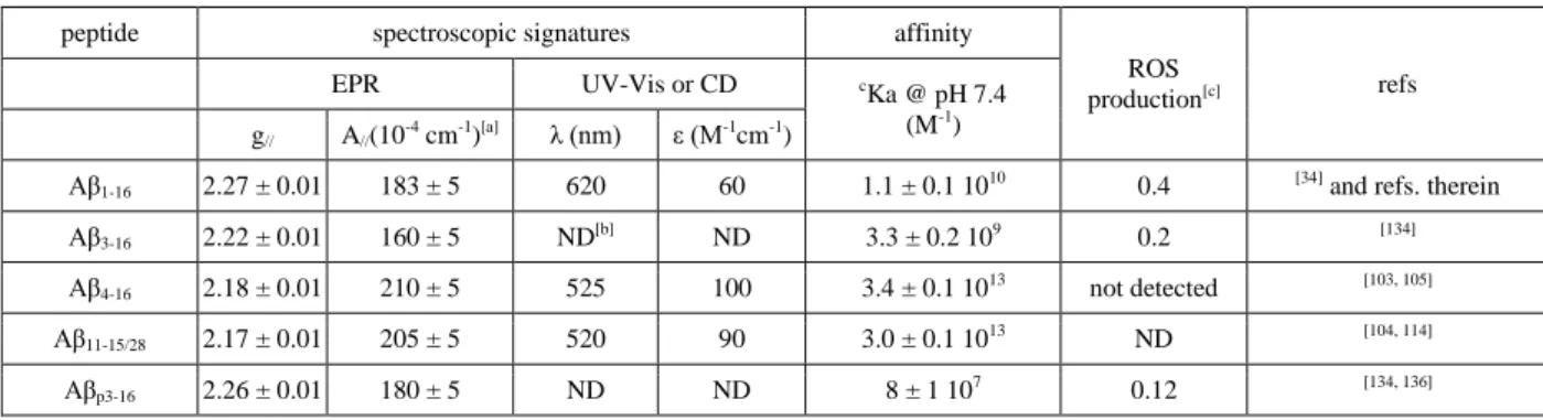
Table 1. Spectroscopic parameters and conditional affinity of Cu(II) bound to a selection of Aβ
peptides and ROS production of the corresponding complex.
peptide spectroscopic signatures affinity
ROS production[c] refs EPR UV-Vis or CD c Ka @ pH 7.4 (M-1) g// A//(10-4 cm-1)[a] λ (nm) ε (M-1cm-1)
Aβ1-16 2.27 ± 0.01 183 ± 5 620 60 1.1 ± 0.1 1010 0.4 [34] and refs. therein
Aβ3-16 2.22 ± 0.01 160 ± 5 ND[b] ND 3.3 ± 0.2 109 0.2 [134]
Aβ4-16 2.18 ± 0.01 210 ± 5 525 100 3.4 ± 0.1 1013 not detected [103, 105]
Aβ11-15/28 2.17 ± 0.01 205 ± 5 520 90 3.0 ± 0.1 1013 ND [104, 114]
Aβp3-16 2.26 ± 0.01 180 ± 5 ND ND 8 ± 1 107 0.12 [134, 136]
[a]A
// parameters refer to 63Cu. [b]ND = not determined. [c]measured as rate of hydroxylation of CCA (in
percentage of hydroxylation of CCA by Cu in buffer only).cKa corresponds to the conditional affinity value, i.e.
the absolute affinity value at a given pH avlue.
14 As reminded in Table 1, N-terminal modifications strongly impact the Cu(II) binding site, both from a structural view or a thermodynamic one. The most striking difference might well be the Cu(II) affinity of the Aβ4/11-16 peptides compared to the one of Aβ1-16. Such more than three orders of magnitude
higher affinity will thus impact Cu(II) trafficking within the synaptic cleft and between peptides. Another key point is that all N-terminally truncated peptides has a weaker (up to a negligible) ability to produce ROS compared to the full-length peptide.
However, from the results and data described above, it appears that some studies are still missing to better decipher the importance of N-terminally truncated Aβ peptides in AD. In particular and as recently studied for Aβ1-16,
[137]
the interference of Zn in Cu(II) binding should be addressed. Aggregation studies of N-terminally truncated Aβ peptides in presence of Cu(II) are also too scarce. It would also be very interesting to study the co-aggregation of several forms of Aβ peptides as recently done for Aβ11-40, Aβ1-40 and Aβ1-42
[113]
but in presence of Cu(II).
As stated above, the difference in affinity and ROS production ability between the full-length and N-terminally truncated peptides is of importance. Indeed, because Aβ4/11-16/40 can (i) remove Cu(II) from
Aβ and (ii) redox silence it, those peptides appear as extremely beneficial in the context of AD in link with oxidative stress, to which the ROS produced by the Cu(II)-Aβ system may contribute.
As a direct consequence, modifying the production of the Aβ peptide to favor cleavage at position 4 or 11 appears as a new therapeutic approach, at least with regards to the interaction with Cu(II). More generally, N-terminally peptides might also be considered as new therapeutic targets.[9]
To briefly conclude and as previously pointed out in seminal works,[103, 104, 111, 134, 136] the interaction of Cu(II) with N-terminally truncated peptides might reshape the current vision of a deleterious impact of Cu(II) binding to amyloid- peptides.
Acknowledgments.
The ERC aLzINK grant (ERC-StG-638712) is acknowledged for financial support. We thank Prof. Peter Faller, students and associated researchers of the “Alzheimer and amyloids” team for their contributions in several of the results described in the present review.
15
References
[1] D. J. Selkoe, Physiol. Rev. 2001, 81, 741-766.
[2] D. M. Holtzman, J. C. Morris and A. M. Goate, Sci. Transl. Med. 2011, 3, 77sr71. [3] M. Goedert and M. G. Spillantini, Science 2006, 314, 777-781.
[4] J. Hardy and D. J. Selkoe, Science 2002, 297, 353-356.
[5] R. B. Maccioni, J. P. Muñoz and L. Barbeito, Arch. Med. Res. 2001, 32, 367-381. [6] J. A. Hardy and G. A. Higgins, Science 1992, 256, 184-185.
[7] C. Reitz, J Alzheimers Dis. 2012, 2012, 11 pages.
[8] E. Karran, M. Mercken and B. De Strooper, Nature Reviews 2011, 10, 698-712. [9] T. A. Bayer and O. Wirths, Acta Neuropathol. (Berl). 2014, 12è, 787-801. [10] R. Kayed and C. A. Lasagna-Reeves, J Alzheimers Dis. 2013, Suppl1:S67-78.
[11] V. Meenakshi, V. Abhishek and T. Vibha, Ann. Indian Acad. Neurol. 2015, 18, 138-145. [12] U. Sengupta, A. N. Nilson and R. Kayed, EBioMedicine 2016, 6, 42-49.
[13] I. Benilova, E. Karran and B. De Strooper, Nat. Neurosci. 2012, 15, 349-357.
[14] E. Gaggelli, H. Kozlowski, D. Valensin and G. Valensin, Chem. Rev. 2006, 106, 1995-2044. [15] K. J. Barnham and A. I. Bush, Chem. Soc. Rev. 2014, 43, 6727-6749.
[16] C. Hureau, Coord. Chem. Rev. 2012, 256, 2164-2174. [17] J. H. Viles, Coord. Chem. Rev. 2012, 256, 2271-2284.
[18] R. Roychaudhuri, M. Yang, M. M. Hoshi and D. B. Teplow, J. Biol. Chem. 2009, 284, 4749-4753. [19] D. G. Smith, R. Cappai and K. J. Barnham, Biochim. Biophys. Acta 2007, 1768, 1976-1990.
[20] M. A. Smith, C. A. Rottkamp, A. Nunomura, A. K. Raina and G. Perry, Biochimica et Biophysica
Acta - Molecular Basis of Disease 2000, 1502, 139-144.
[21] C. Hureau and P. Faller, Biochimie 2009, 91, 1212-1217.
[22] S. Chassaing, F. Collin, P. Dorlet, J. Gout, C. Hureau and P. Faller, Curr. Top. Med. Chem. 2012, 12, 2573-2595.
[23] G. Perry, A. D. Cash and M. A. Smith, J Biomed Biotechnol. 2002, 2, 120-123.
[24] H. Lewis, D. Beher, N. Cookson, N. Oakley, M. Piggott, C. M. Morris, E. Jaros, R. Perry, P. Ince, R. A. Kenny, C. G. Ballard, M. S. Shearman and R. N. Kalaria, Neuropathol. Appl. Neurobiol. 2006, 32, 103-118.
[25] E. B. Portelius, N., M. K. Gustavsson, I. Volkmann, G. Brinkmalm, H. Zetterberg, B. Winblad and K. Blennow, Acta Neuropathol. 2010, 120, 185-193.
[26] E. Portelius, A. J. Tran, U. Andreasson, R. Persson, G. Brinkmalm, H. Zetterberg, K. Blennow and A. Westman-Brinkmalm, J. Proteome Res. 2007, 6, 4433-4439.
[27] C. L. Masters, G. Multhaup, G. Simms, J. Pottgiesser, R. N. Martins and K. Beyreuther, EMBO J.
1985, 4, 2757-2763.
[28] C. L. Masters, G. Simms, N. A. Weinman, G. Multhaup, B. L. McDonald and K. Beyreuther, Proc.
Natl. Acad. Sci. U. S. A. 1985, 82, 4245-4249.
[29] K. Liu, I. Solano, D. Mann, C. Lemere, M. Mercken, J. Q. Trojanowski and V. M. Lee, Acta
Neuropathol. 2006, 112, 163-174.
[30] F. Seifert, H. U. Demuth, T. Weichler, H. H. Ludwig, K. Tittmann and S. Schilling, Bioorg. Chem.
2015, 60.
[31] P. Dorlet and C. Hureau in Étude de complexes de cuivre associés aux maladies
neurodégénratives, Vol. 7 (Ed. P. Bertrand), EDP Grenoble Sciences, 2014, pp. 263-290.
[32] P. Faller, C. Hureau, P. Dorlet, P. Hellwig, Y. Coppel, F. Collin and B. Alies, Coord. Chem. Rev.
2012, 256, 2381-2396.
[33] S. C. Drew and K. J. Barnham, Acc. Chem. Res. 2011, 44, 1146-1155. [34] C. Hureau and P. Dorlet, Coord. Chem. Rev. 2012, 256, 2175-2187. [35] P. Faller and C. Hureau, Actualité Chimique 2011, 356-357, 88-90.
16 [36] C. Hureau, V. Balland, Y. Coppel, P. L. Solari, E. Fonda and P. Faller, J. Biol. Inorg. Chem. 2009, 995-1000.
[37] V. Balland, C. Hureau and J.-M. Savéant, Proc. Natl. Acad. Sci. U. S. A. 2010, 107, 17113-17118. [38] B. Alies, E. Renaglia, M. Rozga, W. Bal, P. Faller and C. Hureau, Anal. Chem. 2013, 85, 1501-1508. [39] I. Zawisza, M. Rozga and W. Bal, Coord. Chem. Rev. 2012, 256, 2297-2307.
[40] A. Conte-Daban, V. Borghesani, S. Sayen, E. Guillon, Y. Journaux, G. Gontard, L. Lisnard and C. Hureau, Anal. Chem. 2017, 89, 2155-2162.
[41] K. Reybier, S. Ayala, B. Alies, S. Bustos Rodriguez, J. Rodrigues, C. Gomes, G. La Penna, F. Collin, C. Hureau and P. Faller, Angewandte Chemie international Edition 2016, 55, 1085-1089.
[42] E. Hellstrand, B. Boland, D. M. Walsh and S. Linse, ACS Chem. Neurosci. 2010, 1, 13-18. [43] P. Faller, C. Hureau and O. Berthoumieu, Inorg. Chem. 2013, 52, 12193-12206.
[44] B. Alies, C. Hureau and P. Faller, Metallomics 2013, 5, 183-192.
[45] M. Bartolini, M. Naldi, J. Fiori, F. Valle, F. Biscarini, D. V. Nicolau and V. Andrisano, Anal. Biochem.
2011, 414, 215-225.
[46] M. del Barrio, V. Borghesani, C. Hureau and P. Faller, Biometals in Neurodegenerative Diseases:
Mechanisms and Therapeutics 2017, 1-30.
[47] V. Tiwari, V. Solanki and M. Tiwari, Frontiers in Life Science 2015, 8, 332-347.
[48] M. Bartolini, C. Bertucci, M. L. Bolognesi, A. Cavalli, C. Melchiorre and V. Andrisano,
ChemBioChem 2007, 8, 2152-2161.
[49] D. Kurouski, R. P. Van Duyne and I. K. Lednev, Analyst 2015, 140, 4967-4680.
[50] L. Hou, H. Shao, Y. Zhang, H. Li, N. K. Menon, E. B. Neuhaus, J. M. Brewer, I. J. Byeon, D. G. Ray, M. P. Vitek, T. Iwashita, R. A. Makula, A. B. Przybyla and M. G. Zagorski, J. Am. Chem. Soc. 2004, 126, 1992-2005.
[51] T. K. Karamanos, A. P. Kalverda, G. S. Thompson and S. E. Radford, Prog. Nucl. Magn. Reson.
Spectrosc. 2015, 88–89, 86-104.
[52] M. Baldus, Eur. Biophys. J. 2007, 36, 37-48.
[53] B. Alies, P. L. Solari, C. Hureau and P. Faller, Inorg. Chem. 2012, 51, 701-708.
[54] C. L. Shen, G. L. Scott, F. Merchant and R. M. Murphy, Biophys. J. 1993, 65, 2383-2395.
[55] G. M. J. A. Klug, D. Losic, Supundi, S. Subasinghe, M.-I. Aguilar, L. L. Martin and D. H. Small, Eur. J.
Biochem. 2003, 270, 4282-4293.
[56] W. E. Klunk, R. F. Jacob and R. P. Mason, Anal. Biochem. 1999, 266, 66-76.
[57] L. P. Jameson, N. W. Smith and S. V. Dzyuba, ACS Chem. Neurosci. 2012, 3, 807-819.
[58] B. Aliès, H. Eury, E. M. Essassi, G. Pratviel, C. Hureau and P. Faller, Anal. Chem. 2014, 86, 11877-11882.
[59] M. Biancalana and S. Koide, Biochim. Biophys. Acta 2010, 1804, 1405-1412.
[60] M. Groenning, L. Olsen, M. van de Weert, J. M. Flink, S. Frokjaer and F. S. Jørgensen, J. Struct.
Biol. 2007, 158, 358-369.
[61] N. Amdursky, Y. Erez and D. Huppert, Acc. Chem. Res. 2012, 45, 1548-1557. [62] S. Noël, S. Cadet, E. Gras and C. Hureau, Chem. Soc. Rev. 2013, 42, 7747-7762. [63] R. Nelson and D. Eisenberg, Curr. Opin. Struct. Biol. 2006, 16, 260-265.
[64] J. I. Guijarro, M. Sunde, J. A. Jones, I. D. Campbell and C. M. Dobson, Proc. Natl. Acad. Sci. U. S. A.
1998, 95, 4224-4228.
[65] N. E. Pryor, M. A. Moss and C. N. Hestekin, International Journal of Molecular Sciences 2012, 13, 3038-3072.
[66] W. F. Goure, G. A. Krafft, J. Jerecic and F. Hefti, Alzheimer's Research & Therapy 2014, 6, 42. [67] M. I. Iuraşcu, C. Cozma, N. Tomczyk, J. Rontree, M. Desor, M. Drescher and M. Przybylski, Anal.
Bioanal. Chem. 2009, 395, 2509-2519.
[68] E. Drolle, F. Hane, B. Lee and Z. Leonenko, Drug Metab. Rev. 2014, 46, 207-223.
[69] L. Guilloreau, L. Damian, Y. Coppel, H. Mazarguil, M. Winterhalter and P. Faller, J. Biol. Inorg.
Chem. 2006, 11, 1024-1038.
17 [71] C. Hureau, Y. Coppel, P. Dorlet, P. L. Solari, S. Sayen, E. Guillon, L. Sabater and P. Faller, Angew.
Chem., Int. Ed. Engl. 2009, 48, 9522-9525.
[72] P. Faller and C. Hureau, Dalton Trans. 2009, 1080-1094.
[73] T. Kowalik-Jankowska, M. Ruta, K. Wisniewska and L. Lankiewicz, J. Inorg. Biochem. 2003, 95, 270-282.
[74] C. Cheignon, M. Jones, E. Atrian-Blasco, I. Kieffer, P. Faller, F. Collin and C. Hureau, Chem. Sci.
2017.
[75] C. Cheignon, P. Faller, D. Testemale, C. Hureau and F. Collin, Metallomics 2016, 8, 1081-1089. [76] C. Cheignon, F. Collin, P. Faller and C. Hureau, Dalton Trans. 2016, 45, 12627-12631.
[77] C. Hureau, H. Eury, R. Guillot, C. Bijani, S. Sayen, P. L. Solari, E. Guillon, P. Faller and P. Dorlet,
Chem. Eur. J. 2011, 17, 10151-10160.
[78] N. Yako, T. R. Young, J. M. Cottam Jones, C. A. Hutton, A. G. Wedd and Z. Xiao, Metallomics 2017. [79] J. T. Pedersen, S. W. Chen, C. B. Borg, S. Ness, J. M. Bahl, N. H. Heegaard, C. M. Dobson, L. Hemmingsen, N. Cremades and K. Teilum, J. Am. Chem. Soc. 2016, 138, 3966-3969.
[80] P. Faller, ChemBioChem 2009, 10, 2837-2845.
[81] C. J. Sarell, S. R. Wilkinson and J. H. Viles, J. Biol. Chem. 2010, 285, 41533-41540.
[82] Z. Lv, M. M. Condron, D. B. Teplow and Y. L. Lyubchenko, Journal of Neuroimmune Pharmacology
2013, 8, 262-273.
[83] F. Hane, G. Tran, S. J. Attwood and Z. Leonenko, PLOS ONE 2013, 8, e59005.
[84] W.-T. Chen, Y.-H. Liao, H.-M. Yu, I. H. Cheng and Y.-R. Chen, J. Biol. Chem. 2011, 286, 9646-9656. [85] C. J. Matheou, N. D. Younan and J. H. Viles, Biochem. J. 2015, 466, 233-242.
[86] J. T. Pedersen, J. Østergaard, N. Rozlosnik, B. Gammelgaard and N. H. Heegaard, J. Biol. Chem.
2011, 286, 26952-26963.
[87] M. Mold, L. Ouro-Gnao, B. M. Wieckowski and C. Exley, Scientific Reports 2013, 3, 1256. [88] S. Jun, J. R. Gillespie, B.-k. Shin and S. Saxena, Biochemistry 2009, 48, 10724-10732.
[89] V. Tõugu, A. Karafin, K. Zovo, R. S. Chung, C. Howells, A. K. West and P. Palumaa, J. Neurochem.
2009, 110, 1785-1795.
[90] S. W. Snyder, U. S. Ladror, W. S. Wade, G. T. Wang, L. W. Barrett, E. D. Matayoshi, H. J. Huffaker, G. A. Krafft and T. F. Holzman, Biophys. J. 1994, 67, 1216-1228.
[91] C. S. Atwood, R. D. Moir, X. Huang, R. C. Scarpa, N. M. Bacarra, D. M. Romano, M. A. Hartshorn, R. E. Tanzi and A. I. Bush, J. Biol. Chem. 1998, 273, 12817-12826.
[92] J. T. Pedersen, C. B. Borg, T. C. T. Michaels, T. P. J. Knowles, P. Faller, K. Teilum and L. Hemmingsen, ChemBioChem 2015, 16, 1293-1297.
[93] J. T. Pedersen, K. Teilum, N. H. H. Heegaard, J. Østergaard, H.-W. Adolph and L. Hemmingsen,
Angew. Chem., Int. Ed. Engl. 2011, 50, 2532-2535.
[94] J. Näslund, A. Schierhorn, U. Hellman, L. Lannfelt, A. D. Roses, L. O. Tjernberg, J. Silberring, S. E. Gandy, B. Winblad, P. Greengard, C. Nordstedt and L. Terenius, Proc. Natl. Acad. Sci. U. S. A. 1994, 91, 8378-8382.
[95] R. Vassar, B. D. Bennett, S. Babu-Khan, S. Kahn, E. A. Mendiaz, P. Denis, D. B. Teplow, S. Ross, P. Amarante, R. Loeloff, Y. Luo, S. Fisher, J. Fuller, S. Edenson, J. Lile, M. A. Jarosinski, A. L. Biere, E. Curran, T. Burgess, J. C. Louis, F. Collins, J. Treanor, G. Rogers and M. Citron, Science (Washington, D.
C., 1883-) 1999, 286, 735-741.
[96] J. T. Huse, K. Liu, D. S. Pijak, D. Carlin, V. M. Lee and R. W. Doms, J. Biol. Chem. 2002, 277, 16278-16284.
[97] C. Harford and B. Sarkar, Acc. Chem. Res. 1997, 30, 123-130.
[98] W. Bal, M. Sokołowska, E. Kurowska and P. Faller, Biochim. Biophys. Acta 2013, 1830, 5444-5455. [99] P. Seubert, C. Vigo-Pelfrey, F. Esch, M. Lee, H. Dovey, D. Davis, S. Sinha, M. Schlossmacher, J. Whaley, C. Swindlehurst, R. McCormack, R. Wolfer, D. J. Selkoe, I. Lieberburg and D. Schenk, Nature
(London) 1992, 359, 325-327.
[100] L. Miravalle, M. Calero, M. Takao, A. E. Roher, B. Ghetti and R. Vidal, Biochemistry 2005, 44, 10810-10821.
18 [101] D. L. Miller, I. A. Papayannopoulos, J. Styles, S. A. Bobin, Y. Y. Lin, K. Biemann and K. Iqbal, Arch.
Biochem. Biophys. 1993, 301, 41-52.
[102] G. Thinakaran and E. H. Koo, J. Biol. Chem. 2008, 283, 29615-29619.
[103] M. Mital, N. E. Wezynfeld, T. Frączyk, M. Z. Wiloch, U. E. Wawrzyniak, A. Bonna, C. Tumpach, K. J. Barnham, C. L. Haigh, W. Bal and S. C. Drew, Angew. Chem. Int. Ed. 2015, 54, 10460-10464.
[104] J. D. Barritt and J. H. Viles, J. Biol. Chem. 2015, 290, 27791-27802.
[105] J. W. Karr, H. Akintoye, L. J. Kaupp and V. A. Szalai, Biochemistry 2005, 44, 5478-5487.
[106] M. Mital, I. A. Zawisza, M. Z. Wiloch, U. E. K. Wawrzyniak, V., W. Wróblewski, W. Bal and S. C. Drew, Inorg. Chem. 2016, 55, 7317-7319.
[107] V. B. Kenche, I. A. Zawisza, C. L. Masters, W. Bal, K. J. Barnham and S. C. Drew, Inorg. Chem.
2013, 52, 4303-4318.
[108] R. De Ricco, D. Valensin, S. Dell’Acqua, L. Casella, C. Hureau and P. Faller, ChemBioChem 2015,
16, 2319-2328.
[109] A. Trapaidze, C. Hureau, W. Bal, M. Winterhalter and P. Faller, J. Biol. Inorg. Chem. 2012, 17, 37-47.
[110] M. Sokolowska, A. Krezel, M. Dyba, Z. Szewczuk and W. Bal, Eur. J. Biochem. 2002, 269, 1323-1331.
[111] N. E. Wezynfeld, E. Stefaniak, K. Stachucy, A. Drozd, D. Płonka, S. C. Drew, A. Krężel and W. Bal,
Angew. Chem. Int. Ed. 2016, 55, 8235-8238.
[112] Y. Bouter, K. Dietrich, J. L. Wittnam, N. Rezaei-Ghaleh, T. Pillot, S. Papot-Couturier, T. Lefebvre, F. Sprenger, O. Wirths, M. Zweckstetter and T. A. Bayer, Acta Neuropathol. (Berl). 2013, 126, 189-205.
[113] J. H. Viles, J. D. Barritt and N. D. Younan, Angew. Chem. Int. Ed. 2017, doi:
10.1002/anie.201704618.
[114] V. Pradines, A. Jurca Stroia and P. Faller, New J. Chem. 2008, 32, 1189-1194.
[115] B. Alies, V. Pradines, I. Alliot, S. Sayen, E. Guillon, C. Hureau and P. Faller, J. Biol. Inorg. Chem.
2011, 16, 333-340.
[116] S. Jawhar, O. Wirths and T. A. Bayer, J. Biol. Chem. 2011, 286, 38825-38832.
[117] Y. Harigaya, T. C. Saido, C. B. Eckman, C. M. Prada, M. Shoji and S. G. Younkin, Biochem.
Biophys. Res. Commun. 2000, 276, 422-427.
[118] J. L. Wittnam, E. Portelius, H. Zetterberg, M. K. Gustavsson, S. Schilling, B. Koch, H. U. Demuth, K. Blennow, O. Wirths and T. A. Bayer, J. Biol. Chem. 2012, 287, 8154-8162.
[119] O. Wirths, H. Breyhan, H. Cynis, S. Schilling, H. U. Demuth and T. A. Bayer, Acta Neuropathol.
(Berl). 2009, 118, 487-496.
[120] D. Schlenzig, R. Rönicke, H. Cynis, H. H. Ludwig, E. Scheel, K. Reymann, T. Saido, G. Hause, S. Schilling and H. U. Demuth, J. Neurochem. 2012, 121, 774-784.
[121] S. Schilling, U. Zeitschel, T. Hoffmann, U. Heiser, M. Francke, A. Kehlen, M. Holzer, B. Hutter-Paier, M. Prokesch, M. Windisch, W. Jagla, D. Schlenzig, C. Lindner, T. Rudolph, G. Reuter, H. Cynis, D. Montag, H. U. Demuth and S. Rossner, Nat. Med. 2008, 14, 1106-1111.
[122] J. L. Frost, B. Liu, M. Kleinschmidt, S. Schilling, H. U. Demuth and C. A. Lemere, Neurodegener.
Dis. 2012, 10, 265-270.
[123] O. Wirths, T. Bethge, A. Marcello, A. Harmeier, S. Jawhar, P. J. Lucassen, G. Multhaup, D. L. Brody, T. Esparza, M. Ingelsson, H. Kalimo, L. Lannfelt and T. A. Bayer, J. Neural Transm. 2010, 117, 85-96.
[124] D. Schlenzig, S. Manhart, Y. Cinar, M. Kleinschmidt, G. Hause, D. Willbold, S. A. Funke, S. Schilling and H. U. Demuth, Biochemistry 2009, 48, 7072-7078.
[125] S. Schilling, T. Lauber, M. Schaupp, S. Manhart, E. Scheel, G. Böhm and H. U. Demuth,
Biochemistry 2006, 45, 12393-12399.
[126] J. M. Nussbaum, S. Schilling, H. Cynis, A. Silva, E. Swanson, T. Wangsanut, K. Tayler, B. Wiltgen, A. Hatami, R. Rönicke, K. Reymann, B. Hutter-Paier, A. Alexandru, W. Jagla, S. Graubner, C. G. Glabe, H. U. Demuth and G. S. Bloom, Nature 2012, 485, 651-655.
19 [128] C. Dammers, K. Reiss, L. Gremer, J. Lecher, T. Ziehm, M. Stoldt, M. Schwarten and D. Willbold,
Biophys. J. 2017, 112, 1621-1633.
[129] S. Tomaselli, K. Pagano, C. D'Arrigo, H. Molinari and L. Ragona, ACS Chem. Neurosci. 2017, 8, 759-765.
[130] J. N. Meißner, Y. Bouter and T. A. Bayer, J Alzheimers Dis. 2015, 45, 471-482.
[131] M. Wulff, M. Baumann, A. Thîmmler, J. K. Yadav, L. Heinrich, U. Knîpfer, D. Schlenzig, A. Schierhorn, J.-U. Rahfeld, U. Horn, J. Balbach, H.-U. Demuth and M. Fändrich, Angew. Chem. Int. Ed.
2016, 55.
[132] Y. Sohma, M. Yamasaki, H. Kawashima, A. Taniguchi, M. Yamashita, K. Akaji, H. Mukai and Y. Kiso, Bioorg. Med. Chem. Lett. 2013, 23, 1326-1329.
[133] C. P. Sullivan, E. A. Berg, R. Elliott-Bryant, J. B. Fishman, A. C. McKee, P. J. Morin, M. A. Shia and R. E. Fine, Neurosci. Lett. 2011, 505, 109-112.
[134] B. Alies, C. Bijani, S. Sayen, E. Guillon, P. Faller and C. Hureau, Inorg. Chem. 2012, 51, 12988-13000.
[135] T. R. Young, A. Kirchner, A. G. Wedd and Z. Xiao, Metallomics 2014, 6, 505-517. [136] S. C. Drew, C. L. Masters and K. J. Barnham, PloS One 2010, 5, e15875.
20 TOC
Cu(II) binding sites and affinity in the Cu(Aβ) complexes involved in Alzheimer’s are described in link with Cu(II) induced ROS formation and Aβ altered aggregation. The peptides under focus are the N-truncated Aβ at positions 3, 4 and 11. This review highlights the paradigm shift from a toxic role of Cu(II) / Aβ interaction to a possible protective when Aβ is a N-terminal truncated peptide.