HAL Id: hal-01737058
https://hal.univ-lorraine.fr/hal-01737058
Submitted on 19 Mar 2018
HAL is a multi-disciplinary open access archive for the deposit and dissemination of sci-entific research documents, whether they are pub-lished or not. The documents may come from teaching and research institutions in France or abroad, or from public or private research centers.
L’archive ouverte pluridisciplinaire HAL, est destinée au dépôt et à la diffusion de documents scientifiques de niveau recherche, publiés ou non, émanant des établissements d’enseignement et de recherche français ou étrangers, des laboratoires publics ou privés.
preeclamptic and intra-uterine growth restriction
pregnancies: third trimester quantification using 3D
power Doppler with comparison to placental vascular
morphology (EVUPA): a prospective controlled study
Jie Duan, Anne-Claire Chabot-Lecoanet, Estelle Perdriolle-Galet, Christophe
Christov, Gabriela Hossu, Aboubaker Cherifi, Olivier Morel
To cite this version:
Jie Duan, Anne-Claire Chabot-Lecoanet, Estelle Perdriolle-Galet, Christophe Christov, Gabriela Hossu, et al.. Utero-placental vascularisation in normal and preeclamptic and intra-uterine growth restriction pregnancies: third trimester quantification using 3D power Doppler with comparison to pla-cental vascular morphology (EVUPA): a prospective controlled study. BMJ Open, BMJ Publishing Group, 2016, 6 (3), pp.e009909. �10.1136/bmjopen-2015-009909�. �hal-01737058�
Utero-placental vascularisation in
normal and preeclamptic and
intra-uterine growth restriction pregnancies:
third trimester quanti
fication using 3D
power Doppler with comparison to
placental vascular morphology
(EVUPA): a prospective controlled study
Jie Duan,1,2Anne-Claire Chabot-Lecoanet,1,2 Estelle Perdriolle-Galet,1,2Christophe Christov,3,4Gabriela Hossu,5Aboubaker Cherifi,5Olivier Morel1,2,6
To cite: Duan J, Chabot-Lecoanet A-C, Perdriolle-Galet E,et al. Utero-placental vascularisation in normal and preeclamptic and intra-uterine growth restriction
pregnancies: third trimester quantification using 3D power Doppler with comparison to placental vascular morphology (EVUPA): a prospective controlled study.BMJ Open 2016;6:e009909.
doi:10.1136/bmjopen-2015-009909
▸ Prepublication history for this paper is available online. To view these files please visit the journal online (http://dx.doi.org/10.1136/ bmjopen-2015-009909). Received 4 September 2015 Revised 26 January 2016 Accepted 17 February 2016
For numbered affiliations see end of article.
Correspondence to Dr Jie Duan;
jieduan131@gmail.com
ABSTRACT
Introduction:Preeclampsia (PE) and intra-uterine growth restriction (IUGR) are two major pregnancy complications related to chronic utero-placental hypoperfusion. Three-dimensional power Doppler (3DPD) angiography has been used for the evaluation of utero-placental vascularisation and three vascular indices have been calculated: the vascularisation index (VI), flow index (FI) and vascularisation-FI (VFI). However, several technical endpoints hinder the clinical use of 3DPD as physical characteristics and machine settings may affect 3DPD indices, and so its clinical significance is not yet clear.
Objectives:The primary objective is to better understand the clinical significance of 3DPD indices by evaluating the relationship between these indices and placental morphometry. Secondary objectives are (i) to determine the impact of machine settings and physical characteristics on 3DPD indices, and (ii) to evaluate physio-pathological placental vascularisation patterns.
Methods and analysis:This is a prospective controlled study. We expect to include 112 women: 84 with normal pregnancies and 28 with PE and/or IUGR (based on our former cohort study on 3DPD indices for PE and/or IUGR prediction (unpublished data)). Within 72 h before planned or semi-urgent caesarean section, utero-placental 3DPD images with five different machine settings will be acquired. Placentas will be collected and examined after surgery and stereological indices (volume density, surface density, length density) calculated. The 3DPD indices (VI, FI and VFI) of the placenta and adjacent myometrium will be calculated. Correlation between Doppler and morphological indices will be evaluated by Pearson or Spearman tests. Agreement between 3DPD indices and morphological indices will be assessed by Bland and Altman plots. The impact of Doppler settings and maternal characteristics on 3DPD
indices will be evaluated with a multivariate linear regression model.
Ethics:The study and related consent forms have been approved by the French Ethics Committee (CPP, Comité de Protection des Personnes) Est III on 4 March 2014.
INTRODUCTION
Preeclampsia (PE) and intrauterine growth restriction (IUGR) are two major pregnancy complications in Europe and are responsible for over 30% of maternal and fetal morbidity and mortality.1These pathologies, which affect 4–7% of pregnancies, are thought to be related to chronic utero-placental hypoperfusion.1
Human placentation is associated with important uterine vascular remodelling that permits a huge increase in uterine blood flow of up to 600 mL/min during pregnancy.2
Strengths and limitations of this study
▪ For the first time to our knowledge, concordance between three-dimensional power Doppler (3DPD) indices with different Doppler settings and placental morphology will be evaluated in this study.
▪ Hospitalised patients with scheduled or semi-urgent caesarean sections will be included in this study to ensure the homogeneity of Doppler acquisition and placental integrity, but severe pathological cases will be excluded.
▪ Impact of different Doppler settings on 3DPD indices will be evaluated in this study.
Major advances in the understanding of utero-placental physiological were made by Ramsey and Donner during the 20th century.3–5The changes due to pregnancy in the uterine spiral arteries and blood flow in the intervillous space are now well characterised.6 Evaluation of utero-placental vascular modification during pregnancy using non-invasive methods such as ultrasound recently became possible. After implantation, trophoblast cells induce vas-cular remodelling, which beginsfirst in the endometrium and then in adjacent myometrium, and can be detected by Doppler imaging.7–9 A rich and dense vascular network develops inside the myometrium under the pla-cental basal plate, which coincides with changes in the ter-minal parts of the spiral arteries’ induced by invasion by extra-villous trophoblast cells. A functional anatomical arteriovenous shunt was also identified in the sub-placental myometrium from the first trimester until the end of pregnancy, and plays a role in gas exchange.10 Unlike the traditional‘in series’ vascular communication model, the intervillous space is connected ‘in parallel’ with the uterine circulation.10 This type of circulation would offset the maternal haemodynamic changes to protect placental villi, and may also provide a dissolved oxygen reservoir to feed the intervillous space when bloodflow is temporally reduced or modified.2Therefore, the development of this rich anatomical vascular network in the myometrium is a crucial step for normal pregnancy development and fetal growth.
Nowadays, the available in vivo methods for the assess-ment of organ vascularisation include ultrasound with or without contrast agent injection, CT scanning with contrast agent injection, MR angiography with contrast agent injection and functional MRI. However, in preg-nant women, traditional radioactive examination or the use of contrast agents is either forbidden or discouraged. Consequently, ultrasound is recommended for non-invasive, in vivo assessment of placental vascularisation.
Until recently, the most promising method to screen for PE/IUGR was uterine artery Doppler velocimetry by 2D pulsed Doppler. This approach, however, has some major disadvantages: the bloodflowing perpendicular to the axis of the ultrasonic beam cannot be studied, and sensitivity is too poor for the study of slow flows. Furthermore, movement (organ movement and adher-ent tissue movemadher-ent) reduces the accuracy and reprodu-cibility of 2D pulsed Doppler acquisition, especially for small vessels and slowflows.
Published data suggest that impaired placental perfu-sion, which is associated with development of PE/IUGR, could be reflected in an increased uterine artery pulsati-lity index (PI). Measurement of the uterine artery PI is influenced by sonographer experience and by gesta-tional age, maternal weight, racial origin and pre-existing diabetes mellitus.11 Using the uterine artery PI, the detection rate of PE before 34 gestational weeks is estimated to be 59% with a 5% false positive rate or 75% with a 10% false positive rate.11 The clinical value of spectral analysis of the uterine artery is still uncertain.
Besides, the uterine artery, as the main afferent vessel of the utero-placental unit, only partially reflects the true haemodynamics of the placenta. Therefore, non-invasive and direct quantification of intervillous space perfusion might provide more valuable information about placen-tal function.
Since 2004, it has been possible to quantify placental and myometrium vascularisation by 3D power Doppler angiography (3DPD).12This method allows the vascular-isation of an organ of interest to be studied and quanti-fied in a non-invasive manner and without the use of contrast agent. Quantification is based on calculation of the ratios of voxels with Doppler signals to the intensity of Doppler signals in the voxels. It provides a new approach to quantify the vascular signals from an organ of interest. Three typical indices of a volume of interest were calculated by this method: the vascularisation index (VI), flow index (FI) and vascularisation-FI (VFI). The feasibility and reproducibility of Doppler signal quantification by calculating VI, FI and VFI were found to be satisfactory in vitro and in vivo.13–15 In a previous study by our group, a good correlation between mea-sured 3DPD indices and actual bloodflow has also been shown in vivo in a sheep model.16 A direct relationship between the number of fetal capillary vessels per villus during the first trimester and placental vascular indices (VI, FI and VFI) has been found in first trimester chori-onic villus samples.17
A significant decrease in placental VI, FI and VFI was observed in preeclamptic patients during thefirst trimes-ter18 as well as the second and third trimesters.19 The decrease in 3D placental indices appeared before anom-alies in uterine artery PI were seen.18 20The 3D placen-tal indices seem to be superior for predicting PE compared with the PI of the uterine artery on 2D Doppler at the end of the first trimester.20 In a larger population (4325 pregnant women), the myometrial VI during the first trimester had the best performance of all types of PE prediction compared with uterine artery PI and maternal biomarkers.21 The 3D Doppler meas-urement has been shown its value and superiority for the detection of maternal hypertensive disorders.
In a nitric oxide treated IUGR rabbit model, the pla-cental indices (VI, FI and VFI) were significantly reduced at 28 days of gestation and accompanied important pla-cental morphological alterations.22 In humans, a reduc-tion in placental VI and VFI was always observed in IUGR during the second and third trimesters.23–25 During the first trimester, all placental 3DPD indices in the case of IUGR were similar to those measured in normal preg-nancies in three studies.20 26 27 However, a positive correlation was confirmed between 3DPD indices and IUGR severity.28
Although 3DPD appears to be a very promising clin-ical approach for pathologclin-ical placental perfusion assess-ment, several technical endpoints still need to be evaluated for optimal use. The main problem with this technique is that the calculated 3DPD indices are
dependent on different technical and physiological para-meters, such asflow rate, distance between the organ of interest and the probe, machine settings, age of the ultrasound system and maternal characteristics.15 29 To reduce the influence of personal characteristics on the 3DPD indices (including body mass index (BMI), and position and depth of the placenta), a new approach was described, known as ‘sub-noise gain’ (SNG), whereby gain is adjusted to the maximum value for examination without artefacts.30The inter- and intra-observer intraclass correlation coefficients for this method were very high (0.96 and 0.98, respectively).31 The reliability of this approach in the evaluation of physio-pathological placen-tal vascularisation needs to be confirmed. Furthermore, as the 3DPD indices are all generated automatically by com-mercial software (VOCAL, Virtual Organ Computer-Aided Analysis, General Electric Healthcare, Austria) using inaccessible raw data, the clinical significance of the indices is still unclear.
OBJECTIVES Primary objective
The main objective of our study is to better understand the clinical signification of 3DPD indices by evaluating the relationship between the indices and placental morphometry in normal and impaired pregnancies.
Secondary objectives
▸ To evaluate the impact of different machine settings (including the SNG setting) on 3DPD indices and the relationship between the 3DPD indices and placental morphometry. This should permit assessment of Doppler sensitivity to different vessels and flows and allow future setting to be optimised for clinical use. ▸ To concomitantly assess in vivo and in vitro normal
and pathological utero-placental vascularisation.
METHODS Trial design
The Etude de la Vascularisation UtéroPlacentaire par echoAngiographie (EVUPA) protocol is a monocentric (Maternité Régionale du CHRU de Nancy, France), pro-spective controlled study.
Study population
To avoid the effect of uterine contractions during deliv-ery on Doppler signals and ensure the integrity of the placenta, pregnant women with a planned or semi-urgent caesarean section will be recruited into this study. Included women will be divided into two groups (normal and pathological) according to current pregnancy issues. Caesarean sections for normal pregnancies are based on national health authority (HAS, Haute Autorité de Santé) recommendations.32 Women with normal pregnancies will be hospitalised on the day before the programmed caesarean section in the maternity unit according to routine protocol. In pathological pregnancies, a
caesarean section might be scheduled in semi-urgent situations after benefit-risk evaluation.
The pathological group will include patients with small for gestational age (SGA) fetuses or IUGR and/or PE diagnosed based on pregnancy monitoring and tests. PE is identified as systolic blood pressure (SBP) ≥140 mm Hg and/or diastolic blood pressure (DBP) ≥90 mm Hg associated with proteinuria >300 mg/24 h after 20 weeks of gestation. Early-onset PE (<34 weeks’ of gestation) and late-onset PE (≥34 weeks’ of gestation) will be identified according to gestational age. IUGR is identified as fetal weight <10th percentile as estimated by ultrasound and based on the personal fetal growth estimation curve (AUDIPOG, France) with an umbilical artery PI >95th percentile, in the absence of malforma-tion, infection or a chromosomal defect. SGA is identi-fied as a birth weight <10th percentile based on the personal fetal growth estimation curve, in the absence of malformation, infection or a chromosomal defect.
The inclusion and exclusion criteria are reported in
table 1. All eligible women will receive an information sheet about this study and explanations by investigators. The informed consent form will be signed and collected if the patient is willing to participate. Patients’ medical history including BMI, smoking history, family history, and gynaecological and obstetric history will be col-lected. Medical information on the current pregnancy will also be collected.
Doppler acquisition
Doppler images will be acquired for all participating women within 72 h before their caesarean section using a Voluson E8 system and 4–8 MHz probe (General Electric Healthcare). All images will be identified by subject code following the same coding rules (initials
Table 1 Study inclusion and exclusion criteria
Inclusion criteria Exclusion criteria Age≥18 years and ≤65 years Fetal anomalies Gestational age≥30 gestational
weeks and≤42 gestational weeks
Maternal or fetal vital urgency With social insurance Multiple pregnancy With written consent for
participation
Non-placental origin IUGR
With medical examination Language barrier Hospitalised pregnant women with
scheduled or semi-urgent caesarean section according to national HAS recommendations or for pathological management (PE and/or IUGR)
Patient under legal protection
Normal pregnancy issues or with PE and/or IUGR (control or pathological group) Normal fetal morphology
HAS, Haute Autorité de Santé; IUGR, intra-uterine growth restriction; PE, preeclampsia.
followed by inclusion order). 3DPD acquisition of utero-placental vascularisation will be carried out first. Basic 3D acquisition settings are pre-programmed in the machine. As it is difficult to acquire all placental volumes during the third trimester, we will use the widest 3D scan angle to obtain as much of the placental volume as possible. Placental position will be noted. To evaluate the impact of different machine settings on 3DPD indices, five settings will be used with modifica-tion each time of pulse repetimodifica-tion frequency (PRF), wall motion filter (WMF) and gain (table 2). A SNG setting will be used as recommended in the literature.30 This setting is obtained by increasing the gain until artefac-tual noise is present, and then slowly reducing it until the noise artefact has just disappeared. To evaluate the effect of attenuation on 3DPD indices, the patient’s BMI and placental location will be recorded.
2D colour Doppler indices of fetal and maternal major vessels (such as the PI and the resistive index of the umbilical artery, middle cerebral artery, ductus venosus and uterine arteries) will be acquired afterwards following ISUOG guidelines.33JD and A-CC-L have been trained on the ultrasound system and will perform all Doppler acquisitions.
Image analysis
All Doppler images will be transferred and stored in CIC-IT (Centre d’Investigation Clinique—Innovation Technologique), Nancy. Each 3D utero-placental volume will be analysed using VOCAL software. The placenta and its entire adjacent myometrium will be each traced with 30° rotation, and the 3DPD vascular indices (VI, FI and VFI) calculated automatically (figure 1). The 2D colour Doppler waveform parameters of each vessel will be noted.
Placenta collection and analysis
After delivery, the umbilical cord will be immediately clamped and cut, and each placenta will be collected and transferred to the laboratory of fetal and placental path-ology for systematic examination. The macroscopic fea-tures of each placenta to be noted are: umbilical cord and membrane insertion, placental size and thickness, and putative presence of thrombosis, infarcts, cysts and
haemorrhage. The placenta will be weighed after the removal of membranes and the remaining length of umbilical cord. The placenta will then be fully immersed in water. The initial and final water level after placenta immersion will be noted and the volume of placenta cal-culated as the final water level minus the initial water level.
The entire placenta will befixed in 4% formaldehyde for about 3 weeks. Placental volume and weight will be measured again after fixation using the same technique as described above. A systematic uniform random sam-pling (SURS) design will be used and six cubes (≈1.5×1.5×1.5 cm3) containing whole placenta thickness will be sampled for each placenta.34 Each tissue cube will be dehydrated in step-increased concentrations of ethyl alcohol. Once the tissue block is embedded in par-affin wax, a 6 µm vertical uniform random (VUR) section containing the entire thickness of the placenta will be taken and mounted on a Superfrost Plus glass slide for staining with H&E (figure 2).
Images will be taken at different magnifications. Certified pathologists will read slides in order to establish a histopathological diagnosis. Stereological parameters such as volume density (Vv), surface density (Sv) and length density (Lv) will be calculated using STEPanizer software.35 Structures to be measured are summarised in
table 3. Correlation coefficients between 3DPD indices and morphological indices will be estimated by Pearson or Spearman tests as appropriate.
Outcomes
Validation of the 3DPD technique as a reliable indicator of utero-placental vascularisation and a better under-standing of utero-placental physio-pathological mechan-isms are expected. If a high degree of correlation between 3DPD indices and placental morphometry is confirmed, 3D Doppler angiography could have an important role in future strategies for placental vascular pathology screening and diagnosis. Earlier screening (during thefirst trimester) for placental vascular patholo-gies could be established and corresponding medical care instituted, which might reduce maternal and fetal morbidity and mortality. An adapted 3DPD setting for clinics will be proposed at the end of this study.
Participant timeline
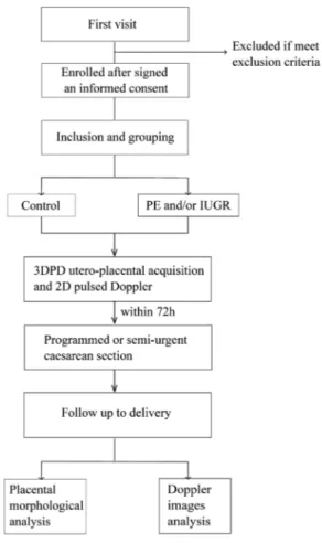
The enrolment of women started in February 2014. In view of the caesarean rate in our hospital, recruitment should be achieved by February 2017. Theflow chart of patient participation is presented infigure 3.
Premature ending of patient participation
Participants will be excluded from the study in the fol-lowing situations:
▸ Lack of 3DPD acquisition
▸ No placental morphological analysis
▸ Patient has an undesirable event related to the study ▸ Withdrawal of consent before the end of the study.
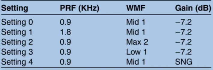
Table 2 3D power Doppler settings
Setting PRF (KHz) WMF Gain (dB) Setting 0 0.9 Mid 1 −7.2 Setting 1 1.8 Mid 1 −7.2 Setting 2 0.9 Max 2 −7.2 Setting 3 0.9 Low 1 −7.2 Setting 4 0.9 Mid 1 SNG
SNG was set by increasing the gain until artefactual noise was present then slowly reducing it to where the noise artefact has just disappeared.
PRF, pulse repetition frequency; SNG, sub-noise gain; WMF, wall motion filter.
Patients will be immediately excluded from the study and replaced with other new participants. Any decision to withdraw consent should not affect the patient’s routine medical care. In the case of an undesirable event related to the study, the patient should be informed and excluded. Revised medical care should be offered.
Follow-up
No specific follow-up has been planned for participants except for routine healthcare after caesarean section. Information on newborns will be collected after delivery, such as gender, birth weight, Apgar score, umbilical cord arterial pH, base excess and lactate value, and neonatal intensive care. Any undesirable event after caesarean section such as maternal or neonatal death, transfusion, hysterectomy, hospitalisation in the intensive care unit, and significant post-partum haemorrhage will be noted and declared.
Sample size consideration
As this, as far as we know, is the first study to evaluate concordance between 3DPD indices and placental morphometry, there is no available reference for sample
size determination. Therefore, the ability to detect dif-ferences in 3DPD indices between normal and patho-logical pregnancies was used for sample size estimation. Based on our previous cohort study on 3DPD indices for PE and/or IUGR prediction (unpublished data of Morel et al), in order to detect a difference in placental FI (the best index in our study) between normal pregnancy and PE (PE prevalence of 3%), with anα of 5% and a power of 80%, a population of 112 pregnant women (28 with PE and/or IUGR and 84 with normal pregnancies) have to be included. As the main objective of this study is to evaluate the correlation between Doppler and morpho-metric indices, different pathologies (such as early onset and late onset PE, and IUGR) will be combined during analysis.
Data collection and management
Participant consent will be obtained after detailed explanation to patients during recruitment. An elec-tronic case report form (e-CRF) will then be created for each patient on a secure website dedicated to clinical research (2008 Clinsight). All information related to patient and pregnancy outcomes mentioned above will
Figure 1 Three-dimensional power Doppler (3DPD) volume analysis. After automated 3DPD acquisition of the utero-placental unit with predetermined machine settings, the volume of interest (VOI) is defined by manual segmentation with 30° rotation. In our study, the placenta and adjacent myometrium will be separately traced (the placenta is shown here). After tracing, the 3D VOI is reconstructed using VOCAL software and 3DPD indices (vascularisation index (VI), flow index (FI) and vascularisation-FI (VFI)) are then calculated and displayed as histograms.
be noted. Each subject will be anonymized as initials fol-lowed by inclusion order. Each investigator will have an individual account allowing them to fill in the e-CRF. Confirmation of participation in this study will be noted in patients’ medical records by the investigators.
Patients’ identities will be kept confidential. The subject code will be used for all related data (case report form, ultrasound and morphological images and undesirable invent declaration form) using the same coding rules (initials followed by inclusion order). One list only with patients’ identities and corresponding subject codes will be stored in the principle investigator’s file in a secure place, together with patient’ consent forms.
Data management will be carried out by CIC-IT, Nancy. Queries will be answered in order to solve questions regarding missing data, unreadable text, and coherence.
Study monitors will be responsible for obtaining written and signed answers by investigators to all
questions. Before the database is frozen, a data review will be conducted in order to identify errors during recruiting and data input. The clinical study coordin-ator, study monitor and data manager will jointly conduct the data review and approve the list of omitted data.
When the queries process has ended, the data manager will declare database closure so that the dataset can be sent to the statistician. The database will be frozen once all corrections required by the investigators have been implemented and all data reviewed.
All ultrasound and morphological images will be anon-ymized and archived in the ARCHIMED database, which has been declared to CNIL (National Commission for Data Protection and Liberties; declaration number: 1410005). All data (numeric and paper) will be archived for 15 years after the end of the study.
Statistical analysis
Statistical analysis will be performed using R software (R Foundation for Statistical Computing, Vienna, Austria). Quantitative results will be expressed as the mean±SD. Correlation coefficients between 3DPD indices and mor-phological indices will be estimated by Pearson or Spearman tests as appropriate. Agreement between 3DPD indices and morphological indices will be assessed
Figure 2 Placental tissue sampling. After fixation, systematic uniform random sampling of placenta will be performed with the help of a plastic grid. Six sample cubes with whole placental thickness will be taken (≈1.5×1.5×1.5 cm3). A vertical uniform random (VUR) section will be prepared and then stained with H&E. Several pictures will be taken at different magnifications for stereology analysis. The volume density (Vv), surface density (Sv) and length density (Lv) of different placental structures will be calculated using STEPanizer software.
Table 3 Stereological indices
Structure Parameter Intervillous space Vv Villi Vv, Sv, Lv Capillary Vv, Sv, Lv Stroma of villi Vv Trophoblast cells Vv Trophoblast knots Vv, Lv
Lv, length density (/μm2); Sv, surface density (/μm); Vv, volume density.
Figure 3 Flowchart of participating women through the study. IUGR, intra-uterine growth restriction; PE, preeclampsia.
by the Bland and Altman plot. Reference data should be morphological results. The impact of ultrasound settings and maternal characteristics on 3DPD indices will be evaluated using a multivariate linear regression model. Differences in 3DPD indices and/or morphological indices between normal and pathological pregnancies will be evaluated with the t test or Mann-Whitney test according to data distribution. p<0.05 will be considered to be significant.
QUALITY CONTROL
Right of access to data and source documents
The Centre Hospitalier Régional Universitaire (CHRU) of Nancy is responsible for obtaining the agreement of all parties involved in the study so as to guarantee direct access to all study sites, source data, source documents and reports so that the sponsor can control data quality and perform an audit.
Investigators will make available the documents and individual data strictly required for monitoring, quality control and audit of the biomedical study to persons allowed access to these, in accordance with the statutory and regulatory provisions in place (Articles L.1121-3 and R.5121-13 of the French Public Health Code).
Any original document or object that allows the exist-ence or accuracy of a data point or information recorded during the study to be proved is defined as a source document.
In accordance with the statutory provisions in place (Articles L.1121-3 and R.5121-13 of the French Public Health Code), the persons having direct access to source data will take every precaution necessary to ensure the confidentiality of information relating to investigational medicinal products, studies, participants, particularly con-cerning their identity, as well as the results obtained. These persons, as the investigators themselves, are subject to professional confidentiality.
The CHRU of Nancy will ensure that each participant has given her written consent for access to her personal data that is strictly required for study quality control.
Study monitoring
The method of data monitoring has been established by DRI (Department of Clinical Research and Innovation) of CHRU, Nancy. A clinical research associate (CRA) will regularly visit the Nancy regional university mater-nity unit of the CHRU for the quality control of reported data entered in the e-CRF. The CRA will verify that the study is being conducted according to protocol and ensure that the data form contains all requested information.
During monitoring, the source documents of each patient should be provided (medicalfiles) and must be consistent with the e-CRF. The CRA will have access to the e-CRF, medicalfiles and all other related documents. The CRA is bound by a duty of confidentiality regarding the reviewed patients’ information.
The risk of the study has been classified as category B by the CHRU of Nancy according to the GT5 work group of the national assembly of the Delegation of Clinical Research and Innovation (DRCI), which is vali-dated by the primary healthcare department (Direction Générale de l’Offre de Soins, DGOS). A proper moni-toring plan including verification of partial patient’ consent forms, inclusion and exclusion criteria, data entry and security, principle endpoint, and undesirable event reports has been established.
POTENTIAL RISKS RELATED TO THE STUDY
The research team does not foresee any special medical risks for participating women or their unborn children. This study follows good clinical practice as defined by the French ministry of health. The only difference related to the study is the addition of utero-placental 3DPD acquisition. The duration of this acquisition does not exceed 5 min. The ultrasound machine is EC (European Community) marked and routinely used in clinic service. Patients with a maternal or fetal vital emergency will not be included in this study.
ETHICAL PERMISSION
The CHRU of Nancy and investigators commit that this research will be conducted in accordance with legislative regulation no. 2004–806 of 9 August 2004, as well as in agreement with Good Clinical Practices (ICH V.4 of 1 May 1996 and Decision of 24 November 2006) and the Helsinki Declaration (Ethical Principles for Medical Research Involving Human Subjects, Tokyo 2004). In order to initiate the research, the CHRU of Nancy as its sponsor has submitted an authorisation request to the competent authority, ANSM (French National Agency for Medicines and Health Products Safety). The compe-tent authority, as defined in Article L. 1123-12, has pro-vided its decision with regard to the safety of individuals who consent to biomedical research, by especially taking into consideration the safety and quality of products used during the research in accordance with, where appropriate, existing repositories, their condition of use and the safety of persons with regard to acts performed and the methods used as well as intended procedures for patient follow-up. Approval of ANSM was obtained on 14 February 2014 and the reference number of the study is 203-A01049-36.
In accordance with Article L. 1123-6 of the Public Health Code, the research protocol has been submitted by the sponsor to the Committee for the Protection of Persons (CPP, Comité de Protection des Personnes). The study and related consent forms have been approved by CPP Est III on 4 March 2014 and the CPP reference number of this study is 13.09.02.
PROTOCOL AMENDMENT
A substantial change is a change that is liable, in one way or another, to modify the assurances made to
participants who consent to the study (modification of an inclusion criterion, extending the inclusion period, participation of new centres, etc).
Once the research has begun, any substantial modi fica-tion thereof on the initiative of the sponsor must obtain, prior to its implementation, approval by the committee and authorisation from the competent authority. In this case, if necessary, the committee ensures that a new consent form is obtained from individuals participating in research.
Any substantial change requires authorisation from ANSM and CPP in accordance with legislative regulation no. 2004–806 of 9 August 2004.
FINAL RESEARCH REPORT
The coordinator and the mandated biostatistician will collaboratively write the final research report. This report will be submitted to each of the investigators for review. Once consensus has been reached, the final version must be endorsed with the signature of each of the investigators and sent to the sponsor as early as pos-sible after the effective end of the research. A report prepared according to the reference plan of the compe-tent authority must be forwarded to the compecompe-tent authority and the CPP within a year after the end of the research.
DISCUSSION
This study aims to evaluate concordance between 3DPD indices and placental morphology. The reliability of 3DPD for the evaluation of utero-placental vascularisation will be validated if a high correlation between 3DPD indices and morphological indices is found. Stereological analysis, which can provide three-dimensional unbiased and quantitative data from two-dimensional cross-sections, has been widely used in placental morphological analysis. When defining our 3DPD indices, we found similarities with stereological indices such as volume density, surface density and length density. Therefore, we will use stereol-ogy for our primary morphological analysis for relation-ship assessment; traditional morphological analysis will also be used for further physio-pathological interpret-ation. As far as we know, this is thefirst study to evaluate the relationship between utero-placental 3DPD and morphology in pregnant women.
Furthermore, the clinical use of 3DPD will also be assessed in this study. The impact of different Doppler settings, including the new SNG setting, on 3DPD indices in women with normal and pathological preg-nancies will be evaluated. To our knowledge, such eva-luations have only been carried out in phantoms and animal models. The homogeneity of 3DPD indices in a specific population, as well as the relationship between 3DPD and morphology with one particular setting, will be considered regarding future suggestions for clinical settings. We anticipate that our study data will inform recommendations about machine settings to provide the
maximum information about utero-placental physiology and pathophysiology while minimising ultrasound expos-ure for the pregnant women and elucidate the impact of maternal physical characteristics such as BMI and pla-cental position on Doppler signals.
Author affiliations
1IADI, Inserm U947, University of Lorraine, Nancy, France
2Pôle de Gynécologie-Obstétrique, Service d’Obstétrique et Médecine Fœtale, CHRU Nancy, Nancy, France
3Service Commun de Microscopie, Faculté de Médecine, University of Lorraine, Vandoeuvre-Lès-Nancy, France
4Laboratory of Fetal and Placental Pathology, CHRU Nancy, Nancy, France 5CHRU Nancy, CIC-IT Inserm CIC 1433, Nancy, France
6PremUp Foundation, Paris, France
Acknowledgements We thank Mrs A. Willm of DRI of the CHRU of Nancy for project monitoring. We thank Mr E. Micard for clinical and images data management. We thank the staff of the feto-placental anatomical laboratory, especially Mr R. Toussaint who kindly provided technical support for placental examination and sampling.
Contributors JD participated in study design and is carrying out or will carry out recruitment, Doppler acquisition, placental examination, data analysis and manuscript writing. A-CC-L is carrying out or will carry out recruitment, Doppler acquisition and data analysis. EP-G and OM are major investigators for clinical assessment, Doppler acquisition and study design. CC is in charge of placental morphological analysis. AC is project manager. GH is in charge of statistical analysis. All authors read and approved the final manuscript.
Funding This study is supported by CPRC founding of CHRU of Nancy. The CIC-IT of Nancy and CHRU of Nancy assure the data management, storage, ultrasound acquisition and placental morphological analysis related costs. J. Duan is sponsored by the China Scholarship Council for her PhD studies at the University of Lorraine in Nancy.
Competing interests None declared. Patient consent Obtained.
Ethics approval ANSM (the French National Agency for Medicines and Health Products Safety) and the Committee for the Protection of Persons (Comité de Protection des Personnes, CPP) approved this study.
Data sharing statement All data generated during the project will be made freely available via CIC-IT, Nancy. Data obtained from this study will be deposited at CIC-IT Nancy where they will be maintained for a minimum of 15 years. There are no security, licensing or ethical issues related to the expected data, and all data used in the project will be generated directly as a result of the project, without any pre-existing data being used.
Trial status This is an ongoing trial. Recruitment began in February 2014. We expect to complete recruitment by February 2017. We plan to publish final results in 2017.
Open Access This is an Open Access article distributed in accordance with the Creative Commons Attribution Non Commercial (CC BY-NC 4.0) license, which permits others to distribute, remix, adapt, build upon this work non-commercially, and license their derivative works on different terms, provided the original work is properly cited and the use is non-commercial. See: http:// creativecommons.org/licenses/by-nc/4.0/
REFERENCES
1. Kaufmann P, Black S, Huppertz B. Endovascular trophoblast invasion: implications for the pathogenesis of intrauterine growth retardation and preeclampsia.Biol Reprod2003; 69:1–7.
2. Burton GJ, Woods AW, Jauniaux E, et al. Rheological and physiological consequences of conversion of the maternal spiral arteries for uteroplacental blood flow during human pregnancy.
3. Ramsey EM, Martin CB Jr, McGaughey HS Jr, et al. Venous drainage of the placenta in rhesus monkeys: radiographic studies. Am J Obstet Gynecol 1966;95:948–55.
4. Donner MW, Ramsey EM. Radioangiographic studies on the dynamics of the blood circulation in the maternal placenta. (Experimental studies on the rhesus monkey).Fortschritte Auf Dem Geb Röntgenstrahlen Nukl1966;104:796–808.
5. Ramsey EM. Uteroplacental circulation during labor.Clin Obstet Gynecol1968;11:78–95.
6. Ramsey EM. Maternal and foetal circulation of the placenta.Ir J Med Sci1971;140:151–68.
7. Jaffe R, Woods JR. Doppler velocimetry of intraplacental fetal vessels in the second trimester: improving the prediction of pregnancy complications in high-risk patients.Ultrasound Obstet Gynecol1996;8:262–6.
8. Jaffe R, Jauniaux E, Hustin J. Maternal circulation in the first-trimester human placenta—Myth or reality?Am J Obstet Gynecol1997;176:695–705.
9. Jurkovic D, Jauniaux E, Kurjak A, et al. Transvaginal color Doppler assessment of the uteroplacental circulation in early pregnancy. Obstet Gynecol 1991;77:365–9.
10. Schaaps J-P, Tsatsaris V, Goffin F, et al. Shunting the intervillous space: new concepts in human uteroplacental vascularization.Am J Obstet Gynecol2005;192:323–32.
11. Poon LC, Nicolaides KH. Early prediction of preeclampsia.Obstet Gynecol Int2014;2014:297397.
12. Mercé LT, Barco MJ, Bau S. Reproducibility of the study of placental vascularization by three-dimensional power Doppler.J Perinat Med
2004;32:228–33.
13. Morel O, Grangé G, Fresson J, et al. Vascularization of the placenta and the sub-placental myometrium: feasibility and reproducibility of a three-dimensional power Doppler ultrasound quantification
technique. A pilot study.J Matern Fetal Neonatal Med
2011;24:284–90.
14. Raine-Fenning NJ, Clewes JS, Kendall NR, et al. The interobserver reliability and validity of volume calculation from three-dimensional ultrasound datasets in the in vitro setting.Ultrasound Obstet Gynecol2003;21:283–91.
15. Raine-Fenning NJ, Nordin NM, Ramnarine KV, et al. Determining the relationship between three-dimensional power Doppler data and true blood flow characteristics: an in vitro flow phantom experiment.
Ultrasound Obstet Gynecol2008;32:540–50.
16. Morel O, Pachy F, Chavatte-Palmer P, et al. Correlation between uteroplacental three-dimensional power liniqu indices and true uterine blood flow: evaluation in a pregnant sheep model.
Ultrasound Obstet Gynecol2010;36:635–40.
17. Rizzo G, Silvestri E, Capponi A, et al. Histomorphometric
characteristics of first trimester chorionic villi in pregnancies with low serum pregnancy-associated plasma protein-A levels: relationship with placental three-dimensional power liniqu ultrasonographic vascularization.J Matern Fetal Neonatal Med2011;24:253–7. 18. Dar P, Gebb J, Reimers L, et al. First-trimester 3-dimensional
power Doppler of the uteroplacental circulation space: a potential screening method for preeclampsia.Am J Obstet Gynecol
2010;203:238.e1–7.
19. Costa J, Rice H, Cardwell C, et al. An assessment of vascularity and flow intensity of the placenta in normal pregnancy and pre-eclampsia
using three-dimensional ultrasound.J Matern Fetal Neonatal Med
2010;23:894–9.
20. Hafner E, Metzenbauer M, Stümpflen I, et al. First trimester placental and myometrial blood perfusion measured by 3D power Doppler in normal and unfavourable outcome pregnancies.Placenta
2010;31:756–63.
21. Hafner E, Metzenbauer M, Stümpflen I, et al. Measurement of placental bed vascularization in the first trimester, using
3D-power-Doppler, for the detection of pregnancies at-risk for fetal and maternal complications.Placenta2013;34:892–8.
22. Lecarpentier E, Morel O, Tarrade A, et al. Quantification of utero-placental vascularization in a rabbit model of IUGR with three-dimensional power Doppler angiography.Placenta
2012;33:769–75.
23. Noguchi J, Hata K, Tanaka H, et al. Placental vascular sonobiopsy using three-dimensional power Doppler ultrasound in normal and growth restricted linique.Placenta2009;30:391–7.
24. Guiot C, Gaglioti P, Oberto M, et al. Is three-dimensional power Doppler ultrasound useful in the assessment of placental perfusion in normal and growth-restricted pregnancies?Ultrasound Obstet Gynecol2008;31:171–6.
25. Pomorski M, Zimmer M, Florjanski J, et al. Comparative analysis of placental vasculature and placental volume in normal and IUGR pregnancies with the use of three-dimensional Power Doppler.Arch Gynecol Obstet2012;285:331–7.
26. Odibo AO, Zhong Y, Longtine M, et al. First-trimester serum analytes, biophysical tests and the association with pathological morphometry in the placenta of pregnancies with preeclampsia and fetal growth restriction.Placenta2011;32:333–8.
27. Odeh M, Ophir E, Maximovsky O, et al. Placental volume and three-dimensional power Doppler analysis in prediction of pre-eclampsia and small for gestational age between Week 11 and 13 weeks and 6 days of gestation.Prenat Diagn2011;31:367–71. 28. Luria O, Barnea O, Shalev J, et al. Two-dimensional and
three-dimensional Doppler assessment of fetal growth restriction with different severity and onset.Prenat Diagn2012;32:1174–80. 29. Raine-Fenning NJ, Nordin NM, Ramnarine KV, et al. Evaluation of
the effect of machine settings on quantitative three-dimensional power Doppler angiography: an in-vitro flow phantom experiment.
Ultrasound Obstet Gynecol2008;32:551–9.
30. Collins SL, Stevenson GN, Noble JA, et al. Influence of power Doppler gain setting on Virtual Organ Computer-aided AnaLysis indices in vivo: can use of the individual sub-noise gain level optimize information?Ultrasound Obstet Gynecol2012;40:75–80. 31. Sanderson J, Wu L, Mahajan A, et al. Selection of the sub-noise
gain level for acquisition of VOCAL data sets: a reliability study.
Ultrasound Med Biol2014;40:562–7.
32. Indications de la césarienne programmée à terme. Méthode Recommandations pour la pratique linique. 2012.
33. Bhide A, Acharya G, Bilardo CM, et al. ISUOG Practice Guidelines: use of Doppler ultrasonography in obstetrics.Ultrasound Obstet Gynecol2013;41:233–9.
34. Mayhew TM. Taking tissue samples from the placenta: an illustration of principles and strategies.Placenta2008;29:1–14.
35. Tschanz SA, Burri PH, Weibel ER. A simple tool for stereological assessment of digital images: the STEPanizer.J Microsc
2011;243:47–59.
controlled study
morphology (EVUPA): a prospective
comparison to placental vascular
quantification using 3D power Doppler with
restriction pregnancies: third trimester
preeclamptic and intra-uterine growth
Utero-placental vascularisation in normal and
Morel Christophe Christov, Gabriela Hossu, Aboubaker Cherifi and Olivier Jie Duan, Anne-Claire Chabot-Lecoanet, Estelle Perdriolle-Galet,
doi: 10.1136/bmjopen-2015-009909
2016 6: BMJ Open
http://bmjopen.bmj.com/content/6/3/e009909
Updated information and services can be found at:
These include:
References
http://bmjopen.bmj.com/content/6/3/e009909#ref-list-1
This article cites 34 articles, 1 of which you can access for free at:
Open Access
http://creativecommons.org/licenses/by-nc/4.0/
non-commercial. See:
provided the original work is properly cited and the use is
non-commercially, and license their derivative works on different terms, permits others to distribute, remix, adapt, build upon this work
Commons Attribution Non Commercial (CC BY-NC 4.0) license, which This is an Open Access article distributed in accordance with the Creative
service
Email alerting
box at the top right corner of the online article.
Receive free email alerts when new articles cite this article. Sign up in the
Collections
Topic
Articles on similar topics can be found in the following collections (122)Radiology and imaging
(352)
Obgyn
Notes
http://group.bmj.com/group/rights-licensing/permissions
To request permissions go to:
http://journals.bmj.com/cgi/reprintform
To order reprints go to:
http://group.bmj.com/subscribe/