HAL Id: hal-03005939
https://hal.archives-ouvertes.fr/hal-03005939
Submitted on 16 Nov 2020
HAL is a multi-disciplinary open access
archive for the deposit and dissemination of
sci-entific research documents, whether they are
pub-lished or not. The documents may come from
teaching and research institutions in France or
abroad, or from public or private research centers.
L’archive ouverte pluridisciplinaire HAL, est
destinée au dépôt et à la diffusion de documents
scientifiques de niveau recherche, publiés ou non,
émanant des établissements d’enseignement et de
recherche français ou étrangers, des laboratoires
publics ou privés.
Soft Hydrothermal Synthesis of Hafnon, HfSiO 4
Paul Estevenon, Thibault Kaczmarek, Mohamed Ruwaid Rafiuddin, Éléonore
Welcomme, Stephanie Szenknect, Adel Mesbah, Philippe Moisy, Christophe
Poinssot, Nicolas Dacheux
To cite this version:
Paul Estevenon, Thibault Kaczmarek, Mohamed Ruwaid Rafiuddin, Éléonore Welcomme, Stephanie
Szenknect, et al.. Soft Hydrothermal Synthesis of Hafnon, HfSiO 4. Crystal Growth & Design,
American Chemical Society, 2020, 20 (3), pp.1820-1828. �10.1021/acs.cgd.9b01546�. �hal-03005939�
Soft hydrothermal synthesis of hafnon, HfSiO
4
Paul Estevenon
†,‡,§, ∥, Thibault Kaczmarek
†,‡, Mohamed Ruwaid Rafiuddin
‡, Eleonore Welcomme
†,
Stephanie Szenknect
‡, Adel Mesbah
‡, Philippe Moisy
†, Christophe Poinssot
†, Nicolas Dacheux
*,‡† CEA, DEN, DMRC, Univ Montpellier, Marcoule, France.
‡ ICSM, Univ Montpellier, CNRS, CEA, ENSCM, Marcoule, France.
KEYWORDS: hafnium silicate, hafnon, wet chemistry route, hydrothermal synthesis, zircon structure type.
ABSTRACT: Despite being a member of the zircon type silicate family, the conditions allowing the hydrothermal synthesis of
HfSiO4 were not well constrained. A multiparametric study was performed in order to follow the synthesis of this phase under soft
hydrothermal conditions and thus to determine the most efficient conditions to form single phase samples. Among the experimental parameters investigated, concentration of reactants, pH of the reactive media, temperature and duration of the hydrothermal treat-ment impacted significantly the formation rate of hafnon and its crystallization state. Pure HfSiO4 was obtained in acid reactive
media with an acidity ranging from CHCl = 1.5 M to pH = 1.0 and for CSi ≈ CHf ≥ 0.21 mol·L -1
. The silicate phase was obtained after a 24-hours treatment at temperatures ranging from 150°C to 250°C. However, the rise of temperature and extension of the duration of the hydrothermal treatment favored the crystallization state of the final HfSiO4 samples.
INTRODUCTION
Hafnon, HfSiO4, is one of the end-members belonging to the
zircon-type silicate group (tetragonal, I41/amd) like ZrSiO4,
CeSiO4, ThSiO4, PaSiO4, USiO4, NpSiO4, PuSiO4 and
AmSiO4. 1,2-4
As a consequence, hafnon is reported to form a solid solution with ZrSiO4.
5-7
It is usually observed as a minor component in natural zircons 8-10 and less frequently as the major phase in natural assemblies where zircon appears as a minor secondary phase.11
Owing to its interesting thermal and electric properties (such as thermal shock resistance, low dilatation coefficient on a wide temperature range, high thermal conductivity, low rela-tive permittivity and low dielectric loss), 12, 13 hafnon is often considered as promising high-temperature refractory material
12
for semiconductor devices 14 or microwave substrate for electronic data transmission.13
Moreover, as zircon-type ceramics (e.g. ZrSiO4 and HfSiO4)
are isostructural with actinide silicates, AnIVSiO4 and due to its
high chemical stability, they have been suggested as potential actinide-specific matrices for the immobilization of radionuclides associated to high level nuclear waste and more specifically of plutonium excess coming from dismantled nuclear weapons.15-19 In this context, (Hf,Pu)SiO4 compounds
were prepared by Burakov et al. using sol-gel method.20 However, these experiments suggest that plutonium content cannot exceed 7 wt.% in such materials.
From a general point of view, the synthesis of HfSiO4 was
reported by high temperature solid state chemistry,1, 5, 7, 13, 21-25 sol-gel methods,12, 26, 27 chemical transport reaction,14, 28, 29 physical vapor deposition 30 and hydrothermal synthesis.31, 32 However, the only hydrothermal syntheses were reported by Caruba et al. under acid conditions at T = 800°C, 75 MPa,31 and by McNeil et al. in very acid conditions, i.e. CHF = 6 mol·L
-1
and CH2SO4 = 0.5 mol·L -1
at T = 850°C,
200 MPa.32 These conditions appear to be quite surprising taking into account the possibility to prepare isostructural silicates, e.g. ZrSiO4,
31, 33-46 CeSiO4, 4, 47-49 ThSiO4, 2, 34, 50-65 USiO4, 2, 59, 60, 66-76 NpSiO4, 2 PuSiO4 2 and AmSiO4 2 under soft hydrothermal conditions (i.e. for T ≤ 250°C). However, it may be also noticed that the formation of amorphous hafnon has been observed by alteration, in aqueous solution, of Hf-bearing borosilicate glasses at 90°C and pH = 1.77 More spe-cifically, we recently reported that modifications of the start-ing pH and elementary concentrations allowed to decrease the temperature of formation of ThSiO4 and CeSiO4 under
hydro-thermal conditions.49, 64 Therefore, the aim of this study was to determine an efficient way of synthesis allowing the formation of HfSiO4 under soft hydrothermal conditions.
MATERIALS AND METHODS
Syntheses
All the reagents used for the materials preparation were sup-plied by Sigma-Aldrich. Na2SiO3·5H2O (95%) and HfCl4
(98%) were used as aqueous silicate and hafnium precursors, respectively. 1.5 and 1.0 mol·L-1 HCl solutions were prepared by dilution of Sigma Aldrich ACS grade mother solutions HCl (37%). 8 mol·L-1 NaOH solution was freshly prepared from Sigma Aldrich ACS grade NaOH (98 %) before the experiments.
The syntheses of HfSiO4 were performed by adapting the
protocol recently developed for ThSiO4. 64
Aqueous mixtures of hafnium at the oxidation state +IV and silicate were pre-pared by dissolving HfCl4 and Na2SiO3·5H2O in 1.5 mol·L
-1
hydrochloric acid (Table S1). At this stage, a silicate excess of 3 mol.% was considered to avoid the formation of hafnium dioxide or hydroxides during the synthesis. The pH was then adjusted to the final value with 8 mol·L-1 NaOH.
All the prepared mixtures were introduced in a 23 mL Tef-lon lined container. The container was placed in a Parr
auto-clave in an oven to reach hydrothermal conditions during 1 to 20 days with a given temperature and under autogenous pres-sure. The final cooling to room temperature was done within one hour. Then, the precipitates were separated from the su-pernatant by centrifugation at 14 000 rpm for 12 min, washed twice with deionized water and once with ethanol. They were finally dried overnight at 60°C in an oven.
Characterization
PXRD data were collected on the resulting powders using the Bruker D8 advance diffractometer equipped with a lynxeye detector and working with Cu Kα radiation (λ = 1.54184 Å) in a reflection geometry (parallel beam). Patterns were recorded between 5° and 100° (2θ) with steps of 0.019° and a total counting time of 2.5 to 3 hours per sample. Pure silicon was used as a standard material to extract the instrumental function. The collected data were refined by the Rietveld method using the Fullprof_suite package.78 During the refinements, different profile and structure parameters were adjusted, such as the zero shift, unit-cell parameters, scale factor, and overall displacement factor. An anisotropic size and strain model was also used in order to consider the broadening effect.
Raman spectra were recorded with a Horiba-Jobin Yvon Aramis device equipped with an edge filter and a Nd:YAG laser (532 nm) that delivered 60 mW at the sample surface. In order to avoid any laser-induced degradation of the compound, the power was turned down by the means of optical filters. The laser beam was then focused on the sample using an Olympus BX 41 microscope with an × 50LMP objective, resulting in a spot area of ∼1 μm2
and a power of 39 mW. For each spectrum, a dwell time of 1 to 30 s was used. Four scans were recorded for each analyzed area in order to minimize the measurement error.
FTIR spectra were recorded with a Perkin-Elmer FTIR Spectrum 100 device in the 300–4000 cm-1 range. Powdered samples were deposited on the surface of an ATR crystal without any prior preparation. The spectra collected in such operating conditions exhibited a resolution lower than 4 cm-1. Four scans were performed to average the measurement error.
SEM observations were directly conducted using a FEI Quanta 200 electronic microscope on small powder samples without prior preparation such as metallization. The electronic microscope was equipped either with an Everhart-Thornley Detector (ETD) or a Back-Scattered Electron Detector (BSED), under high vacuum conditions with a low accelerat-ing voltage (8 kV). These conditions were chosen in order to create a beam deceleration effect that led to high resolution images.
Thermogravimetric analyses were performed to determine the hydration content of the samples prepared at the end of the syntheses. All of these analyses were done between room temperature and 1000°C under air atmosphere thanks to a SETSYS evolution analyzer. These measurements were cou-pled with mass spectroscopy analyses on the residual gaz.
RESULTS AND DISCUSSION
Effect of pH of the starting solution
The effect of pH of the starting mixture on the nature of the resulting precipitate was followed between CHCl = 1.5 mol·L
-1
and pH = 8 considering a starting mixture with a hafnium concentration of 0.21 mol·L-1 and a Si:Hf molar ratio of 1.03. Hydrothermal treatments were performed at 250°C during 24 hours.
All the samples prepared were characterized by PXRD
(Fig-ure 1). From these data, HfSiO4 (zircon-type structure, space
group I41/amd) was formed using a starting mixture whose
acidity was below pH = 1.6. For higher pH values, the precipi-tation of monoclinic HfO2 (space group P21/c) was obtained
due to hafnium hydrolysis leading to the precipitation of the hafnium hydroxides then hafnium oxide by ageing. In these conditions, the unit cell parameters of HfSiO4 reached
a = 6.596(2) Å and c = 5.958(3) Å (i.e. V = 259.2(1) Å3; val-ues calculated by averaging the data obtained on the samples (1) to (4) – syntheses leading to HfSiO4 as single phase). The a
and c lattice parameters were slightly higher and smaller, respectively, than the reference lattice parameters obtained by high temperature methods (a = 6.5725(7) Å and c = 5.9632(4) Å, i.e. V = 257.60(7) Å3 28). According to the recent results already reported for ThSiO4, these variations were attributed to
the insertion of hydroxide groups in the HfSiO4 structure 64, 79
, which may be correlated to the non-ideal crystallization in the soft hydrothermal conditions considered.
Rietveld refinements performed on the PXRD data did not allow to observe any significant variation of the HfSiO4
unit-cell parameters (Table 1) nor of the crystallite size (Figure S1 and Table 1 and Table S2) according to the initial pH of the reactive media.
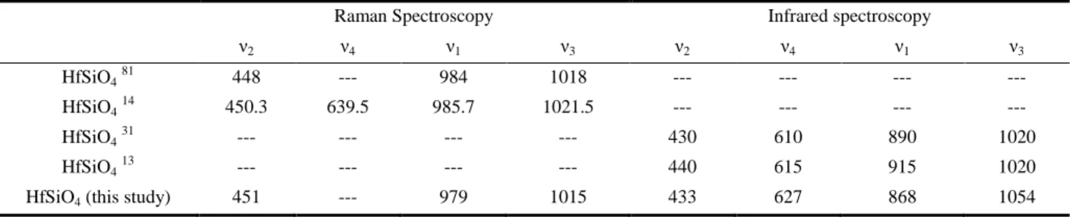
Characterization by Raman and IR spectroscopies (Figure 2) revealed the presence of symmetric and antisymmetric stretch-ing modes of SiO4 at 979 cm
-1
and 1015 cm-1, respectively by Raman (868 cm-1 and 1054 cm-1, respectively by IR spectros-copy). The symmetric bending modes were observed at 451 cm-1. On the contrary, the antisymmetric mode was not observed due to its low intensity (both symmetric and antisymmetric modes were observed at 433 cm-1 and 627 cm-1, respectively by IR spectroscopy) (Table 2).
It worth noting that the positions of the HfSiO4’s ν1 and ν3
vibration bands observed by infrared spectroscopy are signifi-cantly different to the ones reported in the literature (Table 2). However, the values obtained here are in great agreement with the ones reported in the literature for the other zircon type silicate phases (Table S3) and especially for ZrSiO4 (ν1 =
866 cm-1 and ν3 = 1049 cm -1
).80 Moreover, the position of the ν3 vibration bands at around 1050 cm
-1
should be taken with care because this position correspond to the ν4 mode of SiO2.
Additionally, the presence of small amounts of hydroxide groups or water was suggested from the observation of broad bands around 3400 cm-1 and 1630 cm-1 in the IR spectra, which was consistent with the hypothesis proposed regarding the insertion of hydroxide groups in the HfSiO4 lattice already
10
20
30
40
50
60
70
80
90
100
C
HCl= 1.0 M
C
HCl= 1.5 M
HfSiO
4°
°
°
°
°
°
Int
ensi
ty (a.u.)
2·
(degrees)
°
°
°
°
°
°
°
°
°
°
°
°
°
°
°
°
°
°
°
°
°
°
°
°
°
°
°
° °
°
°
°
°
pH = 1.0
pH = 0.5
pH = 2.0
pH = 1.6
pH = 5.0
pH = 3.0
1
2
3
4
5
6
7
8
Figure 1. PXRD patterns recorded for samples prepared under hydrothermal conditions (24 hours, T = 250°C) with hafnium and
silicate concentrations of 0.21 mol·L-1 and for various initial pH values: CHCl = 1.5 mol·L -1
(1), CHCl = 1.0 mol·L -1
(2), pH = 0.5 (3), pH = 1.0 (4), pH = 1.6 (5), pH = 2.0 (6), pH = 3.0 (7) and pH = 5.0 (8). Bragg positions of the characteristic peaks of hafnon were extracted from 28. The presence of HfO2 is pointed out by empty circles in the PXRD patterns.
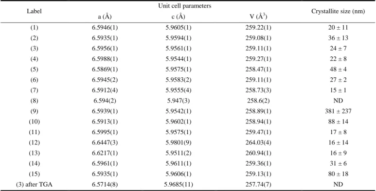
Table 1. Lattice parameters and crystallite size determined by Rietveld refinement for HfSiO4 samples.
Label Unit cell parameters Crystallite size (nm)
a (Å) c (Å) V (Å3) (1) 6.5946(1) 5.9605(1) 259.22(1) 20 ± 11 (2) 6.5935(1) 5.9594(1) 259.08(1) 36 ± 13 (3) 6.5956(1) 5.9561(1) 259.11(1) 24 ± 7 (4) 6.5988(1) 5.9544(1) 259.27(1) 22 ± 8 (5) 6.5869(1) 5.9575(1) 258.47(1) 48 ± 4 (6) 6.5945(2) 5.9583(2) 259.11(1) 27 ± 2 (7) 6.5912(4) 5.9555(4) 258.73(3) 15 ± 1 (8) 6.594(2) 5.947(3) 258.6(2) ND (9) 6.5939(1) 5.9542(1) 258.89(1) 381 ± 237 (10) 6.5913(1) 5.9602(1) 258.94(1) 88 ± 14 (11) 6.5995(1) 5.9575(1) 259.47(1) 17 ± 8 (12) 6.6447(3) 5.9801(9) 264.03(4) 16 ± 14 (13) 6.6217(1) 5.9511(2) 260.94(1) 16 ± 9 (14) 6.5961(1) 5.9611(1) 259.36(1) 31 ± 6 (15) 6.5935(1) 5.9606(1) 259.13(1) 80 ± 18 (3) after TGA 6.5714(8) 5.9685(11) 257.74(7) ND ND : Not Determined
(a) 1200 1000 800 600 400 200 Inte nsity (a. u. ) Wavenumber (cm-1) 3 (b) 3600 3200 1600 1200 800 400 Inte nsity (a. u. ) Wavenumber (cm-1) 3 3 (SiO4) 1 (SiO4) 4 (SiO4) 2 (SiO4)
Figure 2. Raman (a) and IR (b) spectra recorded for HfSiO4
samples prepared under hydrothermal conditions during 24 hours at T = 250°C starting from hafnium and silicate concentrations of 0.21 mol·L-1 and with pH = 0.5 (3).
10 20 30 40 50 60 70 80 90 100 HfSiO4 Inte nsity (a. u. ) 2· (degrees) 9 10 3 11 ° ° ° ° ° ° ° ° ° ° ° ° ° ° CHf = 8.4 x 10 -3 mol·L-1 CHf = 4.2 x 10⁻² mol·L-1 CHf = 0.21 mol·L-1 CHf = 1.0 mol·L-1 ° °
Figure 3. PXRD patterns recorded for samples prepared under hydrothermal conditions (24 hours, T = 250°C) with pH = 0.5 and various hafnium and silicate concentrations (with a molar ratio of Si:Hf = 1.03): CHf = 8.4 × 10⁻³ mol·L-1 (9), CHf =4.2 × 10⁻² mol·L-1 (10), CHf = 0.21 mol·L -1 (11) and CHf = 1.0 mol·L -1 (12). The characteristic Bragg positions of hafnon were extracted from
28
and the XRD lines associated to HfO2 are pointed out by empty
circles in the PXRD patterns.
0.01 0.1 1 0 100 200 300 400 500 600 ˜˜ CHf (mol·L-1) CSi Cryst al siz e (n m)
Figure 4. Variation of the HfSiO4 crystallite size determined by
Rietveld refinement as a function of the starting hafnium and silicate concentrations, obtained for samples under hydrothermal conditions (250°C, 24 hours) and with pH = 0.5.
Table 2. Assignment of the vibration bands (expressed in cm-1) associated to silicate groups observed in the Raman and IR spectra of HfSiO4.
Raman Spectroscopy Infrared spectroscopy
ν2 ν4 ν1 ν3 ν2 ν4 ν1 ν3
HfSiO481 448 --- 984 1018 --- --- --- ---
HfSiO414 450.3 639.5 985.7 1021.5 --- --- --- ---
HfSiO431 --- --- --- --- 430 610 890 1020
HfSiO413 --- --- --- --- 440 615 915 1020
Effect of the starting hafnium and silicate concentrations
Since the concentration of the reactants were crucial parame-ter for the synthesis of ThSiO4 and CeSiO4,
49, 64, 65
it was sus-pected that the concentration of the hafnium and silicate pre-sent in the starting mixture could affect the saturation indexes in solution, and thus the formation of HfSiO4. In this frame,
several syntheses were performed under hydrothermal condi-tions with different concentracondi-tions. The impact of the concen-trations of both reactants was followed between 8.4 × 10⁻³ mol·L-1 and 1.0 mol·L-1, keeping constant the Si:Hf molar ratio (1.03), the initial pH value (pH = 0.5) and the conditions of the hydrothermal treatment (T =250°C, t = 24 hours). As a result, pure HfSiO4 was obtained for CHf
≥ 0.21 mol·L-1
whereas the formation of HfO2 was observed as
a by-product for lower concentrations (Figure 3). In the less concentrated media, the silicate concentrations in solution were not sufficient to fully counterbalance the hydrolysis of hafnium. Therefore, the formation of hafnium silicate was favored for CHf ≥ 0.21 mol·L
-1
.
Moreover, the considered concentration significantly affect-ed the crystallization state of HfSiO4. Specifically, decreasing
the hafnium and silicate ions concentration in the reactive media induced the increase of the crystallite size of HfSiO4
(Figure 4 and Table 1). This behavior is similar to what was observed previously for ThSiO4.
64
It may be attributed to the faster kinetics of nucleation in the more concentrated reactive media, leading to the formation of numerous nucleation cen-ters, whose growth may be hindered by the limited amounts of reactants in the synthesis medium. Additionally, the Rietveld refinements allowed to observe the increase of the HfSiO4’s
lattice parameters and volume cell and the decrease of the crystallite size (Table 1 and Figure S3). Furthermore, the decrease of the crystallite size could be observed in each crys-talline planes (Table S2), especially the low size value ob-tained in the (hkl) planes with l ≠ 0 at high concentration could explain the anisotropic effect which could be observed with the widening of the corresponding peaks.
Impact of the hydrothermal treatment (temperature and duration)
In order to identify the impact of the temperature of the hy-drothermal treatment on the formation of HfSiO4, several
experiments were performed at 150°C, 200°C and 250°C, keeping constant the concentration of the starting hafnium (0.21 mol·L-1), the molar Si:Hf ratio (1.03), the starting pH value (pH = 0.5) and the heating time (t = 24 hours). From PXRD analyses, HfSiO4 was formed whatever the temperature
of synthesis (Figure 5), which was also confirmed by IR spec-troscopy (Figure S3). Additional experiments performed in the same conditions at 100°C did not lead to the formation of HfSiO4 after 24 hours holding time.
However, working at the lower temperatures led to the for-mation of HfSiO4 compounds exhibiting a very strong
aniso-tropic effect (Figure 5). Indeed, the (hkl) reflections involving the c axis were strongly broadened and attenuated whereas the (hk0) reflections were finer and more intense when the tem-perature of the hydrothermal treatment decreased, which un-derlined the formation of platelet crystallites. Considering that the decreasing temperature was associated to slower kinetics of reaction, this phenomenon was assigned to the growth pro-cess of HfSiO4 crystallites, involving first the formation of
bidimensional crystallites and then crystal growth to form
three-dimensional particles. Finally, varying the starting pH value at 150°C or 200°C confirmed that HfSiO4 was always
obtained for pH < 1.6 (Figure S4 and Figure S5), as already discussed for the experiments at 250°C.
Moreover, the preferential growth observed in this study is in agreement with the results reported recently by Calas et al. for the alteration of Hf-bearing borosilicate glasses at 90°C and pH = 1 in aqueous solution, showing the (200) reflection of hafnon as the only observable diffraction peak in their con-ditions.77 The reflections (220), (400) and (420) are invisible due to their low intensity compared to the (200), while the (hkl) peaks with l ≠ 0 are invisible due to an anisotropic effect. Increasing the temperature of the hydrothermal treatment led to the decrease of the unit-cell parameters of HfSiO4 (Figure
S8 and Table 1). No significant variation of the average
crys-tallite size was observed (Figure S8). However, Rietveld refinements performed on the PXRD patterns allowed to con-firm the qualitative results suspected on the anisotropy which could be explained by the crystallite growth along the (hkl) planes with l ≠ 0 (Figure 6 and Table S2) when the tempera-ture of the hydrothermal treatment increased.
10 20 30 40 50 60 70 80 90 100 (112) (332) (204) (431) (224) (512) (411) (312) (321) (103) (301) (202) (211) (101) (420) (400) (220) = 200°C = 150°C Inte nsity (a. u. ) 2· (degrees) HfSiO4 = 250°C 12 13 3 (200) (420) (400) (220) (200)
Figure 5. PXRD pattern and corresponding Miller index obtained for samples prepared under hydrothermal conditions with starting silicon and hafnium concentrations of 0.21 mol·L-1 and pH = 0.5, after hydrothermal treatment performed for 24 hours at T = 150°C (12), at T = 200°C (13) and at T = 250°C (3). The characteristic XRD lines of HfSiO4 were extracted from 28.
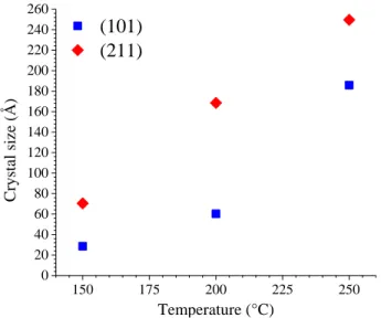
150 175 200 225 250 0 20 40 60 80 100 120 140 160 180 200 220 240 260
(101)
(211)
Temperature (°C) Cryst al siz e (Å)Figure 6. Crystallite size of HfSiO4 along (101) and (211)
crystal-line planes determined by Rietveld refinement as a function of the temperature of the hydrothermal treatment for samples prepared
under hydrothermal conditions (t = 24 hours, pH = 0.5 and CHf = 0.21 mol·L-1).
Thermogravimetric analyses, coupled with mass spectrosco-py, performed between room temperature and 1000°C under air atmosphere, with the samples dried overnight at 60°C, allowed to observe the loss of water in two steps. The first one around 100°C corresponded to the elimination of free water and the second one between 200°C and 850°C was probably associated to the elimination of water inserted in HfSiO4
struc-ture (as H2O or HO
group).
The characterizations performed on the TGA analyses resi-dues allowed to confirm that the final phase obtained corre-sponded to HfSiO4 as single crystalline phase, whatever the
temperature of hydrothermal treatment considered. It worth noting that the HfSiO4 lattice parameters evolved during the
thermogravimetric analysis to reach parameters close to refer-ence values: a = 6.5714(8) Å, c = 5.9685(11) Å and V = 257.74(7) Å3 (against a = 6.5725(7) Å, c = 5.9632(4) Å, i.e. V = 257.60(7) Å3 28) (Table 1). Raman and infrared spec-troscopy characterizations did not lead to significant differ-ences compared to the spectra measured before the thermogravimetric analyses.
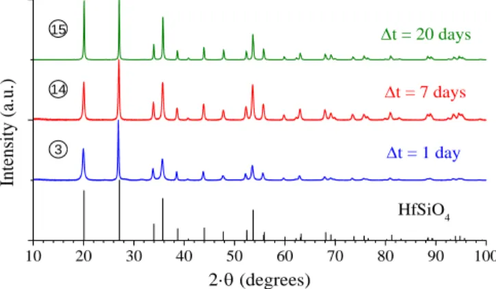
In order to evidence the impact of the duration of the hydro-thermal treatment on the formation of HfSiO4, several
experi-ments were also performed at 250°C, for pH = 0.5 and CHf = 0.21 mol·L
-1
with holding times spread from 1 day to 20 days. From PXRD analysis, it was clear that HfSiO4 was
al-ways single phase and was not degraded by extending the holding time (Figure 7).
Moreover, extending the duration of the hydrothermal treat-ment did not lead to significant change of the HfSiO4 unit cell
parameters determined by Rietveld refinement (Table 1). The HfSiO4 lattice parameters reached a = 6.5937(1) Å,
c = 5.9605(1) Å and V = 259.14(1) Å3 for 20 days of hydro-thermal treatment (Figure 8). As already described, the lattice parameters obtained were slightly different to those obtained by high temperature methods (a = 6.5725(7) Å,
c = 5.9632(4) Å and V = 257.60(7) Å3 28), which might corre-spond to the insertion of hydroxide groups in the HfSiO4
struc-ture.64, 79 The extension of the heating time tended to reduce the anisotropic effect evidenced by PXRD. Therefore, initial HfSiO4 platelets crystallites may evolve to form 3D particles
through crystal growth process.
The determination of the size of the HfSiO4 crystallites
per-formed by Rietveld refinement indicated that increasing the duration of the hydrothermal treatment promoted their growth (Figure 9).
The direct comparison by SEM of the samples prepared at 250°C clearly confirmed the growth of HfSiO4 grains from ~
60 nm after 1 day to ~ 200 nm after 7 days and ~ 450 nm after 20 days (Figure 10). It also exhibited the evolution of the grain morphology with the clear identification of a square based bipyramid morphology characteristic of zircon-type materials, for the hydrothermal treatments extended for 7 days and more.82 This evolution of morphology may be correlated to the crystalline growth of the HfSiO4 grains. Moreover,
HfSiO4 particles also appeared to be very flattened and may be
correlated to the anisotropic effect previously observed. How-ever, it is worth noting that a size discrepancy was observed between the crystallite size determined by XRD method
(Fig-ure 9) and grain size meas(Fig-ured thanks to SEM
.
Therefore, it can be concluded that these grains observed arepolycrystal-based compounds including ThSiO4. 64
Additionally, SEM-EDX characterization allowed to confirm the sample composi-tion, without any metallic insertion in HfSiO4 (Figure S13).
10 20 30 40 50 60 70 80 90 100 HfSiO4 t = 1 day Inte nsity (a. u. ) 2· (degrees) t = 7 days t = 20 days 3 14 15
Figure 7. PXRD patterns recorded for samples prepared under hydrothermal conditions (T = 250°C) starting with pH = 0.5 and CHf = 0.21 mol·L
-1
and for various heating times: 1 day (3), 7 days (14) and 20 days (15). The XRD lines characteristic of HfSiO4
were extracted from 28.
10 20 30 40 50 60 70 80 90 100 Inte nsity (a. u. ) 2· (degrees) 15
Figure 8. PXRD diagram of HfSiO4 prepared under hydrothermal
conditions (T = 250°C, t = 20 days) starting with pH = 0.5 and CHf = 0.21 mol·L-1, and associated calculated and difference
profile obtained by Rietveld refinement.
0 2 4 6 8 10 12 14 16 18 20 0 10 20 30 40 50 60 70 80 90 100
Hydrothermal treatment duration (days)
Cryst
al
siz
e (n
m)
Figure 9. HfSiO4 crystallite size determined by Rietveld
re-finement as a function of the duration of the hydrothermal treatment for samples prepared under hydrothermal conditions
(a)
(b)
(c)
Figure 10. SEM micrographs recorded for HfSiO4 samples
pre-pared under hydrothermal conditions at T = 250°C, starting with pH = 0.5 and CHf = 0.21 mol·L-1, and for heating times of 1 day
(3) (a), 7 days (14) (b) and 20 days (15) (c).
CONCLUSION
The multiparametric study of HfSiO4 synthesis allowed
de-termining an appropriate set of hydrothermal conditions to prepare single phase samples, which are listed below:
- acidic reactive media (typically with pH ≤ 1.6, fixed to pH = 0.5 in this study);
- hafnium and silicate concentrations over 0.21 mol·L-1 with a molar Si/Hf ratio of 1.03 in order to avoid the formation of hafnium dioxide in the final mixtures;
- hydrothermal treatment for 24 hours at T ≥ 150°C. In-creasing the temperature and the duration of the hydrothermal treatment promoted the formation and then the growth of the square based bipyramid feature.
The formation of HfO2 in the place of HfSiO4 at high pH and
low reactant concentration was easily explained by the compe-tition between the formation of hafnium hydroxide and hafni-um silicate. This behavior and more generally the conditions associated to the synthesis of HfSiO4 are very similar to those
of thorite (ThSiO4) in ligand free reactive media.
Moreover, it worth noting that such kind of synthesis of HfSiO4 in soft hydrothermal conditions or hydrothermally
assisted synthesis could be considered as a potential option to increase the solubility of tetravalent actinides in zircon-type
materials or to prepare hafnon based materials doped by divalent or trivalent elements.
ASSOCIATED CONTENT
Supporting Information
The Supporting Information is available free of charge on the ACS Publications website.
Table S1 reporting the parameters considered for all the syntheses of HfSiO4 under hydrothermal conditions. Table S2 gathering the
crystallite size along different (hkl) plan determined by Rietveld refinement for HfSiO4 samples. Table S3 reporting the
assign-ment of the bands associated to silicate groups in zircon type silicates.
Figure S1 representing the variation of the HfSiO4 unit cell
pa-rameters and volume and of the crystallite size as a function of the starting pH. Figure S2 representing the IR spectra obtained for HfSiO4 samples prepared considering various initial pH values.
Figure S3 representing the variation of the HfSiO4 unit cell
pa-rameters and volume as a function of the reactant concentration.
Figure S4 representing the IR spectra obtained for HfSiO4
sam-ples prepared with various hafnium and silicon concentrations.
Figure S5 representing the IR spectra obtained for HfSiO4
sam-ples prepared at various temperatures of hydrothermal treatment. Figure S6 and Figure S7 representing the PXRD patterns ob-tained for HfSiO4 samples prepared at 150°C and at 200°C,
re-spectively, with various starting pH values. Figure S8 represent-ing the variation of the HfSiO4 unit cell parameters and volume
and of the crystallite size versus the temperature of the hydro-thermal treatment. Figure S9 representing the thermogravimetric analyses for HfSiO4 samples prepared at 150°C and 250°C.Figure
S10 representing the IR spectra obtained for HfSiO4 samples
prepared for various heating times at 250°C. Figure S11 repre-senting the variation of the HfSiO4 unit-cell parameters and
vol-ume and of the crystallite size versus the heating time of the hy-drothermal treatment. Figure S12 representing the SEM micro-graphs recorded for HfSiO4 samples prepared at 150°C and
250°C. Figure S13 representing the EDX analysis of HfSiO4.
AUTHOR INFORMATION
Corresponding Author * Prof. Nicolas Dacheux
ICSM, CEA, CNRS, ENSCM, Univ Montpellier Site de Marcoule, Bât. 426 BP 17171, 30207 Bagnols-sur-Cèze France Phone : +33 466339205 Fax : +33 466797611 e-mail : [email protected] Present Addresses §
The European Synchrotron, CS40220, 38043 Grenoble Cedex 9, France.
∥ Helmholtz Zentrum Dresden-Rossendorf (HZDR), Institute of
Resource Ecology, P.O. Box 510119, 01314, Dresden, Germany. Author Contributions
The manuscript was written through contributions of all authors. Notes
The authors declare no competing financial interest.
ACKNOWLEDGMENT
The authors would like to thank R. Podor, J. Lautru and V. Trillaud (from ICSM) for supporting SEM experiments.
500 nm 500 nm 500 nm
REFERENCES
(1) Curtis, C. E.; Doney, L. M.; Johnson, J. R., Some Properties of Hafnium Oxide, Hafnium Silicate, Calcium Hafnate, and Hafnium Carbide. J. Am. Ceram. Soc. 1954, 37, 458-465.
(2) Keller, C., Untersuchungen über die Germanate und Silikate des Typs ABO4 der vierwertigen Elemente Thorium bis Americium.
Nukleonik 1963, 5, 41-48.
(3) Speer, J. A., The actinide orthosilicates. Rev. Mineral. Geochem. 1980, 5, 113-135.
(4) Skakle, J. M. S.; Dickson, C. L.; Glasser, F. P., The crystal structures of CeSiO4 and Ca2Ce8(SiO4)6O2. Powder Diffr. 2000, 15,
234-238.
(5) Ramakrishnan, S. S.; Gokhale, K. V. G. K.; Subbarao, E. C., Solid solubility in the system zircon-hafnon. Mater. Res. Bull. 1969, 4, 323-327.
(6) Hoskin, P. W. O.; Rodgers, K. A., Raman spectral shift in the isomorphous series (Zr1-xHfx)SiO4. Eur. J. Solid State Inorg. Chem.
1996, 33, 1111-1121.
(7) Cota, A.; Burton, B. P.; Chain, P.; Pavon, E.; Alba, M. D., Solution properties of the system ZrSiO4-HfSiO4: a computational and
experimental study. J. Phys. Chem. C 2013, 117, 10013-10019. (8) Hoskin, P. W. O.; Schaltegger, U., The composition of zircon and igneous and metamorphic petrogenesis. Rev. Mineral. Geochem. 2003, 53, 27-62.
(9) Ballard, J. R.; Palin, J. M.; Campbell, I. H., Relative oxidation states of magmas inferred from Ce(IV)/Ce(III) in zircon: Application to porphyrycopper deposits of northern Chile. Contrib. Mineral. Petrol. 2002, 144, 347–364.
(10) Uher, P.; Cerny, P., Zircon in hercynian granitic pegmatites of the Western Carpathians, Slovakia. Geol. Carpathica 1998, 49, 261– 270.
(11) Correia Neves, J. M.; Lopes Nunes, J. E.; Sahama, T. G., High hafnium members of the zircon-hafnon series from the granite pegmatites of Zambézia, Mozambique. Contrib. Mineral. Petrol. 1974, 48, 73-80.
(12) Kanno, Y., Effect of dopants on the formation of hafnon via a sol-gel route. J. Mater. Sci. Lett. 1993, 12, 1807-1809.
(13) Varghese, J.; Joseph, T.; Surendran, K. P.; Rajan, T. P. D.; Sebastian, M. T., Hafnium silicate: a new microwave dielectric ceramic with low thermal expansivity. Dalton Trans. 2015, 44, 5146-5152.
(14) Manoun, B.; Downs, R. T.; Saxena, S. K., A high-pressure Raman spectroscopic study of hafnon, HfSiO4. Am. Mineral. 2006, 91, 1888-1892.
(15) Lutze, W.; Ewing, R. C., Radioactive waste forms for the future; New York: North-Holland; NY 1988.
(16) Meldrum, A.; Zinkle, S. J.; Boatner, L. A.; Ewing, R. C., A transient liquid-like phase in the displacement cascades of zircon, hafnon and thorite. Nature 1998, 395, 56-58.
(17) Meldrum, A.; Zinkle, S. J.; Boatner, L. A.; Wu, M.; Mu, R.; Ueda, A.; Henderson, D. O.; Ewing, R. C., Radiation effects in zircon, hafnon and thorite implications for Pu disposals. Mater. Res. Soc. Symp. Proc. 1998, 540.
(18) Lutze, W.; Ewing, R. C.; Helean, K. B.; Gong, W. L., Zircon: a host phase for the disposal of weapons plutonium. University of New Mexico report 1999.
(19) Ewing, R. C.; Meldrum, A.; Wang, L. M.; Weber, W. J.; Corrales, L. R., Radiation effects in zircon. Rev. Mineral. Geochem. 2003, 388-425.
(20) Burakov, B. E.; Anderson, E. B.; Zamoryanskaya, M. V.; Yagovkina, M. A.; Strykanova, E. E., Synthesis and study of 239
Pu-doped ceramics based on zircon (Zr,Pu)SiO4, and hafnon (Hf,Pu)SiO4.
Mater. Res. Soc. Symp. Proc. 2001, 663, 307-313.
(21) Salt, D. J.; Hornung, G., Synthesis and X-ray study of hafnium silicate. J. Am. Ceram. Soc. 1967, 50, 549-550.
(22) Vasquez, A.; Rodgers, J. D.; Maciel, A.; Fraga, E. R., Nuclear quadrupole interaction in hafnon. Rev. Bras. Fis. 1973, 3, 311-315. (23) Ueno, S.; Jayaseelan, D. D.; Ohji, T.; Lin, H. T., Corrosion and oxidation behavior of ASiO4 (A = Ti, Zr and Hf) and silicon nitride
with an HfSiO4 environmental barrier coating. J. Ceram. Process. Res. 2005, 6, 81-84.
(24) O'Neill, H. S. C., Free energy of formation of zircon and hafnon. Am. Mineral. 2006, 91, 1134-1141.
(25) Chain, C. Y.; Damonte, L. C.; Ferrari, S.; Munoz, E.; Rodriguez Torres, C.; Pasquevich, A. F., PAC study in the HfO2-SiO2 system. J. Alloys Compd. 2010, 495, 527-531.
(26) Kanno, Y., Thermodynamics and crystallographic discussion of the formation and dissociation of zircon. J. Mater. Sci. 1989, 24, 2415-2420.
(27) Jaeger, H.; McBride, S. P., Perturbed angular correlation measurement of the electric field gradient at 181Ta in ZrSiO4 and HfSiO4. Hyperfine Interact. 2007, 177, 51-56.
(28) Speer, J. A.; Cooper, B. J., Crystal structure of synthetic hafnon, HfSiO4, comparison with zircon and the actinide
orthosilicates. Am. Mineral. 1982, 67, 804-808.
(29) Fuhrmann, J.; Pickardt, J., Bildung von HfSiO4-einkristallen
durch chemische Transportreaktion. Z. Anorg. Allg. Chem. 1986, 532, 171-174.
(30) Punchaipetch, P.; Pant, G.; Quevedo-Lopez, M.; Zhang, H.; El-Bouanani, M.; Kim, M. J.; Wallace, R. M.; Gnade, B. E., Hafnium silicate formation by ultra-violet/ozone oxidation of hafnium silicide. Thin Solid Films 2003, 425, 68-71.
(31) Caruba, R.; Baumer, A.; Turco, G., Nouvelles synthèses hydrothermales du zircon : substitutions isomorphiques; relation morphologie-milieu de croissance. Geochim. Cosmochim. Acta 1975, 39, 11-26.
(32) McNeil, A. G.; Linnen, R. L.; Flemming, R. L., Hydrothermal synthesis of columbite-(Mn), Tantalite-(Mn), hafnon and zircon at 800-850°C and 200 MPa. Can. Mineral. 2015, 53, 1073-1081. (33) Mumpton, F. A.; Roy, R., Hydrothermal stability studies of the zircon-thorite group. Geochim. Cosmochim. Acta 1961, 21, 217-238. (34) Frondel, C.; Collette, R. L., Hydrothermal synthesis of zircon, thorite and huttonite. The Am. Mineral. 1957, 42, 759-765.
(35) Valero, R. Mécanismes de la synthèse hydrothermale du zircon. Ph.D. Thesis. Université de Haute Alsace, Mulhouse, France, 1997. (36) Valero, R.; Delmotte, L.; Paillaud, J. L.; Durand, B.; Guth, J. L.; Chopin, T., A new hydrothermal fluoro-zircon. J. Mater. Chem. 1999, 9, 117-123.
(37) Valero, R.; Durand, B.; Guth, J. L.; Chopin, T., Mechanism of hydrothermal synthesis of zircon in a fluoride medium. J. Sol-Gel Sci. Technol. 1998, 13, 119-124.
(38) Valero, R.; Durand, B.; Guth, J. L.; Chopin, T., Hydrothermal synthesis of porous zircon in basic fluorinated medium. Microporous Mesoporous Mater. 1999, 29, 311-318.
(39) Valero, R.; Durand, B.; Guth, J. L.; Chopin, T., Influence des ions fluorures et de la silice amorphe sur la solubilité des gels de zircone et caracteriation des fluroro-complexes de zirconium en milieu moyennement acide. Can. J. Chem. 1999, 77, 2099-2104. (40) Valero, R.; Paillaud, J. L.; Durand, B.; Guth, J. L.; Chopin, T., Rietveld refinement of two fluoro-hydroxy-zircons. Eur. J. Solid State Inorg. Chem. 1998, 35, 735-743.
(41) Kido, H.; Komarneni, S., Hydrothermal processing of zircon. Trans. Mater. Res. Soc. Jpn. 1990, 358-369.
(42) Mosset, A.; Baules, P.; Lecante, P.; Trombe, J. C.; Ahamdane, H.; Bensamka, F., A new solution route to silicates Part 4. Submicronic zircon powders. J. Mater. Chem. 1996, 6, 1527-1532. (43) Caruba, R.; Baumer, A.; Ganteaume, M.; Iacconi, P., An experimental study of hydroxyl groups and water in synthetic and natural zircons: a model of the metamict state. Am. Mineral. 1985, 70, 1224-1231.
(44) Ilyushin, G. D., Phase relations in the Na2CO3-ZrO2-SiO2-H2O
system at 0.1 and 0.05 GPa and 450°C. Inorg. Mater. 2002, 38, 1249-1257.
(45) Ilyushin, G. D.; Dem'yanets, L. N., KOH-ZrO2-SiO2-H2O
hydrothermal system: formation of potassium zirconosilicates and crystallochemical correlations among them. Growth Cryst. 1996, 20, 89-99.
(46) Ilyushin, G. D.; Dem'yanets, L. N., Hydrothermal synthesis of K2ZrSi6O15, K2ZrSi3O9, K2ZrSi2O7, and ZrSiO4 in the system
KOH-(47) Dickson, C. L.; Glasser, F. P., Cerium(III, IV) in cement Implications for actinide (III, IV) immobilisation. Cem. Concr. Res. 2000, 30, 1619-1623.
(48) Estevenon, P.; Kaczmarek, T.; Vadot, F.; Dumas, T.; Solari, P. L.; Welcomme, E.; Szenknect, S.; Mesbah, A.; Moisy, P.; Poinssot, C.; Dacheux, N., Formation of CeSiO4 from cerium (III) silicate
precursors. Dalton Trans. 2019, 48, 10455-10463.
(49) Estevenon, P.; Welcomme, E.; Szenknect, S.; Mesbah, A.; Moisy, P.; Poinssot, C.; Dacheux, N., Preparation of CeSiO4 from
aqueous precursors under soft hydrothermal conditions. Dalton Trans. 2019, 48, 7551-7559.
(50) Fuchs, L. H.; Gebert, E., X-ray studies of synthetic coffinite, thorite and uranothorites. The Am. Mineral. 1958, 43, 243-248. (51) Sinha, D. P.; Prasad, R., On the synthetic preparation and lattice structure of thorite. J. Inorg. Nucl. Chem. 1973, 35, 2612-2614. (52) Clavier, N.; Szenknect, S.; Costin, D. T.; Mesbah, A.; Poinssot, C.; Dacheux, N., From thorite to coffinite: A spectroscopic study of Th1-xUxSiO4 solid solutions. Spectrochim. Acta, Part A 118 2014, 118,
302-307.
(53) Clavier, N.; Szenknect, S.; Costin, D. T.; Mesbah, A.; Ravaux, J.; Poinssot, C.; Dacheux, N., Purification of uranothorite solid solutions from polyphase systems. J. Nucl. Mater. 2013, 441, 73-83. (54) Costin, D. T. Solutions solides d'uranothorite : de la préparation à la dissolution. Ph.D. Thesis. Université de Montpellier 2, Montpellier, France, 2012.
(55) Costin, D. T.; Mesbah, A.; Clavier, N.; Dacheux, N.; Poinssot, C.; Szenknect, S.; Ravaux, J., How to explain the difficulties in the coffinite synthesis from the study of uranothorite? Inorg. Chem. 2011, 50, 11117-11126.
(56) Costin, D. T.; Mesbah, A.; Clavier, N.; Szenknect, S.; Dacheux, N.; Poinssot, C.; Ravaux, J.; Brau, H. P., Preparation and characterization of synthetic Th0.5U0.5SiO4 uranothorite. Prog. Nucl.
Energy 2012, 57, 155-160.
(57) Szenknect, S.; Costin, D. T.; Clavier, N.; Mesbah, A.; Poinssot, C.; Vitorge, P.; Dacheux, N., From uranothorites to coffinite: a solid solution route to the thermodynamic properties of USiO4. Inorg.
Chem. 2013, 52, 6957-6968.
(58) Bauer, J. D.; Labs, S.; Bayarjargal, L.; Morgenroth, W.; Weiss, S.; Curtius, H.; Bosbach, D.; Winkler, B., The crystal structures of synthetic coffinite, USiO4, and uranothorite, UxTh1−xSiO4, analyzed
by Powder Diffr.. DESY annual report 2013.
(59) Labs, S., Secondary uranium phases of spent nuclear fuel - coffinite, USiO4, and studtite, UO4·4H2O - Synthesis,
characterization, and investigations regarding phase stability. Schriften des Forschungszentrums Jülich 2015, 267.
(60) Labs, S.; Hennig, C.; Weiss, S.; Curtius, H.; Zänker, H.; Bosbach, D., Synthesis of coffinite, USiO4, and structural
investigations of UxTh(1-x)SiO4 solid solutions. Environ. Sci. Technol.
2014, 48, 854-860.
(61) Guo, X.; Mesbah, A.; Clavier, N.; Poinssot, C.; Wu, D.; Xu, H.; Dacheux, N.; Ewing, R. C.; Navrotsky, A., Energetics of a uranothorite (Th1-xUxSiO4) solid solution. Chem. Mater. 2016, 28,
7117-7124.
(62) Mesbah, A.; Clavier, N.; Lozano-rodriguez, M. J.; Szenknect, S.; Dacheux, N., Incorporation of thorium in the zircon structure type through the Th1-xErx(SiO4)1-x(PO4)x thorite−xenotime solid solution.
Inorg. Chem. 2016, 55, 11273-11282.
(63) Knyazev, A. V.; Komshina, M. E.; Savushkin, I. A., Synthesis and high-temperature X-ray diffraction study of thorium orthosilicate. Radiochemistry 2017, 59, 225-228.
(64) Estevenon, P.; Welcomme, E.; Szenknect, S.; Mesbah, A.; Moisy, P.; Poinssot, C.; Dacheux, N., Multiparametric study of the synthesis of ThSiO4 under hydrothermal conditions. Inorg. Chem.
2018, 57, 9393-9402.
(65) Estevenon, P.; Welcomme, E.; Szenknect, S.; Mesbah, A.; Moisy, P.; Poinssot, C.; Dacheux, N., Impact of carbonate ions on the synthesis of ThSiO4 under hydrothermal conditions. Inorg. Chem.
2018, 57, 12398-12408.
(66) Fuchs, L. H.; Hoekstra, H. R., The preparation and properties of uranium(IV) silicates. The Am. Mineral. 1959, 44, 1057-1063.
(67) Hoekstra, H. R.; Fuchs, L. H., Synthesis of coffinite - USiO4.
Science 1956, 123, 105.
(68) Mulak, J., Crystal field parameters in USiO4, from temperature
dependence of paramagnetic susceptibility. J. Solid State Chem. 1977, 21, 117-126.
(69) Deditius, A. P.; Pointeau, V.; Zhang, J. M.; Ewing, R. C., Formation of nanoscale Th-coffinite. Am. Mineral. 2012, 97, 681-693. (70) Deditius, A. P.; Utsunomiya, S.; Pointeau, V.; Ewing, R. C., Precipitation and alteration of coffinite (USiO4·nH2O) in the presence
of apatite. Eur. J. Mineral. 2010, 22, 75-88.
(71) Lian, J.; Zhang, J. M.; Pointeau, V.; Zhang, F. X.; Lang, M.; Lu, F. Y.; Poinssot, C.; Ewing, R. C., Response of synthetic coffinite to energetic ion beam irradiation. J. Nucl. Mater. 2009, 393, 481-486. (72) Pointeau, V.; Deditius, A. P.; Miserque, F.; Renock, D.; Becker, U.; Zhang, J.; Clavier, N.; Dacheux, N.; Poinssot, C.; Ewing, R. C., Synthesis and characterization of coffinite. J. Nucl. Mater. 2009, 393, 449-458.
(73) Zhang, F. X.; Pointeau, V.; Shuller, L. C.; Reaman, D. M.; Lang, M.; Zhenxian, L.; Hu, J.; Panero, W. R.; Becker, U.; Poinssot, C.; Ewing, R. C., Structural transitions and electron transfer in coffinite, USiO4, at high pressure. Am. Mineral. 2009, 94, 916-920.
(74) Zhang, J. M.; Lu, F. Y.; Pointeau, V.; Zhang, F. X.; Lang, M.; Poinssot, C.; Lian, J.; Ewing, R. C., Irradiation effects of synthetic coffinite (USiO4) studied by in-situ TEM. Mater. Res. Soc. Symp.
Proc. 2009, 1193, 9-14.
(75) Reynolds, H. S. Synthesis, characterisation and dissolution studies of the uranium mineral coffinite. PhD. Thesis. Royal Melbourne Institute of Technology University, Melbourne, Australia, 2013.
(76) Mesbah, A.; Szenknect, S.; Clavier, N.; Lozano-Rodriguez, J.; Poinssot, C.; Den Auwer, C.; Ewing, R. C.; Dacheux, N., Coffinite, USiO4, is abundant in nature: so why is it so difficult to synthesize?
Inorg. Chem. 2015, 54, 6687-6696.
(77) Calas, G.; Galoisy, L.; Menguy, N.; Jollivet, P.; Gin, S., Incipient formation of zircon and hafnon during glass alteration at 90°C. J. Am. Ceram. Soc. 2019, 102, 3123-3128.
(78) Frontera, C.; Rodriguez-Carvajal, J., FullProf as a new tool for flipping ratio analysis. Phys. B 2003, 335, 219-222.
(79) Frondel, C., Hydroxyl substitution in thorite and zircon. United State Departement of the Interior Geological Survey 1953, Trace Elements Investigation Report 327.
(80) Dawson, P.; Hargreave, M. M.; Wilkinson, G. R., The vibrational spectrum of zircon (ZrSiO4). J. Phys. C: Solid State Phys.
1971, 4, 240-256.
(81) Nicola, J. H.; Rutt, H. N., A comparative study of zircon (ZrSiO4) and hafnon (HfSiO4) Raman spectra. J. Phys. C: Solid State
Phys. 1974, 7, 1381-1386.
(82) Caruba, R.; Baumer, A.; Hartman, P., Crystal growth of synthetic zircon round natural seeds. J. Cryst. Growth 1988, 88, 297-302.
FOR TABLE OF CONTENTS USE ONLY
Soft hydrothermal synthesis of hafnon, HfSiO
4
Paul Estevenon
†,‡,§, ∥, Thibault Kaczmarek
†,‡, Mohamed Ruwaid Rafiuddin
‡, Eleonore Welcomme
†,
Stephanie Szenknect
‡, Adel Mesbah
‡, Philippe Moisy
†, Christophe Poinssot
†, Nicolas Dacheux
*,‡† CEA, DEN, DMRC, Univ Montpellier, Marcoule, France. ‡
ICSM, Univ Montpellier, CNRS, CEA, ENSCM, Marcoule, France.
§ The European Synchrotron, CS40220, 38043 Grenoble Cedex 9, France.
∥ Helmholtz Zentrum Dresden-Rossendorf (HZDR), Institute of Resource Ecology, P.O. Box 510119, 01314, Dresden,
Ger-many.
The conditions allowing the preparation of single phase HfSiO4 has been determined through a multiparametric study by
varying the concentrations of the reactants, the pH of the reactive media or the temperature and duration of the hydrothermal treatment. HfSiO4 was prepared through relatively soft conditions (pH ≤ 1.6, CSi ≈ CHf ≥0.21 mol·L-1, T ≥ 150°C for t ≥ 24