HAL Id: hal-01867962
https://hal.archives-ouvertes.fr/hal-01867962
Submitted on 14 Jan 2019HAL is a multi-disciplinary open access
archive for the deposit and dissemination of sci-entific research documents, whether they are pub-lished or not. The documents may come from teaching and research institutions in France or abroad, or from public or private research centers.
L’archive ouverte pluridisciplinaire HAL, est destinée au dépôt et à la diffusion de documents scientifiques de niveau recherche, publiés ou non, émanant des établissements d’enseignement et de recherche français ou étrangers, des laboratoires publics ou privés.
Contribution from SEM Studies in the Understanding of
Degradation Mechanisms of Copper Green Pigments
from the Louvre Museum’s 21st Dynasty Egyptian
Coffins
Sandrine Pagès-Camagna, Lucile Brunel-Duverger, Christel Doublet, Eric
Laval, Nancy Brodie-Linder, Sandrine Pages-Camagna
To cite this version:
Sandrine Pagès-Camagna, Lucile Brunel-Duverger, Christel Doublet, Eric Laval, Nancy Brodie-Linder, et al.. Contribution from SEM Studies in the Understanding of Degradation Mecha-nisms of Copper Green Pigments from the Louvre Museum’s 21st Dynasty Egyptian Coffins. Mi-croscopy & Microanalysis 2018 Meeting, Aug 2018, Baltimore, MD, United States. pp.2126-2127, �10.1017/S143192761801111X�. �hal-01867962�
Contribution from SEM Studies in the Understanding of Degradation Mechanisms
of Copper Green Pigments from the Louvre Museum’s 21
stDynasty Egyptian
Coffins
Lucile Brunel-Duverger1,2,3, Christel Doublet1, Éric Laval1, Nancy Brodie-Linder 3,4 and Sandrine Pagès-Camagna1,2.
1. Centre de Recherche et de Restauration des Musées de France, Research Department, Paris, France 2. Université Paris Sciences et Lettres, Research Group PCMTH UMR8247 CNRS, Paris, France 3. Université Cergy-Pontoise, Laboratoire de Chimie Biologique, Cergy-Pontoise, France
4. CEA, Laboratoire Léon Brillon, Saclay, France
The 21st Egyptian Dynasty marks a big change in funeral practices. Decorations with Gods’ representations and magic formulas to help the deceased in his journey to afterlife are no longer on walls but directly on the coffin itself; which acts as an entire grave. Coffins were stored in long corridors, resembling mass graves. Egyptian Yellow Coffins are therefore a very specific production only found in the Theban area and reserved for the priests and priestesses of the Amun Temple. The study of these objects, a part of the Vatican Coffin Project, has as a main goal to identify all the materials used in order to determine the manufacturing process. A global view of this characteristic production will allow us to isolate and maybe even identify specific workshops. For this purpose a multiscale and multispectral methodology for analysis has been developed at the Centre de Recherche et de Restauration des Musées
de France. In cultural heritage studies analysis are done in two steps: in-situ ones and those done on
micro-samples which are micro-flakes, having a size of less 1mm². Each sample is performed to answer a precise question about the object. As an invasive procedure, very few are done. They are then unique and also very fragile. A dozen funerary sets from the Egyptian Antiquities Department of the Louvre Museum are under study. SEM imaging and SEM-EDS analysis have proved to be very helpful for the chemical characterization and also the identification of degradation mechanisms of green pigments.
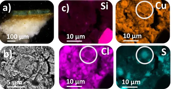
To be studied at the SEM, samples are embedded in epoxy resin, cut and polished with silicon carbide disks (grains size 46, 15 and 5 µm) in order to have a cross-section which makes the paint layers organization visible (figure 1.a). The thickness of the green layer ranges from 10 to 100 µm, but is generally between 25 and 35µm. Arsenic (Kα = 10,5keV) is the louder element in our samples and then require an excitation energy of at least 15,75kV. For SEM-FEG imaging, they are coating with platinum and studying under a 15kV (100 µA) current at a 3,0 mm Working Distance (WD) to optimize the solid angle with the Backscattered-Electron (BSE) detector. For SEM-FEI analysis it is used a carbon coating. Punctual analysis are done under a 20kV current at a 10,0 mm WD. Mapping is still at 10,0 mm WD but with a 15kV current to have a better spatial resolution, and used to calculate an image resolution of 3000 counts/pixel for 2 hours acquisition. The first elementary analyses by SEM-EDS showed that we have in all cases, a copper material where the detection of tin and lead traces proves that the copper used in the fabrication of synthetic green pigments comes from bronze scraps. It also allowed the identification of two possible types of initial pigments. One, with silica amorphous phases (figure 1.c) characteristic of a heat treatment, and the other one without; possibly copper acetate. The SEM-FEG imaging provides new information concerning texture layer and grain morphology. Two groups have also been identified: green samples presenting as compact mass of tiny grains less than 1µm and ones with grain vestiges (2-5 µm) and external crowns (figure 1.b). Those both textures suggest a pigment largely altered and seem to be independent of the initial pigment. It is well known that copper greens are quite unstable, and the right
type of environment (salts, humidity and/or presence of organic materials [1, 2]) is conducive to their alteration. This hypothesis has been confirmed by the presence on SEM-EDS mapping of copper chlorides and copper sulfides in very localized areas of grains (figure 1.c). Other studies in the literature propose, besides newly formed copper chlorides, the possible formation of copper-protein complexes, just like copper-proteinate, possibly altered to copper oxalate [3, 4].
It is also important to note that for all experiments, the intensity of the signal is quite weak in the green areas in comparison to other areas in the samples. This point suggests the presence of organic compounds but they are impossible to identify with this technic. The inability to identify multiphasic material just like organometallic ones; and also the impossibility to determine precisely the nature of the present materials is one of the limits of the SEM as an elementary analytical technic. However, this first analytical step gives us very precious information to go further in the identification of these materials. It helps to proceed to the structural analysis, the next step in the understanding of the degradation mechanisms.
µ-FTIR mapping confirmed the presence of copper chlorides (atacamite Cu2Cl(OH)3 and paratacamite (Cu,Zn)2(OH)3Cl) and a supposition that copper oxalates are also present. The identification of alteration mechanisms enables us to know the precise nature of initial pigments and maybe even the fabrication recipes. To go even further, prepared samples of initial green pigments (copper glass and copper acetate) will be put in chloride and/or oxalic conditions to mimic alteration phenomena and then they will be studied to identify any chemical changes due to the alteration treatment. Follow-up and control will be done with the same analytical methodology as for archaeological samples (SEM and FTIR).
References
[1] S Svarcova, et al, Analytical and Bioanalytical Chemistry (2009)
[2] M Gunn et al, Studies in Conservation (2002)
[3] M Pérez-Alonso, K Castro, JM Madariaga, Analytica Chimica Acta (2006) [4] A Zoppi, et al, Analytical and Bioanalytical Chemistry (2010)
42. This work is labellised by the Fondation des Sciences du Patrimoine. Thanks to the Egyptian Antiquities Department from the Louvre Museum.
Figure 1. Sample example: AF 9592 – Co1 a) Optical Microscope observation of the cross-section b)
SEM-FEG BSE imaging of the green layer texture c) SEM-EDS mapping of the same area as 1.b presenting results of Si, Cu, Cl, and S.