HAL Id: tel-01374865
https://tel.archives-ouvertes.fr/tel-01374865
Submitted on 2 Oct 2016HAL is a multi-disciplinary open access archive for the deposit and dissemination of sci-entific research documents, whether they are pub-lished or not. The documents may come from teaching and research institutions in France or abroad, or from public or private research centers.
L’archive ouverte pluridisciplinaire HAL, est destinée au dépôt et à la diffusion de documents scientifiques de niveau recherche, publiés ou non, émanant des établissements d’enseignement et de recherche français ou étrangers, des laboratoires publics ou privés.
Maria Antonietta Principalli
To cite this version:
Maria Antonietta Principalli. Structure-function studies of the Kir6.2 channel and of its coupling with natural and artificial partners. Molecular biology. Université Grenoble Alpes, 2015. English. �NNT : 2015GREAV014�. �tel-01374865�
THÈSE
Pour obtenir le grade de
DOCTEUR DE L’UNIVERSITÉ GRENOBLE ALPES
Spécialité : Biologie Structurale et NanobiologieArrêté ministériel : 7 août 2006
Présentée par
Maria Antonietta PRINCIPALLI
Thèse dirigée par Dr. Michel VIVAUDOU et codirigée par Jean REVILLOUDpréparée au sein de l’Institut de Biologie Structurale dans l'École Doctorale Chimie et Sciences du Vivant
Étude structure-fonction du canal
Kir6.2 et de son couplage avec des
partenaires naturels et artificiels
Thèse soutenue publiquement le 9 Octobre 2015 devant le jury composé de :
Dr. Pierre-Jean CORRINGER
Président
Prof. Anna MORONI
Rapporteur
Prof. Bruno ALLARD
Rapporteur Dr. Patrice CATTY Membre Dr Michel VIVAUDOU Directeur de thèse Jean REVILLOUD Co-directeur de thèse
Thèse pour obtenir le grade de
DOCTEUR ès SCIENCES DE L’UNIVERSITÉ DE
GRENOBLE
Specialité: Biologie Structurale et Nanobiologie
Presented and defended in public by
Maria Antonietta PRINCIPALLI
On October 9
th2015
Structure-function studies of the Kir6.2 channel and of
its coupling with natural and artificial partners
Thesis directors:
Dr. Michel VIVAUDOU
Jean REVILLOUD
Jury:
President: Dr. Patrice CATTY
Reviewer: Prof. Anna MORONI
Reviewer: Prof. Bruno ALLARD
Examiner: Dr. Pierre-Jean CORRINGER
Acknowledgements
I would like to thank in the first place Prof. Anna Moroni and Prof. Bruno Allard for dedicating their time to the evaluation of this work. I would like to express my gratitude as well to Dr. Patrice Catty, Dr. Pierre-Jean Corringer and Dr. Hugues Nury for being part of my annual evaluation committee, for their precious advices and their optimistic vision of problematic projects.
A special thanks to Michel Vivaudou, my thesis director, for his advices and his sense of humour and for giving me the chance to experience an adventure like no other, scientifically and personally. Thanks to Jean Revilloud, my supervisor in the lab, for his patience, for saving my life during my first (and last) day on a pair of skies, and for teaching me the art of patch-clamping. Many thanks to Christophe Moreau, the most patient person I have ever known, for encouraging me to believe in my theories and for helping me exploring my ideas even though they looked crazy or useless to others. Special thanks to Kasia, my personal English teacher, who helped me in moving the first steps in the lab. Big thanks to Catalina, whose friendship and Latin spirit helped me reach the end of this adventure. Thanks to Laura for being an intelligent and fun co-worker, and to Karla for her calm in panic situations. A particular thanks to Yann, Sonja, Ola, Hubert, Lionel, Céline, Michel T. and Isabelle for their priceless advices and their help.
Un grazie tutto particolare alla banda degli italiani a Grenoble: il Mollica, ‘mamma tuttofare’, DdS e mamma Rocha (italiana adottiva), per avermi salvato dai guai in più di un’occasione. Al piccolo Gabriel per avermi insegnato che non ci si veste mai per bene quando si deve fare da baby-sitter, a Filippo per il suo accento toscano e le sue email memorabili, a Sciaken per saper trasmettere la sua calma e il suo approccio positivo alla vita. Grazie a tutti voi ragazzi! Un grazie speciale a Mizar, con cui ho intrapreso questa e molte altre avventure, amica sincera e cuoca personale, all’occorrenza pronta a darmi una strigliata affettuosa.
Grazie a mamma e papà per avermi supportata e per aver creduto in me, sempre. Grazie ad Alex, per essere rimasto fino alla fine.
“There is nothing like looking, if you want to find something. You certainly usually find something, if you look, but it is not always quite the something you were after.”
ABC
ATP binding cassette; 15 ACh
Acetylcholine; 61 ADP
Adenosine diphosphate; 15 AOX
Alcohol oxidase promoter; 78 ATP
Adenosine triphosphate; 15 cAMP
Cyclic adenosine monophosphate; 59 CFTR
Cystic fibrosis transmembrane conductance regulator; 17 COPI
Coat protein complex I; 34 CTD
Cytoplasmic domain; 33 D2L
Long Dopaminergic receptor 2; 51 DAG
Diacylglycerol; 57 DDM
N-dodecyl β-D-maltopyranoside; 81 DEND
Developmental delay, epilepsy, and neonatal diabetes; 44 DEPC Diethyl pyrocarbonate; 72 DNA Deoxyribonucleic acid; 71 ECF Energy-coupling factor; 21 ECL Extracellular loop; 52 eGFP
Enhanced green fluorescent protein; 79 EGTA
Ethylene glycol tetra-acetic acid; 87 ER
Endoplasmic reticulum; 16 GDP
Guanosine triphosphate; 40 GIRK
G-protein activated inward rectifying potassium channel; 32 GPCR
Human influenza hemagglutinin; 75 hOR
Human olfactory receptor; 51 ICCRs
Ion channel-coupled receptor; 50 ICL Intracellular loop; 52 IP3 Inositol trisphosphate; 57 IPTG Isopropyl β-D-1-thiogalactopyranoside; 75 K-ATP
ATP-sensitive potassium channel; 15 KCO
Potassium channel opener; 48 Kir
Inwardly rectifying potassium channel; 15 LB
Luria-Bertani; 71 LC-CoA
long chain Co-enzyme A esters; 43 M2
Muscarinic receptor 2; 51 MAC
Metal affinity chromatography; 81 MNG-3
Maltose-neopentyl glycol; 81 mRNA
Messenger ribonucleic acid; 73 MRP
Multidrug resistance protein; 17 MSP
Membrane scaffold protein; 80 NBD
Nucleotide binding domain; 17 PBS
Phosphate-buffer saline; 82 PCR
Polymerase chain reaction; 78 P-gp
P-glycoprotein; 24 PHHI
Persistent hyperinsulinemic hypoglycaemia of infancy; 35 PIP2
SDS-PAGE
Sodium dodecyl sulphate - polyAcrylamide gel electrophoresis; 74 SEC Size-exclusion chromatography; 82 SUR Sulfonylurea receptor; 15 TBS Tris-buffer saline; 75 TEVC
Two-electrode voltage-clamp technique; 90 TMD
Transmembrane domain; 17 TRIS
Tris(hydroxymethyl)aminomethane; 75 tRNA
Transfer ribonucleic acid; 83 UTR
Untranslated region; 72 VFTM
Venus flytrap mechanism; 56 YPD
membrane potential across the plasma membrane of excitable cells such as neurons, muscle cells and endocrine cells. In neurons, for example, the membrane potential of the cell at rest is around -70 mV. This value increases to about +50 mV during an action potential and, subsequently, returns to the resting level again. The action potential event is triggered by a depolarization and produced by the activation of voltage-gated ion channels.
Conductance: indicates the capacity of an ion channel to let ions move through its pore. It is measured in Siemens (S).
Gating: the phenomenon or mechanism by which an ion channel opens or closes its pore. Depending on the specific ion channel, this process is regulated by voltage, metabolic state of the cell (ATP/ADP concentration), intracellular ligands and/or second messengers (calcium, cAMP, etc.), or extracellular ligands (neurotransmitters).
Membrane potential: The voltage difference across the plasma membrane of cells. It is negative under resting conditions and becomes positive during an action potential. Its value depends on different factors: the concentration of ions at both sides of the membrane, the permeability of the membrane to these ions, the activity of electrogenic pumps that maintain the ions concentration outside and inside the cells.
Open probability (Po): describes the fraction of time spent by an ion channel in its open conformation.
Preamble ... 15
Introduction ... 17
The ATP-sensitive potassium channel constituent subunits ... 18
K-ATP channel overview ... 18
The Sulfonylurea Receptor: an atypical transporter of the ABC family ... 20
1.2.1 The ABC family ... 23
Kir6 channels ... 29
1.3.1 Inwardly rectifying potassium channels (Kir) ... 29
1.3.2 Inward rectification ... 30
1.3.3 Kir channel structures ... 32
SUR-Kir6 assembly and functional coupling ... 37
SUR-Kir6 association ... 37
SUR-Kir6 functional coupling ... 41
K-ATP channel mechanism and regulation ... 42
Physiological regulation of the K-ATP channel mediated by SUR ... 42
Physiological regulation of the K-ATP channel mediated by Kir6 channels ... 44
K-ATP channel physiology and physiopathology ... 46
K-ATP channel pharmacology ... 48
3.4.1 K-ATP channel inhibitors ... 48
3.4.2 K-ATP channel openers (KCO) ... 50
Ion Channel-Coupled Receptors (ICCRs) ... 53
Principle of the ICCR concept ... 53
ICCR engineering ... 54
Applications of the ICCR technology ... 54
4.4.3 GPCR ligands... 59
4.4.4 GPCR-mediated signalling through G Proteins ... 59
4.4.5 The M2 muscarinic acetylcholine receptor ... 61
Engineering of the GPCR-Kir6.2 fusion to obtain functional coupling... 63
Materials and Methods ... 67
Molecular biology ... 68
Genes and expression vectors ... 68
Mutagenesis ... 69
6.2.1 Site-directed mutagenesis ... 69
6.2.2 Deletion of long fragments ... 70
Subcloning ... 71
Amplification of genetic material ... 73
6.4.1 Transformation of competent bacteria ... 73
6.4.2 Amplification and purification of the DNA plasmid ... 73
6.4.3 Miniprep and Midiprep methods ... 73
RNA preparation ... 74
Biochemistry: Production and purification of recombinant proteins ... 76
Basic techniques of biochemistry ... 76
7.1.1 Protein electrophoresis (SDS-PAGE) ... 76
7.1.2 Western blot technique (immunodetection) ... 76
Expression in Escherichia coli and solubilisation of bacterial membranes ... 77
Expression in the yeast Saccharomyces cerevisiae ... 79
Expression in the yeast Pichia pastoris ... 80
In-vitro synthesis ... 81
Heterologous expression in Xenopus oocytes ... 85
Xenopus laevis oocytes ... 85
Extraction and preparation of oocytes ... 86
RNA microinjection ... 86
Functional characterization of ion channels by electrophysiology techniques ... 87
The patch clamp technique ... 87
9.1.1 Patch clamp configuration ... 88
9.1.2 Data processing and analysis ... 91
The Two-Electrode Voltage-Clamp technique (TEVC) ... 92
9.2.1 Experimental procedure ... 92
9.2.2 Data processing and analysis ... 93
Results ... 95
Project 1: ‘Structure of the ATP-sensitive potassium channel’ ... 96
Relevance of the study ... 96
Project background and experimental approach ... 96
Results: Expression trials ... 97
10.3.1 Expression in E. coli ... 97 10.3.2 Expression in yeasts ... 99 10.3.3 In-vitro synthesis ... 100 Results: Purification ... 101 10.4.1 TMD0-eGFP ... 101 10.4.2 Kir6.2 ... 107
Discussion and perspectives ... 109
Project 2: ‘Study of the functional coupling between SUR and Kir6.2: focus on the SUR interacting region ... 110
Results: Residues Q1342, I1347 and L1350 of SUR1 are essential in transducing
activation by Diazoxide and ADP to Kir6.2 ... 111
Results: Differences in the coupling of Kir6.2 with SUR1 and SUR2A revealed by alanine mutants ... 116
Discussion and perspectives ... 118
Project 3: ‘Role of a cluster of arginines in the N-terminal region of Kir6.2 in its regulation by natural and unnatural partners’ ... 121
Relevance of the study ... 121
Project background and experimental approach ... 122
Results: Substitution of the arginines to confirm their role in ICCR function ... 125
Role of arginine R32 in ICCR function ... 127
Results: role of the Kir6.2 N-terminal arginines in the natural K-ATP channel . 132 12.5.1 Response to the physiological opener MgADP ... 132
12.5.2 Response to pharmacological openers ... 134
12.5.3 Response to the K-ATP channel blocker Glibenclamide ... 136
12.5.4 Whole-cell experiments with TEVC ... 137
Discussion and perspectives ... 138
12.6.1 ICCR regulation ... 138
12.6.2 Finding arginine partners in Kir6.2 C-terminal ... 139
12.6.3 Impact of mutation R27Y and R32A on K-ATP channel regulation ... 140
15
16
1983 by Akinori Noma, it obtained significant notoriety because of its implication in the insulin secretion process. Over the past years, huge progresses have been made regarding the understanding of its function, yet many questions remain to be answered. Among these, this thesis tried to help elucidate two main aspects of the K-ATP channel, its structure and the mechanism used by the two partners to communicate with each other in what is called ‘functional coupling’.
To achieve this, the work was divided in three main projects. First, we attempted to heterologously express and purify the protein to obtain samples suitable for structural studies. Although the precise mechanism is not fully understood, it is known that binding of nucleotides or drugs to the SUR subunit activates the Kir6 channel. Thus, the second project focused on the role of SUR in activation of the channel. The third project is related to the second as it aims to better understand Kir6 regulation by SUR; but this time from the side of the pore-forming subunit. This last objective cannot be easily achieved with classical approaches as the region thought to be involved in Kir6 activation is in tight physical association with SUR and mutations at this level would compromise the general folding of the channel. Ion Channel-Coupled Receptor (ICCRs) technology, developed in our laboratory, provides a unique system to study Kir6.2 mechanistic.
In ICCRs, Kir6 is directly fused at its N-terminus to the C-terminus of a G protein-coupled receptor (GPCR) that tunes its gating depending on ligand binding. Using ICCRs we discovered a cluster of arginines in the Kir6 N-terminus responsible for channel gating modulation. These arginines were subsequently studied in the K-ATP channel to understand their involvement in the regulation of Kir6 gating by its natural partner SUR. The three projects embrace three large families of proteins such as ABC transporters, Kir channels and GPCRs. ABC transporters and Kir channels will be described in detail in the introduction of this manuscript. GPCRs, although a very important family of proteins, will not be described in detail because they are not directly the subject of this work. They are used only as a tool to understand SUR-Kir6 coupling.
17
18
The ATP-sensitive potassium channel constituent subunits
K-ATP channel overview
ATP-sensitive potassium channels result from the unique partnership of two distinct proteins: the Sulfonylurea Receptor (SUR), which belongs to the ATP binding cassette (ABC) transporter family, and the inwardly rectifying potassium channel (Kir6). These particular channels are physiologically up-regulated by MgADP and down-regulated by ATP. This regulation allow the channel to ‘sense’ cell metabolism and, depending on the energy level of the cell, to change the potassium permeability of the membrane, which might result in a variation of the membrane potential. Variations in the membrane potential trigger a certain cellular response depending on the K-ATP localization. Examples of processes regulated by these channels are insulin secretion and regulation of the duration of the action potential in cardiac cells.
K-ATP channel subunits SUR and Kir6 exist in different isoforms depending on the cell type where these are expressed. The SUR protein (~ 170 kDa) exists as three isoforms: SUR1 mostly located in the pancreas and in the brain, SUR2A expressed in cardiac and skeletal muscles and SUR2B in smooth muscle blood vessels. The Kir6 channel (~ 45 kDa) is present in two isoforms: Kir6.1 in smooth muscle and Kir6.2 in pancreas, brain, cardiac and skeletal muscles.
In the vertebrate genome, we can find two Kir6 genes (KCNJ8, KIR6.1 and KCNJ11, KIR6.2) and two SUR genes (ABCC8, SUR1 and ABCC9, SUR2). Isoforms SUR2A and SUR2B derive from alternate splicing of the same gene. Interestingly, the genes for Kir6.2 and SUR1 are located next to each other on human chromosome 11p15.1 while the genes for Kir6.1 and SUR2 are also consecutive on chromosome 12p12.1, suggesting that an elegant gene expression regulation happens already at the transcription level (Nichols, Singh, and Grange 2013).
It takes four SUR and four Kir6 subunits to build a full-length K-ATP channel (Figure 1). The resultant hetero-octamer has a predicted mass of about 950 kDa. Kir6 is in charge of creating the pore, necessary to ensure K+ flow, while the SUR protein finely plays on channel
19
biochemical studies (Inagaki, Gonoi, and Seino 1997; Clement IV et al. 1997) and a low resolution structure of the full-length complex (Mikhailov et al. 2005).
The presence of retention signals (RXR) on both subunits ensures that only properly assembled channels reach the plasma membrane. In particular, during K-ATP assembling, these motifs are masked so that the resultant complex is released from the ER (Zerangue et al. 1999). Physical association between SUR and Kir6 has been confirmed conducting co-immunoprecipitation experiments (Lorenz and Terzic 1999). Moreover, deletion of the last 36 amino acids in the Kir6.2 channel allows the formation of functional channels in absence of the SUR subunit (Tucker et al. 1997).
Figure 1. Cartoon of the K-ATP channel predicted folding. Kir6 and SUR are the constitutive subunits of the channel. Four subunits of the inwardly rectifying K+ channel Kir6 associate with four ATP-binding cassette proteins,
SUR, to form a functional K-ATP channel octamer. Kir6 has two transmembrane helices and a large cytoplasmic domain harboring an inhibitory binding site for ATP. SUR possesses three transmembrane domains (TMD0, 1&2), and 2 cytoplasmic nucleotide binding domains (NBD1&2).
Inward rectifier K+ channel Kir6.2 Sulfonylurea receptor SUR N TMD1 TMD2 C NBD1 NBD2 ATP ADP C N ATP TMD0 TMD0 TMD1 TMD2
20
The Sulfonylurea Receptor: an atypical transporter of the ABC
family
The Sulfonylurea Receptor SUR belongs to the ATP-binding cassette family (ABC). In particular, SUR is located in the ABCC subfamily, together with MRP1 (multidrug resistance protein) and CFTR (cystic fibrosis transmembrane conductance regulator).
Despite their phylogenic correlation, these proteins share very little in terms of function. MRP1 is a multidrug exporter of glutathione conjugates which, taking advantage of ATP hydrolysis, transports substrates (Cole 2014). CFTR, known for its implication in cystic fibrosis, is an ion channel gated by ATP hydrolysis (Vergani et al. 2005). SUR modulates the gating of a K+ channel using its NBDs domains to ‘sense’ ATP and ADP levels inside cells.
High-resolution structures of several bacterial ABC proteins suggest a mechanism by which ATP hydrolysis at the nucleotide-binding domains (NBDs) of ABC proteins is coupled to conformational changes that drive substrate transport (Locher 2009).
So far, the atomic structure of the SUR is not known. Nevertheless, alignments with other members of the ABC family, whose structure have been resolved, show the presence of common features. SUR N-terminus faces the extracellular side of the membrane while its C-terminus is intracellular (Mikhailov et al. 2005). The hydrophobic plot of the SUR sequence shows the presence of three transmembrane domains named TMD0, TMD1 and TMD2. These are interconnected by a long loop (L0) and two hydrophilic domains, the nucleotide binding domains NBD1 and NBD2.
TMD1 and TMD2 are common among ABC proteins and they typically present six spanning α helices. The TMD0 deserves a particular place among TMDs as it is found only in few members of the ABC family such as MRP1–3, MRP6–7 and YCF1 (yeast cadmium factor) and is connected to TMD1 through the long cytoplasmic loop (L0). It is considered an ‘additional domain’ of such members and its function it is not fully understood. Moreover, TMD0s from different ABC proteins show different amino acid sequences, which suggest a variability in their function. The TMD0 in MRP1 seems to be dispensable as its deletion does not affect the transport function or the cellular targeting of MRP1 (Bakos et al. 1998) or the
21
dimerization (Yang et al. 2007). The TMD0 of MRP2 is required for correct cellular targeting but is not necessary for transport function (Fernández et al. 2002).
In the case of the SUR protein, the TMD0 domain is essential for proper addressing of the K-ATP channel at the plasma membrane and has a critical role in Kir6.2 gating modulation (Chan, Zhang, and Logothetis 2003; Fang, Csanády, and Chan 2006). In particular, TMD0 cannot abolish the retention of full-length Kir6 channels (containing the RKR retention signal at the C-terminal) but it sensitively burst the expression of Kir6 channels deleted of the RKR motif. When Kir6.2 tetramers are forced to the cell surface by deleting the retention signal, they show a low maximal open probability in ligand-free solutions (Po(max)). TMD0 alone is able to sensibly stabilized the burst and conducting states of Kir6.2 tetramers raising its Po(max) of 4-fold, suggesting that interactions of transmembrane helices are able to force channel opening (Andrey P. Babenko and Bryan 2003).
NBDs domains are cytoplasmic and present consensus sequences as the ‘ABC signature’ as well as the Walker A (GXXGXGK(S/T)) and Walker B (XXXXDE, where X is a hydrophobic amino acid) motifs. These signatures allow ABC proteins identification. NBDs function consists in sensing the intracellular ATP/ADP ratio, allowing the SUR subunit to modify Kir6 sensitivity to nucleotides.
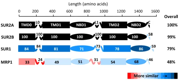
Among all ABC proteins, SUR shares the highest level of similarity with the MRP1 protein. Despite a 48% of sequence similarity (Moreau, Prost, et al. 2005) (Figure 2), that suggests a common folding and so a common function, SUR and MRP1 show very different functions. Over the past years, scientists have taken advantage of such predicted structural similarities to create SUR-MRP chimeras to study SUR2A-Kir6.2 interactions (Rainbow et al. 2004; Dupuis et al. 2008; Lodwick et al. 2014). In particular, this same strategy has been used in the present study to pinpoint SUR1 determinants for functional interaction between SUR1 and Kir6.2.
22
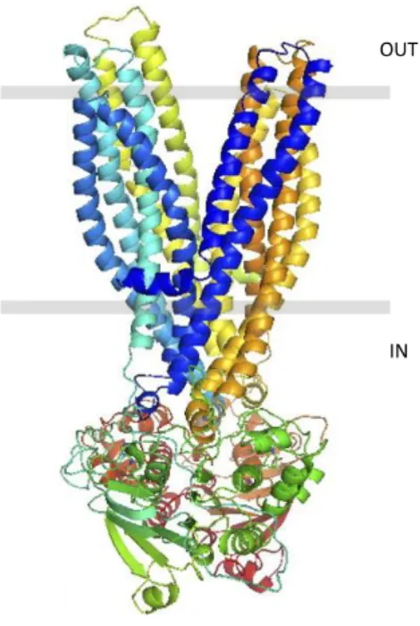
The absence of structural data of proteins containing the TMD0 prevents the creation of full-length SUR models. Nevertheless, Bessadok and colleagues proposed, in 2011, a model of SUR1 in the outward-facing conformation, lacking the TMD0, based on the structures of other ABC transporters, MsbA and Sav1866 (Bessadok et al. 2011). This model is based on ∼20% sequence identity and is supposed to be the physiologically relevant enzymatic state because it is the nucleotide-bound form (Figure 3).
Because full-length models of SUR are not yet feasible, a model of the full K-ATP channel complex remains utopic. The complexity of building such a model is further increased by the absence of solid data describing the interacting regions at SUR-Kir6 interface.
0 200 400 600 800 1000 1200 1400 1600 SUR2A MRP1 SUR1 84 84 81 71 73 78 86 59 33 24 49 51 31 54 68 46 100% 48% 79% TMD0 L0 TMD1 NBD1 L1 TMD2 NBD2 C SUR2B 100 100 100 100 100 100 100 58 99% Overall
Length (amino acids)
More similar
Figure 2. The human cardiac SUR2A isoform displays a high degree of sequence homology with MRP1 protein with the most convergence in domains NBD2 and TMD1 - TMD2 and the most divergence in domains TMD0 and connecting loops L0 and L1. Sequences were aligned individually with SUR2A using ClustalX. Similarity within each domain is specified in percent (Moreau, Prost et al., 2005).
23
1.2.1 The ABC family
The transport of organic and inorganic molecules across cell membrane is essential for living organisms. These molecules often need to be moved against their gradient. Transport against a chemical gradient can be driven by, for example, the free energy change associated with ATP hydrolysis (primary transport), or facilitated by the potential energy of the chemical gradient of another molecule (secondary transport). Primary transporters include a large family of integral membrane proteins referred as “ABC” (ATP-binding cassette) transporters.
These transporters represents the largest protein family identified to date. There are 48 ABC transporters in humans and, depending on their function for human health, many have been correlated to severe disease such as cystic fibrosis, Tangier disease (characterized by a severe reduction in the amount of high density lipoprotein), neonatal diabetes, macular dystrophy. Moreover, some ABC transporters are involved in the drug resistance of bacteria and cancer cells (Wilkens 2015).
Figure 3. Outward-facing conformation model of SUR1 from Bessadok et al., 2011. SUR1 is shown with two transmembrane domains only (TMD1, in shades of blue & TMD2, in shades of yellow), and the two cytoplasmic nucleotide binding domains (NBD1, in shades of green and NBD2, in shades of brown).
IN OUT
24
The existence of methods that allow to closely look at protein structure sensibly improved the understanding of protein functions. On the other hand, obtainment of the atomic structure of proteins, and in particular of membrane proteins, it is a very complex and challenging process. To date, 14 ABC transporters have been structurally characterized:
BtuCD, Vitamin B12 transporter (E. coli), (Locher, Lee, and Rees 2002);
Sav1886, multidrug transporter (S. aureus), (Dawson and Locher 2006);
ModB2C2, Molybdate transporter (A. fulgidus), (Hollenstein, Frei, and Locher 2007); HI1470/1, Metal-Chelate-type transporter, (H. influenzae), (Pinkett et al. 2007); MsbA, lipid ‘flippase’, (S. typhimurium) (Ward et al. 2007);
MalFGK2, Maltose uptake transporter complex (E. coli), (Oldham et al. 2007); P-Glycoprotein, (M. musculus), (Aller et al. 2009);
MetNI, Methionine uptake transporter complex (E. coli), (Kadaba et al. 2008); TM287-TM288, (T. maritime), (Hohl et al. 2012);
HmuUV, heme transporter (Y. pestis), (Woo et al. 2012);
ABCB10, Mitochondrial ABC transporter,(H. sapiens), (Shintre et al. 2013); Atm1-type, ABC exporter,(N. aromaticivorans), (Lee et al. 2014);
Atm1, mitochondrial ABC transporter (S. cerevisiae), (Srinivasan, Pierik, and Lill 2014); McjD, antimicrobial peptide transporter (E. coli), (Choudhury et al. 2014);
These currently available structures have sensibly advanced our knowledge on the transport mechanism and revealed a sensible structural divergence (Beek, Guskov, and Slotboom 2014).
ABC transporters discovered so far are classified into two main groups: exporters and importers. The importers are further divided into class I and class II and a third group, the energy-coupling factor (ECF) family which is structurally and functionally more distinct (Figure 4). Bacteria use both importers and exporters while eukaryotes, with few exceptions, only employ exporters.
25
A canonical ABC transporter is organized in four domains: two TMDs and two NBDs. While the NBDs are highly conserved among all ABC proteins, TMDs show a variable folding. NBDs
Each NBD consists of two subdomains: the RecA-like domain and the α-helical domain. Alignment of NBDs sequences show highly conserved futures (Figure 5):
The A-loop, which contains a conserved aromatic residue responsible for the correct binding of the ATP;
The Walker A motif, a phosphate binding loop with a conserved K;
The Walker B motif, which helps the coordination of Mg2+, and possesses a
conserved E that works as base to polarize water;
The D-loop, important to maintain the geometry of the ATP hydrolysis site;
The H-loop, which assists the positioning of the water molecule, Mg2+, and the
general base;
Josy ter Beek et al. J Gen Physiol 2014
Figure 4. ABC transporters folds. All share a similar general architecture: two NBDs (blue and sky blue) are attached to two TMDs (orange and yellow). In some transporters, additional domains are present (green), which often have a regulatory function. In Type I and II importers, the transported compounds are delivered to TMDs by SBPs (substrate-binding proteins, magenta) located in periplasm (Gram-negative bacteria) or external space (Gram-positive bacteria and Archaea).
26
The Q-loop, which has a conserved Q responsible for allowing the formation of an active site during ATP hydrolysis and is also in contact with the TMDs;
The ABC signature motif (LSGGQ), ABC proteins hallmark.
TMDs
Depending on the transporter class, TM domains have 6 to 10 transmembrane α helices that are arranged in such a way that they form a pore necessary to allow substrate passage. In general, the TMDs do not share sequence similarities but fold similarities, reflecting the need to transport a wide range of molecules.
Transport mechanism
The essential catalytic cycle of ABC transporters consists in a series of steps (Wilkens 2015): Binding of substrate;
Figure 5. NBD structure (MalK dimer of the maltose transporter MalEFGK2). (A) View along an axis
perpendicular to the membrane plane from the trans-side onto the NBDs (The TMDs and SBP have been removed for clarity). Domains and highly conserved sequence motifs are color-coded: green, α-helical domain; light blue, RecA-like domain; faded gray, regulatory C-terminal domain; red, A-loop; magenta, Walker A; orange, Walker B; blue, D-loop; green, H-loop; cyan, ABC motif; yellow, Q-loop. The ATP analogue AMP-PNP is shown in sticks. (B) A closer look onto the nucleotide-binding site. The key amino acids are indicated (see NBDs for details) (Josy ter Beek et al., J Gen Physiol 2014).
27
Binding of 2 Mg-ATP molecules to the NBDs; NBDs dimerization;
Conformational changes into the TMDs to allow the passage of substrates; ATP hydrolysis;
Release of phosphate, ADP and substrates;
NBDs dissociation with subsequent return to the basal state.
This general cycle can be adapted to most ABC proteins but there is still a lot to clarify about number of ATP molecules used, mechanism of NBDs action and motion changes in the TMDs (Linton 2007) (Figure 6).
To illustrate the progresses in deciphering the conformational changes at the TMDs level, it is useful to consider the recent results obtained with the P- glycoprotein (P-gp). The P-gp transporter is an ATP-dependent efflux pump with a wide substrate specificity. It has a great clinical and pharmacological significance as it is implicated in the transport of many drugs. Large amounts of this protein are found in cancer cells conferring them multi-drug resistance.
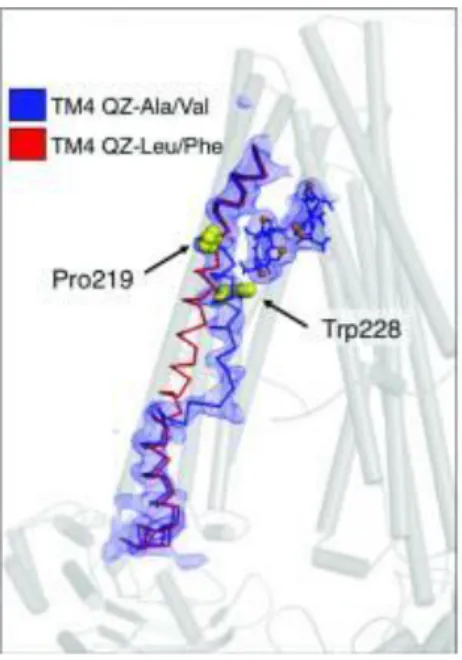
P-gp consists of two pseudosymmetric halves encoded into a single polypeptide. Each half is formed by six transmembrane helices (TMs) and one cytosolic nucleotide-binding domain (NBD) along with interconnecting loops and short helices. The TMs surround a central pocket, which contains multiple binding sites for ligands. Crystal structure studies employing a broad spectrum of ligands show that the binding of certain ligands produces a large conformational change in the fourth transmembrane helix (TM4) (Figure 7), which is postulated to be positioned to potentially transmit a signal to the NBDs (Szewczyk et al.
28
2015). This helix may possess a high degree of flexibility which is consistent with a role in substrate binding. Mutations in this region, which favour a well ordered, straight-helical conformation, disrupt substrate transport (Kodan et al. 2014), reinforcing the predicted role for TM4 in facilitating substrate entry and/or binding. The ligand binding-induced kinking of TM4 begins at Pro219.
Such detailed knowledge of the mechanism of ABC transporters remains restricted mainly to P-gp, the most studied ABC protein. Studies of other ABC proteins, such as SUR, are needed to understand their specific function and pharmacology.
Figure 7. Overview of two different conformational changes in the TM4 mediated by 2 different classes of substrates. The kinking of TM4 in response to ligand QZ-Ala/Val (in stick-and-balls format) is shown (blue ribbon) in comparison to its ‘straight’ topology of the ligand QZ-Leu/Phe co-crystal structures (red ribbon). Essential residues Pro219 and Trp228 are shown as yellow spheres.
29
Kir6 channels
Kir6 channels belong to the inwardly rectifying potassium channels family (Kir channels). Both members of this subfamily, Kir6.1 and Kir6.2, show a common predicted folding. Each subunit folds into a short cytoplasmic N-terminus, two transmembrane α-helices (TM1 and TM2) connected by a loop (H5 loop) and a short helix (pore helix) containing the Kir channels signature (TVGYG for K+ channels, TVGFG for Kir6 channels) and a large
cytoplasmic C-terminal (Antcliff et al. 2005). Full-length Kir6 channels result from the assembly of four subunits that arrange to build a central pore that allows potassium flow. Tetrameric Kir6 channels can result from the assembly of Kir6.1 or Kir6.2 channels only (homotetramer) or from the association of Kir6.1 and Kir6.2 channels (heterotetramers) (Teramoto et al. 2009).
Kir6 channels are widely expressed in mammals. Kir6.1 is mostly expressed in smooth muscle (Inagaki, Inazawa, and Seino 1995) while Kir6.2 is highly expressed in pancreatic cells (Suzuki et al. 1997).
1.3.1 Inwardly rectifying potassium channels (Kir)
Inwardly rectifying potassium channels comprise a large family of voltage-independent potassium channels. The inward rectification is caused by cytoplasmic ion such as polyamines and Mg2+, which block the outward passage of ions in favour of an inward current when the
cell is at rest. Because of their peculiar characteristics, Kir channels stabilize the resting membrane potential close to the K+ equilibrium potential by mediating transport of
potassium across the membrane (Nichols and Lopatin 1997).
Depending on their degree of rectification, they are classified into ‘strong’, for channels that are more sensitive to Mg2+ and polyamines block, or ‘weak’ rectifier for channels that are less
sensitive to these molecules.
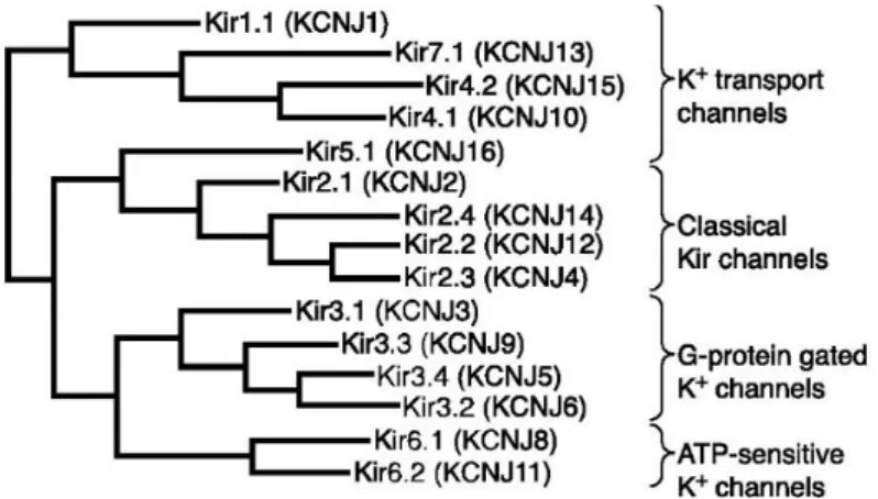
So far, 15 Kir channels have been characterized. They are divided into seven subfamilies, depending on their characteristics such as degree of rectification, conductance, and sensitivity to diverse mediators. These subfamilies can be divided into four groups: Classical Kir channels (Kir2.x), G protein-gated Kir channels (Kir3.x), ATP-sensitive K+ channels (Kir6.x)
30
Kir1 subfamily includes weak rectifiers expressed mostly in the kidney and in the brain involved in transepithelial transport (C. G. Nichols and Lopatin 1997). Kir2 channels are strong rectifiers with high conductance variability. They are involved in the control of the excitability of cardiac and cerebral tissues (Reimann and Ashcroft 1999). Kir3 family is represented by G protein–activated strongly rectifying K+ channels expressed in cardiac,
neuronal and neurosecretory cells. Kir4 channels can exist as homo- or heteromers with Kir5 channels and they are expressed in glia cells, cochlea and kidney. In kidney, heteromers help the activity of Na2+- K+ ATPase by supplying potassium to the extracellular side of the
plasma membrane (Hibino et al. 2010). Kir7.x channels function is still unknown. These channels are expressed in the epithelial tissue.
1.3.2 Inward rectification
In physiological conditions, when the cell membrane is depolarized, Kir channels allow weak outward potassium current while the inward flow of potassium is stronger (Figure 9). This characteristic depends on the voltage-dependent block of outward current by cations (mostly Mg2+ and polyamines) that bind the cytoplasmic side of the pore and obstruct the outward
flux of potassium ions (Bichet, Haass, and Jan 2003).
According to the presence of binding sites for positively charged molecules and ions, we can discriminate between strong rectifiers and weak rectifiers. It has been found that two residues located into the transmembrane helix 1 and in the channel C-terminal respectively are responsible for the binding of Mg2+ and polyamines. In particular, aminoacid at position 171
31
(N171) in Kir1.1 (weak rectifier) is uncharged, while in strong rectifier, such as Kir2.2, it is replaced by an aspartate (Yang, Jan, and Jan 1995; Lu and MacKinnon 1994). By mutating the uncharged asparagine in Kir1.1 into aspartate, it is possible to convert this weak rectifier into a strong rectifier channel.
A proposed mechanism to explain how Kir channels are clogged suggests that polyamines bind deep into the channel pore, at the transmembrane level, whereas the cytoplasmic region is a transient binding site that increases the local concentration of polyamines (Kubo and Murata 2001).
A B
Figure 9. Inward rectification. A, TEVC recordings from Xenopus oocytes expressing strong and weak Kir channels. B, associated current-voltage relations. The protocol of stimulation shown below the current traces consists of voltage steps of 10 mV increments from -140 mV to +50 mV from a holding potential of -50 mV. Oocytes were bathed in a physiological extracellular solution (Bichet, Haass, and Jan 2003).
32
1.3.3 Kir channel structures
The ion channel field has been revolutionized by the resolution of the atomic structure of the KcsA channel from S. lividans in 1998 by Roderick MacKinnon and colleagues (Doyle et al. 1998). The high-resolution structure of this channel provided for the first time the structural basis for the K+ selectivity and conduction that was only hypothesized at the time
from electrophysiological studies. A characteristic hallmark of potassium channels is the combination of high selectivity (at least 10,000 times more permeant to K+ than Na+) and
rapid throughput (up to 108 ions per second). These features allow K+ to pass at such high
rates that the protein seems to represent no limitation at all, while simultaneously acting as a concrete barrier to the smaller Na+ ion.
The amino acid sequence of the KcsA channel core is similar to that of other K+ channels,
including vertebrate and invertebrate voltage-dependent K+ channels, vertebrate inward
rectifier and Ca2+-activated K+ channels, K+ channels from plants and bacteria, and cyclic
nucleotide-gated cation channels (Doyle et al. 1998).
The structure showed that KcsA is a homotetramer of four subunits that arrange to create an inverted cone. Each subunit participate to the formation of the tetramer with two transmembrane α-helices connected by a string of about 30 residues that constitute the pore. This region includes the turret, the pore helix, and the selectivity filter with its characteristic sequence highly conserved among potassium channels, TVGYG. The selectivity filter is structured to create a pile of oxygen rings organized with such a precision that they match the dimensions for coordinating a dehydrated K+ ion (Figure 10).
The first structure at 3.2 Å resolution was followed by a refinement at 2 Å. This new highly detailed view of the channel provided a deeper understanding of the potassium conduction thanks to the removal of the hydration shell (Figure 11).
The new data provided by the refinement let the viewer see K+ ions actually traversing the
33
The inner and outer ion configurations are precisely balanced in terms of free energy, so that potassium ions travel very rapidly along the filter (Miller 2001). These refined X-ray results also provide for the first time the complete inner hydration shell of the ion inside the channel aqueous cavity that occurs at the intracellular side of the selectivity filter, halfway across the membrane. Eight molecules of water are individually visible, with their oxygens packed against the K+ ion. This geometry matches the arrangement of the K+-coordinating oxygens
in the selectivity filter (Figure 11).
Figure 10.KcsA selectivity filter showing the linear array of K+ binding sites. The TVGYG signature is shown in
ball-and-stick representation. At the extracellular and internal ends of the filter, water molecules surround K+ ions. As ions
enter the filter, their hydration shell is progressively replaced by interactions with the backbone carbonyls of the selectivity filter (positions 0 to 4). The filter contains two ions simultaneously, either at positions 1 and 3 (green spheres), or at 2 and 4 (white spheres) (Bichet et al., 2003).
Figure 11. Eight water molecules (red spheres) surround a single K+ ion (green sphere) in the cavity. Residues forming the cavity are shown in ball-and-stick representation. For clarity, only backbone atoms and the side chains facing the cavity (Thr 75, Ile 100, Phe 103, Gly 104 and Thr 107) are shown. The subunit closest to the viewer has been removed for clarity (Zhou et al. 2001).
34
The structure of the eukaryotic Kir2.2 channel, a strong rectifier, was solved later, in 2009, and provided the structural basis of the rectification mechanism (Tao et al. 2009). As predicted earlier, the selectivity filter resembles other potassium channel filters (Kuo et al. 2003) with some important changes. First, the filter sequence TXGYGFR in Kir2.2 and other eukaryotic Kir channels, differs from the canonical signature TXGYGDX (where X represent an aliphatic residue). Moreover, eukaryotic Kir channels contain a conserved pair of cysteine residues flanking the pore region that serve to create a covalent linkage between the segment before the pore region and the segment after the selectivity filter. The resultant disulphide bridge is essential for both channel folding and function (Figure 12).
The pore lining on the intracellular side of the selectivity filter is mainly hydrophobic in most K+ channels but eukaryotic Kir channels constitute an exception as their central region of
the pore (central cavity) contains a polar residue. In Kir2.2 and other strong rectifiers, this
Figure 12. Structure of Kir2.2. A, ribbon representation of the Kir2.2 tetramer side view. B, close-up view of the pore-region of a single subunit (in ribbon representation). Side chains of residues E139, R149 and a pair of disulfide-bonded cysteines (C123 and C155) are shown as sticks and coloured according to atom type: carbon, yellow; nitrogen, blue; oxygen, red; and sulfur, green. The region flanked by the two disulfide-bonded cysteines is coloured in orange (Tao et al., 2009).
35
polar amino acid is an aspartate (D173), whereas in weak rectifiers such as Kir1.1 and Kir6.1 it is asparagine, which confers a reduced ability to bind polyvalent cations.
Another representative channel of the Kir family, the G-protein activated Kir3.2 channel (GIRK2) was crystallized in 2011. Its structure resembles the structure of the Kir2.2 channel with two main distinct characteristics. The first concerns the extracellular pore vestibule. In Kir3.2 channel, the turrets are arranged to create a larger vestibule for ions entry. This structural difference may provide an explanation for observed pharmacological differences between classical inward rectifiers and GIRK channels. Some pore-blocking toxins such as tertiapin inhibit many GIRK channels, including GIRK2, whereas classical inward rectifier channels are not affected. The larger vestibule in GIRK2 would allow tertiapin to fit in, whereas the more restrictive turrets in classical inward rectifiers appear to prevent toxin binding by physically restricting their binding site. The second difference occurs at the interface between the TMDs (transmembrane domains) and CTDs (cytoplasmic domains). In Kir2.2, the CTDs and the TMDs are far from each other compared to the corresponding domains in GIRK2. Here the CTDs and TMDs are tightly packed next to each other (Whorton and MacKinnon 2011) (Figure 13).
Figure 13. (A) Cartoon diagram of the GIRK2 structure. Each subunit of the tetramer is a different color. Unmodeled segments of the turret and N-terminal linker are drawn with dashed lines. (B) A cartoon diagram of key residues that mediate the contacts at the interface between the cytoplasmic and transmembrane domains (Whorton and MacKinnon 2011).
36
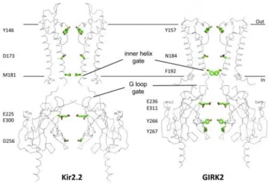
Concerning the gates, the structures of the eukaryotic Kir3.2 and Kir2.2 reveal the presence of two constrictions along the ion conduction pathway that have been proposed to act as gates. The first constriction, the inner helix gate, is located at the end of the inner transmembrane region of the channel. The second gate is represented by the G loop gate, located at the top of the cytoplasmic region of the tetramer (Whorton and MacKinnon 2011) (Figure 14).
The inner helix gate results from the inner helices of the TMDs, which stand above the cytoplasmic region. In Kir2.2 channel, two residues of the TM2, I177 and M181, form two hydrophobic seals, which shrink the pore at the interface between cytoplasm and membrane (Tao et al. 2009). The G loop gate is formed by the G-loop at the top of the CTD, immediately outside the membrane. In the PIP2-free structure of Kir2.2 channel, where CTD
is positioned far away from the TMD, the gates are as well positioned far away from each other. In the structure of Kir2.2 in complex with an analogue of the PIP2, as well as in the
structure of Kir3.2 channel (GIRK2), both gates are closely associated suggesting their possible cooperation (Niescierowicz 2013).
Figure 14.Comparison of the Kir2.2 and GIRK2 Structures. Key gating and rectification residues are highlighted in stick format (Whorton and MacKinnon, 2010).
37
SUR-Kir6 assembly and functional coupling
SUR-Kir6 association
The atypical association between SUR and Kir6 is unique among protein complexes. To communicate properly, both partners need to physically associate first. The association is poorly understood at the molecular level as a high-resolution structure of the K-ATP is still missing. SUR and Kir6 are separately expressed and retained in the ER until their association grants their release. In particular, the RKR sequence present in both proteins (between the helix 11 and the NBD1 in SUR1, and in the C-terminal tail of Kir6.2) is recognized by the protein COPI (coat protein complex I), preventing trafficking to the cell surface when the two partners are separated. Subsequently, when SUR and Kir6 start to associate, the RKR motif on Kir6.2 is masked by the presence of SUR1, preventing COPI binding. At this stage only the RKR motif present on SUR1 is exposed. This motif is inactivated by direct binding of the 14-3-3 protein to this region of the SUR1 or could be indirectly inactivated by 14-3-3 recruitment to a binding site near the distal tail of Kir6.2. (Heusser et al. 2006) (Figure 15).
Heusser et al. J Cell Sci 2006
Figure 15. Retention signals inactivation models. As 14-3-3-binding sites other than the one provided by the distal tail of Kir6.2 remain unknown, the position of the 14-3-3 dimer is hypothetical. Filled circles with white Rs represent active RKR signals, open circles symbolize inactivation of the signal. (A) The signal of SUR1 could be inactivated by direct binding of 14-3-3 to this region of the protein (masking) or (B) be indirectly inactivated by 14-3-3 recruitment to a binding site in the vicinity of the distal tail of Kir6.2 (Heusser et al., 2006).
38
However, Kir6.2 deleted of its last 36 or 26 amino acids (in which the RKR signal is contained) is capable to traffick to the plasma membrane forming functional tetrameric channels in absence of the SUR subunit (Tucker et al. 1997).
Physical contacts between SUR and Kir6 occur at several levels inside the complex in order to favour K-ATP assembly. Despite the lack of a reliable map of these interactions, several regions have been proposed as essential for the association. SUR TMD0 has been proven to strongly associate with Kir6.2 and control its gating. K-ATP channels lacking this domain are not able to traffick to the plasma membrane (Fang, Csanády, and Chan 2006). Mutations in this domain have been correlated with severe diseases such as PHHI (persistent hyperinsulinemic hypoglycaemia of infancy) (Aguilar-Bryan and Bryan 1999), and Cantù Syndrome (van Bon et al. 2012). While for the Cantù Syndrome the molecular mechanism involved in the development of the disease is not known, PHHI onset seems to be directly correlated with the disruption of SUR-Kir6 association due to mutations A116P or V187D in the TMD0.
Moreover, TMD0 has been shown to enhance the expression of Kir6.2 deleted in its last 26 residues. On the other hands, TMD0 alone is incapable of rescuing the expression of non-truncated Kir6.2, suggesting that other regions of the SUR are necessary to mask the Kir6.2 retention signal in the C-terminus. This indicates that other regions of SUR are physically interacting with Kir6.2 (Chan, Zhang, and Logothetis 2003).
In order to understand whether TMD0 affects the gating of Kir6.2, single channel recordings of SUR1+Kir6.2ΔC26, TMD0+Kir6.2ΔC26 and Kir6.2ΔC26 in nucleotide-free solutions have been analysed.
As can be seen in Figure 16, single channel recordings of SUR1+Kir6.2ΔC26 and TMD0+Kir6.2ΔC26 are very similar and differ from the behaviour of the Kir6.2ΔC26 channel. In particular, both SUR1 and TMD0 are able to increase the burst duration and Po of the channel. The values of Po of these two channels are ~ 0.6, which is 4 times the value of Po of Kir6.2ΔC26 channels. This can be explained with the lower frequency of openings and the lower open time of the truncated Kir6.2 (0.76 ms for Kir6.2ΔC26 and 1.8 ms of SUR1+Kir6.2ΔC26 and TMD0+Kir6.2ΔC26).
39
The association between SUR and Kir6 also modifies the ATP sensitivity of Kir6 (Figure 17) (Chan, Zhang, and Logothetis 2003).
SUR1 and TMD0 confer different sensitivities to ATP to Kir6.2ΔC26. The IC50 value for
ATP inhibition of Kir6.2ΔC26 is ~ 100 µM. This value increases when truncated Kir6.2 channels are expressed with TMD0 (IC50= ~ 300 µM) and decreases for SUR1+Kir6.2ΔC26
(IC50= ~ 15 µM), (Chan, Zhang, and Logothetis 2003). It has been shown that ATP is more
likely to bind to the channel when this is in the closed conformation (Enkvetchakul et al. 2000). The lower sensitivity for ATP of TMD0+Kir6.2ΔC26 channels compared to Kir6.2ΔC26 could be explained by the higher (Po) given by the TMD0 domain. In fact, for high values of Po the IC50 is also high. This observation is in agreement with Kir6.2ΔC26
channels having a low Po and a low IC50 compared to TMD0+Kir6.2ΔC26 channels (higher
Po and a higher IC50). On the other hands, this model does not fit with SUR1+Kir6.2ΔC26
channels, which have higher Po but a low IC50. This means that TMD0 alone is not sufficient
to confer to Kir6.2ΔC26 all the characteristics of the K-ATP channel. This differential behaviour of the full SUR and the TMD0 is related to the presence of the NBDs in SUR.
Figure 16. Single channel recordings in ligand-free solution. Single channels were recorded at -80 mV from inside-out patches with 96 mM K+ on both sides of the patches (Chan, Zhang, and Logothetis 2003).
Kir6.2ΔC26
TMD0+Kir6.2ΔC26
40
Another region of SUR (SUR2A specifically) involved in the physical association with Kir6.2 is the segment linking TMD2 with NBD2. This fragment (between residues 1295 and 1358) co-precipitates with Kir6.2 and competes with the full-length SUR for binding to Kir6.2 (Rainbow et al. 2004). The specificity of this interaction was also investigated in experiments performed using chimeric Kir6.2-Kir2.1 channels, as Kir2.1 does not associate with SURs to form functional channels.
Another proposed region of interaction involves residues 196-288 in SUR1 and Kir6.2 residues 28-32. Co-immunoprecipitation studies in Xenopus oocytes expressing Kir6.2 wild type or mutated in the N-terminal (deletion Δ28-32) and residues 196-288 of SUR1 reveal that the interaction between SUR1(196-288) and Kir6.2-Δ2832 is significantly reduced. This supports the idea that residues 196-288 of SUR1 form an essential part of the binding site between SUR1 and the N terminus of Kir6.2 and that this interaction is disrupted by deletion of residues 28-32 in Kir6.2 (Craig et al. 2009).
Other regions of the Kir6 channel participate to the physical association with SUR. Both N- and C-terminal plus the transmembrane helix 1, have been found to physically interact with SUR (Schwappach et al. 2000; Tammaro and Ashcroft 2007; Lodwick et al. 2014). Taken together, these observations are in line with the postulated architecture of the full-length K-ATP channel in which the helix 2 of Kir6, predicted to participate in the formation of the
Figure 17. Dose-response curves for ATP inhibition (Chan, Zhang, and Logothetis 2003). Patches included in the measurements were recorded at -80 mV from inside-out patches with 96 mM K+ on both sides of the membrane.
Kir6.2ΔC26 SUR1+
Kir6.2ΔC26
TMD0+ Kir6.2ΔC26
41
selectivity filter, is buried in the center of the complex and protected from the interaction with SUR.
SUR-Kir6 functional coupling
Beside the physical association, the most suggestive feature of the K-ATP channel is the functional coupling occurring between SUR and Kir6.
With the expression ‘functional coupling’, we refer to the ability of ligands that bind to SUR to trigger Kir6 gating modifications. The SUR domains involved in this process are still largely unknown. To date, only few regions have been linked to ligand-induced functional coupling between SUR and Kir6. One of these is a domain reach in aspartate and glutamate (ED) (Karger et al. 2008). This domain has been proposed to act as an allosteric transducer enabling functional communications between the SUR2A and Kir6.2. The ED consists of a string of 15 aspartate and glutamate residues and it is located in the cytoplasmic loop 6 (CL6) between TMD1 and TMD2. It is positioned far from the postulated binding sites of openers and blockers of the K-ATP. Its main function would be to contribute to the cooperative interaction between the NBDs that is critical for conformational arrangements of MgADP-induced K-ATP channel activation. The ED it is not known to interact physically with Kir6.2 but is an essential part of the allosteric machinery that controls NBDs action and signal transduction to Kir6.2.
Another region of the SUR2A involved in the communication between K-ATP subunits is a region in the proximal C-terminus. Importantly, three residues at this level (E1350, I1310 and L1313) are essential for transmitting activation messages from the SUR2A to the pore-forming subunit. Mutation of these residues resulted in drastic reduction of channel activation by both MgADP and pharmacological openers mediated by SUR2A (Dupuis et al. 2008). The second project of this thesis work examines the role in the activation pathway at this proximal C-terminal region in the SUR1 subunit.
Interestingly, in contrast with what has been observed for the ED, mutation of the three residues do not affect the inhibitory pathway but only the activation network. These results support the theory that multiple transduction pathways between SUR and Kir6 might exist.
42
K-ATP channel mechanism and regulation
Physiological regulation of the K-ATP channel mediated by SUR
Transmembrane ABC proteins display transport activity with few exceptions such as SUR. Binding of MgATP at the SUR nucleotide binding domains (NBDs) results in NBD dimerization and hydrolysis of MgATP at the NBDs that leads to channel opening. Although it has a limited capacity to hydrolyse MgATP, (Bienengraeber et al. 2000), SUR is not a transporter. To date, SUR only known function is the gating regulation of Kir6 channels. SUR brings to the K-ATP channel the capability to be activated by nucleotides when these are in complex with magnesium, as this class of molecules naturally inhibits Kir6. Moreover, SUR is target of drugs that can regulate either opening or blocking of the pore.
K-ATP channels are physiologically regulated by molecules that can target SUR or Kir6 or both. The resultant regulation is very complex and despite a large amount of functional data a lot remain to be clarified.
Regulation by MgADP and other nucleotides associated to Mg2+
K-ATP channel activation mediated by ADP is one of the most important characteristics of the SUR protein. The activation requires the presence of Mg2+ and that both NBDs are intact
(Gribble, Tucker, and Ashcroft 1997). Moreover, studies conducted on SUR1 show that the NBD1 strongly binds ATP rather than ADP, even in absence of Mg2+ and that MgADP,
through binding at NBD2, antagonizes the Mg2+-independent high affinity ATP binding at NBD1. Furthermore, MgADP is more likely to bind to NBD2 than to NBD1 (Ueda, Inagaki, and Seino 1997). MgADP stimulates K-ATP channels with different affinities depending on the SUR isoform present. In particular, SUR1 and SUR2B are more stimulated that SUR2A. Experiments using chimera and mutant SURs suggest that the 42 amino acids at the C-terminal end of SURs (C42) play critical roles in the ADP-mediated activation of K-ATP channels and that the C42 of SUR2A may reduce ADP-mediated channel activation at NBD2 (Matsuoka et al. 2000).
The mechanism by which Mg-ADP binding to SUR is translated into channel opening remains unclear. It is now generally accepted that cooperative interaction between NBDs is essential for Kir6 gating, yet pathways of allosteric inter-subunit communication remain uncertain. Disruption of 15 negatively charged aspartate/glutamate amino acid residues
43
(948–962) of the SUR2A isoform blocks cooperative NBDs interaction and interrupts the regulation of K-ATP channel by MgADP. Moreover, three residues in the SUR2A (E1305, I1310, L1313) are implicated in the transmission of the activation message from SUR to Kir6.2. When these residues are mutated, SUR can no longer activate the channel (Dupuis et al. 2008). Taken together, these data suggest a substantial rearrangement of the SUR general folding and especially at SUR2A/Kir6.2 interface, just after MgADP binding, which triggers conformational changes in Kir6.2 that responds by opening the gate.
Other nucleotides are able to trigger K-ATP channel opening through binding to SUR such as MgATP, MgGDP and MgGTP (Trapp, Tucker, and Ashcroft 1997).
Regulation by Zinc
Applied at either the extracellular or the intracellular side of the membrane, zinc is a potent, reversible activator of K-ATP channels. In particular, zinc performs its action through binding at the extracellular side of SUR1-based K-ATP channels at the level of two histidines (H326 and H332) (Bancila et al. 2005). Moreover, in both SUR1- and SUR2A-based channels, zinc shows an activator intracellular effect (Prost et al. 2004).
Regulation by G proteins
Gα and Gβγ proteins, released after GPCRs activation, have a role in activating K-ATP
channels. In particular, the Gαi1 subunit stimulates SUR1-based K-ATP channels while the
Gβγ2 subunits have been correlated to both SUR1 and SUR2A-based channels activation
44
Physiological regulation of the K-ATP channel mediated by Kir6
channels
Nucleotides
Kir6 channels are strongly inhibited by ATP. ATP binding site in Kir6 does not resemble any of the classical nucleotide binding sites, such as the NBDs of SUR. In particular, ATP cannot be hydrolysed by Kir6. As the structure of Kir6 is still missing, we can only rely on functional data to locate the ATP-binding site. To date, most data agree that this site is formed by both N- and C-terminal domains (Tucker et al. 1998; Proks et al. 1999). With improvements of protein modelling, a putative ATP-binding site in Kir6.2 has been identified (Antcliff et al. 2005). The model, reinforced by experimental tests, locates the binding site for ATP near the top of the intracellular domain (IC). Each of the four binding pockets lies at the interphase between N- and C-terminal domains of the same subunit with a little contribution by the neighbour C-terminal of the subsequent chain (Figure 18).
Once ATP binds, its phosphate tail interacts with R201 and K185 in the C-terminal of one chain and with R50 in the N-terminal of the next chain. The binding of a single ATP
Figure 18. The ATP-binding site. A, side view of the ATP-binding site. For clarity, the TMs of only two subunits and the IC domains of two separate subunits are illustrated. ATP (yellow) is docked into its binding sites. B, Kir6.2 tetramer, top view, with the TMs removed (residues 64–177). The N-terminal domain is shown in ribbon format and the C-terminal domain in backbone format. Different colours represent individual subunits (Antcliff et al., 2005).
45
molecule appears to be sufficient to trigger the conformational changes necessary for channel closure (Markworth, Schwanstecher, and Schwanstecher 2000).
Moreover, other nucleotides can bind in this pocket but with a lower affinity (Dabrowski, Tarasov, and Ashcroft 2004). If phosphates are removed, the binding affinity diminish by several orders of magnitude (Tucker et al. 1998).
Lipids
Like other channels, also Kir6 activity has been shown to be dependent on phosphatidylinositol 4,5-bisphosphate (PIP2). In particular, PIP2 increases channel open
probability in absence of ATP and reduces the inhibitory effect of the ATP by decreasing the channel apparent affinity for this molecule (Hilgemann and Ball 1996; Fan and Makielski 1997). The binding of PIP2 to Kir6.2 has not been precisely located. Nevertheless, mutagenic
experiments have shown that PIP2 effect is carried out through electrostatic interactions of
the polar head with positively charged residues present in the N-terminal (K39, R54), transmembrane (K67) and C-terminal (R176, R177, R301) regions. The aliphatic chain would be anchored at the membrane. The structure of the Kir2.2 channel in complex with a PIP2
analogue suggests an explanation of the effect mediated by this lipid. PIP2 binds at the
interphase between TMD and CTD producing a conformational change in Kir2.2 that would stabilize the channel in an open conformation.
Figure 19. Proposed mechanism of Kir2.2 activation by PIP2. PIP2 (purple sphere) binds at an interface between the
TMD (grey cylinder) and the CTD (grey rectangle) and induces a large conformational change. The flexible linker (green line) contracts (green cylinder), the CTD moves towards and becomes tethered to the TMD, the G-loop (cyan wedge) inserts into the TMD and the inner helix activation gate opens (Hansen et al. 2011).
46
Other lipids of physiological relevance such as the long chain Co-enzyme A esters (LC-CoA) also activate Kir6 channels by decreasing the affinity of these channels to ATP (Gribble et al. 1998). The cholesterol has also been proposed as possible regulator of Kir6 channels but its action remains controversial as some studies suggest an activator effect and others an inhibitory action.
K-ATP channel physiology and physiopathology
The K-ATP channel ability to sense the ATP/ADP ratio inside cells gives it a primary role as metabolic sensor, able to modulate membrane excitability depending on the metabolic state of the cell. At rest, K-ATP activation is translated into membrane hyperpolarization while its inhibition triggers membrane depolarization. Depending on the localization, variations in the membrane potential will provoke different cellular responses.
K-ATP channels in pancreas
K-ATP channel function has been best characterized in pancreatic β cells where its activity is linked to insulin secretion (Figure 20) (Ashcroft, Harrison, and Ashcroft 1984). When the level of glucose in the blood is low, K-ATP channels are partly active at rest and their activity helps to maintain the membrane hyperpolarized. In contrast, when the level of glucose in the blood increases, this is shuttled into the pancreatic β cell where it is be metabolized to produce ATP. ATP level rises, triggering K-ATP channel closure. As the K-ATP channel is highly expressed at the plasma membrane of these cells, channel closure provokes membrane depolarization that in turn activates voltage-dependent Ca2+ channels. These channels allow
influx of Ca2+ that triggers insulin secretion. Thus, K-ATP channel activity in the pancreas is
crucial.
Malfunctions due to K-ATP channel mutations have been correlated with severe pathologies: PHHI characterized by high levels of insulin, type II diabetes involving low levels of insulin secretion despite high levels of glucose in the blood (Gloyn, Siddiqui, and Ellard 2006). Most PHHI mutation are classified as ‘loss of function’ mutations in the gene encoding for SUR1 (Ashcroft 2005). These mutations can be divided in two groups: mutations causing reduced expression of channels (class I) and mutations that reduce channel open probability (class II). SUR1 ‘gain of function’ mutations, resulting in overactive channels, are linked to a rare disease, neonatal diabetes. Depending on the mutations, the severity of the associated
47
pathologies varies from transient neonatal diabetes to neurological disorders (DEND syndrome).
K-ATP channels in cardiomyocytes
K-ATP channels are as well expressed in cardiomyocytes where they appear to be involved in a mechanism of protection against ischemia. During ischemic stress, the concentration of ATP in the cells decreases, triggering K-ATP channel opening and increasing resting K+
permeability of the membrane. The K-ATP channel action favours hyperpolarization and reduces excitability. This change in the membrane potential reduces Ca2+ influx, shortens the
action potential duration and decreases the contraction. This protective effect has been observed in mice knock-out for the Kir6.2 gene (Gumina et al. 2007). Mutations of these K-ATP channels (at the NBD2 of SUR2A) are correlated with cardiomyopathy, ventricular arrhythmia and atrial fibrillation.
K-ATP channels in the central nervous system
K-ATP channels can be found as well in brain tissues. GABAergic neurons preferentially express SUR1 in complex with Kir6.2 while the dopaminergic neurons express both
SUR1-|ATP|↑ |ADP| Vm↑ |Ca2+|↑ Ca2+ [Glucose]↑ G lu t2 KATPchannel Ca 2+ c h a n n e l metabolism Insulin release Insulin
Figure 20. K-ATP role in insulin secretion. Glucose is be metabolized in pancreatic β cells to produce ATP. When ATP level rises, K-ATP channels are inhibited. Channels closure triggers membrane depolarization that in turn activates voltage-dependent Ca2+ channels. Influx of Ca2+ facilitates insulin secretion.