The Journal of Laryngology and Otology
June 1994, Vol. 108, pp. 466-469
CO
2laser repair of the facial nerve: an experimental study in
the rat
JOSEPH C. DORT, M.D.*, MARKUS WOLFENSBERGER, M.D.f, HEIDI FELIX, P H . D 4
Abstract
The facial nerve is often injured by trauma, infection or during the course of tumour resection. Many tech-niques of nerve anastomosis have been described with the current standard nerve repair using the microscope and monofilament suture. The purpose of this study was to evaluate the CO, surgical laser as a tool for facial nerve anastomosis. Following preliminary electrical measurements 36 nerves were anastomosed using either laser or conventional monofilament suture. Laser anastomosis had neither beneficial nor detrimental effects on nerve regeneration. This method of anastomosis may be advantageous when surgical access is limited. In addi-tion this study found that the use of CO, laser as a dissecting or vapourizing tool in proximity to intact facial nerves results in degenerative changes.
Key words: Facial nerve; Laser surgery
Introduction
The facial nerve is often injured by trauma or during the course of tumour resection. The sequelae of facial nerve injury represent significant patient morbidity and there-fore improvements in nerve repair and regeneration are desirable. Complete facial nerve injuries are currently managed by means of micro-anastomosis with or without nerve graft interposition.
Facial nerve recovery remains unpredictable with inconsistent clinical and experimental results (Wickholm et al., 1988; De Medinacelli and Seaber, 1989). This inconsistency may be explained by several local and bio-mechanical factors relating to the nerve repair but the most important factor may be the random regrowth of axons after nerve transection (De Medinacelli and Seaber, 1989). Recent investigations have led to the development of techniques that result in a more predictable recovery in experimental animals (Wickholm et al, 1988; De Medi-nacelli and Seaber, 1989).
The facial nerve presents a unique challenge to the sur-geon because of its complex course from the brain stem to the facial musculature. The more distal mastoid and parotid portions of the nerve are easily amenable to suture repair. However, the labyrinthine and intracranial parts of the facial nerve are more difficult regions in which to place sutures. Sutureless techniques have been used to make ana-stomoses in the labyrinthine and intracranial portions of the facial nerve. However, the continuous pulsation of the cere-brospinal fluid (CSF) is a potential disruptive force to the anastomosis (Fisch and Mattox, 1988). In view of these concerns a method of reconnecting the facial nerve without the use of sutures, such as CO, laser, is desirable.
Experiments have shown that lasers can be used to 'weld' tissue in vivo (Schober et al., 1986). The CO2 laser
has been used to join rat sciatic nerves without evidence of harmful effects (Fischer et al., 1985; Beggs et al., 1986). Laser nerve anastomoses have been found to be weaker than sutured anastomoses up until the fourth post-oper-ative day, after which both anastomoses were equally strong (Maragh et al., 1988). Although several authors have shown that the laser gives a superior neurography result (Fischer et al., 1985; de la Torre et al., 1988; Camp-ion et al., 1990) others have not observed a significant difference between laser and suture repairs (Bailes et al.,
1989; Benke et al, 1989).
Much of the previous work has been done using the rat sciatic nerve model (Fischer et al., 1985; de la Torre et al., 1988; Wickholm et al, 1988; De Medinacelli and Seaber, 1989). This model has been used extensively in peripheral nerve research and is stable and reliable. In addition, the sciatic function index (De Medinacelli et al, 1982) pro-vides a useful indicator of functional recovery after sciatic nerve repair. Nevertheless, it seems rational to use the facial nerve as a model when one is studying the response of that nerve to surgical manipulation. There has been only one study published investigating the use of the CO2
laser in repairing the facial nerve and this was done in a rabbit facial nerve model (Eppley et al, 1989). As pre-viously described there are several advantages to a rat facial nerve model and therefore this has been used in the present study (Gleeson and Felix, 1986; Mattox and Felix,
1987).
This report examines the use of the CO2 laser for facial
nerve anastomosis, comparing the results to suture ana-From the Department of Otolaryngology^: of the University of Zurich, Zurich, Switzerland, the Department of Otolaryngologyt, University of Basel, Basel, Switzerland and the Department of Surgery*, University of Calgary, Calgary, Alberta, Canada.
Presented at the Vllth International Symposium on the facial nerve, Cologne, June 9-14, 1992. Accepted for publication: 8 January 1994.
466
https:/www.cambridge.org/core/terms. https://doi.org/10.1017/S0022215100127124
CO2 LASER REPAIR OF THE FACIAL NERVE: AN EXPERIMENTAL STUDY IN THE RATE 467
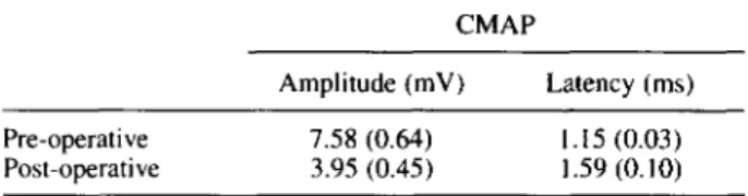
TABLE I
COMPOUND MUSCLE ACTION POTENTIALS! COMPARISON OF PRE-OPERATIVE AND POST-PRE-OPERATIVE RESULTS IN ALL ANIMALS ( ± SEM)
Pre-operative Post-operative CMAP Amplitude (mV) 7.58 (0.64) 3.95 (0.45) Latency (ms) 1.15(0.03) 1.59(0.10)
stomosis. The report also evaluates the safety of using the laser in proximity to the facial nerve.
Materials and methods
Experiments were carried out on male Zbz SIV-50 rats weighing an average of 250 gm. The animals were cared for in accordance with the animal utilization guidelines of the University of Zurich. General anaesthesia was induced with ether and maintained with intraperitoneal pentobar-bital. Through a retroauricular incision the facial nerve was exposed while the buccinator muscle was exposed through a cheek incision (Gleeson and Felix, 1986). Con-stant current, square wave bipolar stimulation of supra-maximal intensity was applied directly to the exposed trunk of the facial nerve. Evoked compound muscle action potentials (CMAP) were measured with silver needle electrodes placed in the buccinator muscle and recorded on paper strips.
Baseline electrophysiological recordings were made in a pilot group of four animals in order to ascertain the opti-mum recording conditions and normal values. These ani-mals were also used to assess the acute effects of the laser when used in proximity to the facial nerve. The nerves were harvested one week after laser dissection, placed in fixative and histologically examined.
Eighteen animals (36 nerves) were placed into one of five experimental groups.
Group I animals (n = 3: 6 nerves) underwent bilateral facial nerve exposure with excision of a 5 mm segment of nerve. This group functioned as a control and as a measure of the inherent regenerative ability of the rat facial nerve. Group II animals (n = 3: 6 nerves) had both facial nerves cut and then gently reapposed without sutures.
In group III animals (n = 4: 8 nerves) the facial nerves were cut and the epineurium was removed for a distance of 3 mm on either side of the transection site. A single per-ineurial traction suture (10-0 nylon) was placed to facili-tate manipulation of the nerve. A series of six laser pulses were then evenly spaced around the circumference of the anastomotic site. A microscope mounted CO2 laser
(2W/50ms/defocussed/0.6 mm spot: Medilas AG, Dietli-kon, Switzerland) was used to make the anastomosis.
In group IV (n = 4: 8 nerves) the facial nerves were cut and prepared as in group III. The nerves were then ana-stomosed by placing three 10-0 nylon perineural sutures equidistant around the periphery of the nerve.
Group V (n = 4: 8 nerves) was identical to group IV except that the anatomosic site was also protected with a single layer of Cargile® (Ethicon) membrane.
At sacrifice (12 weeks post-operatively) facial nerves were harvested for light microscopic evaluation. The nerves were fixed in three per cent glutaraldehyde in 0. 1M phosphate buffer for 24 hours. They were then placed in one per cent osmic acid in 0.1 M phosphate buffer for 120
minutes and then dehydrated in graded concentrations of ethanol and embedded in Epon (Fisher Scientific Company). Semi-thin sections were stained with toluidine blue and microscopically examined. Total counts of nerve fibres were performed by hand on photomontages of the whole nerve cross-section at a magnification of x540.
Statistical evaluation
The results are presented as mean ± SEM. Compari-sons between groups were performed by analysis of vari-ance using the general linear model for unbalvari-anced observations followed where appropriate by Tukey's multiple comparison test (Hassard, 1991).
Results
General observations
Observations made one week post-operatively (in the pilot group) showed that there was some visible charring in the lasered nerves as well as a considerable perianasto-motic fibrous/inflammatory reaction in both sutured and lasered nerves. The charring resolved and the fibrous reac-tion was much less evident at 12 weeks. Group I animals did not demonstrate growth across the 5 mm nerve gap by either electrical or histological criteria.
Electrophysiological observations
Electrophysiological findings are presented in Tables I—II. Compound muscle action potential amplitude (CMAP) and latency are both adversely affected by sur-gery (Table I; P<0.001). However, intergroup compari-sons revealed no significant differences in CMAP regardless of the type of surgical repair performed. Group I animals did not bridge the surgical gap and therefore no CMAP was measureable in this group. The Tables (I—III) are incomplete as several animals died in the peri-oper-ative period.
Histology: qualitative observations
Perineural fibrosis and scarring was found irrespective of whether the laser or suture method was used to ana-stomose the facial nerve. Severe nerve reaction with total degeneration was found even when the laser was used defocussed and at low power (2W/50ms/0.6 mm spot) within 1 mm of the epineurium. Morphology of the distal nerve fibres was abnormal in all groups. Myelin sheaths were thinner than normal and the intraneural organization was disrupted when compared to normal nerves (Figure 1 a and b).
TABLE II
COMPOUND MUSCLE ACTION POTENTIALS (PRE-SACRIF1CE): INTERGROUP COMPARISONS ( ± SEM)
Group Group Group Group II (« = I I I (n •• IV (n V(n = 6) = 5) = 7) = 7) CMAP Amplitude (mV) 3.35 (0.63) 5.10(1.88) 4.14(0.86) 5.06(1.15) Latency (ms) 2.00(0.10) 1.71 (0.18) 1.81 (0.06) 1.69(0.07)
n represents number of nerves.
https:/www.cambridge.org/core/terms. https://doi.org/10.1017/S0022215100127124
468 J. C. DORT. M. WOLFENSBERGER. H. FELIX
FIG. 1
(a) Normal nerve fibres; (b) nerve fibres 12 weeks post-laser anastomosis. Note disruption and variability in myelin sheaths at this stage. (Toluidine blue; x540).
Histology: quantitative observations
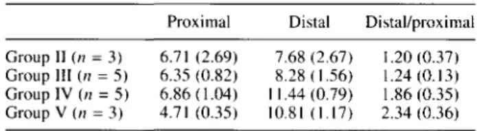
Quantitative nerve fibre data is shown in Table III. There was no significant different in fibre counts between groups. The distal/proximal ratio revealed increased numbers of fibres distal to the anastomosis.
Discussion
In the evaluation of peripheral nerve regeneration fol-lowing traumatic injury it remains difficult to know what parameters to measure. Return of function in the reinner-vated tissue is important and therefore the sciatic func-tional index is a clinically relevant measurement (De Medinacelli etai, 1982). Electrophysiological and histo-logical data provide useful information about the func-tional and structural responses of injured nerves. One drawback to the rat facial nerve model is that there is no very good functional recovery scale. Subprimate mam-mals do not have the richness of facial innervation to allow meaningful interpretation of recovery by obser-vation alone.
Eppley et al. (1989) compared suture and laser-assisted nerve anastomosis in a rabbit facial nerve model. This is the only published work comparing suture and laser ana-stomosis in a facial nerve model. These authors concluded that laser anastomosis resulted in less entrapment of axons at the repair site but unfortunately also produced thermal destruction that could lead to a fibrous reaction. This study showed no electrophysiological differences between laser and suture anastomoses. A quantitative analysis of nerve fibre regeneration was not performed in their study.
The results of our study show that at the three-month post-operative evaluation both laser and suture repairs cause very little grossly visible reaction at the anastomotic site. Microscopically, both types of repair result in
neu-TABLE III
MEAN NERVE FIBRE COUNTS ( 1 O ' ± SEM)
Proximal Distal Distal/proximal
Group II (n = 3) Group III (n = 5) Group IV (n = 5) Group V (/; = 3) 6.71 (2.69) 6.35 (0.82) 6.86(1.04) 4.71 (0.35) 7.68 (2.67) 8.28(1.56) 11.44(0.79) 10.81 (1.17) 1.20(0.37) 1.24(0.13) 1.86(0.35) 2.34 (0.36)
n represents number of nerves.
roma formation and extraneural escape of axons. Tensile strength was difficult to assess in this experiment because none of the repairs were disrupted post-operatively prob-ably because of a tension-free anastomosis and an immo-bile wound.
Compound buccinator muscle action potentials were measured in this study. The overall post-operative action potential amplitudes were significantly less than the pre-operative amplitudes reflecting incomplete and aberrant reinnervation of the buccinator muscle. Comparison of action potential amplitude between groups failed to reveal any differences and therefore there are no electrically measureable differences in reinnervation regardless of anastomotic technique. This conclusion has been reached by others in different animal models. (Bailes et al., 1989; Benke et al., 1989).
Nerve transection and repair causes major disruption of nerve architecture which is not normalized even three months post-operatively. In addition to the late effects of injury, this study also found that using the laser in proxim-ity to the facial nerve resulted in significant nerve fibre degeneration, presumably secondary to the thermal injury caused by the laser.
Several studies have reported quantitative data on nerve fibre regeneration after laser anastomosis (Fischer et al, 1985; Beggs et al., 1986; de la Torre et al., 1988; Maragh etai, 1988; Bailes et al., 1989; Benke et al., 1989; Camp-ion et al., 1990) but none of the studies used a facial nerve model. This information is usually reported in the form of nerve fibre histograms. The hand counting method used in this study, while more tedious, provides a more accurate histological picture. The results show a trend to higher distal fibre counts in the sutured nerves. However these results did not reach statistical significance perhaps due to insufficient numbers of animals. If one assumes that an increased number of distal fibres correlates with a better quality of regenerative response then there is a trend to better regeneration in both the suture alone and the suture/ Cargile® groups.
Conclusions
This study has shown that although the CO, laser is a useful technique for facial nerve anastomosis its use is neither harmful nor beneficial to nerve regeneration in the
https:/www.cambridge.org/core/terms. https://doi.org/10.1017/S0022215100127124
CO2 LASER REPAIR OF THE FACIAL NERVE: AN EXPERIMENTAL STUDY IN THE RATE 469 rat. The technique has the advantage of allowing a faster
anastomosis in a restricted surgical field. It is important to emphasize however that when used as a dissecting tool, even at low power settings, the CO, laser results in degen-eration of adjacent neural tissue. This finding may have particular relevance in the cerebellopontine angle and temporal bone.
Acknowledgement
The authors would like to thank Medilas AG, Dietlikon, Switzerland for lending the surgical laser used in these experiments.
References
Bailes. J. E., Cozzens, J. W., Hudson, A. R., Kline, D. G., Ciric, I., Gianaris, P., Bernstein, L. P., Hunter, D. (1989) Laser-assisted nerve repair in primates. Journal of Neurosurgery 71: 266—272. Beggs, J. L., Fischer, D. W., Shetter, A. G. (1986) Comparative study of rat sciatic nerve microepineurial anastomoses made with carbon dioxide laser and suture techniques. Part 2: A morpho-metric analysis of myelinated nerve fibres. Neurosurgery 18(3): 266-269.
Benke. T. A., Clark, J. W., Wisoff, P. J., Schneider, S., Balasubrama-niam, C , Hawkins, H. K., Laurent, J., Perling, I., Shehab, A. (1989) Comparative study of suture and laser-assisted anastom-oses in rat sciatic nerves. Lasers in Surgery and Medicine 9: 602-615.
Campion, E. R., Bynum , D. K., Powers, S. K. (1990) Repair of per-ipheral nerves with the argon laser: a functional and histological evaluation. Journal of Bone and Joint Surgery 72-A(5): 715-723. de la Torre, J. C , Karaca, M., Merali, Z., Fortin, T , Richard, M. (1988) Laser of razor?: a novel experimental peripheral nerve repair technique. Neurosurgery 22(3):531-539.
De Medinacelli, L., Seaber, A. V. (1989) Experimental nerve recon-nection: importance of initial repair. Microsurgery 10:56—70. De Medinacelli, L., Freed, W. J., Wyatt, R. J. (1982) An index of the
functional condition of rat sciatic nerve based on measurements
made from walking tracks. Experimental Neurolog\ 11: 634-643.
Eppley, B. L., Kalenderian, E., Winkelmann, T., Delfino, J. J. (1989) Facial nerve graft repair: suture versus laser-assisted anastomosis.
International Journal of Oral and Maxillofacial Surgery 10:
50-54.
Fisch, U., Mattox, D. (1988) Microsurgery of the Skull Base, Georg Thieme, Verlag, Stuttgart, pp 104-108"
Fischer, D. W., Beggs, J. L., Kenshalo, D. L., Shetter, A. G. (1985) Comparative study of microepineural anastomoses with the use of CO, laser and suture techniques in rat sciatic nerves. Part 1: Sur-gical technique, nerve action potentials and morpholoSur-gical stud-ies. Neurosurgery 17(2): 300-308.
Gleeson, M. J., Felix, H. (1986) The Caletron ultrasonic surgical aspirator system: an anatomical and physiological study of the effect of its use on the rat facial nerve. Clinical Otolanngologv 11: 177-187.
Hassard, T. H. (1991) Understanding Biostatistics, Mosby Year Book Inc., New York, pp 88-89.
Maragh, H., Hawn, R. S., Gould, J. D., Terzis, J. (1988) Is laser nerve repair comparable to microsuture coaptation? Journal of
Recon-structive Microsurgery 4(3): 189-195.
Mattox, D. E., Felix, H. (1987) Surgical anatomy of the rat facial nerve. American Journal of Otology 8: 43^17.
Schober, R., Ulrich, F., Sander, T, Durselen, H., Hessel, S. (1986) Laser-induced alteration of collagen substructure allows micro-surgical tissue welding. Science 232: 1421-1422.
Wickholm, R. P., Swett, J. E., Torigoe, Y, Blanks, R. H. I. (1988) Repair of severed peripheral nerve: a superior anatomic and func-tional recovery with a new 'reconnection' technique.
Otolar-yngology-Head and Neck Surgery 99: 353-361.
Address for correspondence: Joseph C. Dort, M.D.,
Department of Surgery, Section of Otolaryngology, University of Calgary,
Room 193A, Heritage Medical Research Building, 3330 Hospital Drive N.W.,
Calgary, Alberta, Canada T 2 N 4 N 1 .
https:/www.cambridge.org/core/terms. https://doi.org/10.1017/S0022215100127124