Co-culture of 1-cell outbred mouse embryos on bovine
kidney epithelial cells: effect on development, glycolytic
activity, inner cell mass:trophectoderm ratios and
viability
Greet Leppens
1"
2, David ICGardner
3and
Denny Sakkas
1'Clinic of Sterility, Department of Obstetrics and Gynaecology, Hospital Cantonal University of Geneva, 1211 Geneva 14, Switzerland and 3Institute of Reproduction and Development, Monash University, Monash Medical Centre, Clayton, Victoria 3168, Australia
2To whom correspondence should be addressed at: Laboratoire des Gametes, Policlinique de Steiilite', Hopital Cantonal Universitaire de Geneve, 20 Rue Alcide-Jentzer, 1211 Geneve 14, Switzerland
In an attempt to enhance embryo development, we have co-cultured 1-cell OF1 mouse embryos on bovine kidney epithelial (Madine-Darby bovine kidney; MDBK) cells in a complex medium called complex mouse tubal fluid (cMTF; based on the energy substrate levels found in the mouse oviduct, containing non-essential amino acids, glutamine and EDTA). To determine the quality of the blastocysts obtained, we examined several parameters: morphology, total cell numbers, inner cell mass (ICM):trophectoderm (TE) ratio, glycolytic activity and viability after transfer. A significantly lower number of blastocysts developed on MDBK cells compared with cMTF medium. cMTF blastocysts had a significantly higher glycolytic activity and a lower blastocyst cell number than those grown in co-culture, while both in-vitro groups had higher ICM:TE ratios compared with in vivo. Blastocysts grown on MDBK cells displayed an elevated ICM number compared with those grown in cMTF medium alone. However, the per-centage of fetuses after transfer remained drastically low in both culture groups compared with in-vivo blastocysts. In conclusion, co-culture did not increase the number of zygotes reaching the blastocyst stage. Although co-culture blastocysts show some similarities to in-vivo embryos in cell number and glycolytic activity, no enhancement in viability was observed.
Key words: co-culture/embryo culture/inner cell
mass/metabol-ism/trophectoderm
Introduction
Over the past 20 years considerable progress has been made in culturing preimplantation embryos. However, many problems remain, including retarded development in vitro compared with
in vivo, a decrease in viability (Harlow and Quinn, 1982;
Sakkas et al, 1989) and species-specific blocks at the time of embryonic genomic activation (Goddard and Pratt, 1983; Gandolfi and Moor, 1987; Lawitts and Biggers, 1991a). A main challenge of embryo culture studies is therefore to
enhance embryo development by finding conditions as close as possible to those in vivo. Since the initial embryo culture studies (Whittingham, 1971; Quinn et al, 1982), several successful media modifications have been reported. For example, Chatot et al. (1989) successfully cultured mouse embryos from a blocking strain by elevating the lactate pyruvate ratio and by adding glutamine and EDTA to a medium without glucose, while Gardner and Leese (1990) obtained a glycolytic rate closer to that in vivo by using a culture medium based around the carbohydrate composition of mouse tubal fluid (MTF). Modifications to this medium were made by Gardner and Sakkas (1993), who showed that components such as amino acids, vitamins and growth factors increased embryo development and cell number. Embryo development in medium containing amino acids has also been shown to support a pattern of gene expression similar to that in embryos developed in vivo (Ho et al, 1995). Gardner and Lane (1993) have also examined the role of specific groups of amino acids and found that blastocyst formation and cell number were higher when non-essential amino acids and glutamine were added to the medium. Interestingly, the presence of amino acids in the culture medium was also responsible for an inhibition of embryo development, as amino acids spontaneously break down at 37CC to produce
embryotoxic ammonium ions (Gardner and Lane, 1993; Lane and Gardner, 1994).
Whilst the formulation of a defined medium has led to improvements in embryo development, another approach has been the use of co-culture or medium which was conditioned by cells. The increased interest in this technique followed a report by Gandolfi and Moor (1987), who successfully used co-culture with sheep oviduct epithelial cells to improve the culture and viability of sheep embryos. Similar studies were subsequently conducted by Eyestone and First (1989) on cattle, Ouhibi et al. (1990), Sakkas and Trounson (1990) and Lai
et al. (1992) in mice and Sakkas et al. (1989) in goats. Several
authors (Gandolfi and Moor, 1987; Ellington et al, 1990; Mennillod et al, 1993) have since suggested the presence of embryotrophic factors but have failed to identify them specifically. More conclusive evidence exists for the presence of growth factor receptors such as insulin-like growth factor (IGF)-I, IGF-II, insulin, epidermal growth factor (EGF) and platelet-derived growth factor (Adamson, 1993; Harvey et al, 1995) on early embryos. Therefore if cells are secreting growth factors, these could enhance embryo development
The enhancement of embryo development in co-culture systems has been observed in many cases, but how the cells were acting was not determined. One of the perceived problems of co-culture is the use of inadequate culture medium to 598
Quality of mouse embryos after co-culture
support both the embryo and cells (Leppens and Sakkas, 1995). We have shown previously that Me'ne'zo's B2 medium conditioned by Madine-Darby bovine kidney epithelial (MDBK) cells could support mouse blastocyst formation, while M6n6zo's B2 medium alone was inadequate. In these experiments, a beneficial effect was observed only when poor embryo development occurred in medium alone. This has led to the theory that the cells in co-culture only act by removing embryotoxic factors from poor culture medium (Bavister, 1992). In this study, we have tried to overcome the problem by using cMTF medium, a complex medium which has already been shown to support enhanced mouse embryo development (Gardner and Lane, 1993).
The aim of this study was to show the effect of cMTF medium during co-culture. We have cultured outbred mouse embryos from the 1-cell stage until the blastocyst stage in two conditions: (i) cMTF medium, a medium based on M'l'h supplemented with non-essential amino acids, glutamine and EDTA (Gardner and Lane, 1993; Lane and Gardner, 1994), and (ii) a co-culture system using MDBK cells, with cMTF as the base medium. From the blastocysts obtained, we have examined numerous parameters to determine their quality. We have compared their morphology on days 3 and 5 of culture, determined the glycolytic activity of blastocysts and their respective cell numbers [trophectoderm (TE) and inner cell mass (ICM)] and tested for blastocyst viability after transfer.
Materials and methods
Culture media
In all experiments, a modified MTF medium was used called cMTF (Gardner and Leese, 1990; Gardner and Lane, 1993; Lane and Gardner, 1994). MTF medium was characterized by a reduced sodium chloride concentration (103.4 mM) compared with the original formulation (114.2 mM). The culture medium cMTF had the following composition: 103.4 mM NaCl, 4.78 mM KC1, 1.19 mM KH2PO4, 1.71 mM CaCl2-2H2O, 0.37 mM sodium pyruvate, 4.79 mM sodium lactate, 20 mM NaHCO3, 3.40 mM glucose and 0.01 g/I phenol red. This cMTF medium (pH 7.4, osmolarity 288-292 mosmol/kg) was supplemented with non-essential amino acids used at the con-centration present in Eagle's minimal essential medium (Gibco, Basel, Switzerland), L-glutamine (1 mM) and, where stated, EDTA (100 uM). Embryo collection was performed in a HEPES-buffered modification of cMTF (cMTF buffer; Gardner and Lane, 1993). In this medium, 20 mM NaHCO3 were replaced with 20 mM HEPES, pH 7.4. Antibiotics (100 IU penicillin G, 1000 ug/ml streptomycin sulphate) and 4 mg/ml bovine serum albumin (BSA; Sigma Pharma-ceuticals, Buchs, Switzerland) were added to all culture media. After 48 h of culture, the embryos were placed in fresh medium (without EDTA) to overcome the toxic effect of ammonium ions (Gardner and Lane, 1993; Lane and Gardner, 1994).
Embryo culture
Embryos were obtained from 3-4 week old outbred (OF1) mice (BRL, Fullinsdorf, Switzerland) which exhibit the 2-cell block in vitro (Sakkas et aL, 1993). The females were superovulated by i.p. injection of 5 IU pregnant mare's serum (PMS; Folligon; Veterinaria, Zurich, Switzerland) and an additional injection 48 h later of 5 IU human chorionic gonadotrophin (HCG; Chorulon; Veterinaria). They were then placed individually with OF1 males of proven fertility. The
following morning, the effectiveness of mating was confirmed by the presence of a vaginal plug (day 1). The 1-cell embryos were collected in cMTF buffer at 21-24 h post-HCG. Removal of the cumulus cells was achieved in cMTF buffer containing 1 mg/ml hyaluronidase (Sigma). One-cell embryos were pooled, washed in cMTF buffer and then placed randomly in groups of 35—40 in 80 uJ of the different test media under light mineral oil (Sigma) at 37°C in 5% CO2 in air.
Co-culture system
During these experiments, MDBK cells were used for co-culture. These cells have been used previously in co-culture and in conditioned medium studies (Ouhibi et al., 1990; Menezo et ai, 1993). They have been reported to improve embryo development (Ouhibi et ah, 1990) and share the same embryonic origin (mesoderm) as oviduct cells. In addition, the use of cell lines instead of oviduct cells is preferable in the human as these cells can be checked for contamina-tion, e.g. by viruses. The cells were maintained in Dulbecco's modified Eagle's medium (DMEM; Gibco) supplemented with 10% fetal calf serum (FCS) and antibiotics. Before each experiment, a passage of the MDBK cells was performed (at the same time as PMS injection) and 7X10⁶ cells/ml were seeded in a 96-well plate (Costar, Integra Biosciences, Argovie, Switzerland). The confluency of the cells was examined 4 h before the experiment, and DMEM with 10% FCS medium was removed. The cell monolayers were then washed with cMTF medium, and 80 (il of equilibrated cMTF medium containing EDTA were placed on the cells. This was then covered with light mineral oil. Cell morphology was retained during the culture period in cMTF medium and there was no evidence of cell death. During the experiments, embryos were grown in cMTF medium with EDTA during the first 48 h, after which embryos were placed in cMTF medium without EDTA. The presence of EDTA in the medium has been shown to have a beneficial effect only during the early cleavage stages (Abramczuk et al, 1977; Metha and Kiessling, 1990). Embryos grown in cMTF medium alone were cultured using the same protocol.
Assessment of morphology
Embryo morphology on day 3 of culture (number of embryos 554-cells at 69 h post-HCG) was assessed during the passage from medium with EDTA to medium without EDTA. On day 5, the number of blastocysts at 120 h post-HCG was recorded.
Assessment of glycolytic activity
The glycolytic activity was determined in day 5 blastocysts following the procedure reported by O'Fallon and Wright (1986) and Rieger and Guay (1988). Briefly, the labelled substrate D-5[3H]glucose (Amersham International, Amersham, UK) was dried under nitrogen and resuspended in cMTF buffer to give a nominal concentration of 0.25 uCi/uJ and a final specific activity of 13 Ci/jJ.1. Individual blastocysts were taken up in 3 u.1 (Soccorex; Milian, Geneva, Switzerland) buffer containing 0.019 mM radioactive-labelled glucose and 5.6 mM cold glucose placed previously on the detached lid of a 2 ml polypropylene microcentrifuge vial (Milian). The lid was fitted to the via] which contained 1.5 ml 0.1 N NaOH to act as a water reservoir for the exchange of [3H]2O. Vials were then incubated for 3 h at 37°C. The efficiency of recovery of radiolabelled products was estimated at intervals from 0 to 180 min by placing amounts of [3H]2O in 3 u,l microdroplets of culture medium and culturing them exactly as for the embryo. The recovery efficiency was observed after 3 h of incubation at 37°C. Three sham preparations, containing all reagents but no embryos, were incubated for each experiment to serve as a control for all non-specific measurements of activity due to machine background or spontaneous breakdown of the labelled substrate. Three other samples were prepared by adding 3 u.1 directly
of the radioactive cMTF buffer into the 1.5 ml NaOH solution; these samples served to determine the total concentration of the radioacti vely labelled substance at the moment of the experiment. At the end of the incubation period, 1 ml of the NaOH trap was taken and placed into a 20 ml plastic scintillation vial containing 10 ml organic counting solution (Lumac; Lumagel, Bale, Switzerland) and counted using a Beta spectrophotometer (Packard, Zurich, Switzerland). The amount of substrate metabolized by each embryo was estimated, as described by Rieger and Guay (1988).
Differential labelling of the TE and ICM
Each individual blastocyst used in the above glycolysis test was removed from the lid of the centrifuge vial and used to determine the cell number. The numbers of TE and ICM cells of each individual blastocyst were counted by the differential labelling technique using two polynucleotide-specific fluorochromes (propidium iodide and bisbenzimide; Hoechst 33342; Sigma), as described previously by Handyside and Hunter (1986). Briefly, blastocysts were incubated in pronase 0.5% (Sigma) at 37°C for 5-10 min to remove the zonae pellucidae. They were then washed in buffer and incubated in rabbit anti-mouse spleen cell serum (ICN, Rudolf Streuli AG, Zurich, Switzerland) for 10-15 min at room temperature, washed in buffer and incubated for a further 8 min in guinea pig complement (ICN) diluted to 1:1 in propidium iodide (20 |i.g/ml) (Sigma). Next the embryos were washed in phosphate buffered saline (PBS) and placed overnight at 4°C in bisbenzimide diluted in ethanol (25 ug/ml). The following day, embryos were fixed on a slide with glycerol. Counting was performed using a Zeiss fluorescence microscope.
Assessment of embryo viability
The viability of day 5 embryos was assessed by transferring blastocysts in groups of four to six (depending on the number available at the time of transfer) to the uteri of day 3 pseudopregnant recipients. Transfers were performed between 15:00 and 17:00 h. The same number of blastocysts from cMTF medium or co-culture, or blastocysts generated in vivo and recovered at 96 h after HCG administration, were randomly transferred to opposite uterine homs. Recipient females were examined on day 15 of pregnancy for the number of implantation sites, number of fetuses, normality of fetuses and their relative weight
Statistical analysis
The x2 test was used to compare the percentages of embryos which
reached different morphological stages, i.e. cleavage, compacted and blastocyst. Differences between the number of cells and the glycolytic activity in the embryo at the blastocyst stage were compared using a one-way analysis of variance and Sheffc's F-test for multiple comparisons.
Results
Comparison of embryo development between cMTF medium and co-culture on MDBK cells
Embryo development was assessed on day 3 (the day of changing medium from cMTF with EDTA to a cMTF medium without EDTA) and again on day 5 (Table I). On day 3, the number of embryos with four or more cells was assessed. A significantly (F < 0.001) higher percentage of embryos had progressed past this stage in cMTF medium (75.7%) compared with co-culture (36.2%). On day 5, embryo development remained more advanced in cMTF medium alone. A signi-ficantly (P < 0.001) higher number of blastocysts developed in cMTF medium (37.6%) compared with co-culture (9.0%).
Table L The development of 1-cell OF I mouse embryos to the blastocyst stage in complex mouse tuba] fluid (cMTF) medium and co-culture using Madine-Daity bovine kidney epithelial (MDBK) cells
Culture conditions 69 h post-HCG (day 3) No. of embryos with »4-cells 120 h post-HCG (day 5) No. of blastocysts (%) Control cMTF Co-culture MDBK 3I8/4201 (75.7) I6O/4421(36.2) 150/399b (37.6) 37/41 lb (9.0) Embryos were grown in cMTF medium + 0.1 mM EDTA until 69 h after human chorionic gonadotrophin (HCG) administration and then transferred to cMTF without EDTA. The same media protocol was used for co-culture on MDBK cells. Values were obtained from II experiments and are given relative to the total number of embryos assessed.
•^Values with the same superscript were significantly different: P < 0.001.
control cMTF (98) (54) coculture MDBK (30) (24) in vivo (52) (38)
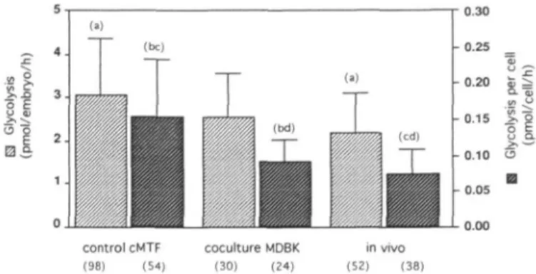
Figure 1. Glycolytic activity (±SD) for blastocysts cultured until 120 h after human chorionic gonadotrophin (HCG) administration in control complex mouse tubal fluid (cMTF) medium and co-culture on Madine-Darby bovine kidney epithelial (MDBK) cells. In-vivo blastocysts were recovered at 96 h after HCG administration. Bars with the same letters in parentheses were significantly different: (a) and (c) at P < 0.0001; (b) and (d) at
P < 0.001. Values in parentheses are numbers of embryos. Glycolytic activity of blastocysts
Day 5 blastocysts obtained from both treatments were analysed for their glycolytic activity. Although blastocyst formation was different between cMTF medium and co-culture, we did not observe any differences in their glycolytic activities per embryo (Figure 1). Glycolysis in cMTF medium (3.1 ± 1 . 3 pmol/ embryo/h) was slightly but not significantly higher than in co-culture (2.6 ± 0.9 pmol/embryo/h). Comparison of the glycolytic activity of blastocysts recovered in vivo (2.2 ± 0.9) showed a significantly {P < 0.0001) lower activity compared with those developed in cMTF medium alone. Despite a poor percentage of blastocysts formed in co-culture, their glycolytic activity was more similar to that in vivo than to that in the cMTF embryos. An analysis of the glycolytic activity per cell showed the same trend but with a statistical difference between co-culture and cMTF medium (Figure 1).
Comparison of cell numbers in blastocysts
For each blastocyst in which the glycolytic activity was previously determined, the cell number was also determined. Total cell numbers were higher in co-culture (26.7 ± 8.8) compared with cMTF medium (21.7 ± 8.7) (Table H). Embryos grown in cMTF medium had significantly lower cell numbers than in-vivo blastocysts (30.2 ± 8.0) (P < 0.0001) taken at
Quality of mouse embryos after co-culture
Table II. Total cell numbers, inner cell mass (ICM) and trophectodermal cell numbers of day 5 blastocysts cultured in control medium (cMTF) or co-culture (MDBK cells) compared with in-vivo day 4 blastocysts
Assessment No. of embryos Control cMTF No. of embryos Co-culture MDBK No. of embryos In vivo Total cell number per embryo (±SD) 64
Trophectoderm (±SD) 64 ICM (±SD) 64 ICM:trophectoderm (±SD)' 64 21.7 ± 8.7t h 13.4 ± 6.T 8.3 ± 4.9£ j 0.8 ± 0.6s 24 24 24 24 26.7 ± 8.8b 14.2 ± 5.^ 12 J ± 6.6e 1.0 ± 0.7h 38 38 38 38 30.2 ± 8.0* 18.8 ± 5.F* 11.3 ± 4.6* 0.6 ± 0 . 3 ^
For abbreviations, see Table I.
•"''Values with the same superscript were significantly different; ••"•«-h/> < 0.001; bP < 0.002; ^P < 0.005.
The ICM:trophectoderm ratio was calculated for each embryo. Mean and SD values were then calculated.
96 h post-HCG, while the total number of cells in co-culture embryos did not differ statistically. The difference in cell numbers of co-culture blastocysts was caused by a higher number of ICM cells (12.5 ± 6.6) when compared with cMTF medium (8.3 ± 4.9) (F < 0.001) (Table II). In contrast to the differences observed with ICM cells, the number of TE cells in the blastocysts did not differ between co-culture (14.2 ± 5.0) and cMTF medium (13.4 ± 6.7). Both culture protocols gave rise to blastocysts with significantly lower numbers of TE cells compared with in-vivo blastocysts (18.8 ± 5.8, P < 0.002) even though they were 24 h older (Table IT). By comparing the ICM:TE ratio, we found that there were no statistical differences between co-culture (1.0 ± 0.7) and cMTF medium (0.8 ± 0.6) embryos. When compared with in-vivo blastocysts (0.6 ± 0.3), we observed the ratio to be significantly higher in both groups (Table II) (P < 0.001).
To examine whether a relationship existed between cell number and glycolytic activity in individual blastocysts, we compared the data obtained from the three different protocols. Surprisingly, the glycolytic activity and the number of cells determined for each blastocyst did not show any correlation (Figure 2). A plot of glycolytic activity compared with total cell number shows widely scattered values in embryos cultured in cMTF medium alone; however, co-culture embryo results were restricted to a smaller area, similar to that observed for in-vivo blastocysts. The glycolytic activity of in-vivo blastocysts was more homogeneous, while that of co-culture blastocysts was less so. Blastocysts developed in cMTF medium alone showed the most scattered levels of glycolytic activity (Figure 2). The glycolytic activity of embryos measured with the radiolabelling technique did not give an overall indication of viability.
Embryo viability
Blastocyst transfers to day 3 pseudopregnant females (Table III) showed a high level of implantation in both co-culture (48.5%) and cMTF medium (47.0%). However, the number of fetuses obtained was very low: only 12.1% were obtained in co-culture and 5.9% in cMTF medium. Control transfers of in-vivo blastocysts showed a high level of fetal development (75%). Both in-vitro treatments failed to enhance embryo viability significantly.
Discussion
To obtain an enhancement in blastocyst formation and embryo viability, a large number of groups have tested a variety of
7O 8
g
o control cMTF y -O.BSx + 19 796, r2 - 0.019 0 1 2 3 4 5 6 7 8 70 cocultura MDBK y - 5.066i * 14.424, r2 - C 0 1 2 3 4 5 6 7 8 0 1 2 3 4 5 6 7 8 Mettboltan (pmol/embryo/h)Figure 2. Relationship between glycolytic activity and total cell number per individual blastocyst after development in complex mouse tubal fluid (cMTF) medium, co-culture on Madine-Darby bovine kidney epithelial (MDBK) cells and when grown in vivo.
medium and co-culture systems. Numerous authors (Whit-tingham, 1971; Chatot et al, 1989; Gardner and Leese, 1990; Lawitts and Biggers, 1991b; Gardner and Sakkas, 1993) have attempted to formulate improved denned media which enable a high level of mouse blastocyst formation. On the other hand, some groups (Ouhibi et al, 1990; Lai et al, 1992) have shown an enhancement in mouse embryo development using co-culture on different cell types. However, as shown by Ouhibi
et al. (1990), certain cell lines are not always beneficial for
embryo development. The main problem during co-culture 601
Table HL Embryo viability after the transfer of day 5 blastocysts grown in complex mouse tuba] fluid (cMTF) or co-culture and day 4 in-vivo blastocysts to day 3 pseudopregnant females
Treatment No. of implantations No. of fetuses Mean weight (g) (±SD) Control cMTF 16/34 (47.0) Co-culture MDBK 16/33 (48.5) In vivo 15/20 (75.0) 2/34 (5.9) 4/33 (12.1) 15/20 (75.0) 0.26 ± 0.038 0.27 ± 0.034 0.30 ± 0.037 For abbreviations, see Table 1. The numbers of implantations and fetuses were assessed on day 15.
studies is to find a complex medium which can support both embryo development and cell growth. In this study we have used a modification of MTF medium containing glutamine, non-essential amino acids and EDTA called cMTF. The com-position of this medium is based on MTF and supports a high level of mouse embryo development (Gardner and Lane, 1993). In using this improved medium we have investigated whether co-culture with MDBK cells could enhance embryo develop-ment further, beyond that observed with cMTF alone. It is evident that embryo development as early as day 3, and then on day 5, is poorer in our co-culture system when compared with cMTF medium alone.
Even though the quantity of co-culture blastocysts was low, their quality may have been improved. Therefore, during this study we have investigated a number of parameters on cultured embryos to determine their quality. Gardner and Sakkas (1993) suggested that the more parameters which are used to examine the efficiency of embryo culture, the greater the understanding gained. Assessments of the development using end-points such as the percentage of embryos which reach the morula or blastocyst stage are inadequate, and must be combined with the use of other parameters to determine the cellular effects and suitability of a particular medium. In fact, the majority of studies (Heyman et al, 1987; Ellington et al., 1990; Ouhibi
et al, 1990; Lai et al, 1992; Minami et al, 1992) have
assessed the effects of co-culture on embryo development using data gathered from very few tests. In these studies only embryo development and cell number were assessed, and in a few cases viability by transferring embryos was also evaluated. However, in our study we have performed multiple tests, including development, glycolytic activity, TE:ICM ratio and viability.
The analysis of the glycolytic activity in blastocysts, by measuring the conversion of [3H]glucose to [3H]2O, showed a
higher activity when blastocysts were cultured in cMTF medium compared with co-culture. Although a lower percent-age of blastocysts was obtained in co-culture, the blastocysts thus obtained had a glycolytic activity closer to that found
in vivo. Furthermore, each blastocyst which had its glycolytic
rate measured, had its cell number determined. No correlation between glycolytic activity per embryo and total cell number was observed. This was also the same when comparing glycolytic activity with the ICM:TE ratio (data not shown). In fact, the data for individual embryos, when comparing glycolytic activity with total cell number, were completely scattered in cMTF medium alone, while in co-culture they 602
were more homogeneous and more similar to the in-vivo blastocyst pattern.
In addition to differences in the glycolytic activity, co-culture blastocysts had a significantly higher ratio of ICM/TE cells when compared with embryos cultured in cMTF medium alone. It is feasible that MDBK cells may secrete factors that act on the development of the ICM. As shown previously by Harvey and Kaye (1990), insulin positively influences the growth of ICM cells in mouse blastocysts. In contrast, insulin does not act on the glycolytic metabolism (Dufranes et al, 1993). Specific growth factors have been shown to act in certain ways. Harvey and Kaye (1990) and Harvey et al. (1995) showed enhanced blastocyst formation by culturing in the presence of EGF and leukaemia inhibitory factor (LIF), as well as an increased development of TE cells by culturing in the presence of EGF. Rappolee et al. (1992) has also shown a positive effect of IGF-II on the ICM. The inference from these previous studies would be that the secretion of any of these growth factors by feeder cells in a co-culture system may also influence embryo development. As proposed by Men6zo et al. (1993), cell lines such as MBDK continually secrete growth factors. Interestingly, the secretion of LIF has been observed by MDBK cells (Plachot, personal communica-tion). Alternatively, M.Lane and D.K.Gardner (personal com-munication) have determined that non-essential amino acids and glutamine stimulate TE development, whereas essential amino acids actively stimulate the development of the ICM in mouse blastocysts. Such observations are consistent with the MDBK cells releasing essential amino acids into the culture medium.
The most important and final criterion used in our study was the transfer of day 5 blastocysts to day 3 pseudopregnant females. No differences were observed between culture groups in the numbers of implantation sites or fetuses, or the weights of the fetuses. Fetal development was reduced greatly when compared with the transfer of day 4 in-vivo control blastocysts. In both culture groups, a high rate of resorption after implanta-tion was evident Successful fetal development depends on the provision of a sufficient number of ICM and TE cells at the time of implantation; an excessive reduction in either lineage diminishes the potential of embryogenesis (Pampfer et al, 1994). In our study both culture groups were well above the ICM:TE ratio of 0.6 observed with in-vivo embryos. This higher ratio reflecting the lower number of TE cells seems to specifically affect post-implantation viability. Reduced fetal development of OF1 strain embryos has also been reported after culture in medium without glucose (Sakkas et al, 1993). The use of outbred strain mice such as OF1 is an interesting model because of their extreme sensitivity to in-vitro culture conditions.
It is clear that certain factors secreted by cells in co-culture can induce differences in embryo development. Therefore some beneficial effect on the quality of co-culture embryos can be observed, even if the number of embryos remains low. Although a number of studies [Gandolfi and Moor (1987) in sheep; Minami et al. (1992) in mice; Mermillod et al. (1993) in cattle] have postulated the existence of a factor that enhances
Quality of mouse embryos after co-culture
embryo development, they have failed to show any direct association between these factors and the embryo.
By analysing a number of parameters on cultured mouse embryos we have a clear idea of the effect of co-culture. Under the current media conditions, co-culture did not increase the number of zygotes reaching the blastocyst stage; however, those blastocysts formed in the presence of somatic cells had an increased ICM and had glycolytic rates closer to in-vivo developed embryos. Therefore further studies are warranted focusing on the mode of action of somatic cells in supporting embryo development.
Acknowledgements
The authors thank Michelle Lane for her assistance and technical advice. This work was supported by the Fonds National Suisse (grant no. 32-39689.93) and The Sir Jules Thorn Charitable Overseas Trust Fund.
References
Abramczuk, J., Solter, D. and Koprowski, H. (1977) The beneficial effect of EDTA on development of mouse one-cell embryos in chemically defined medium. Dev. Biol., 61, 378-383.
Adamson, E.D. (1993) Activities of growth factors in preimplantation embryos.
J. Cell. Biochem., 53, 280-287.
Bavister, B.D. (1992) Coculture for embryo development: is it really necessary?
Hum. Reprod., 7, 1339-1341.
Chatot, C.L., Ziomek, C.A., Bavister, B.D., Lewis, J.L. and Torres, I. (1989) An improved culture medium supports development of random-bred 1-cell mouse embryos in vitro. J. Reprod. Fertil., 86, 679—688.
Dufranes, E., Vanderheyden, I., Robin, D., Delcourt, J., Pampfer, S. and De Hertogh, R. (1993) Glucose and pyruvate metabolism in preimplantation blastocysts from normal and diabetics rats. J. Reprod. Fertil., 98, 169-177. Ellington, J.E., Carney, E.W., Farrell, P.B., Simkin, M.E. and Foote, R.H. (1990) Bovine 1-2-cell embryo development using a simple medium in three oviduct epithelial cell coculture systems. Biol. Reprod., 43, 97-104. Eyestone, W.H. and First, N.L. (1989) Co-culture of early cattle embryos to
the blastocyst stage with oviductal tissue or in conditioned medium. J.
Reprod. Fertil., 85, 715-720.
Gandolfi, F. and Moor, R.M. (1987) Stimulation of early embryonic development in the sheep by co-culture with oviduct epithelial cells. J.
Reprod. Fertil., 81, 23-28.
Gardner, D.K. and Lane, M. (1993) Amino acids and ammonium regulate mouse embryo development in culture. Biol. Reprod., 48, 377-385. Gardner, D.K. and Leese, HJ. (1990) Concentrations of nutrients in mouse
oviduct fluid and their effects on embryo development and metabolism
in vitro. J. Reprod. Fenii, 88, 361-368.
Gardner, D.K. and Sakkas, D. (1993) Mouse embryo cleavage, metabolism and viability: role of medium composition. Hum. Reprod., 8, 288—295. Goddard, MJ. and Pratt, H.P.M. (1983) Control of events during early
cleavage of the mouse embryo: analysis of the '2-cell block'. J. EmbryoL
Exp. Morphol., 73, 111-133.
Handyside, A. and Hunter, S. (1986) Cell division and death in the mouse blastocyst before implantation. Roux's Arch. Dev. Biol., 195, 519-526. Harlow, G.M. and Quinn, P. (1982) Development of preimplantation mouse
embryos in vitro and in vivo. Aust. J. Biol ScL, 35, 187-193.
Harvey, M.B. and Kaye, P.L. (1990) Insulin increases the cell number of the inner cell mass and stimulates morphological development of mouse blastocysts in vitro. Development, 110, 963-967.
Harvey, M.B., Leco, KJ., Arcellana-Panlilio, M.Y., Zhang, X., Edwards, D.R. and Schultz, G.A. (1995) Roles of growth factors during peri-implantation development Mol. Hum. Reprod., 1, see Hum. Reprod., 10, 712-718. Heyman, Y., Menezo, Y., Chesne, P., Camous, S. and Gamier, V. (1987) In
vitro cleavage of bovine and ovine early embryos: improved development using coculture with trophoblastic vesicles. Theriogenology, 27, 59-69. Ho, Y., Wigglesworth, K., Eppig, J J . and Schultz, R.M. (1995) Preimplantation
development of mouse embryos in KSOM: augmentation by amino acids and analysis of gene expression. Mol. Reprod. Dev., 41, 232-238. Lai, Y.M., Stein, D.E., Soong, Y.K., Tang, Y.X., Grifo, J. el at. (1992)
Evaluation of Vero cell co-culture system for mouse embryos in various media. Hum. Reprod., 7, 276-280.
Lane, M. and Gardner, D.K. (1994) Increase in postimplantation development of cultured mouse embryos by amino acids and induction of fetal retardation and exencephaly by ammonium ions. J. Reprod. FertiL, 102, 305-312. Lawitts, J.A. and Biggers, J.D. (1991a) Overcoming the 2-cell block by
modifying standard components in a mouse embryo culture medium. Biol.
Reprod., 45, 245-251.
Lawitts, J.A. and Biggers, J.D. (1991 b) Optimization of mouse embryo culture media using simplex methods. J. Reprod- Fertil., 91, 543-556.
Leppens, G. and Sakkas, D. (1995) Differential effect of epithelial cell-conditioned medium fractions on preimplantation mouse embryo development Hum. Reprod., 10, 1178-1183.
Menezo, YJ.R., Janny, L. and Khatchadourian, C. (1993) Embryo quality and co-culture. In Mastroianni, L., Coellingh Bennink, H J . T , Suzuki, S. and Vermer, H.M. (eds), Gamete and Embryo Quality. The Parthenon Publishing Group, Camforth, UK.
Mermillod, P., Vansteenbrugge, A., Wils, C , Mourmeaux, J.-L., Massip, A. and Dessy, F. (1993) Characterization of the embryotrophic activity of exogenous protein-free oviduct-conditioned medium used in culture of cattle embryos. BioL Reprod., 49, 582-587.
Metha, T.S. and Kiessling, A.A. (1990) Development potential of mouse embryos conceived in vitro and cultured in ethylenediaminetetraacetic acid with or without amino acids or serum. Biol. Reprod., 43, 600-606. Minami, N., Utsumi, K. and Iritani, A. (1992) Effects of low molecular weight
oviductal factors on the development of mouse one-cell embryos in vitro.
J. Reprod. Fertil., 96, 735-745.
O'Fallon, J.V. and Wright, R.W. (1986) Quantitative determination of the pentose phosphate pathway in preimplantation mouse embryos. BioL
Reprod., 34, 58-64.
Ouhibi, N., Hamidi, J., Guillaud, J. and Menezo, Y. (1990) Co-culture of 1-cell mouse embryos on different cell supports. Hum. Reprod, 5, 737-743. Pampfer, S., Wuu, YD., Vanderheyden, I. and De Hertogh, R. (1994) Expression of tumor necrosis factor-a (TNFa) receptors and selective effect of TNFa on the inner cell mass in mouse blastocysts. Endocrinology, 134, 206-212.
Quinn, P., Barros, C. and Whittingham, D.G. (1982) Preservation of hamster oocytes to assay the fertilizing capacity of human spermatozoa. J. Reprod.
FertiL, 66, 161-168.
Rappolee, D.A., Sturm, K.S., Behrendtsen, O., Schultz, G.A., Pedersen, R.A. and Werb, Z. (1992) Insulin-like growth factor II acts through an endogenous growth pathway regulated by imprinting in early mouse embryos. Genes
Dev., 6, 939-952.
Rieger, D. and Guay, P. (1988) Measurement of the metabolism of energy substrates in individual bovine blastocysts. J. Reprod. FertiL, 83, 585-591. Sakkas, D. and Trounson, A.O. (1990) Coculture of mouse embryos with oviduct and uterine cells prepared from mice at different days of pseudopregnancy. J. Reprod. FertiL, 90, 109-118.
Sakkas, D., Batt, P.A. and Cameron, W.N. (1989) Development of preimplantation goat (Capra hircus) embryos in vivo and in vitro. J. Reprod.
FertiL, 87, 359-365.
Sakkas, D., Umer, F , Menezo, Y. and Leppens, G. (1993) Effects of glucose and fructose on fertilization, cleavage, and viability of mouse embryos
in vitro. BioL Reprod, 49, 1288-1292.
Whittingham, D.G. (1971) Culture of mouse ova. J. Reprod, FertiL, 14, 7-21.
Received on August 10, 1995; accepted on November 4, 1995
![Table HL Embryo viability after the transfer of day 5 blastocysts grown in complex mouse tuba] fluid (cMTF) or co-culture and day 4 in-vivo blastocysts to day 3 pseudopregnant females](https://thumb-eu.123doks.com/thumbv2/123doknet/14910421.658227/5.931.56.450.149.247/embryo-viability-transfer-blastocysts-complex-culture-blastocysts-pseudopregnant.webp)