Molecular and culture-based diagnosis of Clostridium difficile
isolates from Coˆte d’Ivoire after prolonged storage at disrupted
cold chain conditions
So¨ren L. Becker
a,b,c,*, Justin K. Chatigre
d, Jean T. Coulibaly
b,c,e,f, Pascal Mertens
g, Bassirou Bonfoh
f,
Mathias Herrmann
a, Ed J. Kuijper
h, Elie´zer K. N’Goran
e,f, Ju¨rg Utzinger
b,cand Lutz von Mu¨ller
a,*
a
Institute of Medical Microbiology and Hygiene, Saarland University, Kirrberger Straße, Building 43, D-66421 Homburg/Saar, Germany; bDepartment of Epidemiology and Public Health, Swiss Tropical and Public Health Institute, P.O. Box, CH-4002 Basel, Switzerland;cUniversity of Basel, P.O. Box, CH-4003 Basel, Switzerland;dDe´partement de Me´decine Interne, Hoˆpital Me´thodiste de Dabou, Dabou, Coˆte d’Ivoire;eUnite´
de Formation et de Recherche Biosciences, Universite´ Fe´lix Houphoue¨t-Boigny, 02 BP 770, Abidjan 02, Coˆte d’Ivoire;fCentre Suisse de Recherches Scientifiques en Coˆte d’Ivoire, 01 BP 1303, Abidjan 01, Coˆte d’Ivoire;gCoris BioConcept, Rue Jean Sonet 4A, B-5032 Gembloux,
Belgium;hDepartment of Medical Microbiology, Leiden University Medical Center, Albinusdreef 2, NL-2333 Leiden, The Netherlands
*Corresponding author: Tel:+49 6841 16-23943; E-mail: [email protected]
Received 23 June 2015; revised 9 August 2015; accepted 12 August 2015
Background: Although Clostridium difficile is a major cause of diarrhoea, its epidemiology in tropical settings is poorly understood. Strain characterisation requires work-up in specialised laboratories, often after prolonged storage without properly maintained cold chain.
Methods: We screened 298 human faecal samples from Coˆte d’Ivoire using a rapid test for C. difficile glutamate dehydrogenase (GDH). GDH-positive samples were aerobically stored at disrupted cold chain conditions (mean duration: 11 days) before transfer to a reference laboratory for anaerobic culture, susceptibility testing, PCR assays and ribotyping.
Results: Sixteen samples (5.4%) had a positive GDH screening test. C. difficile infection was confirmed in six speci-mens by culture and PCR, while no nucleic acids of C. difficile were detected in the culture-negative samples. Further analysis of stool samples harbouring toxigenic C. difficile strains confirmed that both GDH and toxins remained detectable for at least 28 days, regardless of storage conditions (aerobic storage at 48C or 208C). Conclusions: Storage conditions only minimally affect recovery of C. difficile and its toxins in stool culture. A rapid GDH screening test and subsequent transfer of GDH-positive stool samples to reference laboratories for in-depth characterisation may improve our understanding of the epidemiology of C. difficile in the tropics.
Keywords: Coˆte d’Ivoire, Diagnosis, Diarrhoea, Polymerase chain reaction, Rapid diagnostic test, Storage conditions
Introduction
Clostridium difficile is an anaerobic, Gram-positive, rod-shaped and endospore-forming bacterium that may survive under extreme environmental conditions, including high temperatures, toxic chemicals and UV radiation.1 C. difficile is the leading
cause of nosocomial, antibiotic-associated diarrhoea worldwide and there is growing evidence that C. difficile is also a key patho-gen of community-acquired intestinal infections.2–5While a steady increase of C. difficile-associated diarrhoea (CDAD) has been observed in Europe and North America, there is a paucity of epidemiological data from Africa, Asia6and South America7
where diarrhoeal diseases remain important causes of morbidity and mortality.8For example, a search on PubMed/Medline on
June 23, 2015 using the search strategy ‘Clostridium difficile AND Africa’ yielded only 29 hits, 15 studies of which pertained to the bacterium’s prevalence in humans, animals or environmen-tal samples from Africa.9–23
Recent data suggest that CDAD in travellers returning from low- and middle-income countries is considerable, thus highlight-ing the need to deepen our understandhighlight-ing of the epidemiology of C. difficile in Africa and elsewhere in the developing world.24 Additionally, there is ongoing debate whether C. difficile might be regularly transmissible as a zoonotic disease via animals or food products. Zoonotic transmission to humans has been docu-mented for some strains (e.g., ribotype [RT] 078), but no direct food-borne outbreaks have been reported thus far.25,26Strains can be classified into genotypic groups by PCR ribotyping,27
ORIGINAL
ARTICLE
© The Author 2016. Published by Oxford University Press on behalf of Royal Society of Tropical Medicine and Hygiene.
sequence typing28or other genotypic methods.29,30Some hyper-virulent RTs (e.g., RT027) are reported to be associated with a more severe clinical disease, but no data on the currently circulat-ing RTs have been reported from Africa.31
The paucity of epidemiological data from resource-constrained settings is explained by the cumbersome laboratory diagnosis of C. difficile, which has led to its scientific neglect in many tropical areas. Comprehensive diagnostic work-up requires cultural growth of C. difficile on selective media under anaerobic conditions, fol-lowed by subsequent molecular typing in specialised laboratories. It follows that testing strategies for C. difficile vary considerably even in high-income countries. Indeed, a recent multicentre study estimated that approximately 40 000 hospitalised patients with C. difficile infection remain undiagnosed every year in Europe due to the use of insensitive laboratory diagnostic tests.32In con-trast, not even the most basic diagnostic tests for C. difficile are available in many resource-constrained settings. One of the few recent studies pertaining to the clinical relevance of this pathogen stems from Zimbabwe; C. difficile was detected in 8.6% of patients with community-acquired diarrhoea,22which underscores the need for an improved understanding of its occurrence in Africa.
Rapid diagnostic tests (RDTs) for C. difficile have been devel-oped and constitute a useful screening tool to provide point-of-care information, particularly in resource-constrained settings. In common diagnostic algorithms, a C. difficile-specific glutamate dehydrogenase (GDH) assay is employed as a first-line test to identify specimens that warrant further diagnostic work-up.33,34
Though GDH is specific for C. difficile, it does not differentiate pathogenic toxigenic strains from apathogenic non-toxigenic strains. Some rapid membrane tests combine GDH screening with testing for toxins A and B, but the sensitivity of toxin detec-tion is low.35A thorough assessment of GDH-positive samples includes an array of sophisticated tests, including anaerobic toxi-genic culture, followed by genotypic and phenotypic characterisa-tion. While the pure confirmation of C. difficile infection and a differentiation between toxigenic and non-toxigenic strains is pos-sible by stool-based PCR alone, a culture isolate is required for PCR ribotyping and antimicrobial susceptibility testing.
In resource-constrained settings, the transfer of GDH-positive samples to reference laboratories within a country or abroad may take several days. Hence, it is important to understand whether and how prolonged storage, transport conditions, environmental fac-tors and varying temperatures affect the recovery of C. difficile in stool samples. Thus far, it is widely believed that a sensitive diagno-sis needs to be performed on fresh stool samples due to the instability of C. difficile antigens. Indeed, the Association for Professionals in Infection Control and Epidemiology (APIC) currently recommends that samples should be frozen at –708C if testing can-not be performed within 24 hours after stool collection.36However,
storage under controlled freezing conditions is limited in most developing countries.
We conducted a case-control study in south Coˆte d’Ivoire to investigate the epidemiology and diagnostic accuracy of different methods for detection of intestinal pathogens. In this manuscript, we report on the frequency and characterisation of C. difficile strains using a two-step diagnostic algorithm consisting of a GDH screening test, followed by comprehensive sample work-up in a specialised laboratory after prolonged specimen storage at disrupted cold chain conditions. Moreover, we examined the influ-ence of a prolonged storage under standardised aerobic
conditions at varying temperatures on the detection of C. difficile using toxigenic culture and molecular diagnostic techniques.
Materials and methods
Study area and population
The study was conducted in October 2012 in Dabou and 11 surrounding villages, located some 30 km west of Abidjan, the eco-nomic capital of Coˆte d’Ivoire. The study was part of a site assess-ment to identify a suitable setting in Coˆte d’Ivoire for a subsequent multi-country investigation on the aetiology of persistent diarrhoea and persistent abdominal pain.37,38This research is coordinated by
the European research network with the acronym NIDIAG, which aims to develop simple and cost-effective diagnosis-treatment algorithms for three clinical syndromes (i.e., digestive syndromes, persistent fever and neurological disorders) in tropical set-tings.33,38–41In the site assessment reported here, a case-control
approach was adopted. Hence, individuals aged≥1 year presenting with persistent diarrhoea (≥2 weeks) and individuals without any gastrointestinal symptoms (control group) were invited to partici-pate. Definitions put forth by WHO were used to define diarrhoea, i.e., the passing of three or more loose stools within 24 hours.38The
prevalence of various diarrhoeagenic bacteria, helminths, intestinal protozoa and viruses in cases and matched controls has been pre-sented elsewhere.37
Field and laboratory procedures
Fresh stool samples were obtained in the early morning and trans-ferred to the local hospital laboratory in Dabou. Upon arrival, an RDT indicating the presence of GDH (Clostridium K-SeT, Coris BioConcept, Gembloux, Belgium)42was performed to screen for C. difficile. Additionally, several microscopic techniques were employed for the diagnosis of intestinal protozoa and helminth infections (i.e., Baermann funnel, formalin-ether concentration technique, Kato-Katz thick smear and Koga agar plate).37
For later confirmatory testing of C. difficile in a specialised labora-tory, approximately 0.3 g of each stool sample was transferred into a small vial and aerobically stored under ‘real life conditions’ in a fridge without proper maintenance of the cold chain due to power cuts. After a storage period between 8 and 19 days (mean 11 days), the samples were transferred at ambient temperature to the German National Advisory Laboratory for C. difficile in Homburg, Germany. All GDH-positive stool samples were analysed for the pres-ence of C. difficile by anaerobic culture on a selective solid medium (CLO agar, BioMe´rieux, Marcy l’E´toile, France), and by multiplex PCR of stool samples (GenoType CDiff, Hain Lifescience, Nehren, Germany). The multiplex PCR detects the following C. difficile-associated genes: gluD (encoding for GDH), tpi (encoding for triose phosphate isomer-ase), tcdA (encoding for toxin A), tcdB (encoding for toxin B) and cdtA/B (encoding for the binary toxin). Additionally, characteristic deletions in the regulatory gene tcdC and resistance to moxifloxacin (mutations in the gyrA gene) are detected. The PCR assay was regarded as the internal diagnostic reference standard in our study due to stability of culture-independent assays, and results obtained by anaerobic stool culture after the prolonged sample stor-age were compared to this method. Suspected culture-grown col-onies were identified based on typical morphology and distinct odour. Diagnosis of C. difficile was subsequently confirmed by matrix-assisted laser desorption ionisation time-of-flight mass
spectrometry (MALDI-TOF MS with BioTyper software, Bruker Daltonics, Bremen, Germany). Phenotypic characterisation was per-formed by antimicrobial susceptibility testing using Etests (metro-nidazole, moxifloxacin and vancomycin) and disk diffusion assay (clarithromycin and rifampicin), according to previously described standard protocols.32,43Species-specific EUCAST breakpoints (http://
www.eucast.org) were used, if available.44PCR ribotyping using
capillary gel electrophoresis was performed in Homburg, Germany to achieve genotypic differentiation and the results were independ-ently confirmed in Leiden, The Netherlands. Files of untypeable C. dif-ficile strains with previously undescribed ribotype patterns were also sent to Leeds University, UK for analysis using an in-house database.
To further analyse the influence of storage conditions on the performance of diagnostic tests, ten stool samples stemming from symptomatic, hospitalised patients with laboratory-proven toxigenic C. difficile infections were prospectively collected at the Saarland University Medical Center in Homburg, Germany. The faecal samples were split into two aliquots and stored in parallel at 48C and at 208C (ambient temperature) for at least 28 days. Diagnostic testing for C. difficile using toxigenic culture and an RDT for detection of GDH and toxins A/B (C. Diff Quik Check Complete, Alere, Ko¨ln, Germany) was repeated after 7, 14 and 28 days. The resulting RDT line intensities were documented using a semi-quantitative grading scheme, i.e., 3+, strong;
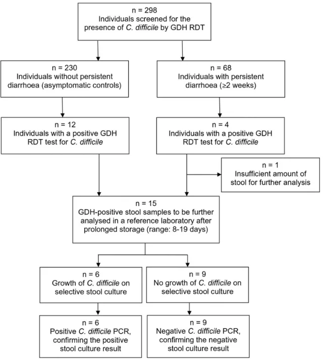
Figure 1. Study flowchart on the occurrence of Clostridium difficile in 298 individuals in Dabou, south Coˆte d’Ivoire, in October 2012. GDH: glutamate dehydrogenase; RDT: rapid diagnostic test.
2+, moderate; 1+, faint; and –, negative. The culture isolates of C. difficile were further characterised by PCR-based detection of toxin genes and ribotyping.
Statistical analysis
Data of the RDT screening results in Coˆte d’Ivoire and of subse-quent laboratory procedures were entered twice and cross-checked in Excel version 14.0 (edition 2010, Microsoft Corp., Redmond, WA, USA). Analysis of C. difficile-specific RT patterns was performed using the software BioNumerics version 7.1 (edi-tion 2013, Applied Maths, Sint-Martens-Latem, Belgium). In brief, a Dice similarity coefficient and the unweighted pair group method with arithmetic mean (UPGMA) algorithm were employed to compare closely related C. difficile isolates and to infer their genetic relatedness.
Ethics statement
The study protocol was approved by the institutional research commissions of the Swiss Tropical and Public Health Institute (Swiss TPH, Basel, Switzerland) and the Centre Suisse de Recherches Scientifiques en Coˆte d’Ivoire (CSRS; Abidjan, Coˆte d’Ivoire). Study approval was given by the Directorate of the Hoˆpital Me´thodiste in Dabou. The study is registered on Current Controlled Trials (http://www.controlled-trials.com; identifier ISRCTN86951400). Individuals aged above 12 months with resi-dency in Dabou or surrounding villages with written informed consent (parents/guardians signing for individuals aged below 18 years) were eligible to participate.
Results
The study flowchart is shown in Figure1. In brief, GDH RDTs were performed on 298 stool samples for screening of C. difficile directly on site. Sixty-eight samples were provided by individuals with per-sistent diarrhoea (≥2 weeks) and 230 specimens originated from individuals without diarrhoea (asymptomatic controls). Positive test results were found on 16 samples. Four faecal specimens stemmed from symptomatic patients with persistent diarrhoea (5.8%). Of these, two patients stated to have received antibiotic treatment with cotrimoxazole and metronidazole, respectively, in the two preceding months. The remaining 12 RDT-positive sam-ples were found in healthy controls (5.2%). Of note, samsam-ples with a faintly discernible test band were also considered as GDH-positive to increase the sensitivity of the screening test (Figure2).
Fifteen GDH-positive samples were available for subsequent culture and molecular diagnosis in a European reference labora-tory. Anaerobic culture yielded six C. difficile isolates which were independently confirmed by direct stool-based PCR. No specific DNA of C. difficile could be amplified in the nine culture-negative samples, thus leading to a 100% concordance between stool cul-ture and direct molecular testing. Among the six isolates, none was a toxigenic strain, as determined by toxigenic culture and PCR for tcdA (toxin A), tcdB (toxin B) and cdtA/B (binary toxin). Four C. difficile isolates originated from individuals without gastro-intestinal disorders (1.7%), while two isolates stemmed from symptomatic patients with persistent diarrhoea (2.9%). The dif-ference between cases and matched controls was not statistically
significant (Fisher’s exact test, p¼0.622) and the lack of toxin production excludes C. difficile as causative agent in the two symptomatic patients.
We assessed all GDH-positive isolates for parasitic co-infections. An infection with intestinal protozoa or helminths was found in five out of nine RDT-positive and culture-negative samples (three Endolimax nana, one Giardia intestinalis, one triple infection with E. nana, Entamoeba coli and hookworm). Among the six culture-positive C. difficile samples, only one parasitic co-infection was detected (triple infection with Blastocystis spp., E. coli and Entamoeba hartmanni).
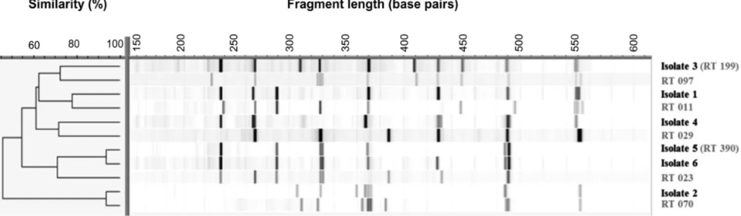
The genotypic differentiation of the six C. difficile isolates via ribotyping determined one isolate as RT199 and one isolate as RT390, while the patterns of the remaining four C. difficile isolates differed from those of the .500 previously described RTs. Hence, these isolates could not be typed in Homburg, Leiden and Leeds and represent new RTs. A dendrogram (Figure3) depicts the spe-cific ribotyping patterns of these C. difficile isolates from Coˆte d’Ivoire in comparison to some closely related RTs. Details on the antimicrobial susceptibility of all isolates are presented in Table1.
Additionally, ten toxigenic C. difficile isolates originating from clinically relevant infections of hospitalised patients in Homburg were analysed to assess the influence of storage conditions on antigen detection and culture of preserved stools. All samples, whether stored at 48C or 208C, tested positive for both GDH and toxins after 7, 14 and 28 days, and C. difficile could always be recovered in toxigenic culture during the study period. In some samples, there was a trend towards reduced signal intensities of RDT after prolonged storage, and this observation was slightly more pronounced if stool samples were stored at ambient tem-perature. However, C. difficile RDTs are designed as qualitative assays, and both GDH and toxins remained detectable in all stool samples. Stability of testing after prolonged storage of
Figure 2. Results given by the rapid diagnostic glutamate dehydrogenase (GDH) screening test (Clostridium K-SeT) employed for diagnosis of Clostridium difficile in Dabou, south Coˆte d’Ivoire in October 2012: negative, faintly positive and strongly positive.
stool samples was confirmed for a variety of clinically important ribotypes (Table2).
Discussion
Our results indicate that the two-step diagnostic algorithm with a point-of-care GDH screening on the spot, followed by prolonged storage under disrupted cold chain conditions and subsequent sample transfer to a reference laboratory for selective anaerobic stool culture and PCR ribotyping is feasible to investigate C. difficile in faecal samples obtained from resource-constrained settings. Importantly, stool culture results were not affected by prolonged storage (up to 19 days) under aerobic atmosphere and varying, non-standardised temperature conditions. Cultural growth of C. difficile was observed in all PCR-confirmed specimens, thus showing an excellent agreement between both methods and confirming the stability of infectious spores of C. difficile over pro-longed time periods. Additional experiments performed on stool samples stemming from hospitalised patients with toxigenic C. difficile infections in Germany further underscored our findings,
as both GDH and toxins remained detectable in the stool over a period of at least 28 days, regardless of storage conditions.
The use of GDH RDT is an easily applicable, rapid and sensitive tool to screen for C. difficile infection and is suitable for use in remote and resource-constrained settings. It is thus of particular importance in areas where prompt diagnosis is crucial and more sophisticated laboratory work-up is not feasible.45Notably, of 15 GDH-positive stool samples in our study, only six revealed C. diffi-cile by culture. However, even samples with a faintly positive test band in the RDT for GDH were included for further diagnostic work-up to maximise the diagnostic sensitivity of the initial screening, acknowledging that this strategy might decrease the specificity of the RDT. On the other hand, we cannot exclude that low amounts of C. difficile may indeed have been present in these culture-negative samples, and unfavourable storage condi-tions (intermittent cold chain, no preservation medium) might have negatively influenced the recovery of bacteria.46However, our finding that all culture-negative samples were also negative for C. difficile when employing a stool-based PCR strongly suggests false-positive RDT results as the more likely explanation for this discrepancy.
Figure 3. Dendrogram showing the specific ribotype (RT) profiles of six Clostridium difficile isolates from Dabou, south Coˆte d’Ivoire, obtained in October 2012, in comparison to related RTs, as determined by capillary ribotyping. Ribotyping displays a high diversity of the isolates. The dendrogram was generated by the software BioNumerics version 7.1 (edition 2013,www.applied-maths.com).
Table 1. Genotypic and phenotypic characterisation of Clostridium difficile isolates from Dabou, south Coˆte d’Ivoire, obtained in October 2012. PCR ribotyping, toxigenic culture and antimicrobial susceptibility testing (S, susceptible; R, resistant) were performed. The results of toxigenic culture were independently confirmed by multiplex PCR for toxin genes (tcdA, tcdB and cdtA/B). The minimal inhibitory concentration (MIC, expressed as mg/ml) is given, if susceptibility testing was performed by Etest
Antimicrobial susceptibility testing (MIC, expressed as mg/ml)
C. difficile Ribotype Toxigenic culture Metronidazole Vancomycin Moxifloxacin Clarithromycin Rifampicin Isolate 1 New (unknown) Non-toxigenic S (0.5) S (1.0) S (1.5) R S Isolate 2 New (unknown) Non-toxigenic S (0.75) S (0.5) S (1.5) S S Isolate 3 RT199 Non-toxigenic S (0.5) S (0.5) S (1.0) S S Isolate 4 New (unknown) Non-toxigenic S (0.75) S (0.75) S (1.5) S S Isolate 5 RT390 Non-toxigenic S (0.5) S (0.5) S (1.5) R S Isolate 6 New (unknown) Non-toxigenic S (0.75) S (0.75) S (1.5) R S
When using an RDT, the resulting line intensity can vary and pale, weak lines may lead to false-positive results. Guideline sys-tems on how to read test results in a reliable, standardised way have been developed e.g., for malaria RDTs, and may also be use-ful to objectify the interpretation of C. difficile-specific RDTs.47With regard to its use as a first-line screening test, the Clostridium K-SeT RDT showed moderate specificity in this study, which underscores the need for a confirmatory test to avoid false-positive results.48
Moreover, GDH assays cannot discriminate between toxigenic and
non-toxigenic infections. However, new research has elucidated that asymptomatic C. difficile carriage per se profoundly alters the intestinal microbial diversity,49and further studies assessing
this infection in both asymptomatic carriers and symptomatic patients from tropical settings will provide additional insights into the pathogenesis of CDAD.
An optimisation of the culture-based recovery of C. difficile from human stool has been identified as an important research need for epidemiological studies.50However, only few investigations have
Table 2. Results of a stool-based rapid diagnostic test (RDT) detecting Clostridium difficile glutamate dehydrogenase (GDH) and toxin A/B, employed on stool samples from symptomatic patients and performed after prolonged aerobic storage at 48C and 208C over a storage period of 28 days. Resulting RDT line intensities are presented using a semi-quantitative grading scheme: 3+, strong; 2+, moderate; 1+, faint; –, negative
Sample Ribotype Toxin genes Day 0 Day 7 Day 14 Day 28 1 001 tcdA, tcdB GDH RDT (48C) 3+ 3+ 3+ 3+ GDH RDT (208C) 3+ 3+ 3+ 3+ Toxin RDT (48C) 3+ 3+ 2+ 2+ Toxin RDT (208C) 3+ 3+ 2+ 1+ 2 005 tcdA, tcdB GDH RDT (48C) 3+ 3+ 3+ 3+ GDH RDT (208C) 3+ 3+ 3+ 2+ Toxin RDT (48C) 2+ 2+ 1+ 1+ Toxin RDT (208C) 2+ 2+ 1+ 1+ 3 005 tcdA, tcdB GDH RDT (48C) 3+ 3+ 2+ 2+ GDH RDT (208C) 3+ 3+ 2+ 2+ Toxin RDT (48C) 3+ 3+ 2+ 2+ Toxin RDT (208C) 3+ 3+ 2+ 2+ 4 011 tcdA, tcdB GDH RDT (48C) 3+ 3+ 3+ 3+ GDH RDT (208C) 3+ 3+ 3+ 3+ Toxin RDT (48C) 2+ 2+ 1+ 1+ Toxin RDT (208C) 2+ 1+ 1+ 1+ 5 013 tcdA, tcdB GDH RDT (48C) 3+ 3+ 3+ 3+ GDH RDT (208C) 3+ 3+ 3+ 3+ Toxin RDT (48C) 1+ 1+ 1+ 1+ Toxin RDT (208C) 1+ 1+ 1+ 1+ 6 014 tcdA, tcdB GDH RDT (48C) 3+ 3+ 3+ 3+ GDH RDT (208C) 3+ 2+ 2+ 2+ Toxin RDT (48C) 3+ 3+ 1+ 1+ Toxin RDT (208C) 3+ 1+ 1+ 1+ 7 017 tcdA, tcdB GDH RDT (48C) 3+ 3+ 3+ 3+ GDH RDT (208C) 3+ 3+ 3+ 2+ Toxin RDT (48C) 2+ 2+ 2+ 2+ Toxin RDT (208C) 2+ 2+ 2+ 2+ 8 027 tcdA, tcdB, binary toxin GDH RDT (48C) 3+ 3+ 3+ 3+ GDH RDT (208C) 3+ 3+ 3+ 3+ Toxin RDT (48C) 3+ 3+ 3+ 3+ Toxin RDT (208C) 3+ 3+ 3+ 3+ 9 029 tcdA, tcdB GDH RDT (48C) 3+ 3+ 3+ 3+ GDH RDT (208C) 3+ 3+ 3+ 3+ Toxin RDT (48C) 3+ 3+ 3+ 3+ Toxin RDT (208C) 3+ 3+ 3+ 3+ 10 029 tcdA, tcdB GDH RDT (48C) 3+ 3+ 3+ 3+ GDH RDT (208C) 3+ 3+ 3+ 3+ Toxin RDT (48C) 3+ 3+ 3+ 2+ Toxin RDT (208C) 3+ 3+ 3+ 2+
addressed this issue with conflicting results, and most studies did not employ independent molecular testing to elucidate ambiguous results obtained by culture and RDT. In contrast to our findings, a previous study reported a rapid decrease in the recovery rate of C. difficile from 26 equine samples kept under aerobic conditions with no C. difficile isolate being culturally detectable after 12 days of aerobic storage at 48C, whereas all isolates could still be recov-ered when stored anaerobically.51Another study found that the
recovery of C. difficile on selective culture is only minimally affected by storage conditions, whereas toxins in the stool samples rapidly become undetectable after repeated freezing at –208C.52 A Canadian study reported no influence of temperature and storage conditions on the recovery of C. difficile kept over a period of 8 weeks.53This finding is in agreement with our results and may be explained by the bacterium’s characteristic formation of durable endospores that persist in the environment before germinating again under optimised growth conditions. Future investigations should thus include environmental examinations and assess the effects of appropriate selective media that may facilitate the con-version from durable C. difficile endospores into the vegetative, cul-tivable form after prolonged storage.
Our study has several limitations. First, the low number of posi-tive samples limits the generalisability of our findings. Second, none of the C. difficile strains in Coˆte d’Ivoire was toxigenic. However, we tried to address these constraints by the additional analysis of toxigenic isolates from clinically relevant infections from Homburg. In all samples, the prolonged storage did not negatively impact on the diagnostic yield of C. difficile stool cul-ture, which underscores the reproducibility of our approach. Third, also healthy controls were tested in our study for the pres-ence of C. difficile, despite many clinical guidelines stating that only symptomatic patients should be tested.3,34While this is true for clinical settings, investigations on the bacterium’s occur-rence in the environment, animals and healthy humans will con-tribute to an improved understanding of the largely unknown epidemiology of C. difficile in Africa. Indeed, a recent study from Coˆte d’Ivoire showed the presence of C. difficile in 12.4% of cooked beef meat sold by street vendors in Abidjan.21
Conclusions
A two-step diagnostic approach consisting of GDH screening on site, followed by in-depth genotypic and phenotypic characterisa-tion in specialised laboratories is a promising strategy to investi-gate C. difficile in tropical settings. The recovery of C. difficile in human faecal samples from Coˆte d’Ivoire remained unaffected by prolonged storage and transport conditions that lacked stand-ardisation and cold chain. This observation is of considerable importance for resource-constrained settings where accurate diagnosis cannot be ascertained and stool samples might be transported over several days to specialised laboratories. The pre-sent pilot study identified only non-toxigenic apathogenic iso-lates, both in symptomatic cases and matched asymptomatic controls. Further studies are warranted to deepen our knowledge of the transmission patterns, strain diversity and clinical relevance of C. difficile in the humid tropics. Our results should also encour-age surveillance studies to CDAD on the African continent to obtain more insights into the global epidemiology of C. difficile infections.
Authors’ contributions: SLB, JU and LvM conceived the study; SLB, JTC, BB, EKN, JU and LvM designed the study protocol; SLB and JKC carried out the clinical assessment; SLB, MH, EJK and LvM carried out the laboratory diagnostic tests, and analysis and interpretation of these data. SLB, JU and LvM drafted the manuscript; JTC, PM, BB, MH and EJK critically revised the manuscript for intellectual content. All authors read and approved the final manuscript. SLB and LvM are guarantors of the paper. Acknowledgements: We would like to thank all the participants and the staff of the Hoˆpital Me´thodiste de Dabou, Coˆte d’Ivoire. We express our gratitude to Ruedi Leuppi and the ‘Ruedi Leuppi Foundation Coˆte d’Ivoire’. The excellent technical laboratory support by Brou K. Jean, Laurent K. Lohourignon and Diabre´ G. Raphael (Dabou, Coˆte d’Ivoire), and Anna Nimmesgern, Sabine Freis and Diana Velten (Homburg, Germany) is greatly appreciated. We are indebted to Ce´line Harmanus (Leiden) and Warren Fawley (Leeds University) for their invaluable help and support to ribotype C. difficile isolates.
Funding: This work is part of the NIDIAG European research network (Collaborative Project), supported by the European Union’s Seventh Framework Programme for research, technological development and demonstration under grant agreement no. 260260. The funders had no role in study design, data collection, data analysis, decision to publish or preparation of the manuscript.
Competing interests: P. Mertens is Director for Research and Development (R&D) at Coris BioConcept (Gembloux, Belgium). The RDT employed in the current study (Clostridium K-SeT) is produced by Coris BioConcept. The company had no role in study design, data collection, data analysis, decision to publish or preparation of the manuscript.
Ethical approval: The study protocol was approved by the institutional research commissions of the Swiss Tropical and Public Health Institute (Basel, Switzerland) and the Centre Suisse de Recherches Scientifiques en Coˆte d’Ivoire (Abidjan, Coˆte d’Ivoire). Study approval was given by the Directorate of the Hoˆpital Me´thodiste in Dabou. The study is registered on Current Controlled Trials (http://www.controlled-trials.com; identifier ISRCTN86951400).
References
1 Nicholson WL, Munakata N, Horneck G et al. Resistance of Bacillus endospores to extreme terrestrial and extraterrestrial environments. Microbiol Mol Biol Rev 2000;64:548–72.
2 Khanna S, Pardi DS, Aronson SL et al. The epidemiology of community-acquired Clostridium difficile infection: a population-based study. Am J Gastroenterol 2012;107:89–95.
3 Bagdasarian N, Rao K, Malani PN. Diagnosis and treatment of Clostridium difficile in adults: a systematic review. JAMA 2015;313:398–408. 4 Leffler DA, Lamont JT. Clostridium difficile infection. N Engl J Med
2015;372:1539–48.
5 Lessa FC, Mu Y, Bamberg WM et al. Burden of Clostridium difficile infection in the United States. N Engl J Med 2015;372:825–34. 6 Ngamskulrungroj P, Sanmee S, Pusathit P et al. Molecular epidemiology
of Clostridium difficile infection in a large teaching hospital in Thailand. PLoS One 2015;10:e0127026.
7 Balassiano IT, Yates EA, Domingues RM et al. Clostridium difficile: a problem of concern in developed countries and still a mystery in Latin America. J Med Microbiol 2012;61:169–79.
8 Murray CJL, Vos T, Lozano R et al. Disability-adjusted life years (DALYs) for 291 diseases and injuries in 21 regions, 1990–2010: a systematic
analysis for the Global Burden of Disease Study 2010. Lancet 2012;380:2197–2223.
9 Rotimi VO, Akindutire D. Faecal carriage of cytotoxigenic strains of Clostridium difficile by adult Nigerians. East Afr Med J 1989;66:319–23. 10 Emeruwa AC, Oguike JU. Incidence of cytotoxin producing isolates of Clostridium difficile in faeces of neonates and children in Nigeria. Microbiologica 1990;13:323–8.
11 Oguike JU, Emeruwa AC. Incidence of Clostridium difficile in infants in rural and urban areas of Nigeria. Microbiologica 1990;13:267–71. 12 Merad SA, Djellout MB. Preliminary epidemiological study of carriers
of Clostridium difficile [in French]. Arch Inst Pasteur Alger 1992;58:169–79.
13 Mwachari C, Batchelor BI, Paul J et al. Chronic diarrhoea among HIV-infected adult patients in Nairobi, Kenya. J Infect 1998;37:48–53. 14 Zulu I, Kelly P, Mwansa J et al. Contrasting incidence of Clostridium
difficile and other enteropathogens in AIDS patients in London and Lusaka. Trans R Soc Trop Med Hyg 2000;94:167–8.
15 Simango C. Prevalence of Clostridium difficile in the environment in a rural community in Zimbabwe. Trans R Soc Trop Med Hyg 2006;100:1146–50.
16 Samie A, Obi CL, Franasiak J et al. PCR detection of Clostridium difficile triose phosphate isomerase (tpi), toxin A (tcdA), toxin B (tcdB), binary toxin (cdtA, cdtB), and tcdC genes in Vhembe district, South Africa. Am J Trop Med Hyg 2008;78:577–85.
17 Simango C, Mwakurudza S. Clostridium difficile in broiler chickens sold at market places in Zimbabwe and their antimicrobial susceptibility. Int J Food Microbiol 2008;124:268–70.
18 Lekalakala MR, Lewis E, Hoosen AA. Clostridium difficile infections in a tertiary hospital: value of surveillance. J Hosp Infect 2010;75: 328–9.
19 Onwueme K, Fadairo Y, Idoko L et al. High prevalence of toxinogenic Clostridium difficile in Nigerian adult HIV patients. Trans R Soc Trop Med Hyg 2011;105:667–9.
20 Rajabally NM, Pentecost M, Pretorius G et al. The Clostridium difficile problem: a South African tertiary institution’s prospective perspective. S Afr Med J 2013;103:168–72.
21 Kouassi KA, Dadie AT, N’Guessan KF et al. Clostridium perfringens and Clostridium difficile in cooked beef sold in Coˆte d’Ivoire and their antimicrobial susceptibility. Anaerobe 2014;28:90–4.
22 Simango C, Uladi S. Detection of Clostridium difficile diarrhoea in Harare, Zimbabwe. Trans R Soc Trop Med Hyg 2014;108:354–7. 23 Jaeggi T, Kortman GA, Moretti D et al. Iron fortification adversely
affects the gut microbiome, increases pathogen abundance and induces intestinal inflammation in Kenyan infants. Gut 2015;64:731–42.
24 Neuberger A, Saadi T, Shetern A et al. Clostridium difficile infection in travelers – a neglected pathogen? J Travel Med 2013;20:37–43. 25 Arroyo LG, Kruth SA, Willey BM et al. PCR ribotyping of Clostridium
difficile isolates originating from human and animal sources. J Med Microbiol 2005;54:163–6.
26 Hensgens MP, Keessen EC, Squire MM et al. Clostridium difficile infection in the community: a zoonotic disease? Clin Microbiol Infect 2012;18:635–45.
27 Gurtler V. Typing of Clostridium difficile strains by PCR-amplification of variable length 16S-23S rDNA spacer regions. J Gen Microbiol 1993;139:3089–97.
28 Joost I, Speck K, Herrmann M et al. Characterisation of Clostridium difficile isolates by slpA and tcdC gene sequencing. Int J Antimicrob Agents 2009;33 Suppl 1:S13–8.
29 Huber CA, Foster NF, Riley TV et al. Clostridium difficile typing methods: challenges for standardization. J Clin Microbiol 2013;51:2810–4. 30 Knetsch CW, Lawley TD, Hensgens MP et al. Current application and
future perspectives of molecular typing methods to study Clostridium difficile infections. Euro Surveill 2013;18:20381.
31 Lessa FC, Gould CV, McDonald LC. Current status of Clostridium difficile infection epidemiology. Clin Infect Dis 2012;55 Suppl 2:S65–70.
32 Davies KA, Longshaw CM, Davis GL et al. Underdiagnosis of Clostridium difficile across Europe: the European, multicentre, prospective, biannual, point-prevalence study of Clostridium difficile infection in hospitalised patients with diarrhoea (EUCLID). Lancet Infect Dis 2014;14:1208–19.
33 Becker SL, Vogt J, Knopp S et al. Persistent digestive disorders in the tropics: causative infectious pathogens and reference diagnostic tests. BMC Infect Dis 2013;13:37.
34 Lu¨bbert C, John E, von Mu¨ller L. Clostridium difficile infection: guideline-based diagnosis and treatment. Dtsch Arztebl Int 2014;111:723–31. 35 Brecher SM, Novak-Weekley SM, Nagy E. Laboratory diagnosis of
Clostridium difficile infections: there is light at the end of the colon. Clin Infect Dis 2013;57:1175–81.
36 APIC. Guide to Preventing Clostridium difficile Infections. Washington D.C.: Association for Professionals in Infection Control and Epidemiology http://apic.org/Resource_/EliminationGuideForm/ 59397fc6-3f90-43d1-9325-e8be75d86888/File/2013CDiffFinal.pdf
[accessed 6 August 2015].
37 Becker SL, Chatigre JK, Gohou JP et al. Combined stool-based multiplex PCR and microscopy for enhanced pathogen detection in patients with persistent diarrhoea and asymptomatic controls from Coˆte d’Ivoire. Clin Microbiol Infect 2015;21:591.e1-591.e10.
38 Polman K, Becker SL, Alirol E et al. Diagnosis of neglected tropical diseases among patients with persistent digestive disorders (diarrhoea and/or abdominal pain ≥14 days): a multi-country, prospective, non-experimental case-control study. BMC Infect Dis 2015;15:338.
39 Yansouni CP, Bottieau E, Chappuis F et al. Rapid diagnostic tests for a coordinated approach to fever syndromes in low-resource settings. Clin Infect Dis 2012;55:610–1.
40 Chappuis F, Alirol E, d’Acremont V et al. Rapid diagnostic tests for non-malarial febrile illness in the tropics. Clin Microbiol Infect 2013;19:422–31.
41 Yansouni CP, Bottieau E, Lutumba P et al. Rapid diagnostic tests for neurological infections in central Africa. Lancet Infect Dis 2013;13:546–58.
42 Van Broeck J, Icyeza ER, Delme´e M. Evaluation of a new glutamate dehydrogenase immunoassay in a two-step algorithm for the diagnosis of Clostridium difficile infection. Poster presented at the Interscience Conference on Antimicrobial Agents and Chemotherapy (ICAAC), San Franciso, USA; 2012.
43 von Mu¨ller L, Halfmann A, Herrmann M. Current data and trends on the development of antibiotic resistance of Clostridium difficile [in German]. Bundesgesundheitsblatt Gesundheitsforschung Gesundheits-schutz 2012;55:1410–7.
44 EUCAST. Breakpoint tables for interpretation of MICs and zone diameters. Version 5.1. Va¨xjo¨, Sweden: The European Committee on Antimicrobial Susceptibility Testing.http://www.eucast.org[accessed 6 August 2015].
45 Shetty N, Wren MW, Coen PG. The role of glutamate dehydrogenase for the detection of Clostridium difficile in faecal samples: a meta-analysis. J Hosp Infect 2011;77:1–6.
46 Nechvatal JM, Ram JL, Basson MD et al. Fecal collection, ambient preservation, and DNA extraction for PCR amplification of bacterial and human markers from human feces. J Microbiol Methods 2008;72:124–32.
47 Van der Palen M, Gillet P, Bottieau E et al. Test characteristics of two rapid antigen detection tests (SD FK50 and SD FK60) for the diagnosis of malaria in returned travellers. Malar J 2009;8:90. 48 Schmidt ML, Gilligan PH. Clostridium difficile testing algorithms: what is
practical and feasible? Anaerobe 2009;15:270–3.
49 Zhang L, Dong D, Jiang C et al. Insight into alteration of gut microbiota in Clostridium difficile infection and asymptomatic C. difficile colonization. Anaerobe 2015;34:1–7.
50 Hink T, Burnham CA, Dubberke ER. A systematic evaluation of methods to optimize culture-based recovery of Clostridium difficile from stool specimens. Anaerobe 2013;19:39–43.
51 Weese JS, Staempfli HR, Prescott JF. Survival of Clostridium difficile and its toxins in equine feces: implications for diagnostic test selection and interpretation. J Vet Diagn Invest 2000;12:332–6.
52 Freeman J, Wilcox MH. The effects of storage conditions on viability of Clostridium difficile vegetative cells and spores and toxin activity in human faeces. J Clin Pathol 2003;56:126–8.
53 Arroyo LG, Rousseau J, Willey BM et al. Use of a selective enrichment broth to recover Clostridium difficile from stool swabs stored under different conditions. J Clin Microbiol 2005;43:5341–3.