Comparative analysis of full-length antigen 11/3 from
Echinococcus multilocularis and E. granulosus
R. FELLEISEN and B. GOTTSTEIN
Institute of Parasitology, University of Berne, Langgass-Strasse 122, P.O. Box 8466, CH-3012 Berne, Switzerland (Received 8 November 1993; revised 10 February 1994; accepted 10 February 1994)
SUMMARY
The recombinant Echinococcus multilocularis antigen II/3-10 is one of the most promising tools for immunodiagnosis of alveolar echinococcosis in human patients. Its nucleic acid sequence represents a part of the E. multilocularis gene encoding the metacestode antigen 11/3, the former being basically present and expressed in both E. multilocularis and E. granulosus. Most (94%) patients with alveolar echinococcosis respond to infection with a marked anti-II/3-10 IgG synthesis; in contrast, most of the cystic echinococcosis patients do not, for some reason, recognize the recombinant antigen. We tackled this problem by generating cDNA derived from both E. granulosus and E. multilocularis full length 11/3 genes, performed by reverse transcription and PCR amplification. Sequence analysis revealed a very high degree of conservation of the primary sequence of the antigen 11/3 in both Echinococcus species. cDNA fragments were subcloned and expressed in E.
coli as fusion proteins with Schistosoma japonicum glutathione S-transferase. Recombinant proteins were affinity purified
and comparatively assessed by ELISA with respect to antibody-binding characteristics. Sera from patients suffering from cystic echinococcosis showed no significant differences in reactivity with the antigens derived from either E. multilocularis or E. granulosus. Therefore, parameters other than some minor differences in the primary sequence seem to be responsible for the lack of antigen 11/3 recognition in cystic echinococcosis.
Note: Nucleotide sequence data reported in this paper have been submitted to the GenBank® data base with the accession numbers U05573 and U05574.
Key words: Echinococcus multilocularis, Echinococcus granulosus, taeniid cestodes, immunodiagnosis, PCR, recombinant antigen II/3, GST-fusion protein, ELISA.
INTRODUCTION
Echinococcosis in humans is caused by a proliferating metacestode following infection with different species of the genus Echinococcus. Alveolar echinococcosis (E. multilocularis) and cystic echino-coccosis (E. granulosus) are both of medical and public health importance. While E. granulosus has a cosmopolitan distribution, E. multilocularis is found in its natural life-cycle only in the Northern hemisphere.
Cystic echinococcosis is characterized by well-delimited cysts formed most frequently in the liver, but also in the lungs and other organs; such hydatid cysts can be removed completely by surgery in most cases. In contrast, alveolar echinococcosis is caused by the tumour-like invasive growth of the parasite, affecting primarily the liver and then other organs by infiltration or metastasis. By the time the disease becomes clinically manifest, the lesions caused by the E. multilocularis metacestode are often too large for complete surgical resection. Therefore, early serological detection of alveolar echinococcosis at the stage of small hepatic infiltrates followed by ap-propriate surgical treatment are important means to reduce mortality (Ammann et al. 1990; Gottstein, 1992).
Immunological evaluation of the recombinant E. multilocularis antigen II/3-10 (Vogel et al. 1988; Miiller et al. 1989) revealed a high diagnostic sensitivity and specificity (Gottstein et al. 1993), thus indicating a high potential value of this antigen for immunodiagnosis.
Recently we characterized this recombinant anti-gen II/3-10 and the corresponding native metacestode antigen using molecular biological and immunochemical methods (Felleisen & Gottstein, 1993). These analyses revealed the presumed ident-ity of antigen II/3-10 with an antigen EmlO recently described by Frosch et al. (1991), which seemed to be related to the antigen EM4 described by Hemmings & McManus (1991). Southern and Northern hybridization analyses and immuno-blotting using a specific anti-II/3-10 hyperimmune serum raised in rabbits demonstrated that sequences encoding antigen II/3-10 seemed to be present not only in E. multilocularis, but also in E. granulosus, and that related antigens were expressed in both species, respectively (Felleisen & Gottstein, 1993).
Although also synthesized by E. granulosus, very few patients (6%) with cystic echinococcosis had serum antibodies against the recombinant II/3-10 antigen (Gottstein et al. 1993). Possibly, the antigens from both Echinococcus species might differ in their
primary sequence, thus serum antibodies from patients with the two diseases might be directed against epitopes differing from those recognized by hyperimmune antibodies. Alternatively, the different reactivities could be caused by other parameters such as differences in post-translational modifications or accessibility or presentation of the antigen to the immune system during the course of infection.
In this paper we describe the PCR directed cloning and comparative sequence analysis of cDNA frag-ments encoding the full-length antigens from both Echinococcus species, their expression in bacteria as recombinant antigens, and the comparative immuno-logical evaluation of these antigens using the sera of alveolar and cystic echinococcosis patients.
MATERIALS AND METHODS
Bacterial strains and parasites
Bacterial strain DH5a was used for propagation of plasmid vector pBluescript KS+, subcloning in pGEX B, and expression of GST-fusion proteins. Echinococcus multilocularis metacestode tissue (clone KF5; Gottstein et al. (1992)) was obtained from ex-perimentally infected C57BL/6J mice. E. granulosus brood capsules were isolated from a fertile lung cyst from a naturally infected cow (Swiss isolate) pro-vided by Dr E. Lanz from the municipal abattoir of Berne.
Reagents used for recombinant DNA techniques Restriction endonucleases and E. coli DNA-polymerase I (Klenow fragment) were obtained from Boehringer, Mannheim, Germany. T4 DNA ligase and MuLV reverse transcriptase were purchased from New England Biolabs, Beverly, MA, USA. Sequenase 2-0-DNA sequencing kit and gene-clean DNA purification kit were supplied by United States Biochemical Corporation, Cleveland, OH, USA. Ultrapure reagents for RNA-isolation were provided by Gibco BRL, Basel, Switzerland. Recombinant RNasin® ribonuclease inhibitor was obtained from Promega, Zurich, Switzerland and native Taq DNA polymerase from Perkin Elmer Cetus, Basel, Switzerland.
(5'-GCATGGCCTTTGCAGGG-3') used for sequencing DNA-fragments cloned in pGEX B was obtained from Synthecell Corp. (Rockville, MD. USA).
Primers RBI (5'-AAA.CAT. A T G . T T G . AAG. AGG-3') and RB2 (5'-AGA.GGA.TCC. AAA. ATT.GC-3') used for polymerase chain reaction (PCR) were synthesized through TIB MOLBIOL, Berlin, Germany. Plasmid pBluescript KS+ and sequencing primers SK and KS were provided by Stratagene, Zurich, Switzerland. As DNA molecular weight standard, bacteriophage A DNA was digested with restriction enzymes EcoR I and Hind III.
Expression vector pGEX B, a derivative of pGEX-3X (Smith & Johnson, 1988) containing a multiple cloning site with additional recognition sequences for Bgl II and Hind III (C. Paranhos, unpublished results), was kindly provided by C. Paranhos, bioMerieux, Marcy-l'Etoile, France.
Recombinant DNA methods
All recombinant DNA methods were carried out according to Sambrook, Fritsch & Maniatis (1989) unless otherwise stated.
Purification of total RNA
For the purification of total RNA a modification of the method described by Glisin, Crkvenjakov & Byus (1974) and Ullrich et al. (1977) was used. Three g of E. multilocularis metacestode tissue and 0-4 g of packed brood capsules from E. granulosus, respectively, were homogenized in 5 volumes of 4 M guanidinum-thiocyanate containing 100 mM Tris-HC1 (pH 7-5) and 1 % 2-mercaptoethanol.
After homogenization, sodium lauryl sarcosinate was added to a final concentration of 0-5%. The samples were layered onto a cushion of 5-7 M CsCl/lOmM EDTA, pH 75, and ultracentrifuged for 24 h in a Beckman SW41 swinging bucket rotor at 32000 rpm. The pelleted total RNA was re-dissolved in diethyl pyrocarbonate (DEPC)-treated water. The total amount of RNA as determined spectrophotometrically was about 750 fig for E.
multilocularis and 80 fig for E. granulosus.
Radioactive material
Radioactive nucleotides a35S-dATP for sequence analyses were supplied by Dupon NEN, Regensdorf, Switzerland.
Nucleic acids
Ultrapure desoxynucleotides and p(dT)15 primer for cDNA synthesis were purchased from Pharmacia, Diibendorf, Switzerland, and Boehringer, Mannheim, Germany, respectively. Primer GST1
Reverse transcription
Total RNA was transcribed into single-stranded cDNA following the protocol of Frohman (1990). Four fig RNA were heated for 3 min at 65 °C, quenched on ice, and transcribed in a total volume of 20 fd of 1 x transcription buffer (50 mM Tris-HCl, pH815, 6mM MgCl2) 40 mM KC1, 1 mM DTT, each dNTP at 1-5 mM) containing 10 U RNasin® ribonuclease inhibitor, 0-5 ju,g p(dT)16 primer, and 125 U MuLV reverse transcriptase. Following in-cubation for 1 h at 42 °C and 30 min at 52 °C, 80 /<!
R. Felleisen and B. Gottstein 12.2kb 4.072 3.054 2.036 1.636 1.018 — 0.510 0.396 0.220
Fig. 1. PCR amplification of full-length antigen 11/3 cDNA. Electrophoretic analysis of amplification products on 1 % agarose gel. Molecular weight
standards A x EcoR I/Hind III (Lane M); amplification from Echinococcus multilocularis (Lanes 1 and 2) and from E. granulosus RNA (Lanes 3 and 4). The results of two independent experiments, respectively, are shown.
of DEPC-treated water were added. The resulting 'cDNA-pool' was used as starting material for PCR amplification.
Polymerase chain reaction (PCR)
Polymerase chain reaction (PCR) was carried out in a 50 /tl reaction volume of 1 x PCR buffer (Perkin Elmer Cetus) using 10 [A of first strand cDNA-pool as template, 25 pmol of primers RBI and RB2, each dNTP at 200 /IM final concentration, and 2 U native Taq polymerase. Samples were overlaid with min-eral oil and amplified in a Perkin Elmer Cetus thermal cycler using the following temperature profile. Denaturing: 94 °C, 45 s; annealing: 45 °C, 25 s; extension: 72 °C, 3 min. Following 30 cycles a final extension step was added for 15 min at 72 °C.
Subcloning and sequencing
PCR amplification products were isolated from 1 % agarose gels using a gene-clean DNA purification kit (USB). Isolated fragments were cloned into the unique EcoR V site of plasmid vector pBluescript KS+. For sequencing, deletion mutants were generated by subcloning appropriate restriction fragments in pBluescript KS+. Sequencing was performed with Sequenase 2-0 (USB) and a-35S dATP following the manufacturer's protocol. Sequencing primers KS and SK located adjacent to the polylinker region were used. DNA-sequences were processed using the GCG-computer program set for VAX/VMS computers (Devereux, Haeberli & Smithies, 1984).
SDS—polyacrylamide gel electrophoresis (SDS-PAGE) and immunoblotting
Protein samples were mixed with an equal volume of 2 x sample buffer (100 mM Tris-HCl, pH 6-8/4% SDS/ 10% glycerol/ 10% 2-mercaptoethanol, 005 % bromophenol blue), boiled for 5 min and separated by SDS-PAGE according to the method of Laemmli (1970) on 8 x 12 cm gels (Hoefer Sci-entific Instruments, Littau, Switzerland). Gels were either stained with Coomassie blue or used for immunoblotting. Transfer of proteins to nitro-cellulose was performed by the Western-blot tech-nique (Towbin, Staehelin & Gordon, 1979). To accomplish the immunoblotting procedure, the filters were saturated with 3 % bovine serum albumin (BSA) in PBS/ 0 3 % Tween. Antisera were incu-bated with the filters at varying dilutions in PBS/ 0-3% Tween overnight at 4°C. Bound antibodies were detected by recombinant protein G (Zymed) conjugated to horseradish peroxidase at 1000-fold dilution for 1 h and developed using the enhanced chemiluminescence system (ECL, Amersham, Zurich, Switzerland).
Reagents used for immunological studies
For detection of GST fusion proteins, polyclonal hyperimmune sera directed against antigen II/3-10 (Felleisen & Gottstein, 1993) and against Fasciola hepatica protease Fcpl fused to GST (Heussler & Dobbelaere, 1994) were used. The antibodies were affinity purified against recombinant II/3-10 antigen and the GST carrier protein, respectively, using the method described by Olmsted (1981).
Horseradish peroxidase conjugated with re-combinant protein G was purchased from Zymed, San Francisco, CA, USA (Cat. No. 10-1223). Alkaline phosphatase-labelled goat anti-human IgG for ELISA was obtained from Sigma, Buchs, Switzerland. Enhanced chemiluminescence detec-tion kit was provided by Amersham, Zurich, Switzerland.
Human sera
For ELISA tests, sera of 28 European patients with clinically and histologically proven alveolar echino-coccosis and 30 patients suffering from cystic echinococcosis, respectively, were taken from a collection of sera used in a previous study (Gottstein et al. 1993). Sera from 30 healthy individuals served as negative controls.
Enzyme-linked immunosorbent assay (ELISA) Enzyme-linked immunosorbent assays (ELISA) in-cluding setting of test parameters were performed basically as described previously (Gottstein et al.
Sad EcoRlNdel i BamHI \ i
Clal Sad Nco/ Sad BamHI Hindlll Kpnl
l i Xhol I (A) (B) ATG TAG pBlue-M31/Gll A l A2 A3 A4 Ptac
BamHI EcoRI Hindlll iSmal | BgUI I
(C) S.japonicum GST
man i
I I
pGEXBFig. 2. Partial restriction map of cDNA clones pBlue M31 and pBlue G i l (A), schematic drawing of the subclones
(A\—4) used for determination of the respective nucleotide sequences (B) and schematic illustration of the restriction
sites contained in the multiple cloning site of pGEX B (C). Sequences originating from primers RBI and RB2 used for PCR amplification are drawn as open boxes. Arrows labelled KS and SK represent sequencing primers located in the polylinker region of pBluescript KS+. Ptac = tac promoter.
1993). ELISA plates (Nunc maxisorb 96F, Nunc, Roskilde, Denmark) were sensitized with purified recombinant antigens at concentrations of 1 /tg/ml.
Absorbance was measured at 405 nm using a Dynatech MR7000 reader coupled to a Macintosh Centris 600 computer with the Biocalc® software (Dynatech, Embrach, Switzerland).
Expression and affinity purification of GST-fusion proteins
Small-scale expression and screening of trans-formants of GST fusion proteins was done according to the method described by Smith & Johnson (1988). For large-scale production of recombinant antigens, overnight cultures of E. coli strain DH5a transformed with parental or recombinant pGEX B plasmids were diluted 1:10 in 500 ml of fresh medium and grown for 75 min at 37 °C under vigorous shaking. Then isopropyl-/?-D-thiogalacto-pyranosid (IPTG) was added to a final concentration of 1 mM and bacteria were grown for an additional 2—6 h, depending on the antigen expressed. Cells were pelleted and resuspended in 25 ml of PBS/1 % Triton X-l 00. Bacteria were lysed by mild sonication and sedimented at 10000^ at 4 °C for 10 min. The pellet (' insoluble fraction') was discarded, the super-natant ('soluble fraction') was used for affinity purification of GST fusion proteins.
Appropriate amounts of the supernatant were mixed with glutathione Sepharose® 4B beads (Pharmacia LKB, Diibendorf, Switzerland). Fol-lowing intensive washing, bound antigens were eluted using 5 mM reduced glutathione (Sigma) in
50 mM Tris-HCl, at a final pH of 75. For ELISA assays the antigens were dialysed overnight against several large volumes of coating buffer (50 mM sodium carbonate, pH 9-6) at 4 °C.
RESULTS
Cloning of cDNA fragments using polymerase chain reaction (PCR)
Reverse transcription carried out on RNA of E. granulosus and E. multilocularis followed by PCR using primers RBI and RB2, derived from the 5'-and 3'-sequences of cDNAs encoding E. multi-locularis antigens EmlO and II/3-10, resulted mainly in populations of cDNA fragments of approximately 1750 bp with both species (Fig. 1), corresponding to the size of the open reading frame published for E. multilocularis clone pEMlO by Frosch et al. (1991). Amplification products were isolated from prepara-tive agarose gels and cloned into the unique EcoR V site of pBluescript KS+.
The 5'- and 3'-ends of the resulting clones were sequenced using primers SK and KS located in the polylinker region of pBluescript KS+. Following restriction analysis, different appropriate restriction enzyme fragments were subcloned into pBluescript KS+ and their respective nucleotide sequences determined (for a schematic overview see Fig. 2B). Various clones from different amplification reactions were analysed in parallel, to eliminate errors introduced by the Taq DNA polymerase itself.
The resulting combined DNA sequences and the derived amino acid sequences are shown in Fig. 3.
RBI —»> 1 IAAACATATGTTGAAGAGGKGTAAGAATAAGACGAATAAGGTCAGGGTGACTACAGCTGAGTCACAGTTAGAGTTTGAGATGCAGAAGGGC MetLeuLysArgSerLysAsnLysThrAsnLysValArgValThrThrAlaGluSerGlnLeuGluPheGluMetGlnLysGly A SerLeuGlyGlnAspLeuPheAspGlnValValArgThrlleGlyLeuArgGluValTrpTyrPheGlylleGlnTyrlleAspLysAsp T A 181 GGC AATCCAACCTTTCTAAG ACTGG ATAAGAAAATTTCG AGTAACG ATTTTGCACCTGGTTCTG AATACGACTTC AAGTTC ATGGTC AAG
GlyAsnProThrPheLeuArgLeuAspLysLysIleSerSerAsnAspPheAlaProGlySerGluTyrAspPheLysPheMetValLys PheTyrProGluAsnValGluGluGluLeuIleGlnThrCysThrlleThrHisPheTyrLeuGlnValLysSerAspIleMetSerGly LysIleTyrCysProThrAspThrAlaValLeuLeuAlaSerTyrAlaCysValAlaLysTyrGlyProTyrAspProGlnSerCysPro T 6 A LysSerLeuProIleAspArgLeuIleThrSerLysGluGlnTyrAspGlnThrAspGluGlnTrpTyrGluArgllelleAlaTyrTyr IleGly LysAspHisHisAspMetSerArgGluAspAlaMetValGlnTyrLeuGlnlleAlaGlnAspLeuGluMetTyrGlyValGluThrPhe C CT C AsnlleLysAsnLysLysGlyThrSerLeuValLeuGlyValAspAlaLeuGlyLeuSerlleTyrGluProGlyAsnLeuLeuAspPro Ser C C T A LysIleGlyPheProTrpSerGluIleArgAsnLeuSerPheHisAspLysLysPhellelleLysProAlaAspLysSerAlaLysGlu Val PhePhePrieLeuValGluLysSerLysIleAsnLysArglleLeuAlaLeuCysThrGlyAsnHisGluLeuTy^MetArgArgArgfLys SerAspSerIleGluValGlnGlnMetLysIleGlnAlaLysGluGluArgGluL$uLysGluAIaGluArgGlnArfirLeuLysGluGl'U A Q ArgLeuGlnArgMetGluAsnGluGlnLysLeuArgGluLeuArgAlaGinMetValGlutrysGluSerAspLeuAlaAspMetLysAs-n G 1081 AAGGCATCTGCCTATGAGAGTAAGATTGCGGAGCTGGAGATGCTGCTACAGCAGGAGCGACATGCGCGTG AGAGTCTICAGAAGAGCCAA LysAlaSerAlaTyrGluSerLysIleAlaGluLeuGluMetLeuLeuGlnGlnGluArgHisAlaArgGluSerLeuGltiLysSerGln A G AspLysLeuAlaGluMetAsnArgLysLeuLysGluGluThrAlaAlaSerAlaGluGluArgAspArgLeuMetAlaGlnArgAspGlu Asn <3 TO ValGlnArgGluValGluAlaGlnLysValAlaMetAaaLysLysGluAlaGluLysAlaGlnAlaGlyAlaGluLeuArgArgMetArg Asa T C O GluLysHisAspAlaLyfHisLysSerGlnValAsnGlySerGlyAspAlaAlaSerGlnAspAspGluSerGluAlaLysGluLeuGlu T y i ' . r > ?x •,:;•;: •• • Gly C VallleProAsnValArgArgThrGluGluSerArgValThrAlaValSerLysAsnGluThrLeuGlnThrLysLeuAlaAsnLeuLys MetGluLeuSerSerThrArgAspGlnSerLysMetArgAspIleAspArgArgHisGluTyrAsnValArgGluGlyAsnAspLysTyr LysThrLeuArgAsnlleArgLysGlyAsnThrMetCysArgValGluGlnPheGluSerMetEnd 1711 CCTCATCTTTCIJGCAATTTTGGATCCTCT j 173 9 - « — RB2
Fig. 3. Combined nucleotide and deduced amino acid sequences of different Echinococcus multilocularis cDNA-clones. Nucleotides different in the corresponding sequence of E. granulosus are depicted in bold type above the respective sequence of E. multilocularis, differences on the level of amino acids are depicted below. Sequences derived from the primers RBI and RB2 used for PCR amplification are boxed. Area of the antigen fragment II/3-10 is shaded in grey.
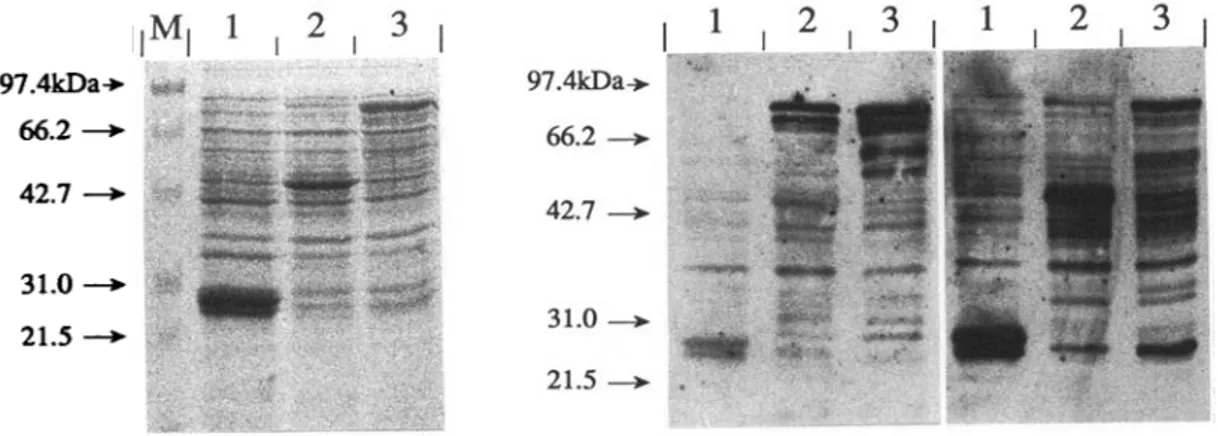
97.4kDa->- tt* 66.2—>- * # 42.7 —i 31.0 —i 21.5 —i 97.4kDa->. f; 66.2 42.7 31.0 21.5
Fig. 4. Expression of Echinococcus multilocularis and is. granulosus antigens as fusion proteins with Schistosoma
japonicum glutathione S-transferase (GST). Total bacterial extracts were boiled with sample buffer and subjected to
SDS-PAGE on 15% gels. Representative clones expressing E. multilocularis (Lane 2) and E. granulosus-derived antigens (Lane 3) and GST carrier protein (Lane 1) are shown. Molecular weight standards (Lane M). Coomassie blue staining (A), and immunoblotting using affinity-purified a-II/3-10 (B) and a-GST specific rabbit hyperimmune sera (C) was performed.
Comparison of the nucleotide sequences derived from the two Echinococcus species revealed 1 nucleo-tide exchange in the 3'-non-coding region and a total of 25 differences with respect to the coding region leading to 98-45 % homology at the nucleotide level. The nucleotide exchanges result in 8 differences in the amino acid sequences leading to 98-57% hom-ology at the amino acid level.
Expression of GST-fusion proteins
E. multilocularis and E. granulosus full length cDNA sequences represented by two clones termed pBlue-M31 and pBlue-Gll, respectively, were subcloned into the expression vector pGEX B using the Hind III recognition sequence of the polylinker of pBluescript KS+ which was ligated into the re-spective site of pGEX B, and following treatment with E. coli DNA polymerase I (Klenow fragment) the Nde I restriction site included in the sequence of amplification primer RBI, which was ligated to the blunt ended BamH I restriction site of pGEX B, respectively (Fig. 2A and C), resulting in an in-frame fusion to the Schistosoma japonicum glutathione S-transferase gene.
Several clones containing the desired sequences were identified, and their nucleotide sequence at the region of fusion to G S T was determined for confirmation. Expression of GST-fusion proteins was monitored by SDS-PAGE and immunoblotting using an anti-II/3-10 specific rabbit hyperimmune serum (Felleisen & Gottstein, 1993). Bacteria transformed with the recombinant plasmids produced fusion proteins of approximately 89 kDa as calculated from the deduced amino acid sequences (Fig. 4). While in the case of the E. granulosus-derived clones the fusion protein of 89 kDa represented an abundant synthesis product, only
minor amounts were seen in the E. multilocularis-derived clones. Here, the most abundant protein found had an apparent molecular weight of approximately 43 kDa, representing a product of degradation at the 3'-end of the fusion protein: while affinity-purified antibodies directed against the GST carrier protein recognized this protein strongly (Fig. 4C), almost no reaction was seen with affinity-purified antibodies directed against II/3-10 (Fig. 4B). Identical patterns were observed with different clones of identical genomic origin (data not shown). Two clones termed pGEX-M31 and pGEX-Gll were selected for further characterization.
Affinity purification of GST-fusion proteins
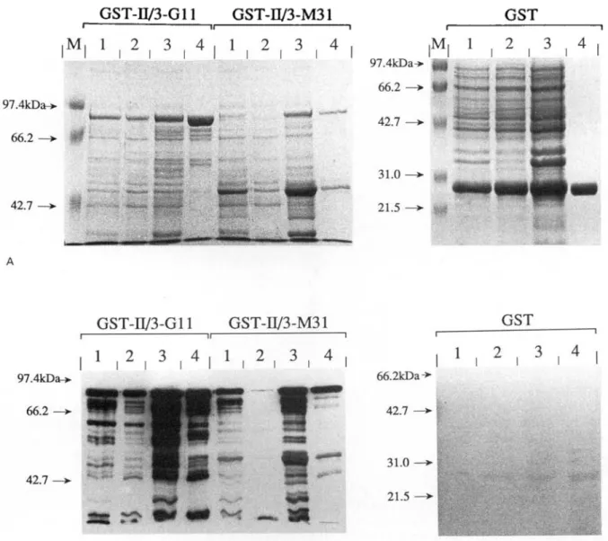
In order to produce the recombinant antigens in amounts and in purity sufficient for ELISA studies, large-scale cultures (500 ml) of bacteria transformed with the parental and the recombinant plasmids were set up. While large amounts were obtained from cultures expressing the GST carrier protein and the E. granulosus-derived fusion protein GST-II/3-G l l , expression of the E. multilocularis-derived fusion protein GST-II/3-M31 was unsatisfactory (Fig. 5). Therefore, the conditions of induction had to be optimized for this particular antigen. Best results were obtained using short induction times (2 h) and a large number of small-scale cultures (4 ml) instead of one large-scale culture (data not shown).
Affinity purification of the antigens is shown in Fig. 5. Reasonable amounts of the GST-II/3-G11 fusion protein and the GST carrier protein were contained in the soluble fraction and could be purified easily by adsorption to glutathione Sepharose®-4B beads. On the other hand, most of the GST-II/3-M31 fusion protein was found in the
R. Felleisen and B. Gottstein
GST-II/3-G11 GST-D/3-M31
GST
2 . 3 . 4M, 1 . 2 . 3 . 4 , 1 . 2 . 3 . 4
42.7GST-II/3-G11 GST-W3-M31
GST
1 . 2 . 3 . 4 , 1 . 2 . 3 . 4
2 , 3 , 4 42.7 66.2kDa-»-42.7 —>- | 31.0 — 2 1 . 5 -BFig. 5. Affinity purification of recombinant antigens. Total bacterial extracts (Lane 1), soluble fractions (Lane 2), insoluble fractions (Lane 3) and proteins bound to glutathione Sepharose beads (Lane 4) were analysed. Molecular weight standard (Lane M). Coomassie blue staining (A), and immunoblotting using affinity purified a-II/3-10 specific rabbit hyperimmune serum (B) was performed. Samples containing antigens GST-II/3-G11 and GST-II/3-M31 were separated on 8% SDS-PAGE gels, samples containing GST carrier protein on 12% gels.
insoluble fraction. Therefore, only limited amounts of this antigen could be purified. T h e antigens were eluted from the beads by competition with free reduced glutathione and, following dialysis, adjusted to a concentration of approximately 1 /tg/ml. The 43 kDa degradation product of antigen G S T I I / 3 -M31 proved to bind more tightly to the purification matrix compared to the full-length product resulting in an enrichment of the latter during elution (data not shown).
Enzyme-linked immunosorbent assay (ELISA)
Affinity-purified recombinant antigens GST-II/3-G l l and GST-II/3-GST-II/3-M31 and the GST-II/3-GST carrier (control) protein were used to sensitize ELISA
plates. Antigens derived from two independent purification procedures were used in each case and were tested with sera from patients with proven alveolar and cystic echinococcosis, respectively, and with sera from healthy blood donors.
No significant differences between homologous antigens derived from two independent purification batches were observed (data not shown), therefore, the mean values of the two tests were used for further considerations. Absorbance values from minor cross-reactions with the purified G S T carrier protein were corrected by subtraction from the value obtained with the recombinant full-length 11/3-antigen.
The reactivities of the E. multilocularis and E.
granulosus patient sera with the two different antigens
correlation coefficient for the respective absorbance values. A value of r = 0-982 was obtained (Fig. 6).
DISCUSSION
Immunodiagnosis of alveolar echinococcosis represents one of the important tools for the detection of infection and disease. The immuno-diagnostic E. multilocularis recombinant antigen 11/3-10 allowed detection of anti-£\ multilocularis antibodies in human patients with high diagnostic sensitivity and specificity (Gottstein et al. 1993). Although homologous antigens are also synthesized by E. granulosus (Felleisen & Gottstein, 1993), very few sera from patients (6%) with cystic echino-coccosis had serum antibodies against the re-combinant II/3-10 antigen (Gottstein et al. 1993).
Therefore, the purpose of the present study was to isolate and to analyse the complete antigen II/3-gene from E. multilocularis and from E. granulosus, and to express the respective sequences in E. coli as recombinant antigens for a comparative immuno-logical characterization and a potential elucidation of the phenomenon of humoral responsiveness and non-responsiveness to the antigen of patients suffering from alveolar or cystic echinococcosis, respectively.
Isolation of full-length cDNA clones was ac-complished by the polymerase chain reaction (PCR). Besides the major amplification products representing full-length fragments, minor smaller amplification products were observed. The latter were probably due to unspecific priming during amplification, because no hybridization was observed in Southern blot analyses using radiolabelled I I / 3 -10 cDNA-fragment as a probe (data not shown), although the possibility cannot be ruled out that they are reflecting alternative splicing of the II/3-mRNA especially in E. granulosus.
Nucleotide sequence analyses of the cDNAs obtained from E. multilocularis and from E. granulosus revealed a considerable high degree of conservation between both species. Only 8 out of 559 amino acids were different, resulting in approxi-mately 98-6% sequence identity. This conservation may suggest a relatively important functional role of the antigen in both Echinococcus species. No differences concerning the nucleotide sequence of the E. multilocularis cDNA clones compared to the published sequences were observed.
The sequence identity of full length antigen II/3 gene (98-6%) was in the same range as those published for antigens Eml3/Egl3 (9829%; Frosch et al. 1993) supporting the concept of a close genetic relationship between both Echinococcus species.
Despite the high degree of conservation in their primary sequence the recombinant antigens ex-pressed as GST-fusion proteins in E. coli behaved rather differently. While the E. granulosus-derived
protein GST-11/3-Gil was expressed in high amounts and mostly in an undegraded soluble form, the synthesis of the E. multilocularis-derived fusion protein GST-II/3-M31 was only very limited and preferentially contained in the insoluble fraction in a degraded form.
Although Smith & Johnson (1988) reported that a high percentage of GST-fusion constructs were at least partly soluble, insolubility of foreign proteins expressed in E. coli is very common (Marston, 1986). The factors influencing solubility and stability of GST-fusion proteins are not well characterized, but the charge and size of the respective protein seem to play an important role (Smith & Johnson, 1988). Calculation of the isoelectric point from the deduced amino acid sequences revealed values of 7-48 and 7-98 for the E. multilocularis and E. granulosus antigens, respectively. But whether this difference could account for the differences in behaviour of the two proteins was not clear. Nevertheless, both antigens were successfully affinity purified from bacterial extracts and used for comparative immuno-logical characterization in ELISA.
Sequence analyses revealed that the differences in the primary sequence of antigens 11/3 between both Echinococcus species were only very limited. But the regions of divergence could potentially represent the most important antigenic regions of the respective proteins. If this would be true, sera from patients with cystic echinococcosis showing no or only low reactivity with antigen II/3 from E. multilocularis should recognize the respective protein from E. granulosus considerably more strongly. But no significant differences in the reactivity of sera from patients either suffering from alveolar or from cystic echinococcosis were observed with the antigens derived from both Echinococcus species. A com-parative investigation with the purified GST-fusion proteins in ELISA demonstrated statistically very good correlations (r = 0-982). Differences in the mean A406 nm values of the sera with the two antigens (0-340±0-294 and 0-209 ±0-201 for E. granulosus and E. multilocularis, antigens, respect-ively) were due to slight variations in technical parameters (e.g. antigen concentration) resulting in a slope of the regression line in Fig. 6 of 0-668.
Sera of healthy blood donors were used to calculate a threshold value for determining positive reactions for both antigens (mean+ 2 S.D.). In this way, only one serum of each patient group was considered borderline negative with the heterologous antigen and positive with the respective homologous antigen (sera pointed by arrow heads in Fig. 6). These findings strongly suggested that the primary se-quence of the antigens was not the crucial cause for the different reactivity of sera from patients with respective diseases.
Nevertheless, the possibility could not be ruled out that conformational epitopes contained in the
231
0.0
0.0 0.2 0.6 0.8 1.0
A405GST-II/3-G11 Fig. 6. Direct comparative analysis by ELISA of purified recombinant antigen GST-II/3-M31 of
Echinococcus multilocularis versus antigen
GST-II/3-G l l of E. granulosus. The investigation was performed using defined sera of E. multilocularis patients (w = 28) and of E. granulosus patients (n = 30). The reactivity of sera of healthy blood donors (n = 30) was used to determine threshold values for each antigen (tv = mean+ 2 S.D.). 0 , E. multilocularis patient sera; • ,
E. granulosus patient sera.
variable regions played an important role for the immunological reactivity of the antigens. In com-puter analyses of the two proteins, no significant differences with respect to hydrophilicity, flexibility and surface probability profiles were observed. Furthermore, the calculated secondary structure of both proteins proved to be very similar (data not shown). Therefore, it was reasonable to assume that the two recombinant fusion proteins had a very similar secondary structure and that as well linear and conformational epitopes were identical or at least very similar.
Antigen 11/3 obviously is localized in the un-differentiated germinal layer of metacestode tissue (Felleisen & Gottstein, 1993). Alveolar echino-coccosis is characterized by the infiltrative tumour-like growth of the parasite with parasite cell protrusions being probably in direct contact with the host tissue and therefore accessible for the host immune system (Mehlhorn, Eckert & Thompson, 1983). During cystic echinococcosis, in contrast, well-delimited cysts are formed which are en-capsulated by host connective tissue surrounding a thick laminated layer, and thus seem to be somehow more inert to recognition by the host immune system and to subsequently induced effector mechanisms. Therefore, antigen 11/3 could be less accessible in cystic echinococcosis, being hypothetically presented to the immune system only in the case of rupture or
leakage of cysts. Ongoing studies are focusing on the potential of antigen I I / 3 to detect E. granulosus cyst damage by serological means.
Other reasons which may help to explain the divergent immunological phenomenon described above include post-translational modifications or processing of the antigen as well as species-specific immune regulatory or modulating mechanisms. These points as well as the actual function of antigen I I / 3 in vivo deserve further clarification.
We thank Ms V. Zimmermann for her expert technical assistance, Dr A. Busato for his support with respect to the statistical evaluation, Dr P. Jacquier for the supply of human sera and Dr E. Lanz for providing bovine E.
granulosus hydatid cysts. This work was supported by a
grant obtained from the Swiss National Science Foun-dation (No. 31-29651.90).
R E F E R E N C E S
AMMANN, R. W., HIRSBRUNNER, R., COTTING, J., STEIGER, u., JACQUIER, p. & ECKERT, j . (1990). Recurrence rate after discontinuation of long-term mebendazole therapy in alveolar echinococcosis (preliminary results). American Journal of Tropical Medicine and
Hygiene 43, 506-15.
DEVEREUX, J. R., HAEBERLI, P. & SMITHIES, O. (1984). A comprehensive set of sequence analysis programs for the Vax. Nucleic Acids Research 12, 387-95. FELLEISEN, R. & GOTTSTEIN, B. (1993). EchinOCOCCUS
multilocularis: molecular and immunochemical
characterization of diagnostic antigen 11/3-10.
Parasitology 107, 335-42.
FROHMAN, M. A. (1990). Rapid amplification of cDNA ends (RACE): user friendly cDNA cloning.
Amplifications 5, 11—15.
FROSCH, P . M., FROSCH, M., PFISTER, T., SCHAAD, V. & BITTER-SUERMANN, D. (1991). Cloning and
characterization of an immunodominant major surface antigen of Echinococcus multilocularis. Molecular and
Biochemical Parasitology 48, 121-30.
FROSCH, P . M., GEIER, C., KAUP, F.-J., MULLER, A. & FROSCH, M. (1993). Molecular cloning of an
echinococcal microtrichal antigen immunoreactive in
Echinococcus multilocularis disease. Molecular and Biochemical Parasitology 58, 301-10.
GLISIN, V., CRKVENJAKOV, R. & BYUS, C. (1974). Ribonucleic acid isolated by cesium chloride centrifugation. Biochemistry 13, 2633.
GOTTSTEIN, B. (1992). Molecular and immunological diagnosis of echinococcosis. Clinical Microbiological
Reviews 5, 248-61.
GOTTSTEIN, B., DEPLAZES, P. & AUBERT, M. (1992).
Echinococcus multilocularis: immunological study on
the ' Ern2-positive' laminated layer during in vitro and
in vivo post-oncospheral and larval development. Parasitology Research 78, 291-7.
GOTTSTEIN, B., JACQUIER, P., BRESSON-HADNI, S. & ECKERT, j . (1993). Improved primary immunodiagnosis of alveolar echinococcosis {Echinococcus multilocularis infection of humans) by Em2plus-ELISA. Journal of
HEMMINGS, L. & MCMANUS, D. P. (1991). The diagnostic value and molecular characterisation of an
Echinococcus multilocularis antigen gene clone. Molecular and Biochemical Parasitology 44, 53-62.
HEUSSLER, V. T. & DOBBELAERE, D. A. E. (1994). C l o n i n g of a protease gene family of Fasciola hepatica by
polymerase chain reaction. Molecular and Biochemical
Parasitology 64, 11-23.
LAEMMLI, u. K. (1970). Cleavage of structural proteins during the assembly of the head of the bacteriophage T4. Nature, London 227, 680-5.
MARSTON, A. o. (1986). The purification of eukaryotic polypeptides synthesized in Escherichica coli. The
Biochemical Journal 240, 1-12.
MEHLHORN, H., ECKERT, J. & THOMPSON, R. C. A. (1983). Proliferation and metastases formation of larval
Echinococcus multilocularis. II. Ultrastructural
investigations. Zeitschrift fiir Parasitenkunde 69, 749-63.
MULLER, N . , GOTTSTEIN, B., VOGEL, M., FLURY, K. &
SEEBECK, T. (1989). Application of a recombinant
Echinococcus multilocularis antigen in an enzyme-linked
immunosorbent assay for immunodiagnosis of human alveolar echinococcosis. Molecular and Biochemical
Parasitology 36, 151-60.
OLMSTED, j . B. (1981). Affinity purification of antibodies from diazotized paper blots of heterogeneous protein samples. Journal of Biological Chemistry 256, 11955-7. SAMBROOK, J., FRITSCH, E. F. & MANIATIS, T. (1989).
Molecular Cloning. A Laboratory Manual. Cold Spring
Harbor, NY: Cold Spring Harbor Laboratory Press. SMITH, D. B. & JOHNSON, K. s. (1988). Single-step
purification of polypeptides expressed in Escherichia
coli as fusions with glutathione S-transferase. Gene 6V,
31^0.
TOWBIN, H., STAEHELIN, T. & GORDON, J. (1979). Electrophoretic transfer of proteins from
polyacrylamide gels to nitrocellulose sheets: procedure and some applications. Proceedings of the National
Academy of Sciences, USA 76, 4350-4.
ULLRICH, A., SHINE, J., CHIRGWIN, J., PICTET, R., TISCHER, E., RUTTER, w. j . & GOODMAN, H. M. (1977). Rat insulin genes: construction of plasmids containing the coding sequences. Science 196, 1313.
VOGEL, M., GOTTSTEIN, B . , MULLER, N . & SEEBECK, T.
(1988). Production of a recombinant antigen of
Echinococcus multilocularis with high
immunodiagnostic sensitivity and specificity.