Treatment challenges associated with bone echinococcosis
Sylvain Steinmetz
1, Guillaume Racloz
1, Richard Stern
1, Dennis Dominguez
1, Mohamed Al-Mayahi
1, Manuel Schibler
2,
Daniel Lew
2, Pierre Hoffmeyer
1and Ilker Uc¸kay
1,2*
1
Orthopaedic Surgery Service, Geneva University Hospitals and Faculty of Medicine, University of Geneva, Geneva, Switzerland;2Service of Infectious Diseases, Geneva University Hospitals and Faculty of Medicine, University of Geneva, Geneva, Switzerland
*Corresponding author. Service of Infectious Diseases, Geneva University Hospitals, 4 Rue Gabrielle Perret-Gentil, 1211 Geneva 14, Switzerland. Tel:+41-22-372-2901; Fax: +41-22-372-9830; E-mail: ilker.uckay@hcuge.ch
Received 3 February 2013; returned 19 August 2013; revised 1 October 2013; accepted 6 October 2013 Objectives: In this literature review, we concentrate on epidemiology and therapy of osseous echinococcosis, with an emphasis on the recurrence risk.
Methods: Literature review 1930 –2012.
Results: We retrieved 200 publications based upon single case reports or case series, mostly from resource-poor settings. Among the 721 rural patients (22% females; median age 37 years), 60% of all reported cases were from the Mediterranean region and almost all patients were immune competent. Echinococcus granulosus was identified as the most frequent species. Most infections involved a single bone (602/721; 83%) and often the spine (321 cases; 45%). In eight cases (8/702; 1%), a secondary bacterial surgical site infection was reported. Surgical intervention was performed in 702 cases (97%), with single intervention in 687 episodes (95%). Complete excision of the lesion was possible in only 117 episodes (16%). Albendazole was by far the most frequently used agent in monotherapy with various dosages, while mebendazole in monotherapy was less frequent (32 cases). The median duration of antihelminthic therapy was 6 months (range 0.7– 144 months). There were 124 recurrences (17%) after a median delay of 2 years (range 0.4 –17 years). In multivariate analysis, the presence of visceral organ involvement increased the odds of recurrence by 5.4 (95% CI 3.1–9.4), whereas the number of surgical interventions, the duration of antihelminthic therapy or the use of hypertonic saline did not influence recurrence.
Conclusions: Bone echinococcosis is a rare parasitic disease. While treatment modalities vary considerably, combined surgical and medical approaches are the standard of care with a 17% risk of recurrence.
Keywords: epidemiology, recurrence, orthopaedic surgery, benzimidazoles
Introduction
Echinococcosis is a worldwide zoonosis infecting a large number of animals and humans.1–3 Human bone involvement is often asymptomatic2,3and estimated to occur at a frequency of 0.5%4 to 4%2of all cases of echinococcosis,5,6most likely by haema-togenous spread5,6 from the liver or a secondary soft organ infected from the liver. Endemic regions include rural areas (sheep farming1,7) of Mediterranean countries, Asia, Africa and Latin America,8 due to a variety of reasons such as migration, chronicity of the disease and absence of spontaneous healing. Nevertheless, a substantial number of physicians and surgeons in less-endemic rich countries may equally witness bone disease once in a lifetime.
Other than the consensus for combined aggressive surgical and medical therapy,9,10 there are no additional detailed
recommendations when confronted with bone hydatid disease. To the best of our knowledge, there are no systematic reviews available. Antihelminthic benzimidazole therapy for many years is frequent.2,10Some surgeons use bone grafts to fill bone cavities, while others insert polymethylacrylate.11Some physicians admin-ister antimicrobial agents for only 3 months,9,12while others prefer 10 years.5Overall, recurrences despite several surgical interven-tions are common.11 In this systematic literature review, we concentrate on the epidemiology and treatment modalities of osseous echinococcosis, with an emphasis on the risk of recurrence.
Methods
We performed a review of the literature from 1930–2012 in the Internet and PubMed database, using the MeSH terms ‘echinococcus’ or ‘hydatid’
#The Author 2013. Published by Oxford University Press on behalf of the British Society for Antimicrobial Chemotherapy. All rights reserved.
combined with either ‘bone’, ‘os*’, ‘Knochen*’ or ‘kemik’. Published original
scientific articles in English, French, German, Italian, Spanish, German or Turkish for human infections were included in an ExcelTMdatabase.
Refer-ences of retrieved articles were further hand-searched for additional papers, while English language abstracts of articles in other languages were excluded. In order to avoid doubling of patients, we included only the last publication from the same author group. Recurrence was defined as progression of the cyst diameter despite therapy and/or the appearance of new bone lesions.
Statistical analyses
The majority of articles were case reports or case series. No prospective cohort studies or randomized trials were found; therefore, a formal meta-analysis regarding outcomes and treatment modalities was not pos-sible. Nevertheless, we attempted to evaluate recurrence risks, even if this approach only considered a collection of retrospective case series. Group comparisons of categorical variables were performed using the Pearson
x2or Wilcoxon rank-sum test. A logistic regression analysis determined as-sociation with the outcome ‘recurrence of bone echinococcosis’. Independ-ent variables with a P value≤0.2 in univariate analysis were introduced stepwise in the multivariate analysis. We included 5 –10 outcome events per predictor variable.13All variables were checked for confounding and interaction. P values≤0.05 (two-tailed) were significant. STATA software (9.0, STATATM, USA) was used.
Results
Our representative case report
An elderly male Spanish immigrant presented at Geneva University Hospitals with a chronic intermittent discharge from the right hip. Twenty-six years previously in rural Spain he was diagnosed with bone echinococcosis (Echinococcus granulosus) of the femur and pelvis without visceral involvement. He was treated without surgery and received albendazole for 2 years. In Switzerland,
after a quiescence of 20 years, he developed chronic bacterial osteomyelitis of the femur and pelvis due to Staphylococcus epider-midis, with a productive sinus tract to the skin and an internal fistula into the abdomen. This bacterial superinfection was new, whereas the size and number of the cysts did not change over .20 years. There were no other signs of active echinococcal disease. Because of the extension of the lesions to weight-bearing bones, surgical resection was not possible. Suppressive therapy with oral ciprofloxacin/rifampicin (600 mg three times a day) stopped the sinus discharge for 2 years, at which time a new intern-al fistula into the abdomen developed. At present, the patient receives occasional suppressive antibiotic therapy in cases of fistula drainage.
Literature review and patients
According to our search criteria, we retrieved 200 publications with 721 different episodes (Table1). Turkey ranked highest with 158 dif-ferent reported cases (22%), followed by Spain with 78 cases (9%) and Germany with 67 reported cases (11%). WHO region 4 (Europe, Turkey and the former Soviet Union) accounted for almost 60% of all reported cases. Most publications concerned one single case (136/200; 68%) and 40 publications between two and nine cases. Only 24 papers (12%) had a substantial experience with ≥10 episodes. Within the publication window from 1930 to 2012, half of the manuscripts were published in the last 16 years (since 1996). The median age of the 721 different patients was 37 years (IQR 29 –45 years). Potential immune suppression was often not addressed, thus the presumption that almost all patients were immune-competent. In the majority of cases, the proven-ance of infection could not be established, besides the fact that the patients lived in rural areas. There were 73 farmers and 6 shep-herds in the reported cases.
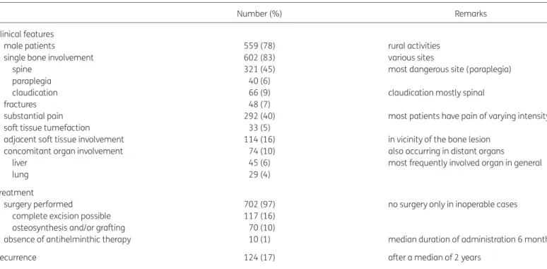
Table 1. Osseous echinococcosis (literature review 1930– 2012; 721 episodes)
Number (%) Remarks
Clinical features
male patients 559 (78) rural activities
single bone involvement 602 (83) various sites
spine 321 (45) most dangerous site (paraplegia)
paraplegia 40 (6)
claudication 66 (9) claudication mostly spinal
fractures 48 (7)
substantial pain 292 (40) most patients have pain of varying intensity soft tissue tumefaction 33 (5)
adjacent soft tissue involvement 114 (16) in vicinity of the bone lesion concomitant organ involvement 74 (10) also occurring in distant organs
liver 45 (6) most frequently involved organ in general
lung 29 (4)
Treatment
surgery performed 702 (97) no surgery only in inoperable cases complete excision possible 117 (16)
osteosynthesis and/or grafting 70 (10)
absence of antihelminthic therapy 10 (1) median duration of administration 6 months
Recurrence 124 (17) after a median of 2 years
Infections, bones and species
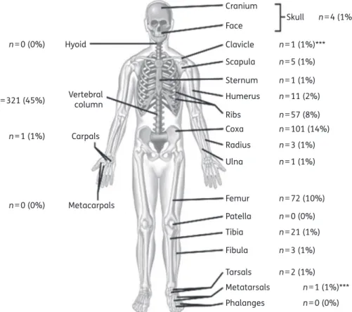
Many older publications did not identify the echinococcosis species. In more recent publications, E. granulosus was identified as the most frequent species with only 11 exceptions: 9 monoinfec-tions due to Echinococcus multilocularis; 1 mixed episode of E. granulosus and E. multilocularis; and 1 mixture of Echinococcus vogeli and E. granulosus. Most infections involved a single bone (602/721; 83%). In 107 episodes, two bones were involved; in 8 epi-sodes, three bones were involved; and in 4 epiepi-sodes, four different bones were involved (Figure1). In two episodes, echinococcosis was in contact with arthroplasty material. The median largest diameter of the bone lesions was 5.5 cm (range 1 –20 cm). Overall, 80% of all cysts were ,10 cm in diameter. In 114 episodes, the parasitic infection involved adjacent soft tissues, together with distant organs in 74 episodes, including: liver (n¼ 45), lung (n¼ 29), spleen (n¼ 2), pancreas (n¼ 2), brain (n ¼1), kidney (n¼ 1), ovaries (n¼ 1) and bladder (n¼ 1).
Surgical treatment
Surgical intervention was performed in almost all cases (702/721; 97%), with the exception of 19 episodes where a surgical approach was deemed not possible due to potential substantial harm to the patient. In 687 episodes (95%), there was only one surgical inter-vention; in seven cases, there were three interventions; in five cases, there were two interventions; and in three cases, there were four interventions. Complete excision of the lesion, as best
as could be reported, was possible only in 117 cases (16%), of which 5 were amputations. The remainder included incomplete debridement, drainage, laminectomy, arthrodesis and hemipel-vectomy.14 In 12 operations, the surgeon injected hypertonic saline at concentrations varying between 3%12 and 40%15or ethyl alcohol15(70%) into the cavity. Very rarely, an anaphylactic reaction was reported after cavity opening.16Despite the large dia-meters of the different lesions and multiple surgical interventions, a secondary bacterial surgical site infection occurred in only eight cases (8/702; 1%). The bone cavities became infected with bacter-ial pathogens in 36 cases (5%), of which 8 were classified as post-interventional surgical site infections and 28 as spontaneous superinfections due to various bacteria (staphylococci, Escherichia coli, Enterobacter spp., Pseudomonas aeruginosa, enterococci and streptococci).17The excised cavities were stabilized by osteosynth-esis in 661 episodes (92%), while fractures occurred in 48 cases. The time of fracture occurrence was, however, not noted. Hence, we ignore whether they occurred early or late in the infection process. In most cases, osteosynthesis was performed with the use of plates or by spondylodesis; in five cases of femoral infection, the surgeons performed a total joint arthroplasty.18Bone grafting was performed in 30 episodes.
Antimicrobial treatment
Since most publications were found in surgical journals, the main focus was the surgical approach. Thus, we often lack detailed
Cranium Skull n=4 (1%) n=1 (1%)*** n=5 (1%) n=1 (1%) n=11 (2%) n=57 (8%) n=101 (14%) n=3 (1%) n=1 (1%) n=72 (10%) n=0 (0%) n=21 (1%) n=3 (1%) n=2 (1%) n=1 (1%)*** n=0 (0%) n=0 (0%) n=1 (1%) n=321 (45%) n=0 (0%) Face Clavicle Scapula Sternum Humerus Ribs Coxa Radius Ulna Femur Metacarpals Carpals Vertebral column Hyoid Patella Tibia Fibula Tarsals Metatarsals Phalanges
Figure 1. Localizations of bone echinococcosis in our literature review 1930– 2012. ***One case each of metatarsal and clavicular involvement mentioned in the literature are not included in our review due to lack of sufficient information.
information about the modalities of antihelminthic therapy for 427 episodes (59%), especially regarding variations of daily doses throughout the therapy course. Albendazole (15 mg/kg/day) in monotherapy was the most frequently used regimen (in a minimum of 235 episodes).15 Nine cases used albendazole in reduced doses (10 mg/kg/day) from the start and in 11 cases, albendazole (15 mg/kg/day) was combined with praziquantel (40 mg/kg/day). Mebendazole was usually combined with alben-dazole5 and praziquantel,9 with mebendazole in monotherapy (10 mg/kg/day) used less frequently (32 cases).5The remaining publications indicated ‘benzimidazole therapy’ or ‘albendazole’ without mentioning the dose. In contrast, the duration of antihel-minthic therapy was almost always noted, with a median duration of therapy of 6 months (range 0.7 –144 months; IQR 3 –8 months). In some cases, the antihelminthic drugs were started 4 weeks prior to surgery.15The benzimidazoles were generally well tolerated, even over a course of 10 years of administration.5
Variables associated with recurrence
Patients were followed for a median of 5.5 years after their last sur-gical intervention (range 0.1 –24 years). Among the 721 episodes, 124 (17%) were reported as recurrent (or progressive infection) after a median delay of 2 years after treatment for the first episode (range 0.4 –17 years). All recurrences occurred at the same bone site and there were no new bone lesions at another lo-cation. Besides clearly evident recurrences (or persistence of infec-tion), patients with bone echinococcosis reported other sequelae: persistent pain was very frequent, followed by fractures in 48 epi-sodes, persistent paraplegia in 14 epiepi-sodes, handicap in 10 cases, death due to progressive disease in 5 cases and pseudarthrosis in 1 case. When adjusting for case mix in multivariate analysis, no single demographic variable was associated with recurrence risk (Tables 2and3), with one exception. The presence of a (visceral) organ involvement increased the OR for recurrence of bone infec-tion by 5.4 (95% CI 3.1–9.4), whereas spine involvement was sig-nificantly associated only in the univariate analysis, but not in the multivariate analysis. In contrast, among therapeutic aspects, no
variable influenced recurrence risk more than another. The number of surgical interventions, the duration of concomitant anti-helminthic therapy or the injection of hypertonic saline failed to reveal superiority compared with other regimens (Table3).
Discussion
Bone echinococcosis is a rare parasitic infection and is difficult to treat. It is probably associated with very high costs, which is evident when we extrapolate the massive investment according to the 200 publications reviewed. It can involve any bone,12but has a predilection for the spine, femur and pelvis. Multiosseous infestation and concomitant soft tissue or organ involvement are frequent, particularly the combination of proximal femur involve-ment with the pelvic girdle.2
Geographically, the disease is endemic almost worldwide in rural areas, especially in sheep farming regions and/or where there is endemicity of stray dogs.8,18,19However, there are hot spots. The Mediterranean region,7,20–22 especially Northern Africa3,7or Turkey10,11,19,23–25together with a few Asian coun-tries12(especially Iran2,26and India14,18),yielded the most publica-tions, although the disease was also reported in more resource-rich areas.27The geographical distribution has probably not changed since 1930, with many authors finding similar distribution patterns although with fewer reported cases than in our present review.2
A combined surgical and medical approach seems to be the basic consensus,9but there are many flaws when it comes to the details. Different authors reveal various therapeutic approaches, especially concerning the duration of antihelminthic therapy com-bined with surgical excision. The majority of authors performed at least one surgical intervention together with 6 months of albenda-zole treatment at oral doses of 15 mg/kg/day.28A minority pre-ferred a ‘second surgical look’.6 Similar to surgery for chronic bacterial osteomyelitis,29,30the number and type of interventions did not influence the recurrence risk, even if the approach was similar to oncologic surgery with margins excised as wide as pos-sible.3Filling excised bony defects with polymethylacrylate beads did not prevent recurrence11 and not even a presumed ‘total excision’ protected from later recurrence. The proportion of surgical excision, which was interpreted as ‘total’ during the first surgery, was only 117/721 episodes (16%), of which 18/117 (15%) recurred. Total excision of echinococcal cysts is difficult, since many may have daughter cysts. In addition, the cysts can be substantial in size or complicated in shape, or even might infest several weight-bearing bones, as was demonstrated in our own case. Radical excision in the spine is difficult because of the absence of distinct anatomic planes and the existence of neural structures.10The parasite grows in the direction of least resistance, infiltrating and damaging the bone like a tumour, with a risk of fracture.7The rigid structure of the bone does not allow formation of adventitia.5 Our evaluation suggests that the duration of concomitant anti-helminthic therapy does not play a role. A duration of ,3 months yielded similar outcomes as compared with administration for .6 months. Likewise, starting a neoadjuvant regimen with albendazole15pre-operatively did not show a benefit in terms of recurrence. The literature is sparse when it comes to recommenda-tions for the choice or duration of antimicrobial chemotherapy for bone echinococcosis. International guidelines exist for visceral organs such as the liver,31 but not for bone. Albendazole and
Table 2. Clinical variables associated with recurrence of bone echinococcosis Recurrence, n¼124 Pa No recurrence, n¼597 Female gender 39 NS 123 Pelvic involvement 13 NS 75 Spine involvement 75 0.019 246 Involvement of .2 bones 9 NS 23 Middle East region 42 0.001 101 Soft tissue involvement 20 NS 94 Organbinvolvement 35 0.001 39
‘Complete’ surgical excision 18 NS 99 NS, not significant.
aPearsonx2test. Only significant values (P,0.05) are displayed. b
Liver, lung, kidney or pancreas involvement concomitant with bone infection.
mebendazole might have the same clinical activity for bone, as suggested by comparative studies between these two agents in alveolar echinococcosis.32However, because of its higher level in blood plasma, albendazole is regarded by many specialists as the first choice for bone treatment.8For visceral organs, a cyclic treat-ment of benzimidazoles in several 1 month courses separated by 14 day intervals is usual,31but its efficacy is unknown for bone infections. To overcome these uncertainties, some authors suggest depending upon the clinical and serological evolution as a guide to the duration of antihelminthic therapy,28although the dormant state of the remission phase can persist for a long time and echinococcal serology can remain falsely elevated27 after treatment.
Despite massive investments of time, resources and surgical interventions, the long-term local recurrence risk of bone echino-coccosis was 17%.2,10,12,14,20,26Local symptomatic recurrences after 10 years have been reported26and we often are not con-cerned whether patients without clinical recurrence are parasito-logically cured or are in remission with a long-lasting control of their infection.20Interestingly, the remission rate and its duration are similar to the recurrence rate for classic bacterial osteomye-litis.29,30This finding is surprising since echinococci, compared with microscopic bacteria with high reproduction rates, are multi-cellular parasites that should be removed when performing surgery. In our formal evaluation, the only variable associated with local recurrence was distant organ involvement. In the presence of concomitant bone and visceral organ infestation, the
incidence was five times greater for local bone recurrence or progression of disease in bone. We were unable to detect any difference among the visceral organs and could not elucidate if recurrences were truly local or reinfections from these organs. Finally, unlike chronic bacterial osteomyelitis in young adults, echi-nococcal infections appear to be more frequently associated with fractures or with the need for osteosynthesis after excision. In our review, 36 of 48 fractures were treated by osteosynthesis and in 30 cases received bone grafts. Of note, the presence of these foreign materials did not seem to increase long-term recurrence, the only exception being arthroplasties performed for an unidentified echinococcal lesion.21,22
Our research has several limitations. (i) Successful cases may have been reported more frequently than therapeutic failures. On the other hand, it is likely that difficult-to-treat cases or cases with particular bone involvement were more often published, whereas uncomplicated complete surgical resections are not mentioned. (ii) Physicians and surgeons in resource-rich countries who rarely see these cases might find them more interesting than surgeons living in endemic areas. Since there are no national registries of bone echinococcosis, we ignore the burden of world-wide disease beneath the ‘tip of the iceberg’. (iii) The literature search was based on the Internet and relied on (Western) European languages. Even if most publications are written in these languages, there might be scientific work in local languages invis-ible on the Internet. (iv) Recurrence for bone hydatid disease not treated by excision may be difficult to interpret. We used the
Table 3. Logistic regression analysis with outcome ‘recurrence of bone echinococcosis’
Univariate analysis OR, 95% CI Multivariate analysis OR, 95% CI Variable female gender 1.4, 0.9–2.2 2.3, 0.7–7.3
age (continuous variable) 1.0, 0.9–1.1 NA
age 35– 50 years compared with ,35 years 1.0, 0.6–1.8 0.8, 0.3–2.3 age .50 years compared with ,35 years 0.7, 0.4–1.1 0.8, 0.3–1.9
pelvic involvement 0.6, 0.3–1.1 0.7, 0.3–1.4
spine involvement 1.7, 1.1 –2.6 1.3, 0.8–2.3
cyst diameter (continuous variable) 1.0, 0.8–1.2 NA
more than one bone involved 1.6, 0.7–3.6 NA
soft tissue involvement 1.0, 0.6–1.7 1.1, 0.5–2.8
visceral organ involvement 5.6, 3.1 –9.4 5.4, 3.1– 9.4
species E. granulosus 1.1, 0.2–5.5 NA
serum CRP value (continuous variable) 0.9, 0.7–1.2 NA
serum eosinophilia .1 G/L 1.1, 0.9–1.3 NA
Treatment modalities
number of surgical interventions 1.0, 0.6–1.8 0.8, 0.3–1.9
presumed total excision 0.9, 0.5–1.5 1.1, 0.6–2.2
bone grafting 0.3, 0.1–1.4 0.4, 0.1–1.7
injection of hypertonic saline 1.4, 0.6–3.0 1.8, 0.2–13.2 duration of antihelminthic therapy 1.0, 0.98–1.01 NA
3 –6 months compared with ,3 months 0.4, 0.1–1.3 0.3, 0.1–1.1 .6 months compared with ,3 months 0.6, 0.3–1.1 0.5, 0.2–1.3 NA, not applicable; CRP, C-reactive protein.
Statistically significant results are shown in bold.
progression of the cyst cavities or reappearance as defining criteria. It is theoretically possible that the parasite remains dormant in once-treated cysts without causing further harm. Whether this should be classified as persistent infection or remission is a philo-sophical question. (v) Our attempt to ‘analyse statistically’ 721 published cases may give only an estimate and by no means repre-sents a valid statistical meta-analysis in proper terms.
Acknowledgements
We thank all our colleagues of the Orthopaedic Surgery Service of Geneva University Hospitals.
Funding
This study was conducted as part of our routine work.
Transparency declarations
None to declare.
I. U. had full access to all of the data in the study and takes responsibility for the integrity of the data and the accuracy of the analysis.
References
1 Bouree P. Hydatidosis: dynamics of transmission. World J Surg 2001; 25: 4 –9.
2 Ebrahimi A, Assadi M, Saghari M et al. Whole body bone scintigraphy in osseous hydatosis: a case report. J Med Case Rep 2007; 1: 93.
3 Zlitni M, Ezzaouia K, Lebib H et al. Hydatid cyst of bone: diagnosis and treatment. World J Surg 2001; 25: 75– 82.
4 Katz AM, Pan CT. Echinococcus disease in the United States. Am J Med 1958; 25: 759–70.
5 Scheuring UJ, Seitz HM, Wellmann A et al. Long-term benzimidazole treatment of alveolar echinococcosis with hematogenic subcutaneous and bone dissemination. Med Microbiol Immunol 2003; 192: 193–5. 6 Apt WL, Fierro JL, Calderon C et al. Vertebral hydatid disease. Clinical experience with 27 cases. J Neurosurg 1976; 44: 72–6.
7 Papanikolaou A. Osseous hydatid disease. Trans R Soc Trop Med Hyg 2008; 102: 233– 8.
8 Song XH, Ding LW, Wen H. Bone hydatid disease. Postgrad Med J 2007; 83: 536–42.
9 Ayles HM, Corbett EL, Taylor I et al. A combined medical and surgical approach to hydatid disease: 12 years’ experience at the Hospital for Tropical Diseases, London. Ann R Coll Surg Engl 2002; 84: 100– 5.
10 O´´zdemir HM, Og˘un TC, Tas¸bas¸ B. A lasting solution is hard to achieve in primary hydatid disease of the spine: long-term results and an overview. Spine 2004; 29: 932–7.
11 Yıldız Y, Bayrakcı K, Altay M et al. The use of polymethylmethacrylate in the management of hydatid disease of bone. J Bone Joint Surg Br 2001; 83: 1005– 8.
12 Zia S, Enam A, Salahuddin I et al. Role of irrigation with hypertonic saline for a recurrent skull base hydatid cyst: case report and review of the literature. Ear Nose Throat J 2010; 89: 22 –6.
13 Vittinghoff E, McCulloch CE. Relaxing the rule of ten events per variable in logistic and Cox regression. Am J Epidemiol 2007; 165: 710–8.
14 Jain S, Chopra P. Cystic echinococcosis of the pelvic bone with recurrences: a case report. Korean J Parasitol 2011; 49: 277– 9.
15 Infanger M, Kossmehl P, Grimm D. Surgical and medical management of rare echinococcosis of the extremities: pre- and post-operative long-term chemotherapy. Scan J Infect Dis 2005; 37: 954– 7.
16 Heinze J, Junginger W, Muller G et al. Anaphylactic shock during excision of an intraosseous Echinococcus granulosus cyst. Anesthesist 1987; 36: 659–61.
17 Uc¸kay I, Harbarth S, Peter R et al. Preventing surgical site infections. Expert Rev Anti Infect Ther 2010; 8: 657– 70.
18 Natarajan MV, Kumar AK, Sivaseelam A et al. Using a custom mega prosthesis to treat hydatidosis of bone: a report of 3 cases. J Orthop Surg (Hong Kong) 2002; 10: 203– 5.
19 Altıntas¸ N. Past to present: echinococcosis in Turkey. Acta Trop 2003; 85: 105–12.
20 Turtas S, Viale ES, Pau A. Long-term results of surgery for hydatid disease of the spine. Surg Neurol 1980; 13: 468–70.
21 Notarnicola A, Panella A, Moretti L et al. Hip joint hydatidosis after prosthesis replacement. Int J Infect Dis 2010; 14: 287–90.
22 Voutsinas S, Sayakos J, Smyrnis P. Echinococcus infestation complicating total hip replacement. A case report. J Bone Joint Surg Am 1987; 69: 1456–8.
23 Sasani M, O´´zer AF. Spontaneous drainage of an asymptomatic recurrent hydatid cyst of the sacrum. Spine 2009; 34: 269– 71.
24 Kural C, Ug˘ras¸ AA, Sungur I et al. Hydatid bone disease of the femur. Orthopedics 2008; 31: 712.
25 Kotil K, Tatar Z, Bilge T. Spinal hydatidosis accompanied by a secondary infection. Case report. J Neurosurg Spine 2007; 6: 585–90.
26 Samadian M, Alavi E, Sharifi G et al. Extension of echinococcal spinal infestation extra- and intradurally after a decade of extinction. J Neurosurg Sci 2010; 54: 143–8.
27 Jensen B, Reuter S, Kratzer W et al. Long-term seropositivity against Echinococcus multilocularis in an epidemiological follow-up study in southwestern Germany (Romerstein). Infection 2001; 29: 310– 4. 28 Bonifacino R, Dogliani E, Craig PS. Albendazole treatment and serological follow-up in hydatid disease of bone. Int Orthop 1997; 21: 127–32.
29 Rod-Fleury T, Dunkel N, Assal M et al. Duration of post-surgical antibiotic therapy for adult chronic osteomyelitis: a single-centre experience. Int Orthop 2011; 35: 1725–31.
30 Uc¸kay I, Jugun K, Gamulin A et al. Chronic osteomyelitis. Curr Infect Dis Rep 2012; 14: 566–75.
31 Guidelines for treatment of cystic and alveolar echinococcosis in humans. Bull World Health Organ 1996; 74: 231–42.
32 Reuter S, Jensen B, Buttenschoen K et al. Benzimidazoles in the treatment of alveolar echinococcosis: a comparative study and review of the literature. J Antimicrob Chemother 2000; 46: 451– 6.