. . . .
. . . .
Clonal restriction and predominance of regulatory
T cells in coronary thrombi of patients with acute
coronary syndromes
Roland Klingenberg
1,2*
, Chad E. Brokopp
3, Audrey Grive`s
4, Anaı¨s Courtier
4,
Milosz Jaguszewski
1, Nicolas Pasqual
4, Eugenia Vlaskou Badra
1, Anika Lewandowski
1,
Oliver Gaemperli
1, Simon P. Hoerstrup
3, Willibald Maier
1, Ulf Landmesser
1,2,
Thomas F. Lu¨scher
1,2†, and Christian M. Matter
1,2†1
University Heart Center, Department of Cardiology, University Hospital Zurich, Ra¨mistrasse 100, CH-8091 Zurich, Switzerland;2
Cardiovascular Research, Zurich Center of Integrative
Human Physiology (ZIHP), Institute of Physiology, University of Zurich, Zurich, Switzerland;3
Regenerative Medicine Center, Department of Cardiothoracic Surgery, University Hospital
Zurich, Zurich, Switzerland; and4
ImmunID Technologies, Grenoble, France
Received 16 July 2013; revised 29 October 2013; accepted 21 November 2013; online publish-ahead-of-print 13 January 2014
This paper was guest edited by Prof. Dr Hiroaki Shimokawa, MD, PhD, Tohoku University Graduate School of Medicine, Japan, (E-mail: [email protected])
Aims Regulatory T cells (Treg) exert anti-inflammatory and atheroprotective effects in experimental atherosclerosis. Treg can
be induced against specific antigens using immunization strategies associated with clonal restriction. No data exist on Treg in combination with clonal restriction of T cells in patients with acute coronary syndromes (ACS).
Methods and results
Among T cell subsets characterized by flow cytometry, Treg (CD4+CD25+CD127low) were twice as frequent in
cor-onary thrombi compared with peripheral blood. Treg prevailed among T cell subsets identified in corcor-onary thrombi. To evaluate clonal restriction, genomic DNA was extracted from coronary thrombi and peripheral blood in order to evalu-ate T cell receptor (TCR) b chain diversity by means of Multi-N-plex PCR using a primer specific for all TCR b V gene segments and another primer specific for TCR b J gene segments. T cell receptor diversity was reduced in thrombi com-pared with peripheral blood (intra-individual comparisons in 16 patients) with 8 gene rearrangements in the TCR common in at least 6 out of 16 analysed coronary thrombi. Compared with age-matched healthy controls (n ¼ 16), TCR diversity was also reduced in peripheral blood of patients with ACS; these findings were independent of peripheral T cell numbers.
Conclusion We provide novel evidence for a perturbed T cell compartment characterized by clonal restriction in peripheral blood and coronary thrombi from patients with ACS. Our findings warrant further studies on Treg as novel therapeutic targets aimed at enhancing this anti-inflammatory component of adaptive immunity in human atherothrombosis.
-Keywords Acute coronary syndromes † Immunity † T cells
Introduction
Atherothrombosis subsequent to plaque rupture or erosion with prominent features of inflammation constitutes the underlying pathophysiology in patients with acute coronary syndromes (ACS).
However, its triggers remain poorly understood and not all
rupture-prone plaques culminate in atherothrombosis.1–3We have
previous-ly identified monocytes/macrophages as the most abundant inflam-matory cell type of innate immunity in coronary atherothrombosis,
co-expressing Toll-like receptor (TLR)-4.4Although less abundant
*Corresponding author. Tel:+41 44 255 2115, Fax: +41 44 255 8701, Email:[email protected]
†Shared contribution.
&The Author 2014. Published by Oxford University Press on behalf of the European Society of Cardiology.
This is an Open Access article distributed under the terms of the Creative Commons Attribution Non-Commercial License (http://creativecommons.org/licenses/by-nc/3.0/), which
permits non-commercial re-use, distribution, and reproduction in any medium, provided the original work is properly cited. For commercial re-use, please contactjournals.permissions@
oup.com
in number, T cells orchestrate the antigen-specific (adaptive) immune response in atherogenesis with prominent effects mediated by dis-tinct T cell subsets throughout the stages of atherosclerosis, including
ACS.2,3,5–8
Among T cell subsets, regulatory T cells (Treg) exert atheropro-tective effects and constitute an inherent anti-inflammatory
compo-nent of adaptive immunity.5–9Identifying a novel mechanistic link
between Treg immunity and lipid metabolism, we recently
demon-strated that selective depletion of Treg (Foxp3+) impacted on lipid
metabolism mediating hypercholesterolaemia and increased
athero-sclerotic lesions.10Furthermore, we and others showed that
athero-protective Treg can be induced in response to immunization with antigen specificity for the apolipoprotein B-100 peptide used in the
vaccine.11–13In humans, Treg were found in increased numbers in
lipid-rich, advanced plaques, whereas reduced numbers of circulating
Treg were found in patients with ACS.14–18From a clinical
perspec-tive, Treg may be an attractive therapeutic target in atherosclerosis due to their anti-inflammatory effects. However, a better under-standing of the role of Treg in ACS is necessary.
T cells orchestrate adaptive immunity to mediate an antigen-specific immune response. Each cell expresses several copies of a single antigen receptor with a unique antigen-binding site. The great variety of antigen specificities in the T cell receptor (TCR) repertoire is generated in the thymus by random recombination of separate inherited TCR a/b gene segments termed V(D)J which encode the variable parts of the heterodimeric receptor. T cell receptor diversity is confined to the complementarity determining regions (CDRs) as the binding site for a peptide presented on the major histocompatibility complex by an
antigen-presenting cell (i.e. dendritic cell).19Recent advances in
multi-plex assay technology made it possible to measure usage frequencies of gene segments V(D)J and enable detection and tracking of specific TCR sequences expressed by clonally restricted T cells. Compared with spectratyping which employs only a limited set of V and J primers, this method enables a more comprehensive analysis that avoids bias
due to transcriptional regulation.20,21
Evidence for an antigen-specific local immune response carried by T cells in unstable atherosclerotic plaque mandates clonal restriction of T cells when compared with peripheral blood. Initial reports on T cells isolated from human atherosclerotic plaques showed a high
TCR diversity indicating a polyclonal origin of T cells.22,23In
circulat-ing CD4+CD28nullT cells from patients with unstable angina
com-pared with patients with stable coronary artery disease, clonal
restriction of T cells was identified using spectratyping.24 Clonal
restriction of T cells was demonstrated using spectratyping of ather-ectomy specimens from coronary plaque compared with peripheral blood mononuclear cells (PBMCs) from patients with ACS and only
to a lesser extent in patients with stable coronary artery disease.25
Similarly, clonal restriction was demonstrated when comparing carotid plaques with PBMC from asymptomatic and symptomatic
patients with .90% carotid artery stenosis.26
However, so far T cell diversity in thrombi of ACS patients has not been investigated. The aims of this study, therefore, were (i) to compare Treg counts in coronary thrombi with PBMC from patients with ACS, (ii) to compare TCR diversity in coronary thrombi with PBMC from patients with ACS, (iii) to compare TCR diversity in PBMC from age-matched healthy subjects with patients with ACS.
Methods
Characteristics of patients and healthy
subjects
Sixteen patients referred to the University Hospital Zurich, Switzerland, for coronary angiography with the diagnosis of ACS between 08/2010 and 11/2011 were enrolled as part of a larger cohort study (SPUM-ACS, clinical trial number NCT01000701). All patients aged 18 years and older presenting within 72 h after pain onset with the main diagnosis of ST-elevation myocardial infarction (STEMI) or non-ST elevation myo-cardial infarction (NSTEMI) were included in the study. Patients had symp-toms compatible with angina pectoris (chest pain, dyspnoea) and at least one of the following inclusion criteria: (i) persistent ST-segment elevation or de-pression, T inversion or dynamic ECG changes, new LBBB; (ii) evidence of positive Troponin based on high sensitive TnT measurement (≥14 ng/L) with rise and/or fall in serial TnT levels. Exclusion criteria comprised docu-mented active autoimmune disease or neoplasm; stent thrombosis; inability to comprehend study, less than 1 year of life-expectancy (for non-cardiac reasons). The Gensini score27was calculated by two interventional cardi-ology fellows (inter-observer variability ¼ 17.4) for each of the 16 patients using the formula severity score× segment location multiplying factor × collateral adjustment factor to determine the extent and severity of coron-ary artery disease in the epicardial coroncoron-ary arteries. In seven patients, the Gensini score differed by more than 10 points based on a different interpret-ation of collateral flow/total occlusion and an expert opinion was consulted from a third experienced senior interventional cardiologist.
Samples from 16 healthy subjects were obtained from a larger cohort study (SuSa Study, AFSSAPS clinical trial number 2010- A00428-31, ImmunID) that were recruited between between 07/2010 and 12/2011 at Optimed Clinical Research, Lyon, France. Individuals were included fol-lowing a detailed medical history, physical exam with vital signs, and blood draw. Exclusion criteria comprised autoimmune disease or neoplasm, inability to comprehend study, treatment with corticosteroids or other immunomodulatory therapy, or vaccination in the three preceding months. All individuals were older than 18 years of age with birth control in place for at least 2 months prior to inclusion in women of childbearing age in the absence of pregnancy or breast feeding. Informed consent was obtained from all individuals with the approval by the Kantonale Ethik-Kommission Zurich, Switzerland (EK-1688), for patients with ACS and the Comite´ de Protection des Personnes CPP Sud-Est III, France (Numero EudraCT : OL039/Susa), for healthy subjects.
Analyses of blood and coronary thrombi
Laboratory parameters were measured using standard protocols. Indi-vidual estimated glomerular filtration rate was calculated using the CKD-EPI formula28based on serum creatinine, gender, and ethnicity. Based on the published trials,29,30coronary thrombi were aspirated using an Export catheter (Medtronic, Tolochenaz, Switzerland) at the site of coronary occlusion from patients with ACS undergoing primary percutaneous coronary intervention (PCI) and were immediately immersed in phosphate-buffered saline (PBS)-containing vials. Peripheral blood was sampled from the inguinal arterial sheath from the same patient and stored in citrate vials. Thrombi and the corresponding periph-eral blood were treated with 50 mL tissue plasminogen activator (0.001% Actilysew; Boehringer Ingelheim Pharma GmbH & Co., Ingelheim, Germany) to remove fibrin in 1 mL 1% Accutasew
(PAA Laboratories, Pasching, Austria), and rotated for 2 h at 378C. Thrombi and blood were then gently dissociated through a 40 mm pore-size cell strainer (BD FalconTM) with a rubber syringe plunger and rinsing with ice-cold
PBS. Half of the cell suspensions were separated for Ficoll-paque centri-fugation and subsequent ImmunTraCkeR bw
Grenoble, France), whereas remaining cells were analysed by flow cyto-metry. In healthy subjects, PBMCs were separated from whole blood (5 mL EDTA) with a density gradient tube (Uni-Sep, Novamed, Jerusa-lem, Israel). Peripheral blood mononuclear cells were prepared and sta-bilized in Easy’IDw
stabilization solution provided by ImmunID, Grenoble following a standard operating procedure.
Analysis of T cell receptor diversity
Human TCR diversity was measured using ImmunTraCkeRbw
tests (ImmunID Technologies, Grenoble, France) and performed as described.31 Peripheral blood mononuclear cells and thrombus samples were frozen and analysed by ImmunID laboratories. Genomic DNA was extracted using standard techniques and Multi-N-plex PCR was performed using an upstream primer specific for all functional members of a given T cell receptor b V segment and a downstream primer specific for a given T cell receptor b J segment (international ImMunoGeneTics information system,www.imgt.org) encoding for the CDR 3 in the human TCR b chain. This assay allows the simultaneous exhaustive detection of V(D)J rearrangements in the same reaction expressed as percentage based on the detected V(D)J combinatorial diversity divided by the expected maximal diversity (276 gene segments) which corresponds to the theor-etical combinatorial diversity obtained with all rearrangements of all func-tional V(D)J genes within the genome. Each Vx – J1, J2, J3, J4, Jn product was separated as a function of its size. Constel’IDw
software (ImmunID Technologies) was used for further analytical studies, including gener-ation of three-dimensional repertoire illustrgener-ation. Numergener-ation and diver-sity of T cells is presented together through NDLw
scoring which enables to determine a correlation between lymphocyte counts and T cell diver-sity. A decreased percentage (i.e. a decreased TCR diversity) defines a state of divpenia (www.divpenia.com). Normal values for TCR b-chain combinatorial diversity were defined based on a cohort of 16 healthy volunteers extracted from a larger study (SuSa Study, AFSSAPS clinical trial number 2010- A00428-31, ImmunID; age range, 42.8 – 74.9 years; 6 females and 10 males).
Flow cytometry
For flow cytometry, thrombus and peripheral blood-derived cell suspen-sions obtained from patients with ACS were centrifuged at 200G or 10 min and resuspended in 1 mL of ice-cold FACS buffer (PBS, 1% foetal calf serum, 0.05% EDTA) with FcR block (Human TruStain FcX, Biolegend, San Diego, CA, USA). Cells were labelled for 1 h at 48C with monoclonal antibodies for extracellular staining of CD3, CD4, CD8, CD28, and a three-color reagent (CD4, CD25, CD127) for identi-fication of Treg (all from BD Biosciences, San Jose, CA, USA) and analysed on a FACScalibur (BD Biosciences). Subpopulation analyses were per-formed using SSC/FSC scatters, differentiating lymphocyte, monocytes, and granulocyte gates (quadrant analysis). Mouse IgG1 was used as isotype control and the proportion of positive cells per lymphocyte gate were determined.
Statistical analysis
For comparisons of continuous and categorical data, we used the non-parametric Mann – Whitney U-test and the Pearson Chi-square test, re-spectively. Intra-individual comparisons of distributions of combinatorial diversity and percentages of cell counts are the result of Wilcoxon signed rank test for paired data. Unless otherwise indicated, data are shown as mean + SEM or relative and absolute frequencies. Pearson’s correlation coefficient was determined for the Gensini score (mean) and TCR diver-sity in PBMC and thrombi, respectively. A two-sided P-value of ,0.05 was considered significant.
Results
Clinical characteristics of patients with
acute coronary syndromes and healthy
subjects
Pairs of coronary thrombi and peripheral blood were obtained from 16 patients with ACS using an aspiration catheter as part of the PCI for a native coronary culprit lesion. Among these, 12 patients were diag-nosed with STEMI, 4 patients with NSTEMI, 11 patients were symp-tomatic for ,24 h, 4 patients had symptoms for 24– 48 h and 1 patient for 48 –72 h. For the evaluation of TCR b chain diversity in per-ipheral blood, 16 patients with ACS were compared with 16 healthy age-matched subjects. The clinical, demographic, and laboratory char-acteristics of patients with ACS and matched healthy subjects are
sum-marized in Table1. Compared with healthy subjects, patients with ACS
. . . .
Table 1 Clinical, demographic, and laboratory
characteristics of patients and healthy subjects
ACS Healthy P-value
Age (years) 58.9 + 2.3 58.7 + 2.3 n.s. Male gender (%) 15 (94) 10 (63) n.s Caucasian ethnicity (%) 16 (100) 16 (100) n.s. Leucocyte count (103/mL) 11.6 + 1.5 5.8 + 0.3 P≤ 0.001 Lymphocyte count (103/mL) 3.2 + 1.2 1.6 + 0.08 n.s. Erythrocytes (106/mL) 4.3 + 0.2 4.5 + 0.1 n.s. Platelets (103/mL) 279 + 25 219 + 8 P , 0.05 Total cholesterol (mmol/L) 5.1 + 0.4 5.6 + 0.3 n.s. Baseline glucose (mmol/L) 9.0 + 1.7 5.2 + 0.08 P≤ 0.01 Serum creatinine (mmol/L) 79.9 + 6.3 72.1 + 2.7 n.s. eGFR (mL/min) 90.1 + 4.9 92.3 + 2.4 n.s. BMI (kg/m2) 28.4 + 2.0 24.6 + 1.1 n.s. Gensini Score 39.7 + 5.7 0 P≤ 0.001 Cardiovascular risk factors
Hypercholesterolaemia 8 (50) 0 P≤ 0.01 Hypertension (%) 4 (25) 0 P , 0.05 Diabetes (%) 2 (13) 0 n.s. Smoking (current, %) 7 (44) 0 P≤ 0.01 Previous medical history
Previous MI 3 (19) 0 n.s. Previous PCI 2 (13) 0 n.s. Medications Aspirin (%) 16 (100) 0 P≤ 0.001 Statins (%) 4 (25) 0 P , 0.05 ACE-I/ARB (%) 2 (13) 0 n.s. b-Blockers (%) 3 (19) 0 n.s. Nitrates (%) 15 (94) 0 P≤ 0.001 Calcium antagonists (%) 0 0 – Diuretics (%) 1 (6) 0 n.s.
Values are shown as mean + SEM or absolute and relative frequencies (in brackets with respect to n ¼ 16 patients), respectively.
N ¼ 12 – 16; n.s. non-significant. ACE-I/ARB, angiotensin converting enzyme-inhibitor/angiotensin receptor blocker; aspirin comprises oral and intravenous formulation; BMI, body mass index; eGFR, estimated glomerular filtration rate; MI, myocardial infarction; PCI, percutaneous coronary intervention.
had increased leucocyte and platelet counts, elevated glucose concen-tration in peripheral blood, presence of coronary artery disease, cardiovascular risk factors, and received medications.
Increased Treg counts in coronary thrombi
compared with peripheral blood in
patients with acute coronary syndromes
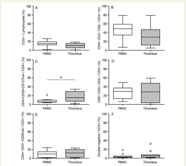
To evaluate T cell subset distribution, flow cytometry was performed for phenotypic characterization of coronary thrombi comparedwith PBMC from patients with ACS. Overall CD3+T cell content
was unaltered and no difference was detected for T helper cells
(CD4+CD3+CD82), but for a trend to numerical reduction vs.
peripheral blood (Figure 1A and B). In contrast, Treg (CD4+
CD25+CD127low) were significantly increased by 2.2-fold in
coron-ary thrombi (Figure1C). Cytotoxic T cells (CD8+CD3+CD42),
CD4+CD3+CD28nullT cells and double negative (CD42CD82
CD3+) T cells were found in similar numbers in coronary
thrombi and PBMC (Figure1D – F). Supplementary material online,
Figures S1 – S6 show representative graphs on how FACS analysis for individual cell subsets was performed. Supplementary material
online, Figure S7 shows a representative immunostaining of CD3+
T cells.
Figure 1 Treg as prominent T cell subset in coronary thrombi vs. peripheral blood from patients with ACS. Flow cytometry of coronary thrombus and PBMC from patients with ACS. Double-staining for T cell subtypes (CD3 with CD8 or CD4, respectively). (A) T cells (CD3+). (B) T helper cells (CD4+CD3+CD82). (C ) Regulatory T cells (CD4+CD25+CD127low). (D) Cytotoxic T cells (CD8+CD3+CD42). (E) CD4+CD3+CD28null T cells. (F ) Double-negative T cells (CD4nullCD8nullCD3+). The bottom and top of the box represent the first and third quartiles (Q1, Q3), and the band inside the box is the median. The whiskers extend to the most extreme data point which is no more than 1.5 times the interquartile range (IQR ¼ Q32Q1) from the box, with individual outliers shown beyond the whisker; n ¼ 9 – 12; *P ¼ 0.0098.
Decreased T cell receptor diversity in
coronary thrombi compared with
peripheral blood in patients with acute
coronary syndromes
Pairwise intra-individual comparisons of coronary thrombi demon-strated a markedly reduced TCR diversity in coronary thrombi vs.
PBMC (reduction by 0.28-fold), shown in Figure 2A – C. Among
V(D)J gene segments identified in coronary thrombi from 16 patients with ACS, 8 rearrangements were found in common in at least 6
thrombi from individual patients (Table2). These gene
rearrange-ments are Vbeta18-J2.3, which is the most frequent (8/16); Vbeta05-J2.7 and Vbeta25-J2.6, both found in common in 7/16 samples; Vbeta27-J2.7, Vbeta24-J2.3, Vbeta24-J2.5, Vbeta15-J2.4, and Vbeta19-J2.5 were found in common in 6/16 samples (IMGT no-menclature). In turn, in peripheral blood from patients with ACS, Vbeta05-J2.7 was the most frequently detected gene rearrangement (13/16). Interestingly, Vbeta05-J2.7 was less common in coronary
thrombi compared with PBMC (Table2). T cell receptor diversity
was only weakly correlated with the extent and severity of epicardial coronary artery disease as assessed by the Gensini score (Supple-mentary material online, Figure S8). Pearson’s correlation coefficient for the comparison of the Gensini score (mean) with TCR diversity in PBMC was 20.418 and 20.058 for the comparison of the score with TCR diversity in coronary thrombus, respectively. These associations were not significant.
Reduced T cell receptor diversity in
circulating T cells of patients with acute
coronary syndromes compared with
healthy subjects
T cell receptor diversity was measured using genomic DNA isolated from PBMCs from 16 healthy subjects and PBMCs from 16 patients with ACS. Compared with matched healthy subjects, patients with ACS had a profoundly reduced TCR diversity in peripheral blood
(re-duction by 0.36-fold), shown in Figure3A – C. Importantly, reduced
TCR diversity did not correlate with overall lymphocyte cell
Figure 2 Reduced T cell receptor diversity in coronary thrombus vs. peripheral blood from patients with ACS. Intra-individual comparisons of TCR diversity expressed as percentage of 276 possible V(D)J gene segment rearrangements in the human TCR b chain (hTRB) in coronary thrombus vs. PBMC (A). Bars indicate means; **P ¼ 0.001. Representative 3-D plots of human TCR b chain diversity in an individual patient with ACS derived from PBMC (B) and coronary thrombus (C ).
number in peripheral blood from patients with ACS and healthy
sub-jects (Figure3D).
Discussion
The present study confers the following key findings: first, we identi-fied Treg as the major T cell subset in aspirated coronary thrombus adjacent to the culprit lesion in patients with ACS. Second, we found a restricted TCR diversity within coronary thrombi compared with peripheral blood of patients with ACS. Third, we show a mark-edly reduced TCR diversity also in peripheral blood of patients with ACS compared with healthy age-matched subjects.
We previously showed an increased immune response at the site of coronary occlusion in patients with ACS compared with peripheral blood, demonstrating elevated levels of serum amyloid A, interleukin
(IL)-6,32myeloid-related protein 8/14,33and expression of TLR-4 on
monocytes as part of innate immunity.4The current study expanded
these previous findings to T cells. Analysis of adaptive immunity carried by T cell subsets in patients with ACS was performed by com-paring coronary thrombi with peripheral blood intra-individually and peripheral blood from healthy subjects.
Treg were reported in increased numbers in lipid-rich, advanced plaques, whereas reduced numbers of circulating Treg were found
in patients with an ACS.14–18This decrease in the circulating Treg
pool is currently unclear as it may be due to either a global Treg defect or increased redistribution between the blood and local
sites of inflammation.34 Interestingly, the latter study18 found
reduced circulating Treg in patients with unstable angina, but increased circulating Treg in patients with acute myocardial infarction (AMI). This may be attributable to insufficient Treg recruitment to the site of the culprit lesion in patients with AMI unlike in unstable angina where sufficient influx of Treg is maintained to control the
in-flammatory reaction in the lesion.34In contrast, we herein
demon-strated a 2.2-fold increase in Treg counts in coronary thrombus
adjacent to the ruptured plaque compared with peripheral blood, suggesting efficient redistribution of the circulating Treg pool to in-flammatory sites in patients with ACS. Furthermore, we found 8 TRB VJ rearrangements common in at least 6 of the 16 analysed thrombus samples. Treg accumulation in non-lymphoid tissues (cor-onary thrombus in our study) is shaped by several mechanisms in-cluding migration and retention of circulating Treg as well as
expansion of Treg clones specific for tissue-specific antigens.35The
local chemokine and cytokine milieu and expression of antigens spe-cific for the tissue promote chemotaxis and clonal expansion of Treg. Temporal changes in these factors may impact on the amount of Treg detected by flow cytometry in our study. It is possible that the in-crease in Treg in coronary thrombi found in the current study reflects a local compensatory response to attenuate inflammation in the sur-rounding pro-inflammatory milieu characterized by elevated concen-trations of pro-inflammatory cytokines in coronary blood distal to
the occluding coronary thrombus.4However, intra-individual
com-parison of TCR diversity in coronary thrombus vs. peripheral blood was reduced in all but three patients in the present study
(Figure2A), demonstrating a consistent pattern of clonal restriction
in thrombus-resident T cells, the majority of which are likely Treg. T cell receptor diversity both in peripheral blood and coronary thrombi, respectively, was not associated with the extent and sever-ity of coronary artery disease in our study as assessed by the Gensini score. This finding suggests that the observed restricted TCR diver-sity in coronary thrombi may reflect differential trapping of antigen-primed T cells from the circulating T cell pool, unrelated to the burden of underlying atherosclerotic disease. To provide definitive cues to the origin of Treg in coronary thrombi, future work should analyse Treg in atherothrombosis using a suitable experimental
model such as the DEREG mouse model10to track labelled Treg in
secondary lymphoid organs, atherosclerotic lesions, and thrombus, respectively. Along those lines, our data are in favour of a post hoc al-teration of T cell subsets as a consequence of the ACS rather than a predisposing factor reflecting an a priori immune imbalance.
The concept of expanding antigen-specific Treg to diminish vascu-lar inflammation and prevent atherothrombotic clinical events by
im-munotherapy7is appealing. Using an immunization strategy, we and
others identified antigen-specific Treg as a critical component of
atheroprotection in mice.11–13However, more data on the origin,
recruitment, and kinetics of Treg during myocardial infarction are needed before such an approach can be evaluated in a clinical trial. The recently completed phase II GLACIER study (NCT01258907) reported no change in inflammatory activity in an index arterial vessel after 12 weeks of treatment with a monoclonal antibody tar-geting oxidized forms of LDL compared with controls, as measured by FDG-PET/CT imaging ([18F ]-2-deoxyglucose positron emission-tomography/computed tomography but showed a good safety profile. Pending full publication of the data, the choice of primary end-point, treatment duration, and patients appear pivotal for future trials
to address efficacy of immunotherapy in atherosclerotic disease.7
Limitations
We could not perform cell-sorting and subsequent repertoire ana-lysis for TCR diversity in Treg only as coronary thrombi are very small with very few T cells present.
. . . .
Table 2 T cell receptor b gene segment
rearrangements in patients with an acute coronary syndrome
V-J rearrangement PBMC Thrombus P-value
Vbeta18-J2.3 7 (44) 8 (50) n.s. Vbeta05-J2.7 13 (81) 7 (44) P ¼ 0.028 Vbeta25-J2.6 3 (19) 7 (44) n.s. Vbeta27-J2.7 11 (69) 6 (38) n.s. Vbeta24-J2.3 10 (63) 6 (38) n.s. Vbeta24-J2.5 8 (50) 6 (38) n.s. Vbeta15-J2.4 6 (38) 6 (38) n.s. Vbeta19-J2.5 3 (19) 6 (38) n.s. Vbeta04-J2.3 9 (56) 5 (31) n.s. Vbeta05-J2.5 8 (50) 5 (31) n.s. Vbeta24-J2.4 7 (44) 5 (31) n.s. Vbeta20-J2.6 3 (19) 5 (31) n.s.
All values are shown as absolute and relative frequencies (in brackets with respect to n ¼ 16 patients), respectively. n.s., non-significant by the Pearson Chi-square test.
Summary and conclusions
Our study is the first to report a restricted TCR diversity in a mark-edly increased cell pool of leucocytes in peripheral blood from patients with ACS when compared with age-matched healthy sub-jects. Together with the profound reduction in TCR diversity identi-fied in coronary thrombi, these findings imply an antigen-specific immune response carried by T cells in patients with ACS. Interesting-ly, patients with rheumatoid arthritis also show a reduced TCR
diver-sity in circulating T cells compared with healthy individuals.36Our
finding adds to the shared features between rheumatoid arthritis and clinical atherosclerosis, suggesting similar autoimmune features in the pathogenesis of atherothrombosis.
In conclusion, we demonstrate a perturbed T cell compartment characterized by clonal restriction of T cells in both peripheral blood and coronary thrombi of patients with ACS. Our data provide novel evidence for antigen-specific adaptive immunity in atherothrombosis with Treg as the prominent T cell subset.
Supplementary material
Supplementary material is available at European Heart Journal online.
Acknowledgements
We are grateful for the excellent technical support and data manage-ment by Silvia Behnke, Maja Franziska Mu¨ller, Christine Lohmann, and members of the local catheter team. The authors would like to thank Orchide´e Filipe-Santos for helpful suggestions and critical reading of the article.
Conflict of interest: A.G., A.C., and N.P. are employed by ImmunID, Grenoble, France.
Funding
The authors received support by the Swiss National Science Foundation (Sonderprogramm Universita¨re Medizin SPUM 33CM30-124112 and Nr. 310030-118353 to T.F.L.); the Swiss Heart Foundation; the Fondation
Figure 3 Reduced T cell receptor diversity in peripheral blood from patients with ACS vs. healthy subjects. Human TCR diversity expressed as percentage of 276 possible V(D)J gene segment rearrangements in the human TCR b chain (hTRB) measured in PBMCs from healthy subjects and patients with ACS (A). N ¼ 16 in each group; ***P , 0.0001. The bottom and top of the box are the first and third quartiles (Q1,Q3), and the band inside the box is the median. The whiskers extend to the most extreme data point which is no more than 1.5 times the interquartile range (IQR ¼ Q3 2 Q1) from the box. Representative 3-D plots of hTRB diversity derived from PBMC from a healthy subject (B) and a patient with ACS (C ). Graph linking peripheral lymphocyte count to human TCR diversity in the human TCR b chain (D). Individual samples are shown. PBMC from healthy subjects (open squares) and patients with ACS (grey circles).
Leducq and the Zurich Heart House—Foundation for Cardiovascular Research, Zurich. Funding to pay the Open Access publication charges for this article was provided by Zurich Heart House. Funding to pay the Open Access publication charges for this article was provided by Zurich Heart House.
References
1. Virmani R, Burke AP, Farb A, Kolodgie FD. Pathology of the vulnerable plaque. J Am Coll Cardiol 2006;47:C13 – C18.
2. Crea F, Liuzzo G. Pathogenesis of acute coronary syndromes. J Am Coll Cardiol 2013; 61:1 – 11.
3. Libby P. Mechanisms of acute coronary syndromes and their implications for therapy. N Engl J Med 2013;368:2004 – 2013.
4. Wyss CA, Neidhart M, Altwegg L, Spanaus KS, Yonekawa K, Wischnewsky MB, Corti R, Kucher N, Roffi M, Eberli FR, Amann-Vesti B, Gay S, von Eckardstein A, Luscher TF, Maier W. Cellular actors, toll-like receptors, and local cytokine profile in acute cor-onary syndromes. Eur Heart J 2010;31:1457 – 1469.
5. Weber C, Noels H. Atherosclerosis: current pathogenesis and therapeutic options. Nat Med 2011;17:1410 – 1422.
6. Bjorkbacka H, Fredrikson GN, Nilsson J. Emerging biomarkers and intervention targets for immune-modulation of atherosclerosis—a review of the experimental evidence. Atherosclerosis 2013;227:9 – 17.
7. Klingenberg R, Hansson GK. Treating inflammation in atherosclerotic cardiovascular disease: emerging therapies. Eur Heart J 2009;30:2838 – 2844.
8. Lahoute C, Herbin O, Mallat Z, Tedgui A. Adaptive immunity in atherosclerosis: mechanisms and future therapeutic targets. Nat Rev Cardiol 2011;8:348 – 358. 9. Ait-Oufella H, Salomon BL, Potteaux S, Robertson AK, Gourdy P, Zoll J, Merval R,
Esposito B, Cohen JL, Fisson S, Flavell RA, Hansson GK, Klatzmann D, Tedgui A, Mallat Z. Natural regulatory T cells control the development of atherosclerosis in mice. Nat Med 2006;12:178 – 180.
10. Klingenberg R, Gerdes N, Badeau RM, Gistera A, Strodthoff D, Ketelhuth DF, Lund-berg AM, Rudling M, Nilsson SK, Olivecrona G, Zoller S, Lohmann C, Luscher TF, Jauhiainen M, Sparwasser T, Hansson GK. Depletion of FOXP3+ regulatory T cells promotes hypercholesterolemia and atherosclerosis. J Clin Invest 2013;123: 1323 – 1334.
11. Klingenberg R, Lebens M, Hermansson A, Fredrikson GN, Strodthoff D, Rudling M, Ketelhuth DF, Gerdes N, Holmgren J, Nilsson J, Hansson GK. Intranasal immuniza-tion with an apolipoprotein B-100 fusion protein induces antigen-specific regulatory T cells and reduces atherosclerosis. Arterioscler Thromb Vasc Biol 2010;30:946 – 952. 12. Wigren M, Kolbus D, Duner P, Ljungcrantz I, Soderberg I, Bjorkbacka H, Fredrikson GN, Nilsson J. Evidence for a role of regulatory T cells in mediating the atheropro-tective effect of apolipoprotein B peptide vaccine. J Intern Med 2011;269:546 – 556. 13. Herbin O, Ait-Oufella H, Yu W, Fredrikson GN, Aubier B, Perez N, Barateau V, Nilsson J, Tedgui A, Mallat Z. Regulatory T-cell response to apolipoprotein B100-derived peptides reduces the development and progression of atherosclerosis in mice. Arterioscler Thromb Vasc Biol 2012;32:605 – 612.
14. de Boer OJ, van der Meer JJ, Teeling P, van der Loos CM, van der Wal AC. Low numbers of FOXP3 positive regulatory T cells are present in all developmental stages of human atherosclerotic lesions. PLoS ONE 2007;2:e779.
15. Mor A, Luboshits G, Planer D, Keren G, George J. Altered status of CD4(+)CD25(+) regulatory T cells in patients with acute coronary syndromes. Eur Heart J 2006;27:2530 – 2537.
16. Han SF, Liu P, Zhang W, Bu L, Shen M, Li H, Fan YH, Cheng K, Cheng HX, Li CX, Jia GL. The opposite-direction modulation of CD4+CD25+ Tregs and T helper 1 cells in acute coronary syndromes. Clin Immunol 2007;124:90 – 97.
17. Cheng X, Yu X, Ding YJ, Fu QQ, Xie JJ, Tang TT, Yao R, Chen Y, Liao YH. The Th17/ Treg imbalance in patients with acute coronary syndrome. Clin Immunol 2008;127: 89 – 97.
18. Ammirati E, Cianflone D, Banfi M, Vecchio V, Palini A, De Metrio M, Marenzi G, Pan-ciroli C, Tumminello G, Anzuini A, Palloshi A, Grigore L, Garlaschelli K, Tramontana
S, Tavano D, Airoldi F, Manfredi AA, Catapano AL, Norata GD. Circulating CD4+
CD25hiCD127lo regulatory T-Cell levels do not reflect the extent or severity of
carotid and coronary atherosclerosis. Arterioscler Thromb Vasc Biol 2010;30: 1832 – 1841.
19. Nikolich-Zugich J, Slifka MK, Messaoudi I. The many important facets of T-cell rep-ertoire diversity. Nat Rev Immunol 2004;4:123 – 132.
20. Pasqual N, Gallagher M, Aude-Garcia C, Loiodice M, Thuderoz F, Demongeot J, Ceredig R, Marche PN, Jouvin-Marche E. Quantitative and qualitative changes in V-J alpha rearrangements during mouse thymocytes differentiation: implication for a limited T cell receptor alpha chain repertoire. J Exp Med 2002;196:1163 – 1173. 21. Venet F, Filipe-Santos O, Lepape A, Malcus C, Poitevin-Later F, Grives A, Plantier N,
Pasqual N, Monneret G. Decreased T-cell repertoire diversity in sepsis: a preliminary study. Crit Care Med 2013;41:111 – 119.
22. Stemme S, Rymo L, Hansson GK. Polyclonal origin of T lymphocytes in human ath-erosclerotic plaques. Lab Invest 1991;65:654 – 660.
23. Oksenberg JR, Stavri GT, Jeong MC, Garovoy N, Salisbury JR, Erusalimsky JD. Ana-lysis of the T-cell receptor repertoire in human atherosclerosis. Cardiovasc Res 1997; 36:256 – 267.
24. Liuzzo G, Goronzy JJ, Yang H, Kopecky SL, Holmes DR, Frye RL, Weyand CM. Mono-clonal T-cell proliferation and plaque instability in acute coronary syndromes. Circu-lation 2000;101:2883 – 2888.
25. De Palma R, Del Galdo F, Abbate G, Chiariello M, Calabro R, Forte L, Cimmino G, Papa MF, Russo MG, Ambrosio G, Giombolini C, Tritto I, Notaristefano S, Berrino L, Rossi F, Golino P. Patients with acute coronary syndrome show oligoclonal T-cell re-cruitment within unstable plaque: evidence for a local, intracoronary immunologic mechanism. Circulation 2006;113:640 – 646.
26. Rossmann A, Henderson B, Heidecker B, Seiler R, Fraedrich G, Singh M, Parson W, Keller M, Grubeck-Loebenstein B, Wick G. T-cells from advanced atherosclerotic lesions recognize hHSP60 and have a restricted T-cell receptor repertoire. Exp Ger-ontol 2008;43:229 – 237.
27. Gensini GG. A more meaningful scoring system for determining the severity of cor-onary heart disease. Am J Cardiol 1983;51:606.
28. Levey AS, Stevens LA, Schmid CH, Zhang YL, Castro AF 3rd, Feldman HI, Kusek JW, Eggers P, Van Lente F, Greene T, Coresh J. A new equation to estimate glomerular filtration rate. Ann Intern Med 2009;150:604 – 612.
29. Svilaas T, Vlaar PJ, van der Horst IC, Diercks GF, de Smet BJ, van den Heuvel AF, Anthonio RL, Jessurun GA, Tan ES, Suurmeijer AJ, Zijlstra F. Thrombus aspiration during primary percutaneous coronary intervention. N Engl J Med 2008;358: 557 – 567.
30. Vlaar PJ, Diercks GF, Svilaas T, Vogelzang M, de Smet BJ, van den Heuvel AF, Anthonio RL, Jessurun GA, Tan ES, Suurmeijer AJ, Zijlstra F. The feasibility and safety of routine thrombus aspiration in patients with non-ST-elevation myocardial infarction. Cath-eter Cardiovasc Interv 2008;72:937 – 942.
31. Manuel M, Tredan O, Bachelot T, Clapisson G, Courtier A, Parmentier G, Rabeony T, Grives A, Perez S, Mouret JF, Perol D, Chabaud S, Ray-Coquard I, Labidi-Galy I, Heudel P, Pierga JY, Caux C, Blay JY, Pasqual N, Menetrier-Caux C. Lymphopenia combined with low TCR diversity (divpenia) predicts poor overall survival in meta-static breast cancer patients. Oncoimmunology 2012;1:432 – 440.
32. Maier W, Altwegg LA, Corti R, Gay S, Hersberger M, Maly FE, Sutsch G, Roffi M, Neidhart M, Eberli FR, Tanner FC, Gobbi S, von Eckardstein A, Luscher TF. Inflam-matory markers at the site of ruptured plaque in acute myocardial infarction: locally increased interleukin-6 and serum amyloid A but decreased C-reactive protein. Cir-culation 2005;111:1355 – 1361.
33. Altwegg LA, Neidhart M, Hersberger M, Muller S, Eberli FR, Corti R, Roffi M, Sutsch G, Gay S, von Eckardstein A, Wischnewsky MB, Luscher TF, Maier W. Myeloid-related protein 8/14 complex is released by monocytes and granulocytes at the site of coronary occlusion: a novel, early, and sensitive marker of acute coron-ary syndromes. Eur Heart J 2007;28:941 – 948.
34. Caligiuri G, Nicoletti A. Tregs and human atherothrombotic diseases: toward a clin-ical application? Arterioscler Thromb Vasc Biol 2010;30:1679 – 1681.
35. Burzyn D, Benoist C, Mathis D. Regulatory T cells in nonlymphoid tissues. Nat Immunol 2013;14:1007 – 1013.
36. Wagner UG, Koetz K, Weyand CM, Goronzy JJ. Perturbation of the T cell repertoire in rheumatoid arthritis. Proc Natl Acad Sci USA 1998;95:14447 – 14452.